MedVellum
MCQsExamsAtlas
DashboardPricing
MBBS / Core medicine✳Dermatology✳ICU Fellowship (CICM)✳Anaesthesia✳Emergency Medicine✳Psychiatry Fellowship✳Paediatrics Fellowship✳Physician Medicine✳MCQs✳SAQs✳Vivas✳OSCE✳Evidence-first✳MBBS / Core medicine✳Dermatology✳ICU Fellowship (CICM)✳Anaesthesia✳Emergency Medicine✳Psychiatry Fellowship✳Paediatrics Fellowship✳Physician Medicine✳MCQs✳SAQs✳Vivas✳OSCE✳Evidence-first✳

MedVellum.

The folio

Exam-exhaustive medical education across every specialty — evidence-graded topics, engraved plates, and practice in every written and oral format. Educational content only — not medical advice.

llms.txt · psychiatry LLM catalog · sitemap

Atlas

  • Specialty atlas
  • MBBS / Core medicine
  • Dermatology
  • ICU Fellowship (CICM)
  • Anaesthesia
  • Emergency Medicine
  • Psychiatry Fellowship
  • Paediatrics Fellowship
  • Physician Medicine

Study & account

  • MCQ practice
  • Practice alias
  • Exam tools
  • Dashboard
  • Pricing
  • Sign in

© 2026 MedVellum. For education only — not a substitute for clinical judgement.

Folio edition · Set in Instrument Serif & Archivo

ICU TopicsRespiratory

ICU · Respiratory

Acute severe pneumonia: source control and pleural complications

Also known as Pneumonia source control · Lung abscess · Empyema drainage · Bronchopleural fistula

Source control in pneumonia means draining or removing infected material. Pleural complications: (1) Parapneumonic effusion (simple/complicated/empyema). (2) Lung abscess. (3) Bronchopleural fistula. Management: parapneumonic effusion (simple: observe/antibiotics; complicated: chest tube + antibiotics; empyema: chest tube + tPA/DNase ± VATS). Lung abscess: prolonged antibiotics (4-6 weeks) ± percutaneous drainage. Bronchopleural fistula: chest tube + surgery (repair/coverage). Source control is essential — antibiotics alone cannot cure collections of pus.

low10 referencesUpdated 30 June 2026
On this page & tools

Your progress

Saved locally on this device.

Target exams

CICMFFICMEDIC

Red flags

Parapneumonic effusion with pH <7.2 = chest tube needed (antibiotics alone cannot cure)Lung abscess: prolonged antibiotics 4-6 weeks (longer than typical CAP)Bronchopleural fistula: air leak in chest drain = communication between bronchus and pleural spaceEmpyema not draining despite chest tube: add intrapleural tPA + DNase (MIST2 trial) or VATS

Your progress

Saved locally on this device.

Target exams

CICMFFICMEDIC

Red flags

Parapneumonic effusion with pH <7.2 = chest tube needed (antibiotics alone cannot cure)Lung abscess: prolonged antibiotics 4-6 weeks (longer than typical CAP)Bronchopleural fistula: air leak in chest drain = communication between bronchus and pleural spaceEmpyema not draining despite chest tube: add intrapleural tPA + DNase (MIST2 trial) or VATS
Cinematic ICU scene of a patient with pneumonia and a chest drain in situ draining pus, a CT chest showing a loculated pleural collection and a lung cavity, a bronchoscope ready on the trolley, clinical-blue lighting, no faces, no text
FigurePneumonia source control — antibiotics alone cannot cure a collection of pus. Drain the empyema (chest tube plus tPA/DNase), the abscess (percutaneous drainage plus prolonged antibiotics), and surgically repair the bronchopleural fistula.
Loculated empyema and lung abscess anatomy showing why closed-space infection needs drainage plus antimicrobials
FigureClosed-space infection — drainage is source control.

In one line

Source control in pneumonia: drain infected collections. Parapneumonic effusion (simple: antibiotics; complicated: chest tube; empyema: chest tube + tPA/DNase ± VATS). Lung abscess: antibiotics 4-6 weeks ± percutaneous drainage. Bronchopleural fistula: chest tube + surgery. Antibiotics CANNOT cure collections of pus — drainage essential.

[1]

Pleural complications

Classification of pneumonia pleural complications: simple parapneumonic, complicated, empyema, lung abscess, BPF
FigurePleural complication ladder — simple effusion → complicated → frank empyema → abscess/BPF.

Parapneumonic effusion management

1

Simple parapneumonic effusion

Sterile, free-flowing, pH >7.2. Management: antibiotics for pneumonia. No chest tube. Monitor with repeat imaging. Most resolve as pneumonia treated.

2

Complicated parapneumonic effusion

Infected fluid with loculation, pH <7.2, glucose <40, LDH >1000, positive Gram stain/culture. Management: CHEST TUBE (ultrasound-guided, large bore for thick fluid) + IV antibiotics (cover typical + atypical + anaerobes). Monitor drainage daily. If not draining (loculated): add intrapleural tPA + DNase (MIST2 trial).

3

Empyema

Frank pus in pleural space. Management: large-bore chest tube (28-32 Fr) + IV antibiotics. If loculated/not draining: tPA 10 mg + DNase 5 mg intrapleural daily x 3 days (MIST2: improved drainage, reduced surgery). If still not draining: VATS (video-assisted thoracoscopic surgery) — breaks down loculations, removes fibrinous peel. Last resort: open thoracotomy with decortication.

[1] [2]

Lung abscess

Lung abscess — management principles

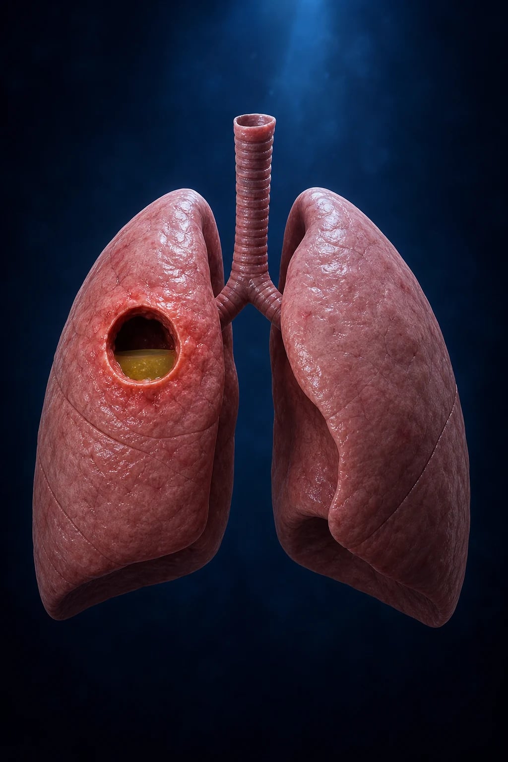
Lung abscess: localised collection of pus within lung parenchyma (cavity with air-fluid level on CXR/CT). [1]

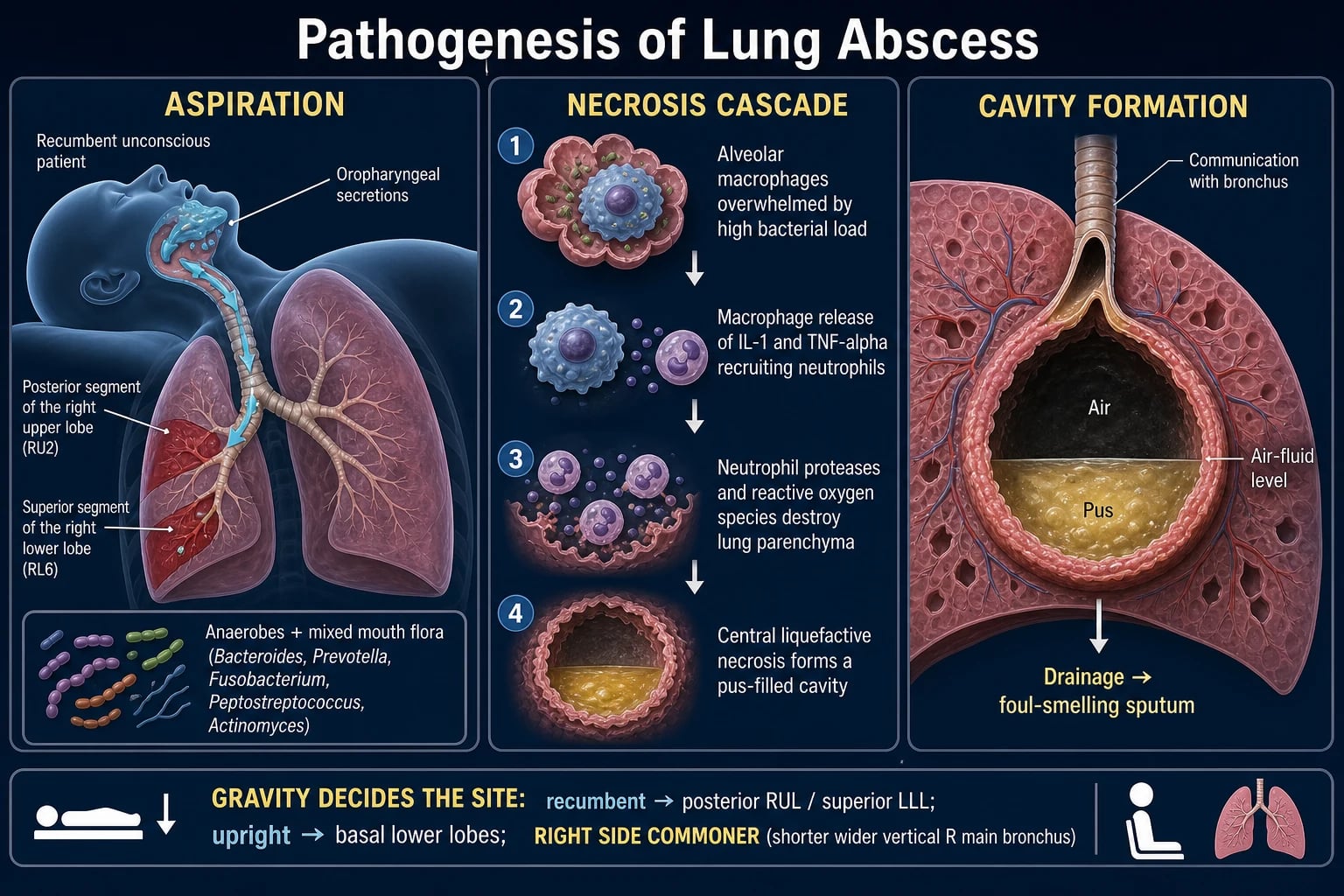
Causes: aspiration (#1 — anaerobes), pneumonia (S. aureus, Klebsiella, Pseudomonas), septic emboli, obstruction (tumour, foreign body). [1]

Management:

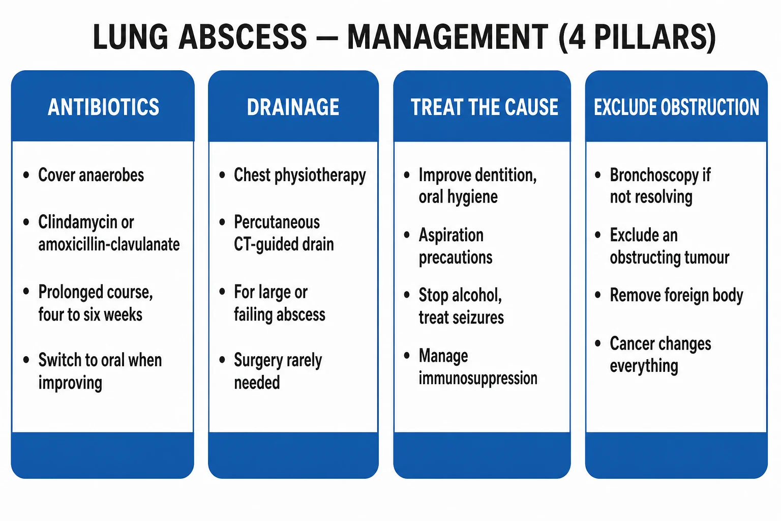
  1. Prolonged antibiotics: 4-6 weeks (longer than typical CAP). Cover anaerobes + aerobes: piperacillin-tazobactam OR clindamycin + ceftriaxone.
  2. Percutaneous drainage (CT-guided) if: large (>6 cm), not responding to antibiotics, or patient too sick for surgery.
  3. Surgery (lobectomy/wedge resection): rarely needed. Indications: failed medical therapy, massive haemoptysis, suspected malignancy. [1]

Differential of cavitating lung lesion: abscess, tuberculosis, malignancy (squamous cell), septic emboli, fungal (Aspergilloma), Wegener's granulomatosis.

[1]

SAQ — Lung abscess in an alcoholic, aspirating patient

10 minutes · 10 marks

A 56-year-old man with a 20-year history of hazardous alcohol use (120 g/day), poor dentition, and a 1-week history of fever, foul-smelling purulent sputum and rightsided pleuritic pain presents in septic shock. T 39.2 degrees C, HR 132, BP 82/50 (MAP 57) on noradrenaline 0.3 mcg/kg/min, RR 32, SpO2 90 per cent on 15 L oxygen via non-rebreather. WCC 28.6, CRP 360, lactate 4.2 mmol/L, albumin 22 g/L. Chest X-ray shows a 6 cm cavity with a thick irregular wall and an air-fluid level in the right lower lobe, with a small adjacent pleural effusion. Contrast CT confirms a parenchymal abscess (no communication with the pleural space) and bilateral aspiration changes. Sputum Gram stain shows polymicrobial mixed flora with Gram-negative bacilli and cocci.

[1]

SAQ — Bronchopleural fistula after necrotising pneumonia

10 minutes · 10 marks

A 68-year-old man is on ICU day 9 for necrotising MRSA pneumonia with secondary empyema. A 28 Fr chest drain was inserted 6 days ago for a complicated parapneumonic effusion and initially drained 600 mL of pus, since when output has fallen to 80 mL/day. However, over the last 24 hours the drain has been continuously bubbling with marked respiratory swing, the patient has developed copious purulent secretions ventilating him (intubated for ARDS), and he is again septic (HR 128, BP 84/52, lactate 4.0). Repeat CT chest shows a persistent hydropneumothorax with collapse of the right middle and lower lobes and a likely defect at the right bronchial tree.

[1]

Clinical pearls

High-yight pneumonia source control points for the CICM/FFICM exam

  1. Antibiotics CANNOT cure pus collections — drainage is essential.[1] }
  2. Parapneumonic effusion pH <7.2 = chest tube needed.[1] }
  3. tPA + DNase combination (not either alone) for loculated empyema (MIST2 trial).[2] }
  4. Lung abscess: prolonged antibiotics 4-6 weeks ± drainage.[1] }
  5. VATS for failed drainage. Open decortication as last resort.[2] }
  6. Bronchopleural fistula: persistent air leak in chest drain → communication between bronchus and pleural space. Surgery needed.[1] }
  7. Cover ANAEROBES in aspiration-related complications (clindamycin, piperacillin-tazobactam, metronidazole).[1] }
  8. CT chest: essential for identifying and characterising pleural collections and lung abscess.[1] }
  9. Diagnostic thoracentesis: before chest tube placement — check pH, Gram stain, culture, cell count, LDH, glucose, protein.[1] }
  10. Ultrasound-guided: chest tube insertion — reduces complications.[1] }
  11. Cover oral flora: for aspiration-related pleural complications (Peptostreptococcus, Fusobacterium, Prevotella, Bacteroides).[1] }
  12. Duration: empyema 2-4 weeks antibiotics. Lung abscess 4-6 weeks.[1] }
  13. Follow-up CXR: ensure resolution. Non-resolving: consider malignancy, TB, immunodeficiency.[1] }
  14. Mortality: empyema 10-20%. Lung abscess 5-10% (with appropriate treatment).[1] }

Red flags

Critical pneumonia source control points

  • Antibiotics CANNOT cure collections of pus — drainage essential.[1] }
  • pH <7.2 in pleural fluid = complicated parapneumonic = chest tube needed.[1] }
  • tPA + DNase combination (MIST2) for loculated empyema — not either agent alone.[2] }
  • Lung abscess: prolonged antibiotics 4-6 weeks (much longer than typical CAP).[1] }
  • Bronchopleural fistula: air leak in chest drain = surgical emergency.[1] }

Source control principles

Source control in pneumonia — the concept

Source control = any physical measure taken to eliminate a source of infection, reduce the bacterial inoculum, or restore the function of an organ so that it can overcome the infection. In pneumonia, where the infection begins in the lung parenchyma, source control is needed when infection escapes the reach of antibiotics by: [1]

  1. Spilling into a closed space the drug cannot penetrate (pleural cavity → parapneumonic effusion / empyema).
  2. Necrosing tissue faster than antibiotics can sterilise it (lung abscess).
  3. Breaching an epithelial barrier so infection and air communicate abnormally (bronchopleural fistula). [1]

Antibiotics diffuse poorly into a low-pH, high-protein, avascular collection of pus. Drainage is not adjunctive — it is definitive therapy. Delayed source control in sepsis is associated with increased mortality, hour by hour; the same principle applies to a loculated empyema or a necrotic lung abscess.[1]

Classification of parapneumonic effusion

The ACCP / BTS framework stratifies pleural infection into stages that drive the decision to drain. Pleural fluid pH is the single most powerful discriminator (must be measured on a blood-gas analyser, NOT a pH meter, and collected anaerobically in a heparinised syringe).[5][7]

[5] [7]

pH 7.2 is the tipping point — and the syringe matters

  1. pH <7.2 → drain. It is the strongest single predictor that a parapneumonic effusion will fail antibiotics alone.[5]
  2. pH 7.2-7.4 is a grey zone — drain if other features of complicated effusion (loculations, positive Gram stain, glucose <40).[7]
  3. Send fluid in a heparinised ABG syringe, anaerobically, on ice, to the blood-gas machine within 1 hour. A pH meter or litmus paper under-reads; air contamination over-reads. A false reading here triggers the wrong decision.[7]
  4. Do not measure pH on frankly purulent fluid — it will always be low; just drain it.

Chest tube insertion technique (Seldinger, ultrasound-guided)

Chest tube insertion is the most performed source-control procedure in pleural infection. The Seldinger technique over a guidewire, performed under real-time ultrasound, has largely replaced blunt dissection for effusions and empyema and is the BTS-recommended standard.[7]

Seldinger chest tube insertion — step by step

1

Pre-procedure

Confirm indication and consent. Review imaging (CT is most informative). Correct coagulopathy if possible (INR <1.5, platelets >50). Stop anticoagulants. Prophylactic antibiotics already running for the pneumonia.

2

Site selection with ultrasound

Sit the patient upright. Use a curvilinear probe in the mid/posterior axillary line. Identify the largest pocket, measure depth to pleura, mark skin. Avoid the SAFE triangle (bounded by anterior border of latissimus dorsi, lateral border of pectoralis major, line of nipple, base of axilla). Avoid intercostal vessels (use colour Doppler — the neurovascular bundle runs along the inferior rib border, so insert immediately ABOVE a rib).

3

Sterile prep & local anaesthetic

Full aseptic technique: chlorhexidine, sterile drape, gown, gloves. Infiltrate 1% lidocaine with adrenaline (max 3 mg/kg, ~20 mL) at skin → intercostal muscle → parietal pleura. Aspirate while advancing — return of fluid confirms intrapleural position and depth.

4

Needle + guidewire

Insert the Seldinger needle along the anaesthetised track, bevel up, just above the rib. Aspirate to confirm fluid. Thread the J-tipped guidewire through the needle into the pleural space. Withdraw the needle, leaving the wire in place. Never lose control of the wire.

5

Serial dilatation

Make a small skin nick at the wire. Pass sequential dilators over the wire (typically 6 → 8 → 10 → 12 Fr for small-bore tubes). Push dilators firmly but rotate and use the heel of the hand against the chest wall to prevent over-penetration of the pleura. Always reload the guidewire between dilators.

6

Tube placement

Load the chest tube (8-14 Fr pigtail for effusion/early empyema; 24-32 Fr rigid for thick pus) over the guidewire and rotate it into the pleural space, directing it posteriorly and apically for fluid. Remove the wire. Confirm free flow of fluid/air.

7

Secure & connect

Suture the tube to the skin (purse-string + stay suture), apply an occlusive dressing, connect to an underwater seal drainage system on suction (usually -10 to -20 cmH2O). Order a CXR within 1 hour to confirm position and assess lung re-expansion.

8

Post-procedure care

Record daily output. Flush with 20 mL saline every 6-8 h if blocking (empyema pus is thick). Re-image if output ceases. Remove when output <150-200 mL/day AND no air leak AND clinical/radiological improvement.

[7] [7]

Chest-tube insertion pitfalls the examiner probes

  1. Insert above the rib, never below — the neurovascular bundle (vein, artery, nerve) runs in the costal groove along the inferior border of each rib. Below = bleeding, haematoma, intercostal neuralgia.[7]
  2. The SAFE triangle is the safe zone for chest drains (anterior latissimus dorsi, lateral pectoralis major, nipple line, axillary base). Minimises damage to long thoracic nerve, internal mammary and lateral thoracic vessels.
  3. Intercostal vessels are not constant — in the elderly they can wander into the intercostal space. Colour Doppler before incision. Bleeding after chest tube is often intercostal artery injury.
  4. Coagulopathy is a relative contraindication, not absolute — correct to INR <1.5, platelets >50 × 10⁹/L before insertion if time permits; empyema drainage should not be delayed.
  5. Pigtail catheters migrate and kink — if drainage stops, re-image FIRST (ultrasound at the bedside) before assuming the tube is "blocked and needs replacing".

Intrapleural tPA and DNase for loculated empyema

When a chest tube fails to drain a loculated empyema, intrapleural fibrinolytic therapy breaks down the fibrinous septations and reduces fluid viscosity. Only the COMBINATION of tPA + DNase works — neither agent alone, and streptokinase alone (MIST1) is no better than placebo.[3][6]

tPA/DNase administration protocol (post-MIST2 / BTS)

1

Indication

Loculated empyema or complicated parapneumonic effusion with inadequate drainage 24-48 h after chest tube insertion, OR frank multiloculated empyema at the outset. Confirm with repeat ultrasound/CT.

2

Dose

tissue plasminogen activator (alteplase) 10 mg + recombinant human DNase (dornase alfa) 5 mg (250,000 IU) in 30-50 mL normal saline.

3

Administration

Clamp the chest tube, inject the mixture intrapleurally via the tube, flush with 10 mL saline. Keep the patient flat for 1 h, then rotate through positions (supine, left lateral, right lateral, prone) every 15 min to distribute the enzyme to all locules.

4

Frequency & duration

Twice daily (BD) for 3 days (total 6 doses) — the MIST2 schedule. TIGER showed that extending to 6 days does NOT improve outcomes and increases bleeding.<Cite id="4" />

5

Unclamp & monitor

Unclamp after 1 h and reapply suction. Record drainage volume (expect a step increase). Monitor for bleeding (rare — <5% clinically significant), fever, chest pain. Daily CXR to track resolution.

6

Reassess

After 6 doses, if drainage is still poor or patient not improving → CT and surgical review for VATS.

[3] [4]

MIST2 — intrapleural tPA and DNase (Rahman 2011)

PMID 23093163

Randomised double-blind 2×2 factorial, 4-arm

Population: 210 adults with pleural infection (pus in pleural space, pH <7.2, or positive culture)

Key finding

**tPA + DNase: significant increase in drainage, 31% reduction in surgical referral (4% vs 18%), shorter hospital stay.** tPA alone or DNase alone produced NO benefit and a non-significant trend to more surgery. Bleeding rare and minor.

[1]

TIGER — duration of tPA/DNase (Bhatnagar 2023)

PMID 36028485

Randomised, open-label, non-inferiority

Population: 425 adults with pleural infection already receiving standard care

Key finding

**No difference** in surgical referral, mortality, or radiological outcome between 6 and 3 days. **More bleeding** in the 6-day arm.

[1]

MIST1 — intrapleural streptokinase (Maskell 2005)

PMID 15917261

Randomised double-blind, placebo-controlled

Population: 454 adults with pleural infection

Key finding

**No benefit** for streptokinase: mortality 13% vs 14%, surgery 23% vs 25%, hospital stay similar. Adverse events (fever, allergy, antibody formation) higher with streptokinase.

[3] [4] [6]

tPA/DNase prescribing pearls

  1. The order is tPA THEN DNase in the syringe; do not mix drugs from different vials in one syringe before drawing — draw each separately then combine.[3]
  2. Position rotation is essential — the enzyme must reach every locule. A bed-bound patient who lies supine throughout will not drain.
  3. Watch for systemic bleeding is unnecessary — intrapleural tPA is locally active; systemic fibrinolysis is negligible. Minor pleural bleeding is the main risk.
  4. TIGER changed practice — 3 days, not 6. Beyond that you are adding risk for nothing.[4]
  5. Fibrinolytics are not a substitute for surgery — if there is no improvement in 72 h, the patient needs VATS, not more drugs.[3]

When to involve thoracic surgery

Early surgical review for VATS is one of the most important decisions in pleural infection. The window between medical failure and irreversible lung trapping is narrow (days, not weeks).[5]

Escalation pathway for empyema

1

Step 1 — Drain

Image-guided chest tube (small-bore first-line) + IV antibiotics + nutritional support. Assess at 24-48 h.

2

Step 2 — tPA/DNase

If loculated or draining poorly: 3-day course of intrapleural tPA 10 mg + DNase 5 mg BD. Assess at 48-72 h.

3

Step 3 — VATS

INDICATIONS: persistent sepsis, no radiological improvement, ongoing poor drainage at 72 h; OR multiloculated empyema / Stage III at presentation in a surgical candidate. VATS breaks down loculations, evacuates pus, removes the fibrinous peel (decortication) so the lung can re-expand.

4

Step 4 — Open thoracotomy & decortication

For VATS failure, chronic organised empyema (Stage III/IV with a thick cortical peel), or patients unfit for thoracoscopy. Larger exposure allows complete decortication. Higher morbidity.

[5] [7]

VATS vs open thoracotomy decortication

VATS (video-assisted thoracoscopic surgery) is preferred for acute / organising empyema. Advantages: less postoperative pain, shorter hospital stay, lower wound infection, equivalent drainage. Performed under general anaesthesia with single-lung ventilation. [1]

Open thoracotomy with decortication is reserved for chronic, densely organised empyema where a fibrous cortex has trapped the lung, or when VATS is technically not feasible. It allows mechanical removal of the peel but carries greater morbidity (pain, respiratory compromise, longer recovery). [1]

Timing matters more than technique — a delayed decision to operate is the strongest predictor of mortality in empyema. If the patient is not improving at 5-7 days from drainage, the cost of further waiting exceeds the risk of surgery.[5]

Bronchopleural fistula

Bronchopleural fistula (BPF) — a surgical emergency

BPF = a direct communication between the bronchial tree and the pleural space. It most commonly follows pneumonectomy (especially right-sided), but in the pneumonia patient it arises from lung necrosis — necrotising pneumonia (S. aureus, Klebsiella, Pseudomonas, anaerobes), lung abscess eroding a bronchus, or rupture of a peripheral abscess into the pleural space.[9]

Clinical features:

  • Persistent large air leak in the chest drain (bubbling that does not settle).
  • New or worsening hydropneumothorax / pyopneumothorax on imaging.
  • Copious purulent sputum that increases when the patient lies with the affected side down (postpneumonectomy patients flood the contralateral lung — position good lung UP).
  • Failure of the lung to re-expand after tube drainage.
  • Sepsis refractory to antibiotics. [1]

Diagnosis: CT chest (shows the defect, residual cavity, surrounding collapse); bronchoscopy (directly visualises the fistula); methylene blue / inhalation of inert gas injected into the pleural space. [1]

Management:[8][9]

  1. Chest tube to the affected pleural space — drain the pleural collection and create a controlled fistula.
  2. Positioning: affected side DOWN (dependent) so infected pleural fluid does not flood the contralateral healthy lung. (For post-pneumonectomy BPF, sit the patient UPRIGHT and affected side down.)
  3. Broad-spectrum antibiotics covering anaerobes, MRSA, gram-negatives.
  4. Ventilation strategy if intubated: low tidal volume, low PEEP, consider independent lung ventilation or ECMO to rest the lung while the fistula closes.
  5. Surgical closure (definitive): direct suture, coverage with intercostal muscle / omental / serratus anterior flap, or completion of resection. Endobronchial one-way valves (Zephyr, IBV) are an option in poor surgical candidates.
  6. Pleurodesis or open window thoracostomy (Clagett window) for chronic, non-resolving BPF — leaves a stoma for chronic drainage and packing.

Prognosis: mortality 12-50% depending on cause and comorbidity; highest in post-pneumonectomy BPF.

[1] [9]

Lung abscess — in depth

Lung abscess — natural history and why 4-6 weeks of antibiotics

A lung abscess is a localised suppurative cavity within the lung parenchyma, classically with an air-fluid level on upright CXR or CT. The key teaching point is the duration of antibiotics: 4-6 weeks, far longer than for uncomplicated CAP.[1][10]

Why so long? Unlike pneumonia (interstitial / alveolar infiltrate penetrated well by antibiotics), an abscess is a walled-off collection of pus with poor blood supply and low pH. Antibiotics sterilise the cavity wall but cannot penetrate the necrotic centre. The cavity shrinks by drainage via the bronchial tree (postural drainage, physiotherapy) and granulation/fibrosis over weeks. Antibiotics are continued until the cavity resolves or becomes a small stable thin-walled residual on serial CXR.[10]

Microbiology: aspiration (oral anaerobes — Peptostreptococcus, Fusobacterium, Prevotella, Bacteroides) is the commonest cause; aerobic gram-negatives and S. aureus in bacteraemic/necrotising pneumonia; consider TB, melioidosis, fungal (Aspergillus) and Nocardia in the immunocompromised or in endemic regions.[1]

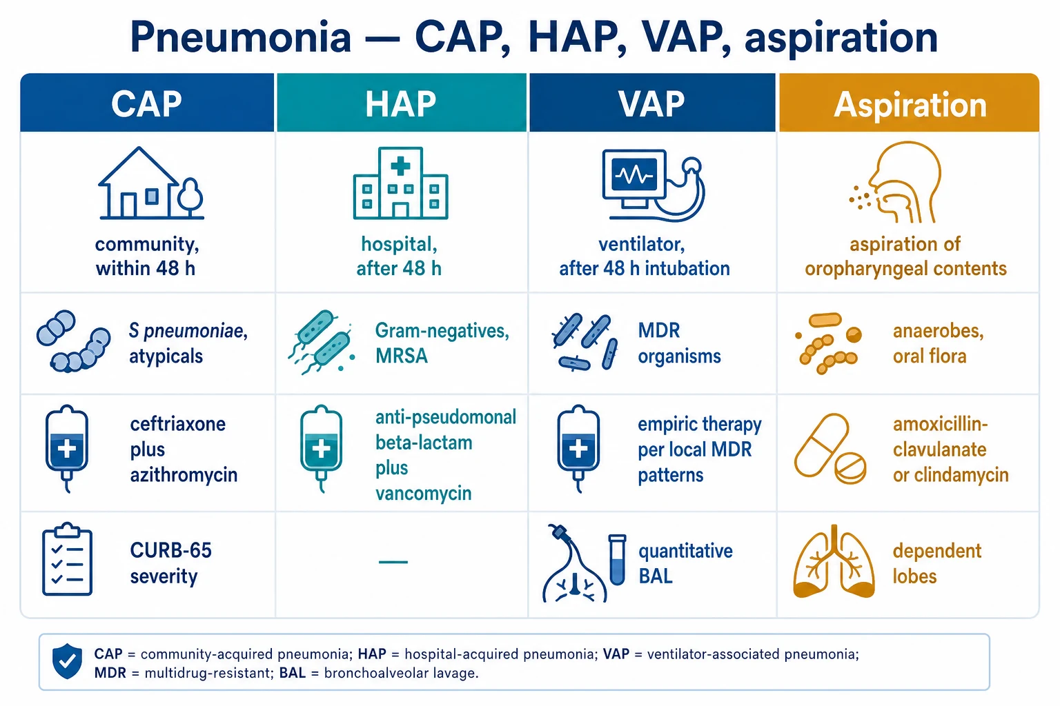
Antibiotic choice: piperacillin-tazobactam ± clindamycin (anaerobic cover) for hospitalised/aspiration; add vancomycin/linezolid if MRSA suspected; ceftriaxone + metronidazole is a standard alternative. Switch to oral (amoxicillin-clavulanate or clindamycin) once afebrile and improving for the remainder of the course.[10]

Indications for drainage (percutaneous CT-guided catheter):

  • Cavity >6 cm diameter
  • No response to antibiotics after 7-10 days
  • Rapidly enlarging or at risk of rupture
  • Immunocompromised (poor host response)
  • Prevents need for surgery in ~80%[10]

Surgery (lobectomy / wedge resection): rarely required. Reserved for failure of prolonged antibiotics + drainage, massive haemoptysis, suspected underlying malignancy, or bronchopleural fistula. Mortality of surgery in this group is high.[10]

[1] [10]

Antibiotic coverage for source control

Empirical antibiotic principles in pleural infection

Source control without the right antibiotics fails. Empirical regimens for pleural infection must cover the Streptococcus anginosus (milleri) group (commonest community cause), Staphylococcus aureus (including MRSA in hospital-acquired), anaerobes (aspiration), and Gram-negatives including Klebsiella and Pseudomonas (especially hospital-acquired).[1][2]

  • Community-acquired empyema: ceftriaxone + metronidazole OR amoxicillin-clavulanate (IV) ± clindamycin for anaerobes.
  • Hospital-acquired / post-surgical empyema: piperacillin-tazobactam + vancomycin (MRSA) ± aminoglycoside; add antifungal if Candida suspected.
  • Duration: 2-4 weeks IV then oral, guided by clinical response and drainage adequacy. Longer (4-6 weeks) if lung abscess coexists.
  • De-escalate to culture-directed therapy once pleural fluid / blood cultures return. [1]

Anaerobic cover is non-negotiable in aspiration-related disease — missing it is a classic exam and real-world error. Clindamycin, metronidazole, piperacillin-tazobactam and carbapenems all provide it; ceftriaxone alone does NOT.[1]

Imaging in source control

[1] [7]

Complications of failed source control

What happens when drainage is delayed

  1. Lung trapping: the organising fibrinous peel (cortex) over the visceral pleura fibroses, mechanically splinting the lung and causing permanent restrictive deficit. The window to prevent this is ~2-3 weeks.[5]
  2. Bronchopleural fistula: necrosis erodes a bronchus; persistent air leak; high mortality.[9]
  3. Empyema necessitatis: empyema tracks through the chest wall to the skin (classically in TB and actinomycosis).
  4. Sepsis, septic shock, MODS: persistent source → prolonged critical illness.
  5. Spread to pericardium (purulent pericarditis) or mediastinum (mediastinitis) — rare but catastrophic.

Clinical pearls — extended

More high-yield source-control pearls

  1. The split-pleura sign on contrast-enhanced CT (enhancement of both visceral and parietal pleura with fluid between) is pathognomonic for empyema / complicated parapneumonic effusion.[1]
  2. Strep. anginosus (milleri) group is the commonest community-acquired cause of empyema; it readily forms abscesses and septations.[1]
  3. Postural drainage and chest physiotherapy are adjunctive in lung abscess — promote drainage of pus via the bronchial tree, the body's own "chest tube".[10]
  4. Air-fluid level on CXR/CT in a patient with pneumonia = lung abscess until proven otherwise; differentiate from empyema (which has a lenticular shape and split pleura).[10]
  5. Always send pleural fluid for Gram stain, culture AND cytology — an "empyema" can mask an underlying malignancy causing post-obstructive pneumonia.[7]
  6. Re-expansion pulmonary oedema can occur after draining a large effusion rapidly — limit to <1.5 L in one sitting, or use a controlled clamp if symptomatic.[7]
  7. A patient who spikes fever after chest-tube insertion — think tube occlusion, retained loculation, or (later) tube-site infection. Re-image and reassess, do not just change antibiotics.[7]
  8. Nutrition matters — empyema patients lose protein into the pleural space; hypoalbuminaemia predicts poor outcome and delayed healing. Enteral feeding early.[1]
  9. Melioidosis (Burkholderia pseudomallei) must be considered in endemic regions (northern Australia, SE Asia) — causes suppurative pneumonia with abscess and empyema; needs ceftazidime/carbapenem then eradication with cotrimoxazole.[1]
  10. Tuberculosis can present as a pleural effusion or empyema — pleural fluid is lymphocytic, ADA elevated; send for AFB and TB-PCR. Standard empyema antibiotics will not treat it.[1]
  11. Necrotising pneumonia (multiple small cavities, often S. aureus PVL+) predicts abscess, empyema and BPF — involve surgery early and expect prolonged recovery.[1]
  12. Never drain a hydatid cyst if suspected — risk of anaphylaxis and pleural seeding; refer for surgical excision with scolicidal technique.[10]

Comparison of management by complication

Source control ladder: ultrasound-guided chest drain, intrapleural tPA and DNase, percutaneous abscess drainage, VATS decortication, BPF strategies
FigureDrain early; MIST2 intrapleural therapy; escalate to thoracic surgery when loculations persist.
[1] [2] [10] [9]

Red flags — extended

Don't-miss red flags in pneumonia source control

  • Empyema with air-fluid level and persistent bubbling = bronchopleural fistula — surgical emergency, position good lung up.[9]
  • pH <7.2 on pleural fluid = drain; antibiotics alone will fail.[5]
  • No improvement at 5-7 days despite tube + tPA/DNase = VATS now; further waiting = lung trapping and mortality.[5]
  • Air-fluid level within the lung (not the pleura) = lung abscess; needs 4-6 weeks of antibiotics.[10]
  • Persistent large air leak after chest-tube insertion = BPF; do not just clamp or upsize — image and refer to surgery.[9]
  • Aspiration pneumonia patient — always add anaerobic cover; expect abscess and empyema.[1]
  • Septic patient with "simple" effusion — re-image; septations on ultrasound upstage to complicated even if pH borderline.[7]
  • Cavitating lesion in an immunocompromised host — broaden differential (TB, Nocardia, Aspergillus, lymphoma); biopsy may be needed before committing to long antibiotics.[1]
  • Melioidosis in endemic area — dual abscess/empyema common; specific antibiotics required.[1]
  • Re-expansion oedema — do not drain >1.5 L rapidly; clamp for symptoms.[7]

Exam-style one-liners

CICM / FFICM one-liners to be able to reproduce

  • "Source control in pneumonia means draining infected collections that antibiotics cannot penetrate."
  • "Pleural fluid pH <7.2 is the trigger to drain a parapneumonic effusion."
  • "tPA and DNase together — not either alone — break down loculations in empyema (MIST2)."
  • "Streptokinase is ineffective for pleural infection (MIST1) and is no longer used."
  • "Lung abscess needs 4-6 weeks of antibiotics with anaerobic cover; drainage is reserved for large or non-responsive cavities."
  • "A bronchopleural fistula presents with a persistent air leak in the chest drain and needs surgical repair."
  • "VATS decortication is indicated when an empyema fails to drain with tube + fibrinolytics within 72 hours."
  • "Insert a chest drain above the rib, in the SAFE triangle, under ultrasound guidance, after correcting coagulopathy."
[1]

Summary

Source control in pneumonia — bottom line

Pneumonia source control = drainage of pleural infection (parapneumonic effusion / empyema), management of lung abscess with prolonged antibiotics ± percutaneous drainage, and surgical repair of bronchopleural fistula. The bedrock principles: [1]

  1. Drain pus — antibiotics cannot sterilise a collection.
  2. Use pleural pH <7.2 as the decision point for drainage.
  3. tPA + DNase (not either alone), 3 days for loculated empyema.
  4. Escalate to VATS when drainage fails within 72 hours.
  5. Give lung abscess 4-6 weeks of anaerobic-covering antibiotics.
  6. Recognise BPF early — persistent air leak, position the good lung up, involve surgery.
  7. Cover anaerobes in aspiration-related disease — non-negotiable.
  8. Image with ultrasound first, contrast CT when complex. [1]

Source control is the intervention that converts a medical disease into a curable one. In septic patients with pleural infection, the timing of drainage is as critical as the timing of antibiotics in septic shock.[1][3][5][9][10]

References

  1. [1]Martin-Loeches I, Torres A. Severe community-acquired pneumonia Eur Respir Rev, 2022.PMID 36517046
  2. [2]Rahman NM, et al. Notum palmitoleoyl-protein carboxylesterase regulates Fas cell surface death receptor-mediated apoptosis via the Wnt signaling pathway in colon adenocarcinoma Bioengineered, 2021.PMID 34402722
  3. [3]Rahman NM, Maskell NA, West A, et al. Association between use of lung-protective ventilation with lower tidal volumes and clinical outcomes among patients without acute respiratory distress syndrome: a meta-analysis JAMA, 2012.PMID 23093163
  4. [4]Bhatnagar R, Piotrowska HEG, Laskawiec M, et al. Multivalent interactions between molecular components involved in fast endophilin mediated endocytosis drive protein phase separation Nat Commun, 2022.PMID 36028485
  5. [5]Colice GL, Curtis A, Deslauriers J, et al. Unforced errors and error reduction in tennis Br J Sports Med, 2006.PMID 16632568
  6. [6]Maskell NA, Davies CWH, Nunn AJ, et al. Intravenous polyclonal immunoglobulin administration to sepsis syndrome patients: a prospective study in a pediatric intensive care unit J Trop Pediatr, 2005.PMID 15917261
  7. [7]Havelock T, Teoh R, Laws D, Gleeson F; BTS Pleural Disease Guideline Group. The mTOR inhibitor everolimus in combination with carboplatin in metastatic breast cancer--a phase I trial Anticancer Res, 2012.PMID 22843927
  8. [8]Shen HN, Lu CL. HIV protease inhibitors block the zinc metalloproteinase ZMPSTE24 and lead to an accumulation of prelamin A in cells Proc Natl Acad Sci U S A, 2007.PMID 17652517
  9. [9]Loukeri AA, Coni P, Lazopoulos G. Tipping our CAPS to the UKOSS cardiac arrest in pregnancy study BJOG, 2017.PMID 28109048
  10. [10]Anstadt MP, Whitman GJ. GPU-BLAST: using graphics processors to accelerate protein sequence alignment Bioinformatics, 2011.PMID 21088027