ICU · Respiratory
Pulmonary contusion and blunt chest trauma
Also known as Pulmonary contusion · Blunt chest injury · Flail chest · Blunt aortic injury
Pulmonary contusion is bruising of the lung parenchyma from blunt chest trauma (motor vehicle crash, fall, crush, blast). Alveolar-capillary disruption → alveolar haemorrhage + interstitial/peribronchial oedema → impaired gas exchange (V/Q mismatch → true shunt) + reduced compliance → progressive hypoxaemia that develops and WORSENS over 24-48h (worsens before improving). Often associated with rib fractures (1 association), flail chest (≥3 consecutive ribs fractured in ≥2 places — paradoxical breathing), pneumothorax, haemothorax, blunt aortic injury and cardiac (myocardial) contusion. Management: oxygen (escalate via HFNC/NIV), analgesia (epidural or thoracic paravertebral block for rib fractures), JUDICIOUS fluids (avoid overload — worsens contusion), NIV if moderate respiratory distress, intubation + lung-protective ventilation (Vt 6 mL/kg PBW) if severe. Monitor for: ARDS (develops in ~20% of severe contusions), pneumonia, respiratory failure — mortality 10-25%. CXR may be NORMAL initially (lags 24-48h); CT chest is more sensitive and quantifies contusion volume.
On this page & tools
Your progress
Saved locally on this device.
Target exams
Red flags
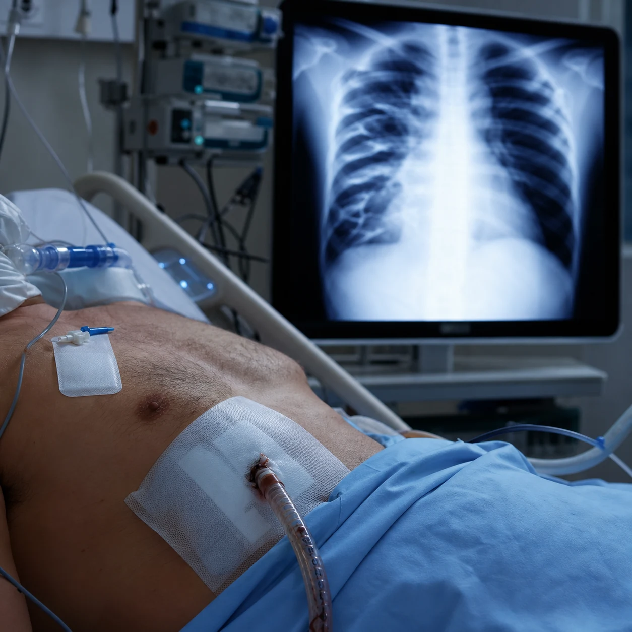
Blunt chest trauma — the key injuries and their distinguishing features
| Injury | Mechanism | Hallmark features | First-line management |
|---|---|---|---|
| Pulmonary contusion | Direct impact → alveolar-capillary disruption | Hypoxaemia WORSENS over 24-48h; patchy infiltrates under the injury; CXR lags CT | Oxygen, NIV, judicious fluids, lung-protective ventilation if intubated |
| Flail chest | ≥3 consecutive ribs fractured in ≥2 places | Paradoxical chest wall movement; severe pain; respiratory failure | Aggressive analgesia (epidural), NIV, surgical rib fixation (selected) |
| Blunt cardiac injury | Direct impact, heart crushed between sternum and spine | Troponin elevated, arrhythmia (AF, PVCs, heart block), ECG changes | ECG monitoring 24-48h, troponin, echo, treat arrhythmia |
| Traumatic aortic injury | Deceleration → intimal tear / partial transection | Widened mediastinum, apical cap, BP differential | CT angiography → TEVAR (preferred) or open repair |
| Tension pneumothorax | Air under pressure, one-way valve | Hypotension, tracheal deviation, absent breath sounds, distended neck veins | IMMEDIATE needle decompression (don't wait for CXR), then chest drain |
| Haemothorax | Bleeding into pleural space | Dullness, decreased breath sounds, hypovolaemia | Large-bore chest drain (28-36 Fr); thoracotomy if >1500 mL or >200 mL/h |
| Tracheobronchial injury | Crush/deceleration, tears at carina | Massive continuous air leak, non-re-expanding pneumothorax, surgical emphysema | Bronchoscopy, surgical repair, double-lumen tube |
| Diaphragmatic injury | Penetrating thoracoabdominal or blunt rupture (L>R) | Delayed herniation; bowel sounds in chest; NG tube curled in chest | Surgical repair (laparotomy/thoracotomy) |
Pathophysiology
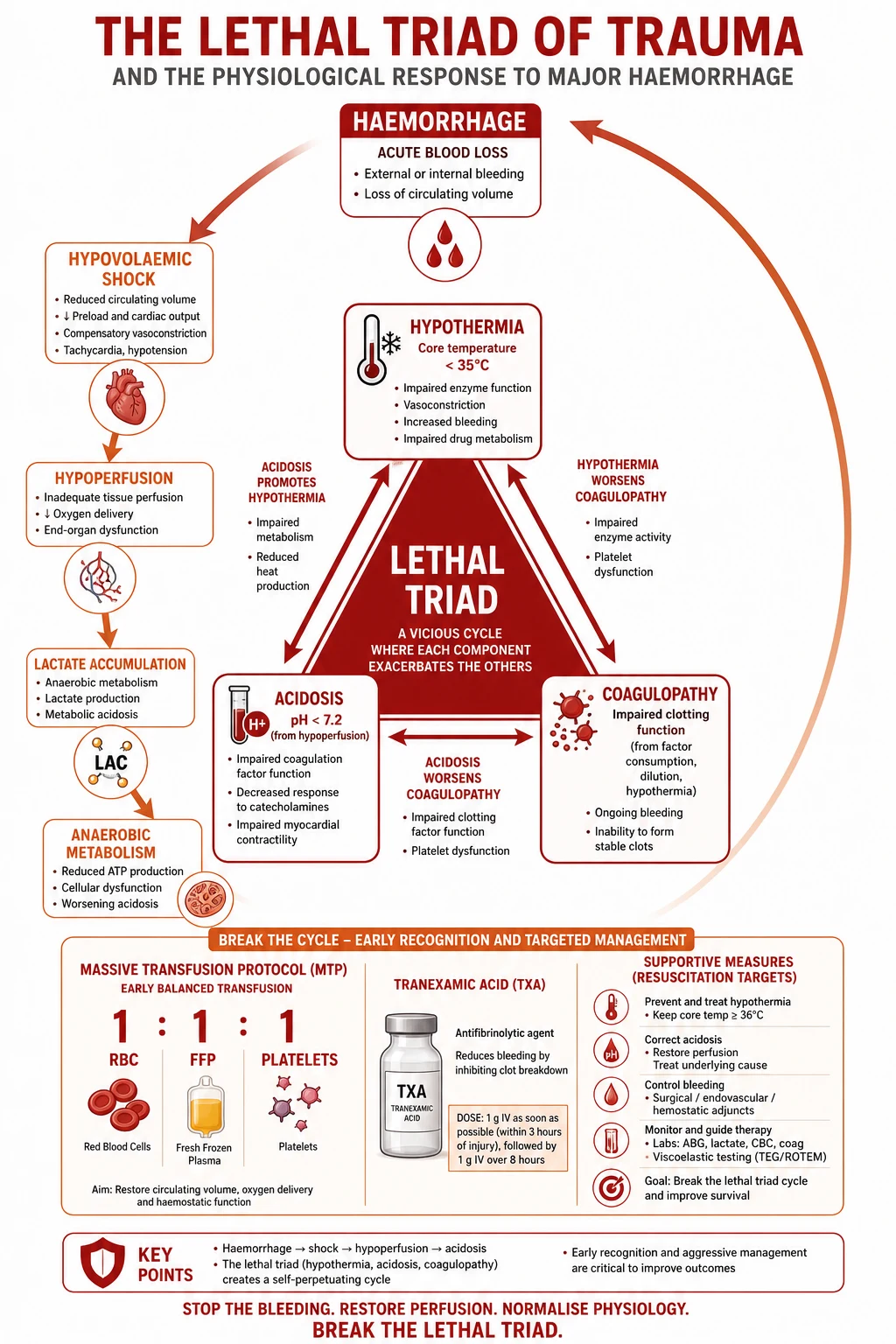
Pulmonary contusion is, in essence, a bruise of the lung parenchyma produced by blunt energy transfer. The injury cascade — much loved by examiners because it explains both the clinical course and the management — is:[2] }[4] }
- Blunt force energy deposition. A moving object (steering wheel, dashboard, blast wave, fall from height) strikes the chest wall. The ribs transmit the energy directly to the underlying lung. Compression of the thorax between the anterior chest wall and spine generates a pressure wave that propagates through fluid-filled alveoli. Spalling (tearing at the gas-liquid interface), inertial effects (different rates of acceleration of the lung hilum versus the parenchyma), and implosion (gas-bubble collapse then rebound) all disrupt the alveolar-capillary membrane.[4] }
- Alveolar-capillary disruption. Capillary and alveolar epithelial tearing allows red blood cells, protein-rich fluid and inflammatory mediators to pour into the alveolar spaces and interstitium → intra-alveolar haemorrhage and peribronchial/interstitial oedema. The alveolar septa fracture; type II pneumocytes are damaged, reducing surfactant production.[2] }
- Oedema, atelectasis and surfactant loss. Proteinaceous oedema and blood fill alveoli and small airways; surfactant is washed out and inactivated → alveolar collapse and atelectasis. Pulmonary lymphatic drainage is overwhelmed. Capillary leak from the systemic inflammatory response adds further interstitial oedema over the next 24-48h.[3] }
- V/Q mismatch → true shunt → hypoxaemia. Blood continues to perfuse consolidated, non-ventilated alveoli → low or zero V/Q units and true shunt. The hypoxaemia is relatively refractory to supplemental oxygen (shunt physiology). Hypoxic pulmonary vasoconstriction partially compensates but is incomplete.
- Reduced compliance and increased work of breathing. Stiff, oedematous, consolidated lung plus painful rib fractures → shallow rapid breathing, splinting, atelectasis, fatigue → ventilatory failure (rising PaCO₂) superimposed on the hypoxaemia.[10] }
- The 24-48h evolution. Because the oedema and inflammatory response take hours to develop, the clinical and radiographic picture WORSENS over the first 24-48h before it improves. Resolution begins around day 3-5 and is usually complete by 7-10 days (unless complicated by ARDS or pneumonia). This delayed course is the single most testable fact about pulmonary contusion and is why a normal initial CXR must never reassure the clinician.[2] }[7] }
Flail chest compounds the problem. When ≥3 consecutive ribs fracture in ≥2 places, a free-floating chest wall segment moves paradoxically — inward on inspiration, outward on expiration — creating inefficient, wasted ventilation, worsening the underlying contusion, and producing severe pain that limits cough and deep breathing. The triad of flail chest = (1) bony instability, (2) pulmonary contusion beneath, (3) pain-induced splinting.[9] }[11] }
Clinical features
The clinical picture reflects a combination of the parenchymal injury itself, the associated chest wall injury, and concurrent multi-system trauma.[8] }
- Hypoxaemia (the cardinal feature): SpO₂ <92% on room air, tachypnoea, respiratory distress. May be ABSENT on arrival and develop over hours.
- Chest wall bruising, abrasions, seat-belt sign over the area of impact; palpable rib crepitus and step deformity; subcutaneous emphysema (suggests pneumothorax or airway injury).
- Rib fractures / flail segment: localised tenderness, crepitus, paradoxical chest wall movement.
- Decreased breath sounds over the contused area (consolidation/atelectasis) or globally (pneumothorax, haemothorax, large effusion).
- Haemoptysis (blood in the airways from alveolar haemorrhage) — may be the earliest clue.
- Tachypnoea, tachycardia, accessory muscle use — work of breathing is markedly increased from pain, stiffness and shunt.
- Hypotension: consider tension pneumothorax, massive haemothorax, cardiac tamponade or haemorrhagic shock from associated injuries (do NOT attribute hypotension to the contusion alone).
- Signs of associated injury: muffled heart sounds/tamponade (cardiac contusion), upper-limb hypertension with BP differential or pulse deficit (blunt aortic injury), neurological deficit (spinal injury), abdominal tenderness (spleen/liver injury). [1]
Imaging
Chest X-ray (CXR). First-line but insensitive early. As oedema develops over 24-48h it shows patchy, non-segmental airspace opacities underlying the area of injury (non-segmental because contusion follows the blow, not a bronchovascular distribution — a useful discriminator from aspiration, which is segmental/dependent). Look also for: rib fractures, flail segment, pneumothorax, haemothorax, subcutaneous emphysema, widened mediastinum (aortic injury), sternal fracture.[2] }
CT chest. The most sensitive modality — detects contusion immediately, characterises its extent and volume, and identifies associated injuries (aortic, sternal, spinal). CT patterns: patchy or confluent ground-glass/consolidation in a non-segmental distribution beneath the impact zone, often with rib fractures, pleural effusion/haemothorax and pneumothorax. Contusion volume as a fraction of total lung volume correlates with the risk of ARDS and the need for mechanical ventilation; >20-28% contused volume is a high-risk threshold.[6] }[3] } Automated deep-learning volumetry is now described.[5] }
CT pulmonary angiography (CTPA). Performed when widened mediastinum, apical cap, mechanism suggestive of deceleration, or BP differential raises suspicion of blunt aortic injury.[19] }
POCUS / lung ultrasound. Useful at the bedside: B-lines, subpleural consolidation, absent lung sliding (pneumothorax), and pleural fluid. Useful for serial monitoring and procedures but operator-dependent. [1]
Echocardiography. Performed when blunt cardiac injury is suspected (new murmur, arrhythmia, troponin rise, haemodynamic instability) to assess wall-motion abnormalities and tamponade.[18] }
CXR versus CT chest in pulmonary contusion
| Feature | CXR | CT chest |
|---|---|---|
| Sensitivity early (0-6 h) | Low — often normal | High — detects contusion immediately |
| Typical finding | Patchy non-segmental infiltrates developing 24-48h | Ground-glass/consolidation under impact zone |
| Quantification of contusion volume | Poor | Excellent (volume % predicts ARDS) |
| Rib/flail assessment | Good | Excellent |
| Detects aortic injury | Indirect only (widened mediastinum) | Definitive (with CTPA) |
| Role | Bedside screening, serial monitoring | Definitive diagnosis + prognostication |
Management
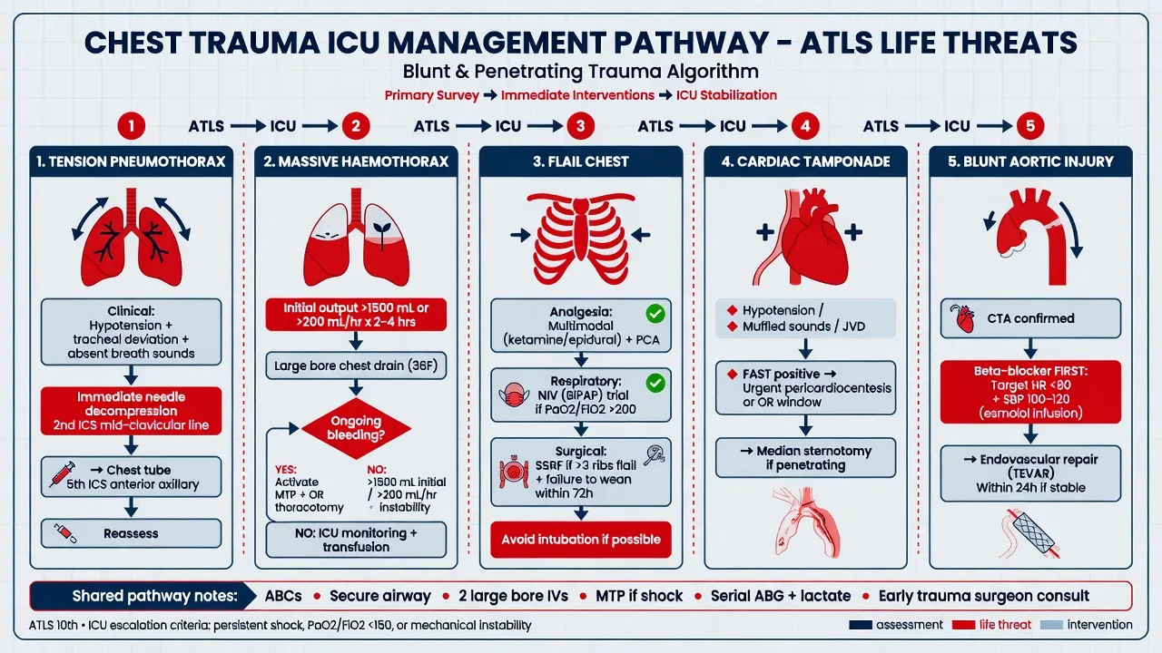
The EAST practice management guideline (Simon 2012) frames care around respiratory support, analgesia and a conservative fluid strategy — there is no specific pharmacotherapy that reverses the contusion.[1] }
Stepwise ICU management of pulmonary contusion / blunt chest trauma
- PRIMARY SURVEY (ABCDE) — airway (with cervical spine control), breathing (high-flow oxygen; assess and immediately decompress tension pneumothorax; drain large haemothorax), circulation (2 large-bore IVs; blood not crystalloid for haemorrhagic shock; control bleeding), disability (GCS), exposure
- IMAGING & WORK-UP — CXR (rib #, pneumo/haemothorax, widened mediastinum, contusion); CT chest (definitive for contusion volume, aortic injury, sternal/spinal fracture); FAST/CT abdomen (30-50% have multi-system trauma); ECG + troponin (blunt cardiac injury); ABG (baseline gas exchange, lactate)
- RESPIRATORY SUPPORT — escalate as needed — (a) Low-flow oxygen if mild. (b) HFNC if moderate hypoxaemia. (c) NIV (CPAP/BiPAP) for moderate respiratory distress / flail chest — improves oxygenation, splints the flail segment, reduces work of breathing. (d) Intubate if: PaO₂/FiO₂ <200 despite NIV, exhaustion, rising PaCO₂, altered mental status, shock, or failure to clear secretions
- LUNG-PROTECTIVE VENTILATION if intubated — Vt 6 mL/kg predicted body weight, plateau pressure <30 cmH₂O, driving pressure <15, PEEP titrated to oxygenation, permissive hypercapnia if brain injury allows. The contused lung is vulnerable to ventilator-induced lung injury (VILI) — barotrauma, volutrauma, atelectrauma, biotrauma. Use the same strategy as for ARDS
- ANALGESIA — the MOST IMPORTANT modifiable intervention — inadequate analgesia → splinting → atelectasis → pneumonia → respiratory failure. Options: (a) Epidural (thoracic T5-T8, local anaesthetic ± opioid) — best for bilateral/multilevel rib # and flail chest. (b) Thoracic paravertebral / serratus anterior plane block — unilateral, less hypotension, preserves neurological exam. (c) PCA (morphine/fentanyl). (d) Multimodal: paracetamol, NSAIDs (if no contraindication), ketamine infusion. Avoid sole reliance on systemic opioids (respiratory depression, delirium)
- JUDICIOUS FLUIDS — AVOID OVERLOAD — the contused lung has disrupted capillaries; excess crystalloid → hydrostatic pressure + capillary leak → worse oedema → worse hypoxaemia. Use the MINIMUM fluid to maintain perfusion (MAP ≥65, urine output, lactate). If hypovolaemic from blood loss, transfuse blood products, not crystalloid. Aim for a neutral-to-negative fluid balance. Vasopressors are preferred over crystalloid boluses once the patient is warm and coagulopathy corrected
- CHEST PHYSIOTHERAPY & PULMONARY TOILET — incentive spirometry, deep breathing, coughing, secretion clearance (aided by good analgesia), early mobilisation — prevents atelectasis and pneumonia
- TREAT ASSOCIATED INJURIES — chest drain for pneumo/haemothorax; CTPA + TEVAR for blunt aortic injury; ECG monitoring 24-48h for blunt cardiac injury; surgical rib fixation for selected flail chest; evaluate for TBI, abdominal and spinal injury
- MONITOR & RE-EVALUATE 24-48h — serial CXR/CT (watch for evolution and ARDS), serial ABG, continuous SpO₂, serial troponin if BCI suspected. The patient who looks well at hour 6 may decompensate at hour 30
- COMPLICATION SURVEILLANCE — watch for ARDS (~20% of severe contusions), pneumonia (contused lung is prone to infection), respiratory failure, atelectasis, and fat embolism (if long-bone fracture present). Antibiotics are NOT prophylactic — treat documented infection
Fluid strategy in pulmonary contusion — dry vs wet
| Strategy | Rationale | Risk |
|---|---|---|
| Conservative / "dry" (preferred) | Minimise capillary leak + hydrostatic pressure on the contused, leaky lung | Under-resuscitation if active haemorrhage not addressed |
| Liberal crystalloid | Easier, faster venous access | Worsens oedema, hypoxaemia, ARDS, prolonged ventilation — AVOID |
| Blood product resuscitation | Restores oxygen-carrying capacity without large crystalloid volume | Transfusion reactions (TRALI/TACO) — balance risk |
| Goal-directed (lactate, urine output, MAP) | Targets perfusion, not arbitrary volume | Requires monitoring; vasoactives often needed |
Analgesia options for rib fractures / flail chest
| Technique | Best for | Advantages | Cautions |
|---|---|---|---|
| Thoracic epidural | Bilateral / multilevel rib #, flail chest | Superior analgesia, best pulmonary function, reduced pneumonia/intubation | Hypotension (sympathectomy), coagulopathy, masks spinal injury |
| Paravertebral / SAP block | Unilateral rib # | Less hypotension, unilateral, preserves neuro exam, continuous catheter option | Technically harder; patchy coverage |
| PCA opioid | Adjunct / epidural contraindicated | Reliable, titratable | Sedation, delirium, respiratory depression, ileus |
| Multimodal (paracetamol ± NSAID ± ketamine) | All patients | Opioid-sparing, well tolerated | NSAID: renal/bleeding caution; ketamine: emergence |
Indications for intubation / ventilation
Associated injuries
Blunt chest trauma rarely occurs in isolation. Actively screen for the "deadly dozen" thoracic injuries.[8] }
Associated injuries to actively exclude in blunt chest trauma
| Injury | Clue / screening | Definitive test | Key management |
|---|---|---|---|
| Rib fractures / flail chest | Tenderness, crepitus, paradoxical movement | CXR/CT | Aggressive analgesia (epidural); NIV; rib fixation (selected) |
| Pneumothorax / tension | Decreased breath sounds, tracheal deviation, shock | CXR/POCUS (clinical for tension) | Needle decompression → chest drain |
| Haemothorax | Dullness, decreased breath sounds, hypovolaemia | CXR/CT | Large-bore chest drain; thoracotomy if >1500 mL or >200 mL/h |
| Blunt cardiac injury | ECG changes, troponin rise, arrhythmia, murmur | ECG, troponin, echo | Monitor 24-48h; treat arrhythmia; support pump failure |
| Traumatic aortic injury | Widened mediastinum, apical cap, BP differential | CTPA | BP control (SBP 100-120); TEVAR (preferred) |
| Diaphragmatic rupture | Bowel sounds in chest, NG tube coiled in chest | CT; often missed | Surgical repair |
| Tracheobronchial injury | Massive air leak, non-re-expanding pneumothorax | Bronchoscopy | Surgical repair; double-lumen tube |
| Oesophageal injury (rare in blunt) | Subcutaneous emphysema, pleural effusion | Contrast study/EGD | Surgical repair; broad-spectrum antibiotics |
| Stemal fracture | Direct impact; crepitus | Lateral CXR/CT | Assess for BCI; analgesia; fixation if displaced |
Blunt cardiac injury (BCI) — the heart is compressed between the sternum and spine; most present with arrhythmia (AF, PVCs, heart block) and troponin elevation, occasionally with wall-motion abnormality, right ventricular dysfunction, valvular injury, or (rarely, fatally) cardiac rupture/tamponade. Screen with admission ECG and troponin; if both are normal the risk of clinically significant BCI is very low. Abnormal ECG → continuous monitoring 24-48h. Elevated troponin or haemodynamic instability → echo. Most arrhythmias are self-limited.[18] }
Blunt thoracic aortic injury — ~90% die at scene; survivors reach hospital with a contained, partial-thickness tear. Suspect from mechanism (high-speed deceleration, fall >3 m) and CXR clues: widened mediastinum (>8 cm), obscured aortic knob, apical cap, depressed left main bronchus, NG tube deviation, tracheal deviation, loss of aortopulmonary window, and first/second rib fractures. Confirm with CTPA. Control SBP to 100-120 mmHg (reduce shear stress) and refer urgently for TEVAR (thoracic endovascular aortic repair — preferred) or open repair.[19] }
Complications and prognosis
- ARDS — develops in ~20% of severe contusions (direct/pulmonary ARDS, "baby lung" that is small and stiff). Treat with lung-protective ventilation, prone positioning, and the full ARDS protocol. Pulmonary contusion–related ARDS may be a steroid-responsive phenotype in selected cases.[20] }
- Pneumonia — contused, blood-filled lung is a perfect culture medium; pain limits cough. Prevent with analgesia + chest physiotherapy + pulmonary toilet. Antibiotics only for documented infection (NOT prophylactic).
- Respiratory failure — from shunt + fatigue + pain; the common pathway to intubation.
- Atelectasis — from splinting and mucus plugging; reversible with analgesia and physiotherapy.
- Empyema / clotted haemothorax — if undrained haemothorax becomes infected.
- Pulmonary embolism — trauma is prothrombotic; VTE prophylaxis once bleeding controlled.
- Fat embolism syndrome — 24-72h after long-bone fracture: respiratory distress + confusion + petechial rash.
- Mortality — 10-25% for severe contusion (driven by ARDS, pneumonia, and associated TBI/aortic injury); higher in the elderly and with flail chest or multi-system trauma.[2] }[17] }
Prognosis / outcomes
Pulmonary contusion — key outcome data
- Contusion volume on CT >20-28% of total lung volume predicts ARDS, need for mechanical ventilation and higher mortality.[6] }
- ARDS complicates ~20% of severe pulmonary contusions (direct-lung ARDS); once present, treat per the ARDS protocol.[2] }[20] }
- Mortality 10-25% for severe contusion — driven by ARDS, pneumonia, and associated TBI / blunt aortic injury rather than the contusion alone.[2] }
- Rib fractures in the elderly (age ≥65, ≥3 ribs): mortality 10-20%, predominantly from pneumonia and respiratory failure — justifies aggressive analgesia and ICU/HDU admission.[17] }
- Surgical rib fixation (flail chest / severely displaced fractures): meta-analyses show reduced ICU stay, ventilation days, pneumonia and tracheostomy; the EAST/STAG guidelines support fixation in selected severe flail chest.[12] }[13] }[14] }
SAQ — Flail chest with pulmonary contusion in an elderly fall
10 minutes · 10 marks
A 74-year-old woman is brought to the emergency department after a high-speed motor vehicle crash. She was the restrained front-seat passenger. On arrival she is in respiratory distress: GCS 14, RR 32, SpO2 88 per cent on 15 L oxygen via non-rebreather, BP 142/88, HR 116. Chest examination reveals paradoxical movement of the right anterior chest wall (4th to 8th ribs) with palpable crepitus, decreased breath sounds on the right base and right-sided subcutaneous emphysema. Initial CXR shows fractures of the right 4th to 8th ribs in two places each (flail segment), a small right pneumothorax, and patchy opacification of the right lower lobe. CT chest confirms a 25 per cent pulmonary contusion volume, no aortic injury, no sternal fracture. CT abdomen and brain are unremarkable. Troponin normal, ECG shows sinus tachycardia. She is in increasing pain and worsening hypoxaemia.
SAQ — Pulmonary contusion with widened mediastinum (blunt aortic injury)
10 minutes · 10 marks
A 45-year-old man is brought in by ambulance after a high-speed motorcycle crash into a tree (estimated speed 100 km/h, prolonged extrication). On arrival: GCS 13, RR 28, SpO2 90 per cent on 15 L oxygen via non-rebreather, BP 168/95 in the right arm and 110/70 in the left, HR 110. He complains of severe retrosternal and interscapular chest pain. CXR shows a widened mediastinum (9 cm at the aortic knob), an obscured aortic knob, an apical cap, depression of the left main bronchus, and left-sided pulmonary contusion with rib fractures. FAST scan is negative. CT chest confirms a traumatic aortic injury at the isthmus with a pseudoaneurysm and a 22 per cent pulmonary contusion volume. He is agitated and hypertensive.
Clinical pearls
Red flags
Pulmonary contusion vs ARDS vs cardiogenic pulmonary oedema — quick discriminator
| Feature | Pulmonary contusion | ARDS | Cardiogenic pulmonary oedema |
|---|---|---|---|
| Trigger | Blunt chest trauma | Direct/indirect lung insult (sepsis, aspiration, etc.) | LV failure (volume overload, MI, valve) |
| Onset | Hours, peaks 24-48h | Over days | Hours, may be acute |
| Distribution | Non-segmental, under impact zone | Diffuse, bilateral, dependent | Perihilar "bat-wing", bilateral |
| PCWP / filling pressures | Normal | Normal | Elevated |
| Response to oxygen | Partial (shunt physiology) | Poor (shunt) | Good (with diuresis/CPAP) |
| Diuretic response | Minimal / may worsen | None | Marked improvement |
| Key management | Conservative fluids, analgesia, lung-protective vent | Lung-protective vent, prone, conservative fluids | Diurese, treat cause, CPAP/NIV |
Pitfalls and exam tips
- Do NOT give a fluid challenge to a hypoxic blunt-chest-trauma patient to "improve perfusion" before excluding and treating the contusion — crystalloid worsens the oedema. Resuscitate with blood products if shocked, and use vasopressors once bleeding is controlled.[2] }
- A normal early CXR is meaningless. The contusion evolves radiographically over 24-48h; always CT a high-energy blunt chest injury.[2] }[6] }
- Pain control is therapeutic, not just comfort. Inadequate analgesia → splinting → atelectasis → pneumonia → respiratory failure. The epidural/paravertebral catheter is one of the most important "interventions" you can place.[16] }[15] }
- Avoid over-ventilating the contused lung. Even small tidal volumes damage the contused, surfactant-depleted alveoli; apply the 6 mL/kg PBW ARDS rule prophylactically.[1] }[3] }
- Don't miss the aortic injury. A widened mediastinum in any deceleration mechanism mandates CTPA — this is the injury that kills the "stable" trauma patient on day 2.[19] }
- Reassess at 24-48h. Serial CXR/CT, ABG and troponin. The patient who is comfortable at hour 6 can decompensate at hour 30 — observation and clear escalation criteria save lives.[2] }[7] }
References
- [1]Simon BJ, Cushman J, Barraco R, et al. Management of pulmonary contusion and flail chest: an Eastern Association for the Surgery of Trauma practice management guideline J Trauma Acute Care Surg, 2012.PMID 23114493
- [2]Cohn SM, DuBose JJ. Pulmonary contusion: an update on recent advances in clinical management World J Surg, 2010.PMID 20407767
- [3]Rendeki S, Molnar C, Szegedi I, et al. Pulmonary contusion J Thorac Dis, 2019.PMID 30906578
- [4]Allen GS, Coates NE. Pulmonary contusion: a collective review Am Surg, 1996.PMID 8895709
- [5]Sarkar N, Jood K, Vinod A, et al. Pulmonary contusion: automated deep learning-based quantitative visualization Emerg Radiol, 2023.PMID 37318609
- [6]Van Diepen MR, Deunk J, Heijnen NR, et al. Classification methods of pulmonary contusion based on chest CT and the association with in-hospital outcomes: a systematic review of literature Eur J Trauma Emerg Surg, 2024.PMID 39251438
- [7]Stolz A, Murphy P, Marín LA, et al. [Pulmonary contusion] Rozhl Chir, 2017.PMID 29320209
- [8]Yamamoto L, Schroeder C, Morley D, Belisle C. Thoracic trauma: the deadly dozen Crit Care Nurs Q, 2005.PMID 15732422
- [9]Perera TB, Fox N, Ricketts K, et al. Flail Chest 2026.PMID 30475563
- [10]Davignon K, Lo K, Evans DC. Pathophysiology and management of the flail chest Minerva Anestesiol, 2004.PMID 15173695
- [11]Parham AM, Yarbrough DR III, Redding JS. Flail chest syndrome and pulmonary contusion Arch Surg, 1978.PMID 354592
- [12]Sawyer E, Winder M, Radoi MP, et al. Surgical Rib Fixation of Multiple Rib Fractures and Flail Chest: A Systematic Review and Meta-analysis J Surg Res, 2022.PMID 35390577
- [13]Senekjian L, Belton K, Raju R, et al. Rib Fracture Fixation: Indications and Outcomes Crit Care Clin, 2017.PMID 27894495
- [14]de Moya M, Bramos T, Agarwal S, et al. Rib fixation: Who, What, When? Trauma Surg Acute Care Open, 2017.PMID 29766079
- [15]Majercik S, Wilson E, Gardner S, et al. Chest Wall Trauma Thorac Surg Clin, 2017.PMID 28363365
- [16]Duch P, Hansen M. Epidural analgesia in patients with traumatic rib fractures: a systematic review of randomised controlled trials Acta Anaesthesiol Scand, 2015.PMID 25683770
- [17]Bulger EM, Arneson MA, Mock CN, Jurkovich GJ. Rib fractures in the elderly J Trauma, 2000.PMID 10866248
- [18]Marcolini EG, Bodson T. Blunt Cardiac Injury Emerg Med Clin North Am, 2015.PMID 26226863
- [19]Mouawad NJ, Wong VL, Doben AR, et al. Blunt thoracic aortic injury - concepts and management J Cardiothorac Surg, 2020.PMID 32307000
- [20]Lee SY, Yang KY, Huang YT, et al. Acute respiratory distress syndrome from pulmonary contusion may be a steroid-treatable clinical phenotype of the disease J Formos Med Assoc, 2021.PMID 33281017