ICU · Respiratory
Pulmonary Embolism — Massive, Submassive, Thrombolysis & Embolectomy
Also known as Pulmonary embolism · PE · Massive PE · Submassive PE · High-risk PE · Intermediate-risk PE · PEITHO trial · MAPPET-3 trial · Systemic thrombolysis · Catheter-directed thrombolysis · Surgical embolectomy · McConnell sign · PESI / sPESI · PERT
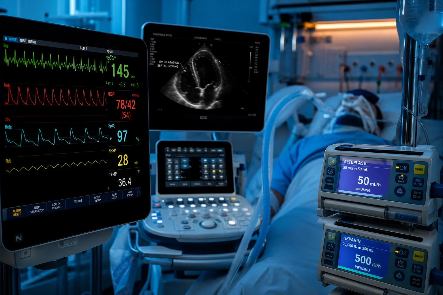
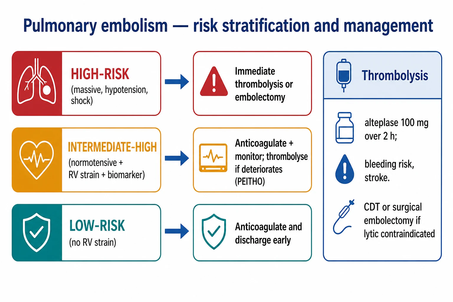
Pulmonary embolism is classified by haemodynamic impact. High-risk (massive) PE — shock, hypotension (SBP <90), or arrest — needs immediate systemic thrombolysis (alteplase 100 mg over 2 hours, or 50 mg bolus in cardiac arrest) or surgical/catheter embolectomy if lysis is contraindicated, with VA-ECMO as a bridge for refractory arrest. Intermediate-high (submassive) PE — normotensive with RV strain on echo and raised biomarkers — is treated with anticoagulation and close monitoring; the PEITHO trial (NEJM 2014) showed systemic lysis prevented haemodynamic decompensation but increased major bleeding and haemorrhagic stroke without a mortality benefit, so thrombolysis is reserved for deterioration. Catheter-directed thrombolysis and surgical embolectomy are options when systemic lysis is contraindicated. Anticoagulate with heparin then a DOAC for 3-6 months (provoked) or lifelong (unprovoked).
On this page & tools
Your progress
Saved locally on this device.
Target exams
Red flags
Definition and classification
Pulmonary embolism is the obstruction of the pulmonary arterial tree by material (usually thrombus, occasionally fat, air, amniotic fluid, or tumour) that has embolised — most often from a deep vein thrombosis of the lower limb or pelvis. The 2019 ESC guidelines classify PE by a two-step risk stratification that pairs the haemodynamic status (the first step, performed at the bedside) with markers of right-ventricular dysfunction (imaging) and myocardial injury (biomarkers).[1]
This classification has displaced the older loose terms "massive" and "submassive," although they persist in the exam and in conversation. The mapping is: [1]
- High-risk PE (≈ "massive") — haemodynamic instability: shock, hypotension (SBP <90 mmHg, or a drop of at least 40 mmHg from baseline sustained for >15 minutes), or cardiac arrest.
- Intermediate-risk PE (≈ "submassive") — normotensive, but with evidence of RV dysfunction and/or myocardial injury. Split into:
- Intermediate-high — both RV dysfunction (echo/CT) and positive biomarkers (troponin/BNP).
- Intermediate-low — either RV dysfunction or positive biomarkers (not both).
- Low-risk PE — normotensive, no RV dysfunction, no biomarker elevation; sPESI = 0. [1]
ESC risk stratification — click each stage (click each)
Intermediate-low PE
Normotensive with EITHER RV dysfunction OR raised biomarkers (not both). Treatment: anticoagulation. Monitor; usually ward-level care adequate. Low threshold for escalation if clinical change.
Risk stratification tools
PESI and sPESI (prognostic, not diagnostic)
The Pulmonary Embolism Severity Index (PESI) is an 11-variable prognostic score predicting 30-day mortality, derived and validated by Aujesky.[8] Its simplified form, sPESI (Jiménez, 2010), retains six equally weighted variables and is the workhorse of the ESC algorithm.[9]
Biomarkers and imaging (the second step)
Once haemodynamic stability is confirmed, the next step is to look for right-ventricular dysfunction and myocardial injury: [1]
- Troponin (I/T) and BNP / NT-proBNP — markers of RV pressure overload and myocyte stretch. Elevation stratifies risk and (when both are raised) upgrades the patient to intermediate-high.
- Echocardiography — the bedside test for RV dysfunction in the unstable and a risk tool in the stable. Look for RV dilatation, hypokinesis, the McConnell sign (free-wall hypokinesis with apical sparing), paradoxic septal motion / septal bowing (the D-shaped septum), tricuspid regurgitation, a plethoric IVC, and rarely a thrombus-in-transit.[7]
- CTPA — the diagnostic gold standard; an RV/LV diameter ratio >0.9 (or >1.0) on CT denotes RV dysfunction and indicates intermediate risk.
Step 1 — Haemodynamics
Bedside, first
- Shock, hypotension (SBP <90 or drop >=40 for >15 min), or arrest = HIGH-RISK
- Normotensive = proceed to step 2 (sPESI, biomarkers, imaging)
- This single step decides whether to lyse now or to work up
Step 2 — sPESI
Prognosis
- 0 = low risk (mortality ~1%)
- >=1 = not low risk — needs further stratification
- Used to select candidates for home treatment
Step 3 — RV + biomarkers
Sub-stratify
- RV dysfunction: echo (McConnell, septal bowing) or CT (RV/LV >0.9)
- Myocardial injury: troponin; stretch: BNP/NT-proBNP
- BOTH positive = intermediate-high; one positive = intermediate-low; none = low
Pathophysiology
The embolus produces two interlocking injuries: [1]
- Mechanical obstruction of the pulmonary arterial bed raises pulmonary vascular resistance. The right ventricle, a thin-walled chamber adapted to a low-pressure circuit, faces an acute afterload increase. It dilates, its wall tension and oxygen demand rise, and its coronary perfusion falls — producing ischaemic RV dysfunction. This is the RV "failing pump" of acute cor pulmonale.
- Hypoxaemia from ventilation-perfusion mismatch, shunt (perfusion of non-ventilated, injured units) and low mixed-venous oxygen (from the falling cardiac output). Hypoxaemia and acidosis further raise pulmonary vascular resistance, creating a decompensating spiral toward RV failure, systemic hypotension, and pulseless electrical activity (PEA) arrest. [1]
The physiology of the crashing RV
Clinical presentation
- Symptoms — sudden-onset dyspnoea (the most common), pleuritic chest pain, haemoptysis, cough, syncope/pre-syncope (a red flag for high-risk PE — it signals transient low cardiac output).
- Signs — tachypnoea, tachycardia, hypoxaemia (usually), a low-grade fever, signs of DVT (calf swelling/tenderness), raised JVP with a clear lung field (the obstructive-shock signature), and in the extreme, shock or PEA arrest.
- Predisposing factors — recent surgery or immobilisation, active cancer, pregnancy/postpartum, oestrogen therapy, obesity, smoking, inherited thrombophilia (factor V Leiden, antithrombin/protein C/S deficiency, antiphospholipid syndrome), prior VTE, long-haul travel. [1]
Investigations
Diagnostic pathway — stable vs unstable patient
Pre-test probability (Wells / YEARS)
Wells score stratifies PE likelihood (low/medium/high, or PE-likely/unlikely). Combined with D-dimer it safely rules out PE in low-probability patients without imaging. YEARS algorithm uses D-dimer with a single imaging decision point.
D-dimer
High sensitivity, low specificity. A normal D-dimer in a low-probability patient RULES OUT PE. A raised D-dimer is not diagnostic (infection, malignancy, pregnancy, surgery all raise it). Useless in the high-probability or already-unstable patient — do not wait.
CTPA (stable patient)
The gold standard — visualises filling defects in the pulmonary arteries down to the segmental level. Also gives RV/LV ratio (>0.9 = RV dysfunction) and an alternative diagnosis. Contrast load — caution in renal impairment.
Bedside echo (unstable patient)
The unstable patient cannot go to CT. RV dilatation + McConnell sign + septal bowing + plethoric IVC = acute cor pulmonale — treat as massive PE on this finding. A thrombus-in-transit (right heart) is pathognomonic when seen.
ECG
Sinus tachycardia is most common. S1Q3T3, right-axis deviation, right-bundle-branch block, T-wave inversion in V1-V4 (RV strain) — all non-specific but support the diagnosis. A rightward axis with dominant R in V1 raises suspicion.
Bloods
Troponin and NT-proBNP (for risk stratification), FBC/coagulation/renal/liver, lactate (a marker of low output). Group and hold if lysis/embolectomy likely. Pregnancy test in women of reproductive age.
V/Q scan (alternative)
Where CTPA is contraindicated (severe contrast allergy, renal failure, pregnancy). Ventilation-perfusion mismatch confirms PE. Requires a reasonable chest X-ray.
Management — high-risk (massive) PE
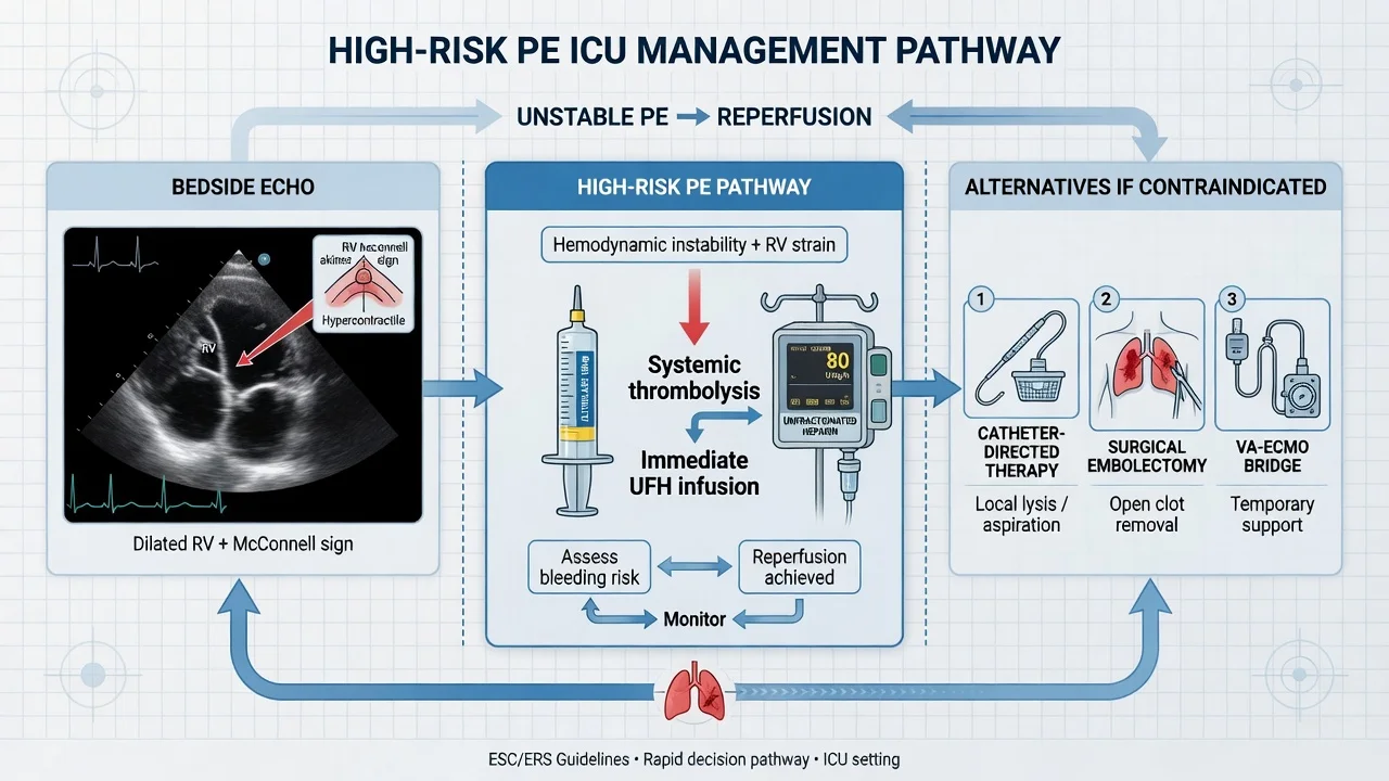
This is a time-critical, RV-salvage emergency. The priority is to restore the pulmonary circulation before the RV fails irreversibly.[1]
Massive PE — the first 60 minutes
1. Recognise and call for help
Obstructive shock or arrest + PE suspicion. Activate the PE response team (PERT). Do NOT delay for CTPA in the crashing patient — an echo is enough.
2. Confirm at the bedside (echo)
Bedside echocardiogram: RV dilatation, McConnell sign (free-wall hypokinesis, apical sparing), septal bowing (D-shaped septum), plethoric IVC. This is diagnostic of acute cor pulmonale in the right clinical context.
3. Give systemic thrombolysis
ALTEPLASE 100 mg IV over 2 hours (ESC-recommended for the perfusing high-risk patient). In CARDIAC ARREST, give a 50 mg IV bolus. If lysis contraindicated, go straight to embolectomy.
4. Anticoagulate with heparin
Unfractionated heparin (UFH) 80 U/kg bolus then 18 U/kg/h infusion — short half-life, reversible. Hold/omit the heparin during the alteplase infusion per local protocol. Do NOT use LMWH here.
5. Supportive — cautious fluids, vasopressors
Avoid large-volume fluids (worsens RV dilatation and septal shift, reduces LV preload). Small (250 mL) boluses only. Noradrenaline for vasopressor support; dobutamine/milrinone for the failing RV with caution.
6. Oxygenation and ventilation
High-flow oxygen; intubate cautiously (positive pressure drops venous return and preload in the RV-dependent circulation — be ready for peri-intubation arrest). Use lung-protective settings, avoid high intrathoracic pressure.
7. Rescue therapies if failing
Surgical embolectomy or catheter-directed thrombolysis/thrombectomy if lysis contraindicated/failed. VA-ECMO (ECPR) for refractory PEA arrest — a bridge to definitive clot removal.
Thrombolysis for massive PE
Alteplase (recombinant tissue plasminogen activator, rt-PA) is the thrombolytic of choice. The ESC-recommended regimen for a high-risk PE that is perfusing is 100 mg IV over 2 hours.[1] In cardiac arrest, where a 2-hour infusion is impractical, give a 50 mg IV bolus (some protocols use 50 mg as a single bolus, others repeat to 100 mg). Tenecteplase (weight-based single bolus) is an alternative where licensed.[4]
Systemic thrombolysis — the numbers
Management — intermediate-high (submassive) PE
The patient is normotensive but has RV dysfunction and raised biomarkers. The evidence-based management is anticoagulation plus close monitoring, with thrombolysis held in reserve.[1][2]
The decisive trial is PEITHO (Meyer, NEJM 2014): 1006 patients with intermediate-risk PE (RV dysfunction on echo/CT plus troponin/BNP elevation) were randomised to single-bolus tenecteplase versus placebo, on top of standard anticoagulation. Thrombolysis halved the primary outcome of haemodynamic decompensation at 7 days (1.6% vs 5.0%) but increased major bleeding (6.3% vs 1.5%) and haemorrhagic stroke (2.4% vs 0.2%), with no difference in mortality.[2] The earlier MAPPET-3 trial (Konstantinides, NEJM 2002) had shown alteplase reduced the need for treatment escalation in submassive PE.[3]
Conclusion: systemic thrombolysis is not routine for submassive PE. It is reserved for the patient who deteriorates haemodynamically — a falling blood pressure, rising lactate, or worsening RV function. The Chatterjee meta-analysis (JAMA 2014) confirmed the trade-off across 16 trials: a mortality benefit was offset by a large increase in major bleeding and intracranial haemorrhage.[4]
Catheter-directed thrombolysis vs systemic thrombolysis vs embolectomy
When full-dose systemic lysis is contraindicated, has failed, or carries an unacceptable bleeding risk, reperfusion strategies with less systemic fibrinolysis come into play. The choice is guided by local expertise, clot location, and bleeding risk.[5][6]
Systemic thrombolysis
Alteplase 100 mg/2 h
- First-line for HIGH-RISK (massive) PE that is perfusing; 50 mg bolus in arrest
- Rapid, no special equipment, available everywhere
- Highest bleeding risk: ~6% major bleed, ~2% intracranial haemorrhage
- Absolute contraindications exist (haemorrhagic stroke, active bleeding, recent surgery)
Catheter-directed lysis (CDT)
US-assisted, EKOS
- Lower-dose rt-PA delivered directly into the clot (total ~20-24 mg vs 100 mg)
- SEATTLE II: RV/LV ratio improved; no major ICH in intermediate/massive PE
- OPTALYSE-PE: 4-24 mg rt-PA over 2-6 h reduced clot burden with low bleeding
- For submassive PE with low bleeding risk, or massive PE with high systemic bleed risk
Surgical embolectomy
Open, cardiothoracic
- For large CENTRAL/saddle clot, lysis contraindicated or failed, thrombus-in-transit
- Requires cardiothoracic surgery + cardiopulmonary bypass — takes time to mobilise
- Choi 2020: in-hospital mortality ~20% but better than failing medical therapy in the right patient
- Also removes paradoxical emboli / right-heart thrombus
Catheter thrombectomy
Aspiration/rheolytic
- Mechanical clot removal (FlowTriever, AngioJet) — little or no lytic
- Attractive when lysis is absolutely contraindicated
- Emerging evidence; the PERFUSE/FLASH registries support safety
VA-ECMO for refractory PE-induced arrest
When massive PE produces refractory cardiac arrest (typically PEA — pulseless electrical activity) or shock unresponsive to lysis and vasopressors, veno-arterial ECMO — often as extracorporeal cardiopulmonary resuscitation (ECPR) — bypasses the obstructed pulmonary circuit, restoring systemic perfusion and unloading the failing RV. It is a bridge to definitive clot removal (embolectomy, CDT, or surgical pulmonary embolectomy), not a treatment of the clot itself.[11]
The systematic review by O'Malley (Resuscitation 2020) of extracorporeal life support for massive PE reported survival-to-discharge rates of roughly 50-70% in selected patients — striking for a condition with otherwise near-100% mortality in refractory arrest.[11]
Refractory PE arrest — the ECPR pathway
1. Standard ACLS + suspect PE
PEA arrest with a compatible history (DVT, perioperative, immobile). Give the 50 mg alteplase bolus early. Echo if feasible: acute cor pulmonale.
2. Activate ECPR
If ROSC not achieved and a reversible PE cause is identified, alert the ECMO team. VA-ECMO is most effective when cannulated within ~30-60 min of arrest.
3. Cannulate (VA)
Percutaneous femoral vein to femoral artery (or surgical central cannulation). Confirm flow, decompress the RV, restore perfusion.
4. Definitive clot removal
Once on ECMO with stable perfusion: surgical embolectomy (central clot) or catheter-directed thrombolysis/thrombectomy. VA-ECMO is a bridge, not the cure.
5. Wean
As RV function recovers (echo-guided) and clot burden resolves, wean VA-ECMO and decannulate. Anticoagulation continued throughout (heparin in the circuit).
Anticoagulation
Anticoagulation is the cornerstone of every PE, regardless of severity. It prevents clot propagation and recurrence; it does not dissolve the existing clot (that is the role of lysis or endogenous fibrinolysis).[1]
Unfractionated heparin (UFH)
When lysis/embolectomy likely
- 80 U/kg bolus then 18 U/kg/h infusion (weight-based), titrate to aPTT 1.5-2.5x
- Short half-life (~60 min), fully reversible with protamine — preferred when thrombolysis or surgery is planned
- Renal-safe; preferred in haemodynamic instability or renal impairment
- Use for high-risk (massive) PE and the peri-arrest
LMWH (enoxaparin)
Standard stable PE
- 1 mg/kg SC twice daily (1.5 mg/kg once daily if CrCl <30)
- Predictable, no monitoring, lower HIT risk than UFH
- Preferred over UFH for stable intermediate/low-risk PE (per ESC)
- NOT used during the systemic lysis infusion window
DOACs (rivaroxaban/apixaban)
Long-term
- Rivaroxaban 15 mg BD x3 weeks then 20 mg OD; Apixaban 10 mg BD x7 days then 5 mg BD
- EINSTEIN-PE (Büller, NEJM 2012): rivaroxaban non-inferior to warfarin+LMWH for PE, less major bleeding
- No monitoring, no dietary restriction — first-line for most stable PE
- Avoid in pregnancy, severe renal/liver impairment, antiphospholipid syndrome, active cancer (LMWH preferred in cancer)
Warfarin / fondaparinux
Selected
- Warfarin: overlap with parenteral heparin (5+ days) until INR 2-3; used where DOAC unsuitable
- Fondaparinux: 5/7.5/10 mg SC daily by weight; option in heparin allergy / HIT history
- Mandatory bridging from heparin for warfarin; not needed for DOACs
Duration of anticoagulation
- Provoked PE (transient/reversible risk — surgery, immobilisation, oestrogen): 3 months.[1]
- Unprovoked PE (no reversible trigger): at least 3 months, then reassess; most guidelines favour indefinite given high recurrence off anticoagulation.
- PE in cancer: indefinite while cancer is active (LMWH or a DOAC per guidelines).
- Recurrence on anticoagulation: investigate adherence, malignancy, antiphospholipid syndrome; consider dose escalation / LMWH.
The PE response team (PERT)
The Pulmonary Embolism Response Team (PERT) is a rapid-response multidisciplinary model (intensivist, haematology, interventional cardiology/radiology, cardiothoracic surgery, imaging, pharmacy) activated for the moderate-to-high-risk PE. Its aim is to coordinate risk stratification and reperfusion decisions rapidly — particularly the judgement-laden choices about CDT, embolectomy, and ECMO — and to avoid the delays inherent in serial single-specialty consultations. PERT activation is associated with faster decision-making and is recommended by the ESC for intermediate-high and high-risk PE where the reperfusion strategy is not straightforward.[1]
Evidence and landmark trials
PEITHO
NEJM 2014
1006 intermediate-risk PE (RV dysfunction + biomarkers) — single-bolus tenecteplase vs placebo, + anticoagulation
Key finding
Haemodynamic decompensation 1.6% vs 5.0% (p=0.002). Major bleeding 6.3% vs 1.5%; haemorrhagic stroke 2.4% vs 0.2%. No mortality difference.
Practice change
Systemic thrombolysis NOT routine for submassive PE — reserved for deterioration
MAPPET-3
NEJM 2002
256 submassive PE (RV strain on echo/ECG) — alteplase + heparin vs heparin alone
Key finding
Less treatment escalation (escalation to rescue lysis/ventilation) with alteplase (10% vs 25%, p=0.004). No mortality difference.
Practice change
Early evidence that lysis reduces escalation in submassive PE — set the stage for PEITHO
Chatterjee meta-analysis
JAMA 2014
16 RCTs, 2115 PE — thrombolysis vs anticoagulation alone
Key finding
Mortality reduced (1.4% vs 3.0%, OR 0.50). Major bleeding increased 2.9-fold; intracranial haemorrhage ~2%. Largest benefit in high-risk PE.
Practice change
Quantified the lysis risk-benefit — drives the restriction of lysis to high-risk PE
OPTALYSE-PE
JACC 2018
101 intermediate-risk PE — 4 arms of US-assisted catheter-directed lysis (rt-PA 4-24 mg over 2-6 h)
Key finding
Reduced clot burden and RV/LV ratio across all arms. Major bleeding low; no intracranial haemorrhage. Lowest-dose arm as effective.
Practice change
Lower-dose catheter lysis is effective for intermediate-risk PE — supports CDT
SEATTLE II
JACC 2015
150 massive + submassive PE — single-arm ultrasound-facilitated catheter-directed lysis (mean rt-PA ~24 mg)
Key finding
RV/LV ratio improved at 48h (1.92 to 1.53). Major bleeding 10%, but NO intracranial haemorrhage (vs ~2% with systemic lysis).
Practice change
CDT improves RV strain with markedly less intracranial bleeding than systemic lysis
EINSTEIN-PE
NEJM 2012
4833 symptomatic PE — rivaroxaban (15 mg BD x3wk then 20 mg OD) vs enoxaparin + warfarin
Key finding
Non-inferior for recurrent VTE (2.1% vs 1.8%). Major bleeding lower (1.1% vs 2.2%). No excess ICH.
Practice change
Single-drug DOAC (rivaroxaban) became first-line long-term therapy for PE
Complications
- Haemorrhagic complications of thrombolysis — the feared one is intracranial haemorrhage (~2% with systemic lysis); also major gastrointestinal and access-site bleeds. Manage with cessation of lysis + heparin, cross-clamping/clotting factors, and urgent imaging.
- Heparin-induced thrombocytopenia (HIT) — falling platelet count 5-10 days after heparin exposure; switch to argatroban/bivalirudin/fondaparinux.
- Reperfusion / re-perfusion pulmonary oedema — after CDT or embolectomy.
- Recurrent PE / clot propagation — anticoagulation failure; investigate adherence, malignancy, antiphospholipid syndrome, or an IVC filter need.
- Post-PE syndrome — persistent dyspnoea and functional limitation after PE (up to 50%); a precursor of CTEPH in some.
- Chronic thromboembolic pulmonary hypertension (CTEPH) — the late, progressive consequence of unresolved emboli (below). [1]
Follow-up — chronic thromboembolic pulmonary hypertension (CTEPH)
CTEPH develops in roughly 0.4-4% of PE survivors within 2 years. It is a treatable cause of pulmonary hypertension: organised, fibrous thrombus organises in the pulmonary arteries, raising pulmonary vascular resistance and causing progressive RV failure. Suspect it in any patient with persistent or new dyspnoea after a PE.[13]
CTEPH surveillance and management
1. Suspect — new dyspnoea after PE
Any patient with unexplained dyspnoea 2+ months after a PE warrants CTEPH screening — do not attribute it to deconditioning without investigation. Risk factors: larger perfusion defect, recurrent PE, elevated D-dimer, antiphospholipid syndrome, splenectomy, non-O blood group.
2. Screen — V/Q scan (NOT CTPA)
V/Q lung scan is the screening test of choice (at least one segmental mismatched perfusion defect = positive). Echocardiography estimates pulmonary artery pressure and RV function. Right-heart catheterisation confirms the haemodynamics (mPAP >20, PVR >2 WU).
3. Confirm disease — CT pulmonary angiography
CTPA + digital subtraction angiography defines the location and operability of the organised thrombus — essential for surgical planning.
4. Pulmonary endarterectomy (PEA)
Pulmonary (thrombo-)endarterectomy is the POTENTIALLY CURATIVE treatment for operable (proximal, segmental-level) disease. Performed on cardiopulmonary bypass with deep hypothermic circulatory arrest. Expert centre required.
5. Non-operative disease — BPA + riociguat
Balloon pulmonary angioplasty (BPA) for distal/inoperable disease. Riociguat (soluble guanylate cyclase stimulator) is the licensed medical therapy. Lifelong anticoagulation.
6. Lifelong anticoagulation
All CTEPH patients receive lifelong anticoagulation (warfarin traditionally, DOACs increasingly). Recurrence prevention is paramount.
Prognosis
PE outcomes by risk class
- Untreated PE mortality is ~30%; with anticoagulation it falls to under 8%. High-risk PE carries the bulk of mortality.[1]
- Recurrent PE risk off anticoagulation is ~50% at 1 year for unprovoked PE — the rationale for indefinite therapy.
- CTEPH develops in 0.4-4% within 2 years — screen symptomatic survivors.[13]
- Post-PE syndrome (persistent dyspnoea/exertional limitation) affects up to half of survivors and is under-recognised.
- Prognostic markers: sPESI, troponin, BNP, echo RV function, lactate, and the presence/extent of clot burden on CTPA.
Exam practice
SAQ — Massive PE in cardiac arrest
10 minutes · 10 marks
A 62-year-old woman (BMI 34, on oral oestrogen) is brought to the ED after a collapse at home. She is diestressed and then loses consciousness. Initial rhythm: sinus tachycardia deteriorating to PEA. Bedside echo during CPR shows a dilated right ventricle with free-wall hypokinesis and apical sparing, a D-shaped septum, and a plethoric IVC. End-tidal CO2 is 14 mmHg. She weighs 90 kg. ROSC has not been achieved after 8 minutes. There is no history of bleeding, stroke, or recent surgery.
Clinical pearls
Red flags
References
- [1]Konstantinides SV, Meyer G. The 2019 ESC Guidelines on the Diagnosis and Management of Acute Pulmonary Embolism Eur Heart J, 2019.PMID 31697840
- [2]Meyer G, Vicaut E, Danays T, Agnelli G, et al.; PEITHO Investigators. Fibrinolysis for patients with intermediate-risk pulmonary embolism N Engl J Med, 2014.PMID 24716681
- [3]Konstantinides S, Geibel A, Heusel G, Heinrich F, et al. Heparin plus alteplase compared with heparin alone in patients with submassive pulmonary embolism N Engl J Med, 2002.PMID 12374874
- [4]Chatterjee S, Chakraborty A, Weinberg I, Kadakia M, et al. Thrombolysis for pulmonary embolism and risk of all-cause mortality, major bleeding, and intracranial hemorrhage: a meta-analysis JAMA, 2014.PMID 24938564
- [5]Tapson VF, Sterling K, Jones N, Elder M, et al. A Randomized Trial of the Optimum Duration of Acoustic Pulse Thrombolysis Procedure in Acute Intermediate-Risk Pulmonary Embolism: The OPTALYSE PE Trial JACC Cardiovasc Interv, 2018.PMID 30025734
- [6]Piazza G, Hohlfelder B, Jaff MR, Ouriel K, et al. A Prospective, Single-Arm, Multicenter Trial of Ultrasound-Facilitated, Catheter-Directed, Low-Dose Fibrinolysis for Acute Massive and Submassive Pulmonary Embolism: The SEATTLE II Study JACC Cardiovasc Interv, 2015.PMID 26315743
- [7]McConnell MV, Solomon SD, Rayan ME, Come PC, et al. Regional right ventricular dysfunction detected by echocardiography in acute pulmonary embolism Am J Cardiol, 1996.PMID 8752195
- [8]Aujesky D, Obrosky DS, Stone RA, Auble TE, et al. Derivation and validation of a prognostic model for pulmonary embolism Am J Respir Crit Care Med, 2005.PMID 16020800
- [9]Jiménez D, Aujesky D, Moores L, Gómez V, et al. Simplification of the pulmonary embolism severity index for prognostication in patients with acute symptomatic pulmonary embolism Arch Intern Med, 2010.PMID 20696966
- [10]EINSTEIN–PE Investigators; Büller HR, Prins MH, Lensin AW, et al. Oral rivaroxaban for the treatment of symptomatic pulmonary embolism N Engl J Med, 2012.PMID 22449293
- [11]O'Malley TJ, Choi JH, Maynes EJ, Wood CT, et al. Outcomes of extracorporeal life support for the treatment of acute massive pulmonary embolism: A systematic review Resuscitation, 2020.PMID 31790756
- [12]Choi JH, O'Malley TJ, Maynes EJ, Weber MP, et al. Surgical Pulmonary Embolectomy Outcomes for Acute Pulmonary Embolism Ann Thorac Surg, 2020.PMID 32151576
- [13]Yang J, Madani MM, Mahmud E, Kim NH, et al. Evaluation and Management of Chronic Thromboembolic Pulmonary Hypertension Chest, 2023.PMID 36990148