ICU · Respiratory / ventilation
Pulmonary Hypertension & Cor Pulmonale
Also known as Pulmonary hypertension · PH · Cor pulmonale · Right ventricular failure · Acute cor pulmonale · Pulmonary arterial hypertension · PAH · WHO groups · CTEPH · Vasopressin for RV failure
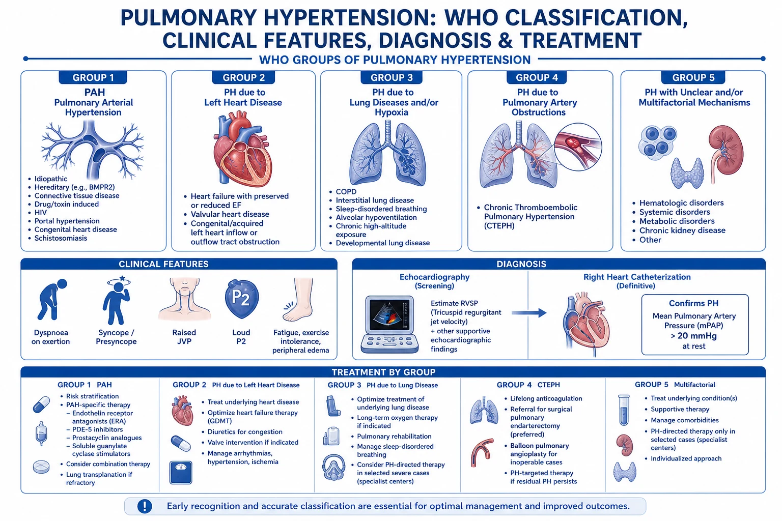
Pulmonary hypertension (a mean pulmonary artery pressure above 20 mmHg) is classified into five WHO groups: pulmonary arterial hypertension (group 1), left heart disease (2), lung disease and hypoxia (3), chronic thromboembolic (4), and multifactorial (5). Cor pulmonale is the right-ventricular hypertrophy, dilation, and failure from the pulmonary hypertension, classically from lung disease (group 3). The thin-walled right ventricle tolerates volume but not pressure: a rising pulmonary vascular resistance causes it to dilate and fail, the septum bows into the left ventricle (the D-shaped septum), the left-sided output falls, and tricuspid regurgitation worsens. The ICU management optimises the preload (diurese, do not overfill), reduces the afterload (inhaled nitric oxide, prostacyclin, PDE5 inhibitors), supports the contractility (milrinone, dobutamine), and maintains the systemic blood pressure to preserve the RV coronary perfusion — vasopressin is preferred, as it raises the systemic vascular resistance without raising the pulmonary.
On this page & tools
Your progress
Saved locally on this device.
Target exams
Overview & definition
Pulmonary hypertension (PH) is a mean pulmonary artery pressure (mPAP) above 20 mmHg (the 6th World Symposium, 2018, lowered the threshold from 25). It is classified into five WHO groups, which drive the cause-specific treatment. Cor pulmonale is the right-ventricular hypertrophy, dilation, and failure that results from the pulmonary hypertension — classically from chronic lung disease (group 3) but applicable to any cause. In the ICU, the patient with PH decompensates into acute or acute-on-chronic right-ventricular failure, a high-mortality state driven by a vicious cycle of rising afterload, falling output, and RV ischaemia.[1][1]
The WHO classification
| Group | Cause | Example |
|---|---|---|
| 1 | Pulmonary arterial hypertension (PAH) | Idiopathic, heritable, connective tissue disease (systemic sclerosis), congenital heart disease, portal hypertension, drugs |
| 2 | PH from left heart disease (postcapillary) | LV systolic/diastolic dysfunction, mitral/aortic valve disease |
| 3 | PH from lung disease and hypoxia | COPD, interstitial lung disease, sleep apnoea, chronic hypoventilation |
| 4 | Chronic thromboembolic PH (CTEPH) | Recurrent or organised pulmonary embolism |
| 5 | Multifactorial | Haematological, systemic, metabolic |
Cor pulmonale classically refers to group 3 (lung-disease PH), but the pathophysiology — RV overload from a high PVR — applies to any group.[1]
Group 1 — pulmonary arterial hypertension (PAH)
Pre-capillary PH (mPAP >20, PAWP ≤15, PVR >2 WU) in the absence of other causes of pre-capillary PH. The pulmonary arterioles show the plexiform lesion — a disordered proliferation of endothelial cells around a thin-walled channel. The 2022 ESC/ERS guidelines divide group 1 into a working clinical set:[2]
- Idiopathic PAH (IPAH) — formerly "primary pulmonary hypertension"; no identifiable cause. Female:male ~4:1, median age at diagnosis ~50 (a bimodal distribution in the elderly is now recognised). The prognosis untreated is dire (median survival ~2.8 years from diagnosis in the historic NIH registry).[2]
- Heritable PAH — autosomal-dominant with incomplete penetrance (~20%). BMPR2 mutations cause ~70% of familial PAH and ~20% of apparently sporadic IPAH. Other genes: ACVRL1 and ENG (hereditary haemorrhagic telangiectasia with juvenile-onset severe PAH), SMAD9, KCNK3, CAV1, EIF2AK4 (the latter causes pulmonary veno-occlusive disease).[2]
- Drug- and toxin-induced PAH — classically the anorexigens (fenfluramine, dexfenfluramine, benfluorex — all withdrawn) which produced an epidemic in the 1990s; also amphetamines, dasatinib (chronic myeloid leukaemia), l-tryptophan, some chemotherapeutics. Latency of months to years after exposure.[2]
- Associated PAH (APAH) — PAH occurring with a predisposing condition: connective tissue disease (systemic sclerosis carries the highest risk — screen annually with echocardiography; also SLE, mixed connective tissue disease, rheumatoid arthritis), HIV, portal hypertension (porto-pulmonary hypertension — a contraindication to liver transplant if severe, a reason to transplant if mild), congenital heart disease (the Eisenmenger physiology — long-standing left-to-right shunt with eventual reversal), and schistosomiasis (the most common cause of PAH worldwide in endemic regions, where eggs embolise to the pulmonary bed).[2]
- Group 1' — PVOD/PCH (pulmonary veno-occlusive disease and pulmonary capillary haemangiomatosis) — rare, distinguished by pulmonary oedema on imaging, nodular ground-glass changes, and prominent septal lines with a near-normal PAWP. PAH-targeted therapy causes pulmonary oedema — a critical pitfall.[2]
Group 2 — PH from left heart disease
The most common form of PH worldwide, and a post-capillary problem. The pulmonary venous pressure rises, transmits back to the capillaries and arteries, and — over time — a fixed, "reactive" increase in PVR develops (combined post- and pre-capillary PH). The 2022 ESC/ERS haemodynamic subtypes are:[2]
- Isolated post-capillary PH (IpcPH) — mPAP >20, PAWP >15, PVR ≤2 WU. Pure back-pressure.
- Combined post- and pre-capillary PH (CpcPH) — mPAP >20, PAWP >15, PVR >2 WU. The fixed remodelling makes this group harder to treat and identifies worse outcomes. Causes: LV systolic dysfunction (HFrEF), LV diastolic dysfunction / HFpEF (the commonest single driver — hypertension, diabetes, obesity, atrial fibrillation), mitral and aortic valve disease (especially mitral stenosis), and congenital/acquired cardiovascular causes (e.g., 3rd-degree AV block with pacemaker syndrome).[2]
Group 3 — PH from lung disease and hypoxia (the home of cor pulmonale)
The classification cor pulmonale traditionally points to. Chronic alveolar hypoxia drives hypoxic pulmonary vasoconstriction (the Euler-Liljestrand mechanism), medial hypertrophy and intimal fibrosis of the pulmonary arterioles, with secondary polycythaemia and volume overload. Subgroups:[2]
- Obstructive lung disease — COPD (mild PH is common in advanced COPD; a disproportionately high mPAP in a COPD patient should prompt a search for an additional cause — overlap, CTEPH, or sleep apnoea). Severe asthma is a rare cause.
- Restrictive lung disease — idiopathic pulmonary fibrosis (worse prognosis when PH develops), other interstitial lung diseases, pneumoconioses.
- Combined pulmonary fibrosis and emphysema (CPFE) — relatively preserved lung volumes but severely impaired DLCO, and disproportionately severe PH; a phenotype to recognise.
- Sleep-disordered breathing, alveolar hypoventilation syndromes — obesity hypoventilation, neuromuscular weakness, cervical cord injury. Often partially reversible with NIV.
- Chronic high-altitude exposure and developmental lung disorders. The therapy is the underlying disease plus long-term oxygen (the NOTT and MRC trials showed oxygen improves survival in hypoxaemic COPD). PAH-specific drugs are not recommended routinely and may worsen outcomes by worsening V/Q matching.[1][2]
Group 4 — chronic thromboembolic pulmonary hypertension (CTEPH)
CTEPH is the potentially curable PH. Organised, fibrous thrombus adheres to the pulmonary arterial wall and obstructs flow; in addition, a secondary vasculopathy develops in the unobstructed vessels (resembling PAH) and contributes to the raised PVR — which is why medical therapy can help even after surgery. CTEPH develops in roughly 0.4-4% of PE survivors within 2 years. Risk factors: larger or persistent perfusion defect on imaging, recurrent PE, antiphospholipid syndrome, splenectomy, non-O blood group, infected pacemaker leads / ventriculo-atrial shunt, thyroid replacement therapy. [1][2]
- Diagnosis: V/Q scan is the screening test (at least one segmental mismatched perfusion defect); CTPA defines the proximal/distal extent and operability; right-heart catheter confirms haemodynamics. Never attribute new dyspnoea after PE to deconditioning without a V/Q.
- Pulmonary endarterectomy (PEA) is the potentially curative treatment for proximal, operable disease — performed on cardiopulmonary bypass with deep hypothermic circulatory arrest at an expert centre. Operability is a surgical, not radiological, decision.
- Balloon pulmonary angioplasty (BPA) for surgically non-operable (distal) disease or residual post-PEA disease.
- Riociguat (sGC stimulator) — the only approved drug for inoperable or persistent/recurrent CTEPH (CHEST-1 trial).[7][2]
Group 5 — PH with unclear or multifactorial mechanisms
Heterogeneous. Haematological (chronic haemolytic anaemias — sickle cell disease affects ~10% of adults, myeloproliferative disorders, chronic thrombocythaemia), systemic (sarcoidosis, vasculitides), metabolic (glycogen storage disease, Gaucher, thyroid disorders), chronic renal failure on haemodialysis, and fibrosing mediastinitis. Management is of the underlying condition; PAH therapy is largely untested here.[2]
The 2022 ESC/ERS haemodynamic definitions (the new mPAP threshold)
The 6th World Symposium (2018, Nice) and the 2022 ESC/ERS guidelines lowered the diagnostic mPAP threshold from 25 to 20 mmHg (the old threshold missed early disease; a normal mPAP is now <20). PAWP is measured at end-expiration. A PVR >2 Wood Units defines pre-capillary PH. The complete set:[2]
Pre-capillary PH
Groups 1, 3, 4, 5
- mPAP >20 mmHg
- PAWP ≤15 mmHg
- PVR >2 Wood Units
- The pathology is in the pulmonary arteries/arterioles — responds to PAH-targeted therapy
Isolated post-capillary (IpcPH)
Group 2 — pure back-pressure
- mPAP >20 mmHg
- PAWP >15 mmHg
- PVR ≤2 Wood Units
- Treat the left heart — PAH drugs unproven and may harm
Combined post- and pre-capillary (CpcPH)
Group 2 — fixed remodelling
- mPAP >20 mmHg
- PAWP >15 mmHg
- PVR >2 Wood Units
- Worst of both — a fixed pulmonary vasculopathy atop a failing LV; diuresis + treat LV; PDE5i sometimes used
Exercise PH (new in 2022)
Replaces the old "borderline" group
- mPAP/CO slope >3 mmHg/L/min between rest and exercise
- A formal resting RHC may look normal — earlier diagnosis in symptomatic patients with normal resting pressures
The pathophysiology — why the RV fails
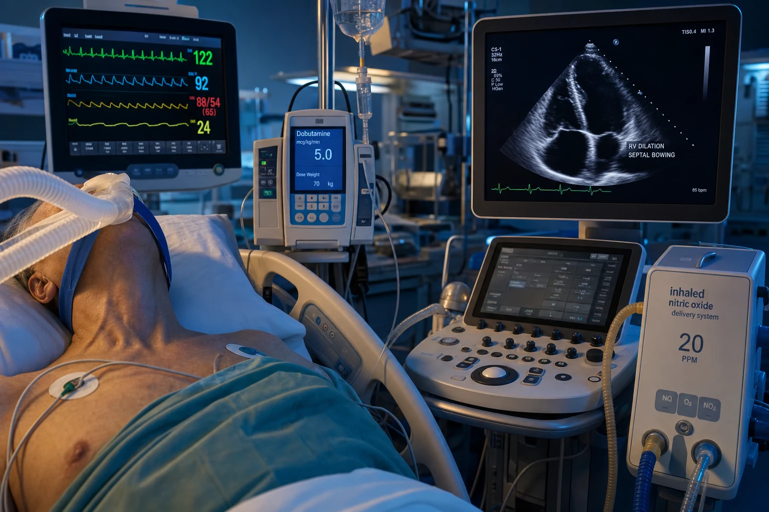
The right ventricle is thin-walled and volume-tolerant but pressure-intolerant:[1]
- A rising pulmonary vascular resistance (PVR) increases the RV afterload.
- The RV works harder, hypertrophies (chronic), then dilates and fails (acute or end-stage chronic).
- The interventricular septum bows into the LV (the D-shaped septum) — ventricular interdependence — reducing the LV preload and the systemic cardiac output.
- The dilating RV develops tricuspid regurgitation, worsening the venous congestion.
- The falling systemic pressure reduces the RV coronary perfusion (the RV perfuses in both systole and diastole at normal pressures, but at high RV pressures it becomes dependent on the aortic-to-RV pressure gradient) — the RV becomes ischaemic, which worsens its function, completing the vicious cycle.
- The result: a falling cardiac output, rising CVP, systemic congestion (hepatomegaly, ascites, peripheral oedema), hypoxaemia, and death if the cycle is not broken.[1]
Vascular-level mechanisms — why the pulmonary arteriole narrows
The clinical PVR is the integrated output of four overlapping vascular processes. Each is a therapeutic target:[2][11]
- Vasoconstriction — sustained smooth-muscle contraction driven by imbalanced vasoactive mediators: excess endothelin-1 (a potent constrictor and mitogen), deficient prostacyclin and nitric oxide (the vasodilators), and reduced soluble guanylate cyclase / cGMP signalling. The basis for endothelin receptor antagonists, prostacyclin analogues, PDE5 inhibitors, and sGC stimulators.[2]
- Vascular remodelling — medial hypertrophy of the smooth muscle, intimal fibrosis, and the pathognomonic plexiform lesion of PAH (a glomeruloid tuft of disorganised proliferating endothelial cells) — the result of disordered endothelial apoptosis and growth-factor signalling. Remodelling is slowly reversible with therapy and is the rationale for long-term combination treatment.
- In-situ thrombosis — endothelial injury promotes platelet aggregation and microthrombosis; the narrowed lumen amplifies the resistance. The basis for lifelong therapeutic anticoagulation in selected PAH (idiopathic/heritable) and all CTEPH.
- Inflammation — perivascular inflammatory infiltrates (macrophages, T-cells, mast cells) drive cytokine-mediated proliferation, prominent in connective-tissue-disease PAH and in schistosomiasis. The Euler-Liljestrand mechanism (hypoxic pulmonary vasoconstriction) is the normal physiological response that matches perfusion to ventilation; it becomes pathological in group 3 PH when chronic alveolar hypoxia sustains it. Unlike the systemic circulation, hypoxia constricts (not dilates) the pulmonary arteriole — the single most important physiology fact for the ICU patient, because a falling PaO₂ raises the PVR and worsens RV failure.[1]
The geometric cascade — RV ischaemia is the killing step
The chain from a raised PVR to cardiovascular collapse follows a predictable geometric sequence:[11]
- Raised PVR → raised RV afterload → RV pressure overload.
- RV dilates to maintain stroke volume (the Frank-Starling reserve of the thin wall is small) → tricuspid annulus stretches → functional tricuspid regurgitation.
- The interventricular septum flattens and bows into the LV in systole (the D-shaped / "D sign" septum on the parasternal short-axis echo) → reduced LV diastolic compliance → reduced LV preload → falling systemic cardiac output.
- The systemic pressure falls → the aortic-to-RV pressure gradient collapses → the RV, now operating at high wall stress, becomes ischaemic (RV perfusion occurs in both systole and diastole at normal RV pressures; at high RV pressures the systolic gradient reverses and the RV subendocardium is starved).
- Ischaemic RV → worse contractility → more dilatation → more TR → more septal shift → lower output → lower coronary perfusion → the vicious cycle of RV failure. Without intervention the cycle is fatal. Breaking any one link can rescue the patient: drop the PVR (iNO, prostacyclin, PDE5i), raise the systemic pressure (vasopressin — preserves the RV perfusion gradient), reduce the RV wall stress (inotropes, mechanical support), or restore sinus rhythm (the atrial kick).[1][11]
Triggers of acute decompensation
A patient with chronic PH decompensates when something increases the PVR or reduces the RV contractility:[1]
- Hypoxia and acidosis — both are potent pulmonary vasoconstrictors.
- Arrhythmia — atrial fibrillation is poorly tolerated (the failing RV depends on the atrial kick).
- Sepsis — the septic vasodilation drops the systemic pressure (reducing the RV perfusion) and the inflammatory injury impairs the RV.
- Excessive fluid — the volume-intolerant RV dilates further, the TR worsens.
- A new pulmonary embolism — an acute rise in PVR on top of the chronic PH.
- Positive-pressure ventilation — the increased intrathoracic pressure reduces the RV venous return and raises the RV afterload.[1]
Diagnosis — how to confirm and classify the PH
The diagnosis proceeds in two stages: (1) suspect PH (the screening test is echocardiography), and (2) confirm and characterise it (the gold standard is right-heart catheterisation, RHC). No PAH-specific therapy is started without an RHC.[2]
Echocardiography — the screening test
Echocardiography cannot diagnose PH (it estimates, not measures, pressure) but it raises the suspicion and assesses the RV. Key measurements:[2]
- Estimated RV systolic pressure (RVSP / PASP) — derived from the tricuspid regurgitation jet velocity using the modified Bernoulli equation (4v²) plus the estimated right atrial pressure. A TR jet >3.4 m/s (RVSP ≈50 mmHg) suggests PH. Caveats: underestimates in severe TR (the jet is "cut short"); overestimates in the elderly and athletes; impossible in ~10% (no TR jet).
- TAPSE (tricuspid annular plane systolic excursion) — the M-mode longitudinal excursion of the lateral tricuspid annulus; <17 mm signals RV systolic dysfunction. A simple, reproducible, prognostic measurement.
- RV dysfunction signs — RV dilatation (RV:LV basal diameter ratio >1), paradoxical septal motion / D-shaped septum on the parasternal short-axis view (the hallmark of RV pressure overload), McConnell's sign (free-wall hypokinesis with apical sparing — historically "specific" for acute PE but seen in any acute RV overload), reduced RV S' (<10 cm/s), reduced RV fractional area change (<35%), TAPSE/PASP ratio (RV-pulmonary coupling — a low ratio is a strong prognostic marker).
- Pericardial effusion and a plethoric IVC (no collapsibility) — markers of advanced, high-risk disease.
- Left-heart assessment — LV size and function, atrial size, valvular disease — to identify post-capillary (group 2) causes.
- The acute RV in the ICU — bedside echo is essential: a dilated RV with a D-shaped septum and a low TAPSE in the right clinical context is acute cor pulmonale.
Right-heart catheterisation (RHC) — the gold standard
RHC is mandatory before starting PAH-targeted therapy. It confirms the diagnosis, characterises pre- vs post-capillary PH, and allows a vasoreactivity test. Measured directly:[2]
- mPAP — the defining measurement (>20 mmHg = PH).
- PAWP (pulmonary artery wedge pressure) — distinguishes pre-capillary (≤15) from post-capillary (>15).
- Cardiac output / index (thermodilution or Fick) and PVR (PVR = (mPAP − PAWP) / CO, in Wood units) — >2 WU defines pre-capillary / combined disease.
- Right atrial pressure — a marker of RV failure; high RAP is a poor prognostic sign.
- Vasoreactivity test — inhaled nitric oxide (or IV epoprostenol or adenosine) at the time of RHC; a positive response (the Barst criteria: ≥10 mmHg fall in mPAP to ≤40 mmHg, with normal/high cardiac output) identifies the ~6-10% of IPAH/HPAH patients who respond to high-dose calcium-channel blockers — a uniquely favourable prognosis. Always repeat after 3 months; CCB non-responders must not be left on CCBs.[2]
- Discriminating PVOD — an RHC pattern of pre-capillary PH with a near-normal PAWP but clinical/imaging features of pulmonary oedema; do not start PAH therapy without recognising PVOD.[2]
Diagnostic workup of suspected pulmonary hypertension
1. Suspect PH
Exertional dyspnoea, fatigue, syncope (a late, pre-terminal sign of low output), chest pain (RV ischaemia), or peripheral oedema in a patient with a known risk factor (CTD, CHD, portal HTN, family history, prior PE, anorexigen exposure). Auscultation: a loud P2, a tricuspid regurgitation murmur, a RV heave. ECG: RV hypertrophy, RAD, RBBB, P pulmonale.
2. Echocardiography (screening)
Estimate RVSP from the TR jet velocity; assess RV size, TAPSE, septal motion, LV and valvular function. The echo quantifies the suspicion and the RV — it does not confirm PH. A normal echo in a high-suspicion patient does not exclude PH (underestimation is common).
3. Identify the cause — the second-line workup
PFTs + DLCO (obstructive/restrictive, low DLCO out of proportion), overnight oximetry / sleep study (OSA/OHS), V/Q scan (CTEPH — NOT CTPA as the screen), CT pulmonary angiography + high-resolution CT (define CTEPH operability and ILD), HIV serology, connective-tissue-disease screen (ANA, anti-centromere, anti-Scl-70), hepatitis and liver ultrasound (porto-pulmonary HTN), schistosomiasis serology in endemic areas, 6-minute walk test (functional capacity and prognosis), and cardiopulmonary exercise testing where available.
4. Right-heart catheterisation (confirm)
The mandatory gold standard before any PAH-targeted therapy. Confirms mPAP, PAWP, PVR, CO/CI, RAP; performs vasoreactivity testing. Classifies pre- vs post-capillary; identifies the CCB responder subset of IPAH/HPAH. NEVER start PAH drugs without an RHC.
5. Risk-stratify and assign therapy
Use the ESC/ERS three-stratum (low / intermediate / high) risk — derived from WHO functional class, 6MWD, BNP/NT-proBNP, RA area, CI, SVI, and mixed venous SvO2. Low risk → oral monotherapy or combination; high risk → initial triple therapy including parenteral prostacyclin. Refer early to a PH expert centre and a transplant centre.
Biomarkers and the ECG
- NT-proBNP / BNP — released from the strained RV; prognostic (high or rising = high risk; falling on therapy = good response). Use to monitor.
- Troponin — a marker of RV ischaemia; elevated in decompensated PH and prognostic.
- Urate, creatinine, bilirubin, hyponatraemia — each adds prognostic information (cardiorenal-hepatic congestion).
- ECG — RV hypertrophy (R/S ratio >1 in V1), right-axis deviation, right bundle branch block, P pulmonale (peaked P waves >2.5 mm in II), and right-heart strain patterns. A normal ECG does not exclude PH. [1]
Imaging — CT, V/Q, and cardiac MRI
- V/Q scan — the screening test of choice for CTEPH; one or more segmental mismatched perfusion defects is positive. CTPA is the next step to define the disease and assess operability.
- CTPA + high-resolution CT — defines the CTEPH extent (webs, bands, abrupt vessel cutoffs, systemic collateral supply), identifies ILD and CPFE, and flags PVOD/PCH (centrilobular ground-glass nodules, septal lines, mediastinal lymphadenopathy).
- Cardiac MRI — the gold standard for RV volumes, mass, and function (better than echo for the dilated RV); quantifies RV stroke volume, ejection fraction, and late gadolinium enhancement of the RV insertion points (a feature of chronic pressure overload).
Management — the five principles

- Optimise the preload. The failing RV is preload-dependent but volume-intolerant. Diurese the volume-overloaded patient (furosemide) to reduce the RV dilatation and the TR; give cautious fluid only to the clearly under-filled. Target a CVP of about 10-14 mmHg (not higher — congestion worsens the RV).[1]
- Reduce the afterload (the pulmonary vasodilators).
- Inhaled nitric oxide or inhaled prostacyclin — selective pulmonary vasodilators (no systemic hypotension).[1]
- IV prostacyclin (epoprostenol) — potent, but causes systemic vasodilation.
- PDE5 inhibitors (sildenafil) — oral; reduce the PVR.
- Endothelin receptor antagonists (bosentan) and soluble guanylate cyclase stimulators (riociguat) — for PAH and CTEPH.[1][1]
- Calcium channel blockers — only for the vasoreactive idiopathic PAH subset (responders on the vasodilator challenge at right-heart catheter).[1]
- Support the RV contractility (inotropes).
- Milrinone (PDE-3 — inotropy plus pulmonary and systemic vasodilation; the SVR fall may need a vasopressor).
- Dobutamine (beta-1 — contractility; also vasodilates).
- Levosimendan (calcium sensitiser).
- Adrenaline for the severe low-output state (raises the PVR via alpha — a drawback).[1]
- Maintain the systemic blood pressure — the RV coronary perfusion depends on the aortic-to-RV pressure gradient. If the systemic pressure falls, the RV is ischaemic. Vasopressin is preferred — it raises the systemic vascular resistance without raising the PVR (unlike noradrenaline, which raises both). Noradrenaline is a second-line; the alpha-1 pulmonary vasoconstriction is the drawback.[1]
- Maintain the sinus rhythm and correct the hypoxia and acidosis. Restore sinus rhythm (cardiovert the AF — the atrial kick matters). Give oxygen, ventilate, and correct the acidosis — both hypoxia and acidosis are pulmonary vasoconstrictors.[1]
Additional measures
- Avoid high intrathoracic pressure — if the patient is ventilated, use a low PEEP, a low plateau pressure, and a permissive-hypercapnia approach within reason (severe acidosis worsens the PVR).[1]
- Treat the underlying cause — CTEPH (pulmonary endarterectomy), PAH (specific therapy), LV failure (if postcapillary).[1]
- Mechanical support — VA-ECMO for refractory RV failure (a bridge to recovery or transplant), or the Impella RP (a percutaneous right-ventricular assist device).[1]
- An atrial septostomy — a palliative right-to-left shunt that decompresses the RV at the cost of systemic hypoxia, for selected refractory cases.[1]
Group-specific chronic therapy — the cause drives the drug
The five principles above manage the failing RV; the underlying cause is treated with group-specific chronic therapy. The general rule: PAH-targeted drugs are proven only for group 1 (and riociguat for group 4) — using them in groups 2 and 3 is largely ineffective or harmful.[2]
Group 1 (PAH) — combination therapy is now the standard
Three interacting pathways are targeted; the 2022 ESC/ERS guidelines recommend initial oral combination therapy (e.g., an ERA + a PDE5i) for most newly diagnosed low- or intermediate-risk patients, escalating to triple therapy (adding a prostacyclin) in high-risk disease:[2][3]
- Endothelin receptor antagonists (ERAs) — block the vasoconstrictor and mitogenic effects of endothelin-1. Bosentan (dual ETA/ETB; the original; LFT monitoring mandatory — hepatotoxicity), ambrisentan (selective ETA; once daily; no LFT monitoring needed), macitentan (dual; the SERAPHIN trial reduced the composite morbidity/mortality endpoint).[4][9]
- PDE5 inhibitors — block cGMP breakdown, augmenting NO signalling. Sildenafil (20 mg TDS; the SUPER-1 trial) and tadalafil (40 mg OD; longer half-life).[8]
- Prostacyclin pathway — mimics prostacyclin (vasodilation + anti-platelet + anti-proliferation). Epoprostenol (continuous IV; the only therapy proven to improve survival in IPAH — Barst 1996; central-line required, abrupt cessation causes rebound PH), treprostinil (IV/SC/inhaled/oral), iloprost (inhaled), selexipag (oral IP-receptor agonist; GRIPHON).[5][10]
- Soluble guanylate cyclase stimulators — riociguat stimulates sGC directly, augmenting cGMP independent of NO. Cannot be combined with PDE5 inhibitors (additive hypotension). PATENT-1 (PAH) and CHEST-1 (CTEPH).[6][7]
- Calcium channel blockers (high-dose nifedipine, diltiazem, amlodipine) — only for the ~6-10% vasoreactive responder subset of IPAH/HPAH, confirmed by repeat RHC at 3 months. Not used in APAH or non-responders.[2]
ERA — endothelin antagonists
Bosentan, ambrisentan, macitentan
- Block the mitogenic and vasoconstrictor endothelin pathway
- Bosentan: LFT monitoring monthly (hepatotoxicity, teratogenic)
- Macitentan (SERAPHIN): reduced morbidity/mortality in PAH
- Side effects: anaemia, peripheral oedema, headache, teratogenic (REMS programme)
PDE5 inhibitors
Sildenafil, tadalafil
- Augment NO–cGMP signalling — pulmonary vasodilation
- Sildenafil 20 mg TDS (SUPER-1); tadalafil 40 mg OD
- Side effects: headache, flushing, hypotension, visual disturbance
- Avoid with nitrates / riociguat (severe hypotension)
Prostacyclin pathway
Epoprostenol, treprostinil, iloprost, selexipag
- The most potent — vasodilation + anti-platelet + anti-proliferation
- IV epoprostenol is the ONLY therapy proven to improve survival in IPAH (Barst 1996)
- Drawback: continuous IV requires a central line — abrupt cessation causes rebound, fatal PH
- Selexipag (GRIPHON): oral IP-receptor agonist; the practical oral option
sGC stimulator
Riociguat
- Stimulates soluble guanylate cyclase directly (NO-independent cGMP)
- Approved for PAH (PATENT-1) AND CTEPH (CHEST-1)
- NEVER combined with a PDE5 inhibitor — additive hypotension
- Side effects: hypotension, GI, headache; contraindicated in pregnancy
Group 2 — left-heart disease
Treat the left-heart cause — optimise GDMT for HFrEF (ARNI, beta-blocker, MRA, SGLT2i), rate/rhythm control for AF, valve repair/replacement, or the underlying cardiomyopathy. Diurese congestion. PAH-specific drugs have not shown benefit and may worsen outcomes by dropping the systemic pressure; sildenafil is sometimes used in carefully selected CpcPH patients with a clear vasculopathic component, under specialist guidance.[2]
Group 3 — lung disease and hypoxia
Treat the underlying lung disease and correct hypoxia — long-term oxygen therapy (the NOTT and MRC trials: improved survival in hypoxaemic COPD, the only intervention proven to do so), NIV for chronic hypoventilation/OHS, immunosuppression for inflammatory ILD, transplantation referral for advanced ILD. PAH-targeted therapy is not routinely recommended — it worsens V/Q matching (vasodilating poorly ventilated lung units worsens shunt) and has shown no benefit. A subset with severe "out-of-proportion" PH may be referred to a PH centre for trial therapy.[1][2]
Group 4 — CTEPH
Always assess operability at an expert centre.[1][2]
- Pulmonary endarterectomy (PEA) — the potentially curative treatment for proximal (main, lobar, proximal segmental) operable disease. Performed on cardiopulmonary bypass with deep hypothermic circulatory arrest. Operability is a surgical, not radiological, decision. Lifelong anticoagulation after surgery.
- Balloon pulmonary angioplasty (BPA) — for distal/surgically inaccessible disease, residual PH post-PEA, or in patients unfit for surgery. Staged sessions; reperfusion oedema is the main complication.
- Riociguat (CHEST-1) — for inoperable disease, or persistent/recurrent PH after PEA/BPA. The only approved drug for CTEPH.[7]
- Lifelong anticoagulation in all CTEPH — the recurrence rate without it is high. Consider inferior vena cava filter only if anticoagulation fails or is contraindicated.
Group 5 — multifactorial
Treat the underlying condition (e.g., hydroxyurea and exchange transfusion for sickle-cell-related PH, immunosuppression for sarcoid, treatment of myeloproliferative disorder). PAH-targeted therapy is largely untested; specialist referral on a case-by-case basis.[2]
Transplant and palliation
- Bilateral lung transplantation (or heart-lung for complex congenital disease) is the final option for progressive disease despite optimal therapy — early referral is essential given the long wait-list times. Post-transplant, the RV often recovers as the afterload normalises.[2]
- Atrial septostomy — a palliative, graded right-to-left atrial shunt decompressing the RV at the cost of systemic hypoxia; for selected refractory cases or as a bridge to transplant.
The ICU patient with decompensated PH — a focused approach
When a patient with known PH (or newly suspected) presents to the ICU in acute RV failure, the structured approach below keeps the priorities in order. The single most common error is fluid-loading the failing RV.[1][11]
The first hour in decompensated pulmonary hypertension
1. Recognise and assess
Hypoxaemia, hypotension, tachycardia, raised JVP, cool peripheries, oliguria, a gallop, a loud P2, a TR murmur, peripheral oedema. Bedside echo: dilated RV, D-shaped septum, low TAPSE, plethoric IVC, often TR. The picture is right-sided shock with congestion.
2. Correct hypoxia and acidosis (the PVR drivers)
Oxygen to target SpO2 ≥92-96% (hypoxic pulmonary vasoconstriction raises the PVR). Ventilate the failing patient with low PEEP (high PEEP compresses the pulmonary vasculature) and lung-protective settings. Correct severe acidosis — bicarbonate for pH <7.15 if ventilation is controlled.
3. Diurese, do NOT fluid-load
The failing RV is volume-intolerant. IV furosemide (often a continuous infusion) to decongest; reassess with echo and the JVP. A cautious 250-500 mL crystalloid bolus only in the clearly under-filled (use dynamic indices, not the CVP alone). Excess fluid dilates the RV further and shifts the septum.
4. Restore and maintain sinus rhythm
Atrial fibrillation drops the cardiac output abruptly — the failing RV needs the atrial kick. Chemical (amiodarone) or electrical cardioversion early. Avoid pure negative inotropes (e.g., non-dihydropyridine CCBs, high-dose beta-blockers).
5. Reduce the RV afterload
Inhaled nitric oxide (5-20 ppm) or inhaled prostacyclin (e.g., nebulised epoprostenol/iloprost) — selective pulmonary vasodilation, no systemic hypotension. Continue the patient's chronic PAH drugs (do NOT stop); consider IV prostacyclin in the most severe.
6. Support the RV contractility and systemic pressure
Milrinone or dobutamine for inotropy (both vasodilate — expect to need a vasopressor). Vasopressin (preferred — raises SVR without raising PVR) or noradrenaline as second-line. Target a MAP that preserves RV coronary perfusion (typically ≥65 mmHg, individualised).
7. Mechanical support and the cause
Refractory shock → VA-ECMO (bridge to recovery, decision, or transplant) or Impella RP. Simultaneously treat the trigger — sepsis, PE, arrhythmia, pneumonia, anaemia, non-adherence to PAH drugs. Refer early to the PH and transplant centres.
Landmark trials in pulmonary hypertension
AMBITION
NEJM 2015
500 treatment-naïve PAH — initial ambrisentan + tadalafil combo vs either monotherapy
Key finding
First treatment-failure composite (death, hospitalisation, progression, unsatisfactory response) reduced by 50% with initial combination vs monotherapy (18% vs 31%, p=0.0002). Hospitalisation for worsening PAH 2% vs 4-8%.
Practice change
Initial oral combination (ERA + PDE5i) became first-line for most newly diagnosed PAH — replacing stepwise monotherapy
SERAPHIN
NEJM 2013
742 PAH — macitentan (3 mg or 10 mg) vs placebo, mean ~2 years follow-up
Key finding
Composite morbidity/mortality endpoint reduced with macitentan 10 mg (31.4% vs 46.4%, HR 0.55). One of the first PAH trials powered for long-term outcome.
Practice change
Macitentan (dual ERA) became a standard oral therapy — confirmed a morbidity/mortality benefit
GRIPHON
NEJM 2015
1156 PAH (the largest PAH RCT to date) — selexipag (oral IP-prostacyclin agonist) vs placebo, titrated to 1600 mcg BD
Key finding
Composite morbidity/mortality reduced (27% vs 41.6%, HR 0.60). Benefit across functional classes and background therapies.
Practice change
Selexipag — the practical oral prostacyclin — added to combination therapy; enabled oral triple therapy
PATENT-1
NEJM 2013
443 PAH — riociguat (sGC stimulator) vs placebo over 12 weeks
Key finding
6MWD improved by 36 m; PVR fell 22%; NT-proBNP fell. Benefit in treatment-naïve and pre-treated patients.
Practice change
Riociguat approved for PAH — the first sGC stimulator; cannot be combined with PDE5 inhibitors
CHEST-1
NEJM 2013
261 inoperable or persistent CTEPH — riociguat vs placebo over 16 weeks
Key finding
6MWD improved by 46 m; PVR fell ~25%; NT-proBNP fell. The first drug shown to help CTEPH.
Practice change
Riociguat approved for inoperable or persistent/recurrent CTEPH — the ONLY approved drug for CTEPH
SUPER-1
NEJM 2005
278 PAH — sildenafil (20, 40, 80 mg TDS) vs placebo over 12 weeks
Key finding
6MWD improved by 45-50 m across all doses; no dose-response — 20 mg TDS is the dose. Improved haemodynamics and QoL.
Practice change
Sildenafil became the first oral PDE5 inhibitor for PAH; the 20 mg TDS dose adopted
BREATHE-1
NEJM 2002
213 PAH — bosentan (dual ERA) vs placebo over 16 weeks
Key finding
6MWD improved by 44 m with bosentan; placebo fell by 8 m. Improved Borg dyspnoea and WHO functional class.
Practice change
Bosentan — the first oral PAH drug — transformed PAH from untreatable to a chronic disease; LFT monitoring required
Barst — epoprostenol
NEJM 1996
81 primary pulmonary hypertension — continuous IV epoprostenol vs conventional therapy
Key finding
12-week 6MWD improved with epoprostenol (+32 m) and fell with placebo (-29 m). Survival improved (0 vs 8 deaths at 12 weeks).
Practice change
Continuous IV epoprostenol became the first therapy proven to IMPROVE SURVIVAL in IPAH — the foundational RCT
SAQ — Decompensated pulmonary arterial hypertension in systemic sclerosis
10 minutes · 10 marks
A 52-year-old woman with limited cutaneous systemic sclerosis (CREST syndrome) and known WHO Group 1 pulmonary arterial hypertension presents to ICU with a 5-day history of progressive dyspnoea, abdominal distension and syncope. On examination she is cool, clammy and cyanotic: HR 118 in atrial flutter with variable block, BP 84/52, JVP distended to the angle of the jaw, a loud pulmonary component to the second heart sound, a tricuspid regurgitation murmur, a right ventricular heave, hepatomegaly and marked peripheral oedema. SpO2 88 per cent on 10 L oxygen via Hudson mask. Bedside echocardiography shows a severely dilated right ventricle (RV to LV basal diameter ratio 1.4) with a D-shaped interventricular septum bowing into the LV throughout the cardiac cycle, TAPSE 10 mm, severe tricuspid regurgitation, a small underfilled LV, a pericardial effusion (1 cm) and a plethoric non-collapsible IVC. Lactate 4.8 mmol/L, creatinine 168 (baseline 90), NT-proBNP 8500.
SAQ — Chronic thromboembolic pulmonary hypertension after pulmonary embolism
10 minutes · 10 marks
A 58-year-old man presents with 4 months of progressive exertional dyspnoea (now NYHA III) and two episodes of exertional presyncope. Eighteen months ago he was admitted with a bilateral proximal pulmonary embolism, treated with apixaban for 6 months then stopped. He is a non-smoker with no other past history. On examination: BP 124/78, HR 96 in sinus rhythm, JVP raised at 6 cm, loud P2, tricuspid regurgitation murmur, right ventricular heave. ECG shows right-axis deviation, R/S ratio greater than 1 in V1, and P pulmonale. Echocardiography estimates RV systolic pressure at 78 mmHg with a dilated RV, TAPSE 14 mm, and D-shaped septum. NT-proBNP 680.
Clinical pearls for the exam and the bedside
[1]Red flags
When to refer and to whom
- To a pulmonary hypertension expert centre — any newly diagnosed or suspected PH, for RHC, classification, and the initiation of PAH-targeted therapy. PAH drugs should not be started in a non-expert setting.[2]
- To a CTEPH team (PEA surgeon + PH expert) — any confirmed CTEPH, for an operability assessment. Operability is a surgical decision, not a radiological one.[2]
- To a transplant centre — early referral for progressive disease on optimal therapy, given the long wait-list times and the unpredictable decompensation trajectory. Bilateral lung (or heart-lung) transplant is the final option.[2]
- To obstetric medicine — any PH patient contemplating or discovered to be pregnant, for multidisciplinary planning.[2]
Summary — the ten things to remember
- PH is mPAP >20 mmHg; pre-capillary adds PVR >2 WU and PAWP ≤15. The 2022 ESC/ERS change.[2]
- The five WHO groups drive the therapy. Group 1 (PAH) and group 4 (CTEPH) get specific drugs/surgery; groups 2 and 3 are treated by the underlying cause.[2]
- The thin-walled RV tolerates volume but not pressure — the central pathophysiology.[1]
- Echo screens; RHC confirms. No PAH drug without an RHC.[2]
- V/Q screens for CTEPH; PEA is potentially curative.[7]
- The ICU management — preload, afterload, contractility, systemic pressure, rhythm.[1]
- Vasopressin is the preferred vasopressor — raises SVR without the PVR.[1]
- Inhaled NO / inhaled prostacyclin are the selective pulmonary vasodilators.[1]
- Hypoxia and acidosis are pulmonary vasoconstrictors — correct them first.[1]
- Refer early — PH centre for therapy, transplant centre for progression.[2]
References
- [1]Ware LR, Kim CS, et al. A Narrative Review on the Administration of Inhaled Prostaglandins in Critically Ill Adult Patients With Acute Respiratory Distress Syndrome Ann Pharmacother, 2024.PMID 37589097
- [2]Humbert M, Kovacs G, Hoeper MM, et al. 2022 ESC/ERS Guidelines for the diagnosis and treatment of pulmonary hypertension Eur Respir J, 2023.PMID 36028254
- [3]Galie N, Barbera JA, Frost AE, et al. (AMBITION) Initial Use of Ambrisentan plus Tadalafil in Pulmonary Arterial Hypertension N Engl J Med, 2015.PMID 26308684
- [4]Pulido T, Adzerikho I, Channick RN, et al. (SERAPHIN) Macitentan and morbidity and mortality in pulmonary arterial hypertension N Engl J Med, 2013.PMID 23984728
- [5]Sitbon O, Channick R, Chin KM, et al. (GRIPHON) Selexipag for the Treatment of Pulmonary Arterial Hypertension N Engl J Med, 2015.PMID 26699168
- [6]Ghofrani HA, Galie N, Grimminger F, et al. (PATENT-1) Riociguat for the treatment of pulmonary arterial hypertension N Engl J Med, 2013.PMID 23883378
- [7]Ghofrani HA, D'Armini AM, Grimminger F, et al. (CHEST-1) Riociguat for the treatment of chronic thromboembolic pulmonary hypertension N Engl J Med, 2013.PMID 23883377
- [8]Galie N, Ghofrani HA, Torbicki A, et al. (SUPER-1) Sildenafil citrate therapy for pulmonary arterial hypertension N Engl J Med, 2005.PMID 16291984
- [9]Rubin LJ, Badesch DB, Barst RJ, et al. (BREATHE-1) Bosentan therapy for pulmonary arterial hypertension N Engl J Med, 2002.PMID 11907289
- [10]Barst RJ, Rubin LJ, Long WA, et al. A comparison of continuous intravenous epoprostenol (prostacyclin) with conventional therapy for primary pulmonary hypertension N Engl J Med, 1996.PMID 8532025
- [11]Price LC, Wort SJ, Finney SJ, Marino PS, Brett SJ Pulmonary vascular and right ventricular dysfunction in adult critical care: current and emerging options for management: a systematic literature review Crit Care, 2010.PMID 20858239