ICU · Respiratory / ventilation
Pulmonary Oedema — Cardiogenic vs ARDS
Also known as Pulmonary oedema · Cardiogenic pulmonary oedema · Non-cardiogenic pulmonary oedema · ARDS vs cardiogenic · PAWP · Pulmonary artery wedge pressure · BNP · CPAP for pulmonary oedema · 3CPO trial · Bat-wing infiltrates
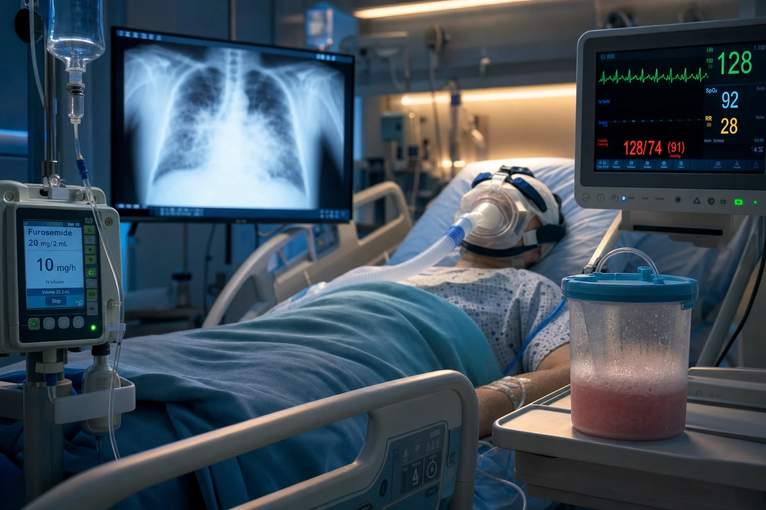
Pulmonary oedema is fluid in the alveoli from one of two mechanisms: cardiogenic (hydrostatic — raised pulmonary capillary pressure from left-heart failure, PAWP over 18 mmHg) or non-cardiogenic (increased permeability — ARDS, a damaged alveolar-capillary membrane, PAWP under 18 mmHg). The distinction is made on the PAWP, the BNP, the echocardiogram, and the CXR pattern. Cardiogenic oedema is treated with CPAP/NIV, diuretics, and vasodilators; ARDS is treated with lung-protective ventilation, PEEP, proning, and a conservative fluid strategy. The oedema-fluid-to-serum protein ratio distinguishes them at the bedside (under 0.65 cardiogenic, over 0.65 ARDS).
On this page & tools
Your progress
Saved locally on this device.
Target exams
Overview & definition
Pulmonary oedema is fluid in the alveoli from one of two mechanisms:[2]
- Cardiogenic (hydrostatic) — the left-heart pressure rises (LV failure, mitral valve disease, volume overload) and pushes fluid out of the capillaries into the alveoli, despite an intact alveolar-capillary membrane. The pulmonary artery wedge pressure (PAWP) is over 18 mmHg.
- Non-cardiogenic (increased permeability — ARDS) — the alveolar-capillary membrane is damaged (by sepsis, pneumonia, aspiration, trauma), and protein-rich fluid leaks into the alveoli, despite a normal or low PAWP (under 18 mmHg).
The distinction drives the treatment — cardiogenic oedema responds to diuresis and CPAP; ARDS does not, and is treated with lung-protective ventilation and PEEP.[2]
The diagnostic distinction
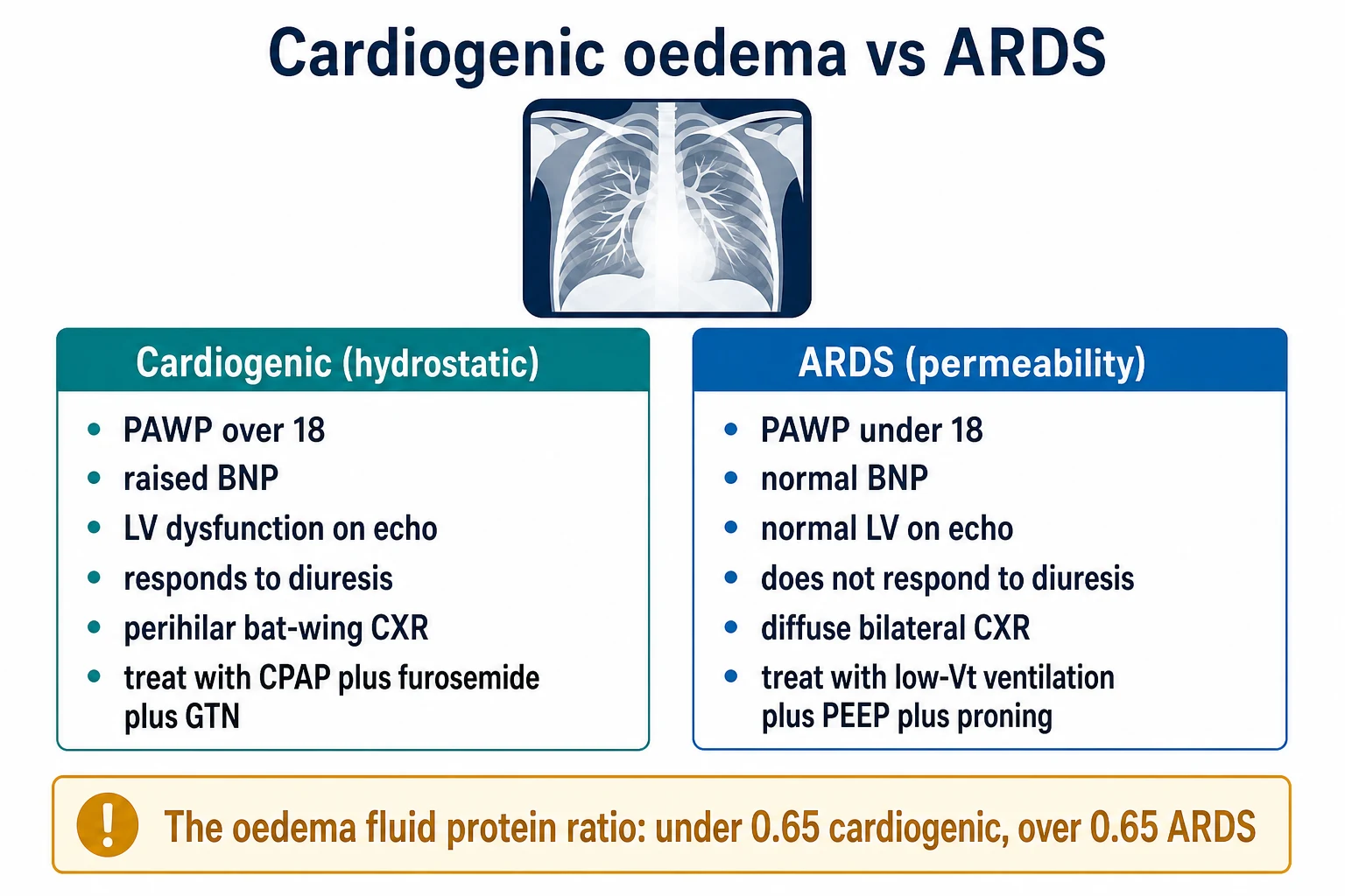
| Feature | Cardiogenic (hydrostatic) | ARDS (permeability) |
|---|---|---|
| PAWP | Over 18 mmHg | Under 18 mmHg |
| Onset | Gradual (hours) | Acute (within 7 days of a known insult) |
| CXR | Perihilar bat-wing, cardiomegaly, pleural effusions, Kerley B lines | Diffuse bilateral infiltrates, no cardiomegaly, no effusions |
| BNP | High (over 400 pg/mL) | Low or normal (under 200 pg/mL) |
| Echo | LV dysfunction, raised filling pressures (E/e-prime) | Normal LV (but may show RV dilatation) |
| Response to diuresis | Improves | Does not improve |
| Oedema-fluid-to-serum protein ratio | Under 0.65 (a low-protein transudate) | Over 0.65 (a high-protein exudate — the barrier leaks protein) |
| Cause | LV failure, valve disease, volume overload | Sepsis, pneumonia, aspiration, trauma, pancreatitis |
The bedside oedema-fluid-to-plasma protein ratio (if suctioned from the ETT) is under 0.65 for cardiogenic (a transudate — the intact barrier holds back protein) and over 0.65 for ARDS (a protein-rich exudate — the damaged barrier leaks it).[2]
Cardiogenic pulmonary oedema
The management of cardiogenic pulmonary oedema reduces the preload, the afterload, and the pulmonary congestion:[1][2]
- Oxygen — high-flow, for the hypoxaemia.
- CPAP/NIV — CPAP is first-line: it recruits collapsed alveoli, reduces the work of breathing, and the positive intrathoracic pressure reduces the venous return (preload) and the LV afterload. The 3CPO trial (NEJM 2008) found CPAP and NIV both effective (reduced intubation and the breathlessness), with no clear superiority of BiPAP over CPAP.[1]
- Diuretics — furosemide IV (reduces the preload, the pulmonary congestion, and the hydrostatic pressure; also a venodilator in the acute phase).
- Vasodilators — glyceryl trinitrate (GTN) if the patient is hypertensive (reduces the preload and the afterload); do NOT use if hypotensive.
- Inotropes and mechanical support — for the low-output cardiogenic oedema (cardiogenic shock): dobutamine, milrinone, IABP, Impella, or VA-ECMO (see the cardiogenic shock topic).
- Treat the cause — ACS (revascularisation), atrial fibrillation (rate control), valvular (surgery).[2]
ARDS (non-cardiogenic) — already covered
ARDS does not respond to diuresis (the barrier is broken, not the pressure). The management is lung-protective ventilation (low Vt, adequate PEEP), proning in severe disease (PROSEVA), a conservative fluid strategy (the FACTT trial — a slightly negative fluid balance improves oxygenation and reduces ventilator days), and ECMO for refractory cases (EOLIA). See the ARDS and refractory-hypoxaemia adjuncts topics for the full strategy.[2]
[1]SAQ — Flash pulmonary oedema from acute coronary syndrome
10 minutes · 10 marks
A 72-year-old man is brought to the emergency department by ambulance at 0300 with sudden onset of severe dyspnoea, orthopnoea and a cough producing pink frothy sputum. He has a history of hypertension and type 2 diabetes. On arrival he is diaphoretic, terrified, sitting upright and gasping: RR 38 with accessory-muscle use, SpO2 84 per cent on a 15 L non-rebreather mask, BP 210/118, HR 128 in sinus tachycardia, bilateral coarse inspiratory crackles to the apices and widespread expiratory wheeze. ECG shows 4 mm of ST elevation in leads V1 to V4 with reciprocal depression in II, III and aVF. CXR shows a normal-sized cardiac silhouette with perihilar bat-wing infiltrates, Kerley B lines and small bilateral pleural effusions. Venous blood gas: pH 7.22, lactate 4.2 mmol/L. Troponin pending.
SAQ — Cardiogenic oedema versus ARDS in a septic, hypoxic patient
10 minutes · 10 marks
A 68-year-old woman is admitted to ICU with severe community-acquired pneumonia and septic shock. On day 2 she develops progressive hypoxaemia (PaO2/FiO2 130 on 15 L oxygen via non-rebreather) and bilateral diffuse infiltrates on CXR. She is on noradrenaline 0.3 mcg/kg/min for MAP 65, has received 4.5 L of crystalloid in the first 24 hours, and her lactate has fallen from 5.2 to 2.4 mmol/L. Her past history includes hypertension and ischaemic heart disease. Bedside echocardiography shows a left ventricular ejection fraction of 35 per cent with regional wall motion abnormalities, E/e-prime 16, and a small pericardial effusion. NT-proBNP returns at 4200 pg/mL. The team is divided on whether she has cardiogenic pulmonary oedema or ARDS.
Red flags
Pathophysiology — the Starling equation decides where the fluid goes
Pulmonary oedema is, at its core, an imbalance between the forces pushing fluid OUT of the pulmonary capillaries and the forces keeping it IN. The Starling equation governs transcapillary fluid flux:[2]
Net fluid flux = Kfc × [ (Pc - Pi) − σ (πc - πi) ]
| Term | Meaning | What raises it (favours oedema) |
|---|---|---|
| Pc (capillary hydrostatic pressure) | The pressure pushing fluid OUT of the capillary into the interstitium/alveolus | Cardiogenic oedema — raised left-heart pressures transmit backwards to the pulmonary capillaries |
| Pi (interstitial hydrostatic pressure) | A slightly negative pressure pulling fluid out | Decreased by alveolar collapse; small effect |
| πc (capillary oncotic pressure) | Albumin holding fluid IN the vessel | Hypoalbuminaemia lowers it (contributes, rarely the sole cause) |
| πi (interstitial oncotic pressure) | Small, pulls fluid out | Raised by protein leaking in (ARDS) |
| Kfc (filtration coefficient) | How leaky the membrane is per unit pressure | ARDS — inflammatory damage massively increases Kfc |
| σ (reflection coefficient) | How well the membrane holds back protein (1 = perfect barrier, 0 = freely leaky) | ARDS — σ falls toward 0; protein gushes out |
The two mechanisms act on DIFFERENT terms:
- Cardiogenic raises Pc (hydrostatic) while the membrane is intact (σ ≈ 1, Kfc normal) — the fluid is a low-protein transudate.
- ARDS (permeability) raises Kfc and lowers σ (the barrier is destroyed) while Pc is normal or low — the fluid is a high-protein exudate. [1]
This is why PAWP and the oedema-fluid protein ratio line up so neatly: cardiogenic oedema is a pressure problem with an intact filter; ARDS is a broken filter at normal pressure.[2]
The three stages of cardiogenic oedema formation
- Interstitial oedema — fluid first accumulates in the peribronchovascular interstitial space (cuffing), seen as Kerley B lines (short horizontal lines at the lung bases, abutting the pleura) and subpleural fluid. Lymphatic drainage increases up to 10-fold to compensate.
- Alveolar flooding — once the lymphatic capacity is overwhelmed and interstitial pressure exceeds alveolar pressure (~18-25 mmHg Pc), fluid pours into the alveoli, producing the classic perihilar bat-wing pattern and pink frothy sputum.
- Airway flooding — frothy, blood-tinged secretum in the ETT/mask; gas exchange collapses as alveoli fill. [1]
The three zones of West and why cardiogenic oedema is basilar/bat-wing
Blood flow is greatest in the dependant (basal) lung (West zone 3, where Pa > Pv > PA). Because Pc is highest at the bases, hydrostatic oedema accumulates there first, producing the perihilar bat-wing and Kerley B pattern. ARDS, by contrast, is a diffuse barrier injury — infiltrates are bilateral and uniform, not gravity-concentrated (though dependent regions still tend to be worse because of perfusion and atelectrauma). [1]
The role of BNP (brain natriuretic peptide)
BNP (and the inactive fragment NT-proBNP) is released by ventricular myocytes in response to wall stretch (volume/pressure overload). It is the single best biochemical discriminator between cardiogenic and non-cardiogenic pulmonary oedema.[7]
| BNP (pg/mL) | Interpretation | Likelihood |
|---|---|---|
| Under 100 | Heart failure UNLIKELY | >90% negative predictive value — points strongly to a non-cardiac cause (ARDS, PE, pneumonia) |
| 100-400 | Grey zone | Possible heart failure; need echo and clinical context (renal failure, obesity, and older age all raise BNP independently) |
| Over 400 | Heart failure LIKELY | Points to cardiogenic oedema |
The Breathing Not Properly study (the BNP evidence base)
Breathing Not Properly 2002 — BNP for the diagnosis of heart failure (PMID 11919308)
Design
Multicentre prospective study, 1586 patients presenting to ED with acute dyspnoea, BNP measured at bedside
Finding
A BNP cut-off of 100 pg/mL had a sensitivity 90%, specificity 76%, and accuracy 83% for diagnosing congestive heart failure as the cause of dyspnoea
At 400 pg/mL
Positive predictive value for heart failure >90% — a single number that powerfully separates cardiogenic from non-cardiogenic dyspnoea
What it changed
BNP/NT-proBNP became a first-line investigation in acute dyspnoea; it is the biochemical pillar of the cardiogenic vs ARDS distinction
BNP pitfalls the examiner will probe
Conditions that falsely raise or lower BNP — the high-yield traps
| Falsely LOW BNP (can mask cardiogenic oedema) | Falsely HIGH BNP (can mimic cardiogenic) |
|---|---|
| Obesity (BMI >30) | Renal failure (reduced clearance; GFR <60 roughly doubles BNP) |
| Flash pulmonary oedema (BNP has not yet risen — takes ~1-2 h from onset of stretch) | Age >75 (upper reference range rises) |
| Heart failure with preserved EF treated with sacubitril/valsartan (BNP degraded by neprilysin; use NT-proBNP instead) | Sepsis/critical illness with myocardial dysfunction (BNP rises from ventricular stress even without chronic heart failure) |
| Mitral stenosis / LA myxoma (the LV is not stretched — BNP may be normal despite florid cardiogenic oedema) | Pulmonary embolism with RV strain (right-sided stretch raises BNP) |
| Atrial fibrillation (atrial stretch releases ANP/BNP) |
The key teaching point
BNP answers the question: 'is the left ventricle overstretched?' In cardiogenic oedema it is (BNP high). In ARDS the LV is usually unstretched (BNP low/normal) — UNLESS there is coincident septic cardiomyopathy, cor pulmonale from severe ARDS, or volume overload, all of which can muddy the water. The BNP is an aid, never a substitute for the echo and the clinical picture.[7]
Echocardiography — the definitive bedside discriminator
In modern ICU practice the PAWP (requiring a pulmonary artery catheter) has largely been replaced by point-of-care echocardiography as the primary tool to separate cardiogenic from non-cardiogenic oedema. Echo is non-invasive, repeatable, and gives both structural and haodynamic information.[2]
Echo findings — cardiogenic vs ARDS
| Echo modality / finding | Cardiogenic pulmonary oedema | ARDS |
|---|---|---|
| LV ejection fraction | Reduced (HFrEF) OR preserved with raised filling pressures (HFpEF) | Usually normal |
| LV chamber size | Dilated (chronic) or thick-walled/hypertrophied | Normal |
| E/e-prime ratio (tissue Doppler) | Raised (>14) — indicates raised LV filling pressure (the echo surrogate for PAWP) | Normal (<8) |
| Left atrial size | Enlarged (volume index >34 mL/m²) — chronic pressure overload | Normal |
| Mitral regurgitation | Functional (apical tethering) or structural (the cause) | Absent |
| Regional wall motion abnormalities | Common (ischaemic origin) | Absent (unless coexisting ischaemia) |
| RV size / function | Usually normal (or dilated if MR/MS) | May be dilated/hypokinetic — acute cor pulmonale from high PEEP/hypoxia/pulmonary vascular injury; RV failure in ARDS predicts mortality |
| IVC | Distended, non-collapsible (high right-sided pressures) | Variable; may be small in septic/volume-depleted ARDS |
| Pericardial effusion | May be present | Usually absent |
| B-lines (lung ultrasound) | Diffuse, bilateral, symmetric (≥3 per field in 8 zones) | Diffuse but often patchy/asymmetric; may coexist with consolidations and pleural effusions |
Lung ultrasound — the eight-zone B-line count
Diffuse bilateral B-lines (comet-tail artefacts arising from the pleura) indicate interstitial syndrome. In cardiogenic oedema they are symmetric and resolve with diuresis; in ARDS they are often patchy and coexist with subpleural consolidations and non-homogeneous pleural movement. A lung-ultrasound protocol can supplement or replace the CXR for diagnosing and monitoring pulmonary oedema.[2]
Diagnostic ladder — the bedside workup
Distinguishing cardiogenic from non-cardiogenic oedema at the bedside
- HISTORY AND EXAMINATION — Cardiogenic: orthopnoea, PND, hypertension, ischaemia/valve history, gallop (S3), bilateral crackles, raised JVP, frothy pink sputum. ARDS: a known trigger (sepsis, pneumonia, aspiration, trauma, pancreatitis, transfusion) within the last week, fever, septic picture, NO JVP elevation, crackles but no frothy sputum.
- ECG — Ischaemia, AF, LV hypertrophy point cardiogenic; sinus tachycardia only is non-specific.
- CXR — Bat-wing perihilar infiltrates, cardiomegaly, Kerley B lines, pleural effusions = cardiogenic. Diffuse bilateral infiltrates, normal heart size, no effusions = ARDS. (Note: ARDS CXR is the basis of the Berlin severity grading.)
- BEDSIDE BLOODS — BNP/NT-proBNP (over 400 → cardiogenic; under 100 → non-cardiogenic), troponin (ischaemia), lactate (sepsis/perfusion).
- ECHOCARDIOGRAPHY — The definitive tool. Low EF or high E/e-prime + dilated LA = cardiogenic. Normal LV but possibly dilated RV = ARDS. Perform within minutes of presentation.
- LUNG ULTRASOUND — Diffuse symmetric B-lines (cardiogenic) vs patchy B-lines with consolidations (ARDS).
- ARTERIAL BLOOD GAS — Calculate P/F ratio. If under 300 with PEEP ≥5 and bilateral infiltrates within 7 days of a trigger, and oedema not fully explained by the heart → ARDS (Berlin).
- (PAWP / oedema fluid protein ratio) — If doubt remains: PAWP over 18 = cardiogenic; oedema-fluid-to-plasma-protein ratio under 0.65 = cardiogenic, over 0.65 = ARDS.
The Berlin definition of ARDS — when 'non-cardiogenic oedema' becomes ARDS
Berlin definition (2012) — the four criteria for ARDS
| Criterion | Requirement | Common errors |
|---|---|---|
| Timing | Within 1 week of a known clinical insult OR new/worsening respiratory symptoms | Chronic fibrotic disease or subacute (>1 week) is NOT ARDS |
| Imaging | Bilateral opacities on CXR or CT, not fully explained by effusion, atelectasis, or nodules | Unilateral infiltrate is NOT ARDS |
| Origin of oedema | Respiratory failure not fully explained by cardiac failure or fluid overload — echo if any cardiac cause is plausible | Cardiac failure and ARDS can COEXIST; the question is whether the oedema is FULLY explained by the heart |
| Oxygenation (with PEEP ≥5) | Mild: P/F 200-300; Moderate: P/F 100-200; Severe: P/F ≤100 | P/F MUST be measured on PEEP ≥5 (or CPAP ≥5); a P/F on nasal prongs does not count |
The Berlin definition (replacing the 1994 AECC definition) explicitly embeds the cardiogenic-vs-ARDS distinction into criterion 3 — the oedema must not be fully explained by the heart. Note the PAWP <18 criterion was REMOVED; echo is now preferred. Note also that cardiogenic and ARDS oedema can coexist (e.g., sepsis-induced ARDS in a patient with chronic HFrEF).[8]
Cardiogenic pulmonary oedema — the management ladder
Treating acute cardiogenic pulmonary oedema — step by step
- SIT THE PATIENT UPRIGHT, HIGH-FLOW OXYGEN — Upright posture pools fluid in the dependant bases and drops preload; titrate O2 to SpO2 ≥92% (or ≥88% if COPD).
- START CPAP / NIV EARLY — CPAP 5-10 cmH2O (start 5, titrate up) is first-line. It recruits alveoli, reduces work of breathing, and the positive intrathoracic pressure reduces venous return (preload) and LV afterload (by reducing transmural LV pressure). Escalate to BiPAP if CO2 retention or persistent distress. (3CPO: CPAP and NIV both reduce intubation and breathlessness; no clear superiority of BiPAP over CPAP.)[1]
- IV FUROSEMIDE — 40-80 mg IV (or 1-2.5× the oral dose if on chronic loop diuretic). Acts as a venodilator within minutes (relieving preload before the diuresis even starts) and then produces a brisk diuresis, dropping intravascular volume and pulmonary capillary pressure. Use a continuous infusion if poor response to boluses.
- IV NITRATES (GTN) IF HYPERTENSIVE — Sublingual GTN (0.4-0.8 mg) or IV infusion (start 10-20 µg/min, titrate). Venodilates (drops preload) and arteriodilates (drops afterload, boosting cardiac output). DO NOT USE if systolic <110 — these patients are in cardiogenic shock and need inotropes/vasopressors/mechanical support, not vasodilators.[2]
- MORPHINE — Historically given for anxiolysis and venodilation; CURRENT GUIDANCE (NICE, ESC) is to AVOID routine morphine — it is associated with increased need for intubation and ICU admission, and respiratory depression is dangerous. Reserve for the severely anxious/distressed patient already on NIV, in small doses.
- TREAT THE PRECIPITANT — ACS (revascularisation — PCI/CABG), arrhythmia (rate/rhythm control — cardiovert unstable AF, beta-blocker/digoxin if stable), valvular catastrophe (mitral regurgitation, endocarditis — surgery), hypertensive emergency (labetalol/nicardipine), volume overload (dialyse if AKI), severe anaemia, thyrotoxicosis.
- IF HYPOTENSIVE (CARDIOGENIC SHOCK) — The oedema PLUS hypotension = cardiogenic shock. Switch to inotropes/vasopressors (dobutamine, milrinone, noradrenaline), mechanical support (IABP, Impella, VA-ECMO), and urgent cardiology/critical care. See the cardiogenic shock topic.
- DEFINITIVE THERAPY — Mechanical (CRT, LVAD, valve repair), coronary revascularisation, or transplantation depending on the underlying cardiomyopathy. Diuresis is a bridge, not a cure.
Mechanism of each therapy in cardiogenic oedema — what it actually does
| Therapy | Preload | Afterload | Pulmonary congestion | Work of breathing | Notes |
|---|---|---|---|---|---|
| CPAP/NIV | ↓ (reduced venous return) | ↓ (reduced LV transmural pressure) | ↓ (recruited alveoli) | ↓↓↓ | First-line; 3CPO supports it |
| IV furosemide | ↓↓ (venodilation then diuresis) | ↔ / ↓ | ↓↓ | ↓ | Cornerstone; bolus or infusion |
| IV GTN | ↓↓↓ | ↓↓ | ↓↓ | ↓ | Only if hypertensive |
| Inotropes (dobutamine/milrinone) | ↔ | ↓ (milrinone) or ↑ (dobutamine) | ↓ (improved CO drops filling pressures) | ↔ | For low-output/shock |
| IABP / Impella / VA-ECMO | ↓ | ↓ | ↓ (unloading) | ↔ | Rescue in cardiogenic shock |
| Morphine | ↓ (mild venodilation) | ↔ | ↔ | ↓ (anxiolysis) | AVOID routine use — respiratory depression, increased intubation |
Why morphine has fallen out of favour
Multiple observational studies (notably Peacock 2008, ADHERE registry) found morphine associated with increased ICU admission, increased need for mechanical ventilation, and higher mortality in acute decompensated heart failure. While these are confounded by indication, current NICE (CG187) and ESC heart-failure guidelines recommend AVOIDING routine morphine. The haemodynamic rationale (venodilation, anxiolysis) is weak compared with CPAP + GTN + furosemide. Reserve morphine for the rare severely distressed patient already safely on NIV.[2]
The 3CPO trial — the CPAP evidence in detail
3CPO 2008 — NIV in acute cardiogenic pulmonary oedema (PMID 18614781)
Design
Multicentre RCT, 1069 patients with acute cardiogenic pulmonary oedema across 26 UK EDs; three arms — standard O2 vs CPAP vs NIV (BiPAP)
Intervention
CPAP 5-7.5 cmH2O (titrated to 10) vs NIV (IPAP 10-15, EPAP 4-8, titrated up) vs standard high-flow O2, all for at least 3 h
Primary outcome
7-day mortality: 9.8% (standard O2) vs 9.5% (CPAP) vs 9.8% (NIV) — NO significant difference between any arms
Secondary outcomes
NIV and CPAP produced SIGNIFICANTLY greater improvements in breathlessness, heart rate, acidosis (pH), and hypercapnia at 1 h; CPAP and NIV both reduced the need for intubation vs standard O2
Why mortality was neutral
Powered for a large mortality difference that was not present; the trial showed NIV improves physiological endpoints and reduces intubation but does not change short-term death
What it changed
CPAP/NIV became first-line for ACPO (better physiology, fewer intubations, no mortality penalty); no clear superiority of BiPAP over CPAP. Use whichever is available; CPAP is simpler
Reading 3CPO correctly — the examiner's trap
3CPO is often misread as 'NIV does not work'. The truth: NIV/CPAP improved breathlessness, heart rate, pH, and hypercapnia, and reduced intubation — it works clinically. What it did NOT do is reduce 7-day mortality. So the correct conclusion is 'NIV/CPAP is first-line for acute cardiogenic pulmonary oedema because it improves physiology and reduces intubation; mortality benefit is unproven.'[1]
When BiPAP beats CPAP in cardiogenic oedema
Use BiPAP/NIV rather than CPAP when there is CO2 retention (the patient is tiring, PaCO2 rising) — the pressure support augments ventilation and clears CO2. In pure cardiogenic oedema with normal CO2, CPAP is sufficient and simpler. Beware the theoretical concern that high IPAP can worsen ischaemia/infarct size in the ACS patient — but 3CPO did not confirm harm.[1]
ARDS (non-cardiogenic oedema) — the management ladder
ARDS does NOT respond to diuresis, CPAP-for-preload, or GTN. The management is entirely about protecting the lung, recruiting it, and treating the underlying cause.[2]
Treating ARDS (non-cardiogenic oedema) — the evidence-based ladder
- TREAT THE TRIGGER — Antibiotics for pneumonia/sepsis, source control (drain collections, stop the transfusion, manage pancreatitis). ARDS is a consequence; without resolving the trigger, ventilation is futile.
- LUNG-PROTECTIVE VENTILATION — Vt 6 mL/kg predicted body weight, plateau pressure <30 cmH2O, driving pressure <15 cmH2O, permissive hypercapnia (pH >7.20), FiO2 titrated to SpO2 88-95%. ARDSNet (2000) reduced mortality from 40% to 31%.[3]
- ADEQUATE PEEP — Start with the ARDSNet PEEP/FiO2 table; in moderate-severe ARDS, favour the HIGHER-PEEP table (LOVS/LIVES meta-analysis showed mortality benefit in moderate-severe but not mild ARDS).[9]
- CONSERVATIVE FLUID STRATEGY — Once resuscitated (NOT during active shock), target a neutral/slightly negative balance with furosemide and fluid restriction (FACTT). Improves oxygenation, increases ventilator-free days, no mortality difference but better outcomes.[5]
- PRONE POSITIONING — For SEVERE ARDS (P/F <150 on FiO2 ≥0.6, PEEP ≥5), proning ≥16 h/day within 7 days of onset. PROSEVA reduced 28-day mortality from 32.8% to 16.0% (a 50% relative reduction).[4]
- CONSIDER DEXAMETHASONE — DEXA-ARDS (2020): dexamethasone 20 mg/day ×5 days then 10 mg ×5 days then 5 mg ×2 days in moderate-severe ARDS within 72 h increased ventilator-free days and reduced mortality. (Note: routine corticosteroids for ARDS remain debated; dexamethasone now favoured over methylprednisolone.)
- VV-ECMO — For refractory severe ARDS (P/F <50 >3 h, <80 >6 h, or pH <7.25 with PaCO2 ≥60 >6 h) despite optimisation. EOLIA showed a strong trend to benefit; modern practice is to refer early.[6]
- AVOID HARM — No routine paralysis (ROSE — no mortality benefit, more sedation); no high-Vt ventilation; no driving pressure >15; no excessive PEEP causing RV failure.
Why diuresis does NOT fix ARDS (but a conservative fluid strategy does)
This is a subtle distinction the examiner will probe. Aggressive diuresis does not reverse ARDS oedema — the barrier is broken and leaks protein-rich fluid regardless of hydrostatic pressure. However, a conservative fluid strategy (FACTT) helps by reducing total body water, lowering interstitial hydrostatic pressure, and improving lung function / ventilator-free days. The mechanism is different: in cardiogenic oedema, diuresis removes the excess hydrostatic pressure (the cause); in ARDS, conservative fluids reduce the volume of fluid available to leak through the broken barrier (a secondary contributor), without repairing the barrier itself. That is why ARDS is treated with ventilation, PEEP, and proning — not furosemide.[5]
The key ventilatory difference
How ventilation differs — cardiogenic vs ARDS
| Aspect | Cardiogenic oedema | ARDS |
|---|---|---|
| First-line ventilatory support | CPAP / NIV (non-invasive, low pressure) | Invasive lung-protective ventilation (intubate if P/F <150 or tiring) |
| Tidal volume | 6-8 mL/kg PBW (standard) | 6 mL/kg PBW (mandatory, reduce to 4 if Pplat/delta P high) |
| PEEP | 5-10 cmH2O (CPAP) — enough to recruit | 5-15+ cmH2O — titrated to FiO2, compliance, driving pressure |
| Plateau pressure | Keep <30 | Strictly <30 (the ARDSNet target) |
| Permissive hypercapnia | Usually not needed (CO2 normal) | Allowed (pH >7.20) to permit low Vt |
| Proning | Not indicated | Indicated if P/F <150 (PROSEVA) |
| Paralysis | Not routine | Not routine (ROSE) — only for severe asynchrony/refractory |
| ECMO | VA-ECMO (for cardiogenic shock) | VV-ECMO (for refractory hypoxaemia) |
| Fluid strategy | Diurese (drop preload/congestion) | Conservative (FACTT) — but resuscitate shock first |
The landmark trials — what each changed
Cardiogenic and ARDS trials — the high-yield evidence map
| Trial (year) | Population | Intervention | Key result | What it changed |
|---|---|---|---|---|
| 3CPO (2008) | Acute cardiogenic pulmonary oedema (n=1069) | CPAP vs NIV vs standard O2 | No mortality difference; NIV/CPAP improved physiology & reduced intubation | CPAP/NIV first-line for ACPO |
| Breathing Not Properly (2002) | Acute dyspnoea in ED (n=1586) | BNP at bedside | BNP >100 = CHF (90% sens) | BNP/NT-proBNP first-line in dyspnoea |
| ARDSNet (2000) | ALI/ARDS (n=861) | Vt 6 vs 12 mL/kg PBW | Mortality 31% vs 40% | Lung-protective ventilation = standard of care |
| FACTT (2006) | ALI/ARDS (n=1000) | Conservative vs liberal fluids | No mortality difference; more VFDs, better oxygenation with conservative | Conservative fluid strategy in ARDS (after resuscitation) |
| PROSEVA (2013) | Severe ARDS P/F <150 (n=466) | Prone ≥16 h vs supine | 28-day mortality 16% vs 32.8% | Early, prolonged proning for severe ARDS |
| LOVS/LIVES meta-analysis (2010) | ALI/ARDS (~3700) | Higher vs lower PEEP | Mortality benefit in moderate-severe, not mild | Use higher-PEEP table in moderate-severe ARDS |
| ROSE (2019) | Moderate-severe ARDS (n=1006) | Routine 48-h cisatracurium vs usual care | No mortality benefit; fewer VFDs | No routine paralysis in ARDS |
| DEXA-ARDS (2020) | Moderate-severe ARDS (n=277) | Dexamethasone vs placebo | More VFDs; lower mortality | Dexamethasone considered in moderate-severe ARDS |
| EOLIA (2018) | Very severe ARDS (n=429) | VV-ECMO vs conventional | Mortality 35% vs 46%, p=0.09 (NS) | ECMO referral for refractory severe ARDS (early) |
| Berlin definition (2012) | Consensus | New ARDS definition | Replaced AECC; removed PAWP criterion; added severity grading | Modern definition of ARDS |
Differential diagnosis of acute pulmonary oedema — beyond the binary
The cardiogenic-vs-ARDS split covers most cases, but the ICU examiner expects a broader differential, because the treatment diverges:[2]
Causes of acute pulmonary oedema — by mechanism
| Mechanism | Examples | Clue |
|---|---|---|
| Cardiogenic (hydrostatic) | Acute decompensated HFrEF, hypertensive emergency, ACS, mitral/aortic regurgitation, mitral stenosis, AF with rapid ventricular response | Raised JVP, S3, cardiomegaly, BNP high, echo low EF / high E/e-prime |
| Permeability (ARDS) | Sepsis, pneumonia (bacterial/viral/fungal), aspiration, trauma, pancreatitis, transfusion (TRALI), burns, inhalation injury, near-drowning | Known trigger within 7 days, fever, septic picture, BNP normal, LV normal |
| Volume overload | Iatrogenic fluid loading, renal failure, post-TURP (glycine), high-output AV fistula | Iatrogenic history, dilutional state, oliguria |
| Negative-pressure / re-expansion | Upper airway obstruction (snoring, laryngospasm), rapid thoracocentesis of a large effusion, post-pleural drainage | Healthy heart, precipitating event, transient |
| Neurogenic | Severe head injury, SAH, status epilepticus (catecholamine surge → transient LV stunning) | Neurological event, transient, BNP may be high from stunning |
| High-altitude | Rapid ascent (HAPE) | Exposure history, descent cures it |
| Transfusion-related (TRALI) | Within 6 h of transfusion | Donor antibody screen, resolves in 48-72 h |
| Toxic / inhaled | Smoke inhalation, nitrogen dioxide, opioid overdose (hypoxic), salicylate | Exposure history |
High-yield differentials the examiner loves
- Flash pulmonary oedema in renal artery stenosis — Pickering syndrome: bilateral RAS causes recurrent flash pulmonary oedema with a normal EF; treat with revascularisation. The clue is refractory hypertension + recurrent flash oedema + abdominal bruit.
- Mitral stenosis — A mechanical obstruction at the mitral valve transmits pressure backwards; the LV is NOT stretched, so the BNP may be normal despite florid cardiogenic oedema. Echo is essential. Treat the valve.
- Negative-pressure pulmonary oedema — Upper airway obstruction (post-extubation stridor, laryngospasm) generates huge negative intrathoracic pressure, drawing fluid into the alveoli. Treat with relief of the obstruction + CPAP; resolves in 24-48 h.
- Neurogenic pulmonary oedema — A catecholamine surge after SAH/TBI/status epilepticus causes transient LV stunning (Takotsubo-like) and hydrostatic + permeability oedema. Treat the cause; usually self-limiting over 48-72 h.
- TRALI — Acute permeability oedema within 6 h of transfusion; the donor has anti-leucocyte antibodies. Treat supportively (lung-protective ventilation); diurese cautiously; resolves in 48-72 h. Leucocyte reduction of future transfusions. [1]
Complications of pulmonary oedema and its treatment
Complications — what to watch for
| Complication | Mechanism | Prevention / management |
|---|---|---|
| Hypotension from over-diuresis / GTN | Excessive preload reduction | Stop GTN, fluid challenge, inotrope if cardiogenic shock |
| AKI from diuresis | Hypovolaemia, nephrotoxins | Monitor U&E, creatinine; titrate diuretic to clinical response, not a fixed dose |
| PEEP-induced hypotension (cardiogenic and ARDS) | Reduced venous return, increased RV afterload | Trial PEEP reduction, give fluid if hypovolaemic, inotrope if RV failure |
| RV failure in ARDS | High PEEP increases pulmonary vascular resistance | Keep plateau pressure <30, driving pressure <15; consider prone positioning |
| NIV failure / asynchrony | Poor mask fit, agitation | Check mask, sedate cautiously, escalate to intubation if failing |
| Ventilator-associated pneumonia | Prolonged intubation, ARDS | Head-up 30°, oral hygiene, subglottic suctioning, daily sedation holds |
| Electrolyte disturbance | Furosemide (hypokalaemia, hypomagnesaemia, hyponatraemia), hypercapnia | Daily U&E/Mg; replete; correct acid-base |
| Barotrauma / volutrauma | ARDS ventilation | Strict Vt 6 mL/kg, plateau <30, driving pressure <15 |
Monitoring the response
How to know the pulmonary oedema is resolving
- CLINICAL — Reduced work of breathing, reduced accessory-muscle use, falling respiratory rate, less frothy sputum, improved oxygen saturation, falling heart rate, normalising blood pressure, patient able to lie flat.
- GAS EXCHANGE — Rising PaO2/FiO2 ratio; falling lactate; correcting acidosis (pH).
- CXR / LUNG ULTRASOUND — Resolving bat-wing infiltrates (cardiogenic); reducing B-line count; re-aeration.
- HAEMODYNAMICS — Falling JVP, stabilising BP, improving urine output (sign of restored perfusion and effective diuresis).
- WEAN NIV/CPAP — Once SpO2 ≥92% on low FiO2 and respiratory rate <25, trial periods off CPAP; transition to high-flow nasal cannula then standard O2.
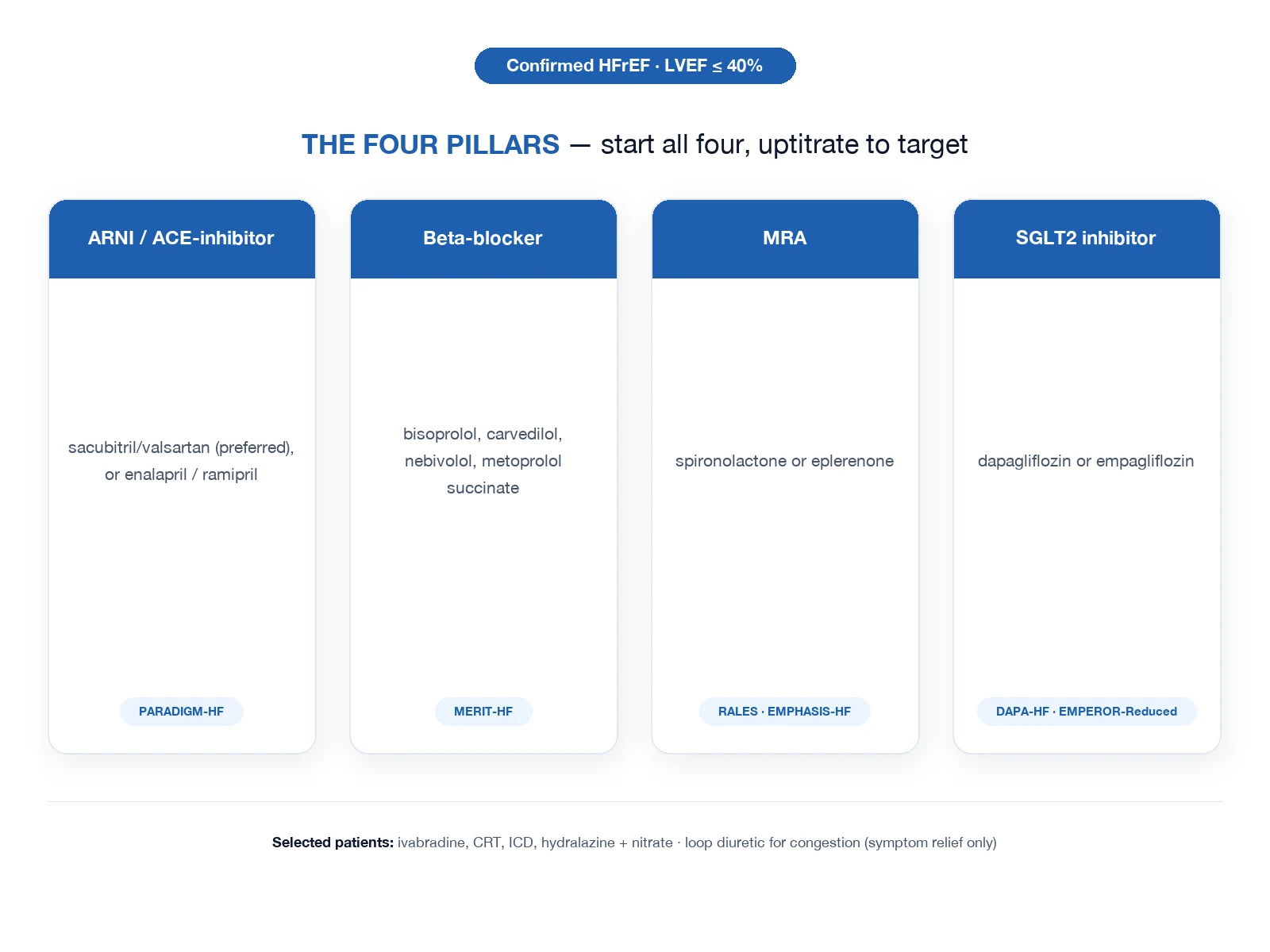
- TRANSITION TO MAINTENANCE — Oral loop diuretic + ACE-inhibitor/ARB + beta-blocker + MRA per heart-failure guidelines (the 'fantastic four' that reduce mortality in HFrEF). Investigate and treat the underlying cardiomyopathy.
Clinical pearls
Additional red flags
The one-page revision summary
Cardiogenic vs ARDS — the complete one-glance table
| Domain | Cardiogenic (hydrostatic) | ARDS (permeability) |
|---|---|---|
| Mechanism | Raised left-heart pressure; intact barrier | Damaged alveolar-capillary membrane; normal pressure |
| Starling term | Pc raised | Kfc raised, σ falls |
| PAWP | >18 mmHg | <18 mmHg |
| BNP | High (>400) | Normal/low (<100-200) |
| Echo | Low EF or high E/e-prime + dilated LA; normal RV | Normal LV; may be dilated RV (cor pulmonale) |
| CXR | Bat-wing perihilar, cardiomegaly, Kerley B, effusions | Diffuse bilateral, no cardiomegaly, no effusions |
| Onset | Gradual (hours) unless flash | Acute (within 7 d of trigger) |
| Fluid | Transudate (protein <0.65 of plasma) | Exudate (protein >0.65 of plasma) |
| Response to diuresis | Yes | No (but conservative fluids help) |
| First-line ventilatory | CPAP / NIV | Lung-protective ventilation |
| Pharmacology | Furosemide + GTN (if hypertensive) | PEEP + treat cause; ± dexamethasone |
| Advanced | Inotropes / VA-ECMO / mechanical support | Proning / VV-ECMO |
| Key trial | 3CPO (CPAP/NIV) | ARDSNet + PROSEVA + FACTT |
| Mortality driver | The underlying heart disease | The lung injury + the trigger |
Exam pitfalls
Common exam errors — and the correct answer
| Pitfall | Wrong answer | Correct answer |
|---|---|---|
| '3CPO showed NIV does not work' | True | False — it showed no MORTALITY benefit; NIV/CPAP improved physiology and reduced intubation. Use it first-line. |
| 'BNP is high in ARDS' | True | False — BNP is normal/low in pure ARDS; it rises only with coincident RV strain, septic cardiomyopathy, or volume overload. |
| 'ARDS responds to furosemide' | True | False — ARDS is permeability; diuresis does not fix it. A conservative fluid STRATEGY (FACTT) helps by reducing total body water, not by repairing the barrier. |
| 'Use actual weight for Vt' | True | False — use PREDICTED body weight (height and sex based). |
| 'PAWP is required for the Berlin definition' | True | False — Berlin REMOVED the PAWP criterion; echo is preferred. |
| 'Give morphine routinely in ACPO' | True | False — NICE/ESC advise AGAINST routine morphine; it increases intubation. |
| 'CPAP and BiPAP have very different outcomes' | True | False — 3CPO showed no clear superiority of BiPAP over CPAP; use whichever is available. |
| 'Proning helps all ARDS' | True | False — PROSEVA benefit is in SEVERE ARDS (P/F <150); mild ARDS is not proned routinely. |
| 'Morphine is a venodilator and so is safe in cardiogenic oedema' | True | Misleading — it is a weak venodilator and an anxiolytic, but respiratory depression and the association with worse outcomes make it non-routine. |
The exam oral — a structured answer
[1] [1]References
- [1]Gray A, Goodacre S, Newby DE, et al.; 3CPO Trialists. Noninvasive ventilation in acute cardiogenic pulmonary edema N Engl J Med, 2008.PMID 18614781
- [2]Ingbar DH Cardiogenic pulmonary edema: mechanisms and treatment - an intensivist's view Curr Opin Crit Care, 2019.PMID 31116110
- [3]The Acute Respiratory Distress Syndrome Network. Ventilation with lower tidal volumes as compared with traditional tidal volumes for acute lung injury and the acute respiratory distress syndrome N Engl J Med, 2000.PMID 10793162
- [4]Guérin C, Reignier J, Richard JC, et al.; PROSEVA Trial Group. Prone positioning in severe acute respiratory distress syndrome N Engl J Med, 2013.PMID 23688302
- [5]Wiedemann HP, Wheeler AP, Bernard GR, et al.; NHLBI ARDS Clinical Trials Network. Comparison of two fluid-management strategies in acute lung injury N Engl J Med, 2006.PMID 16714767
- [6]Combes A, Hajage D, Capellier G, et al.; EOLIA Trial Group. Extracorporeal Membrane Oxygenation for Severe Acute Respiratory Distress Syndrome N Engl J Med, 2018.PMID 29791822
- [7]Maisel AS, Krishnaswamy P, Nowak RM, et al.; Breathing Not Properly Multinational Study Investigators. Rapid measurement of B-type natriuretic peptide in the emergency diagnosis of heart failure N Engl J Med, 2002.PMID 12124404
- [8]ARDS Definition Task Force; Ranieri VM, Rubenfeld GD, Thompson BT, et al. Acute respiratory distress syndrome: the Berlin Definition JAMA, 2012.PMID 22797452
- [9]Briel M, Meade M, Mercat A, et al. Higher vs lower positive end-expiratory pressure in patients with acute lung injury and acute respiratory distress syndrome: systematic review and meta-analysis JAMA, 2010.PMID 20197533