ICU · Respiratory
Acute pulmonary oedema: cardiogenic vs non-cardiogenic differentiation
Also known as Pulmonary oedema · Cardiogenic pulmonary oedema · Non-cardiogenic pulmonary oedema · ARDS vs heart failure · Acute respiratory failure with bilateral infiltrates · Flash pulmonary oedema · Negative pressure pulmonary oedema · Re-expansion pulmonary oedema · Neurogenic pulmonary oedema · High-altitude pulmonary oedema
Acute pulmonary oedema: fluid in alveolar spaces → bilateral infiltrates + hypoxaemia. TWO types: CARDIOGENIC (hydrostatic — from LV failure, acute MR, mitral stenosis, volume overload; PCWP 18 mmHg) and NON-CARDIOGENIC (increased permeability — ARDS, sepsis, trauma, aspiration, transfusion; PCWP <18 mmHg). Differentiation CRITICAL — treatment differs. CARDIOGENIC: oxygen, NIV/CPAP (rapid response), IV loop diuretics, vasodilators (GTN if SBP 110), inotropes (cold/shocked), IABP/Impella/VA-ECMO for refractory. NON-CARDIOGENIC: treat underlying cause, lung-protective ventilation (Vt 6 mL/kg PBW, Pplat <30, driving pressure <15), prone, conservative fluids (FACTT). KEY tools: BNP/NT-proBNP (high = cardiogenic), echocardiography (reduced EF/cardiogenic), POCUS (bilateral B-lines, IVC plethoric, reduced LV), lung ultrasound, CT (cardiogenic: central/perihilar, pleural effusions; non-cardiogenic: peripheral/diffuse), PCWP (cardiogenic 18). SPECIAL FORMS: negative pressure (NPPE), re-expansion, neurogenic, high-altitude (HAPE), TRALI.
On this page & tools
Your progress
Saved locally on this device.
Target exams
Red flags
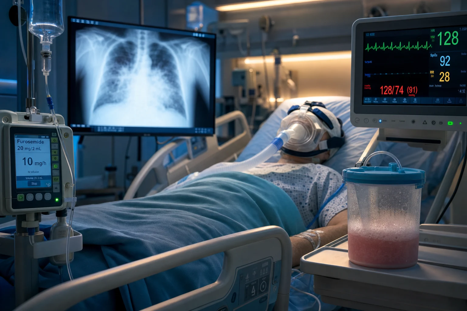
The pathophysiology — hydrostatic forces vs permeability leak
Acute pulmonary oedema develops when fluid accumulates in the interstitium and alveoli faster than lymphatics can clear it. The Starling equation governs transcapillary fluid flux: net filtration = the hydraulic pressure pushing fluid OUT minus the oncotic pressure pulling it BACK, multiplied by the capillary permeability coefficient. Two fundamentally different insults disturb this balance, producing the two phenotypes.[1]
In cardiogenic (hydrostatic) oedema, left-sided filling pressures rise — left atrial pressure (LAP) → pulmonary venous pressure → capillary hydrostatic pressure (Pc). Once Pc exceeds the plasma oncotic pressure (~25 mmHg) and the lung's interstitial pressure, fluid is pushed across an INTACT alveolar–capillary membrane into the interstitium, then into the alveoli. The fluid is a TRANSUDATE (protein-poor), and the membrane architecture is preserved — which is why the lung recovers rapidly once pressure is relieved. The classic PCWP threshold of 18 mmHg comes from the older pulmonary artery catheter literature: above this, hydrostatic oedema is likely.[1]
In non-cardiogenic (permeability) oedema, an insult — sepsis, pneumonia, aspiration, major trauma, pancreatitis, massive transfusion — destroys the integrity of the alveolar–capillary membrane. Endothelial and epithelial injury, neutrophil activation, cytokine surge and loss of surfactant produce a leaky barrier. Fluid AND protein pour into the interstitium and alveoli at a NORMAL or LOW pulmonary capillary pressure. The fluid is a protein-rich EXUDATE. This is the ARDS lung — stiff, heavy, non-compliant, with bilateral opacities that are NOT caused by cardiac failure. The diagnostic standard remains the Berlin definition: timing within 1 week of insult, bilateral opacities not fully explained by effusions/atelectasis/nodules, oedema not fully explained by cardiac failure or fluid overload (and if measured, PCWP < 18 mmHg), and hypoxaemia graded by the PaO2/FiO2 ratio (mild 300-200, moderate 200-100, severe < 100).[9]
The crucial implication at the bedside: lowering the pressure (diuresis, vasodilators, CPAP) resolves cardiogenic oedema within minutes to hours, but does NOT resolve ARDS. ARDS demands treatment of the cause, lung-protective ventilation to avoid further injury, and time for the membrane to heal. [1]
Cardiogenic pulmonary oedema — mechanisms, triggers, treatment
Mechanisms — three haemodynamic routes to high pulmonary venous pressure
- LV systolic failure (HFrEF) — the largest single cause. Extensive MI, decompensated chronic HFrEF, myocarditis, sepsis-induced cardiomyopathy, takotsubo. The LV cannot generate enough forward stroke volume, end-diastolic volume and pressure rise, this back-pressures the left atrium, the pulmonary veins, and ultimately the capillary bed.
- LV diastolic failure (HFpEF) — common in the elderly, hypertensive, diabetic, obese, AF population. A stiff LV with preserved EF cannot accept diastolic filling at normal pressures; small volume loads or tachycardia (loss of diastolic filling time) push LAP sharply up.
- Valvular and mechanical — acute mitral regurgitation (papillary muscle/chordal rupture post-MI, endocarditis), severe mitral stenosis, prosthetic valve thrombosis or obstruction, and (rarely) a left atrial myxoma. These cause pulmonary oedema with a NORMAL LV EF — the obstruction or regurgitation is upstream of the LV cavity, so the LV itself may contract normally.[4]
Common precipitants
ACS, new or fast AF, hypertensive emergency, acute regurgitant lesion, renal failure / volume overload, drug non-adherence, NSAIDs / negative inotropes (CCB, most antiarrhythmics, thiazolidinediones), thyroid disturbance, anaemia, pneumonia/sepsis, and high-output states. Identifying and treating the trigger is as important as treating the oedema itself. [1]
Treatment — a sequenced ladder
The first 30-60 minutes aim to relieve work of breathing, improve oxygenation, drop preload and afterload, and identify the precipitant. Sit the patient upright, give oxygen to target SpO2 92-96%, start continuous ECG monitoring, secure IV access, place a urinary catheter (the urine output is the single best index of decongestion), and classify the haemodynamic phenotype (warm-wet vs cold-wet) at the bedside.[3]
- NIV/CPAP early — CPAP 5-10 cmH2O (or BiPAP IPAP 10-15/EPAP 5-8) for the wet patient in respiratory distress. Mechanism: positive intrathoracic pressure reduces venous return (preload), reduces LV transmural pressure (afterload), recruits alveoli, and unloads respiratory muscles. The 3CPO trial showed faster relief of dyspnoea, hypercapnia and acidosis with NIV; CPAP = BiPAP. Do NOT wait for exhaustion.
- IV loop diuretic — furosemide IV. For a patient NOT on a loop diuretic, give 20-40 mg IV. For a patient already on an oral loop, give 1-2.5x the total daily oral dose IV (DOSE trial: high-dose vs low-dose and bolus vs continuous — no significant difference in the co-primary endpoints; high-dose achieved greater fluid loss at the cost of more transient renal dysfunction). Reassess urine output at 2 h and double if response is poor. Target 3-5 L/day net negative.
- Vasodilator (if SBP > 110) — IV nitroglycerin (GTN) 10-200 mcg/min, titrated to symptoms and BP; venodilates (drops preload) and arteriodilates (drops afterload), ideal for hypertensive pulmonary oedema. Avoid in the hypotensive cold patient, RV infarct, severe aortic stenosis, HOCM, and recent PDE-5 use.
- Morphine — sparingly or not at all. Small doses (1-3 mg IV) for distressed/anxious patients may reduce preload and catecholamine surge, but the ADHERE registry linked morphine to increased mortality (a confounded observational signal, but it has shifted practice away from routine morphine in ADHF).
- Inotrope + vasopressor (cold-wet, cardiogenic shock) — noradrenaline to restore a perfusing MAP > 65 mmHg (preferred over dopamine — SOAP-II: more arrhythmia with dopamine), plus an inotrope (dobutamine 2.5-10 mcg/kg/min, or milrinone if beta-blocked / RV failure). Every inotrope raises myocardial oxygen demand and arrhythmia risk — a bridge to a definitive decision.
- Mechanical circulatory support (refractory shock) — Impella (active LV unloading), VA-ECMO (full heart-and-lung support). The IABP has lost routine status after IABP-SHOCK II (no mortality benefit in MI-shock), but is still used as a bridge in selected centres. Importantly, VA-ECMO does NOT unload the LV — retrograde flow increases afterload and can worsen LV distension and pulmonary oedema; add an Impella (ECPELLA) if the LV is dilating on serial echo.
- Treat the trigger — ACS (revascularise), AF (rate or rhythm control), pneumonia/sepsis (antibiotics, source control), renal failure (dialysis if overloaded), MR/VSD (urgent surgery), severe mitral stenosis (mitral balloon valvuloplasty or surgery), prosthetic valve thrombosis (surgery or thrombolysis). [1]
Cardiogenic vs non-cardiogenic pulmonary oedema
| Feature | Cardiogenic | Non-cardiogenic (ARDS) |
|---|---|---|
| Mechanism | Hydrostatic (high pressure) | Permeability (leaky capillaries) |
| Cause | Heart failure, volume overload, mitral stenosis | Sepsis, pneumonia, trauma, aspiration, transfusion |
| BNP/NT-proBNP | HIGH (>300 NT-proBNP) | LOW or normal |
| Echocardiography | Reduced EF (HFrEF) or diastolic dysfunction (HFpEF) | Usually normal EF |
| CXR/CT pattern | Central, perihilar, pleural effusions, Kerley B lines | Diffuse, peripheral, no effusions (usually) |
| PCWP | >18 mmHg | <18 mmHg |
| Oedema fluid protein | Low (transudate — protein-poor) | High (exudate — protein-rich) |
| Response to diuretics | RAPID improvement (minutes-hours) | Slow/no response |
| Response to CPAP/NIV | RAPID (minutes — pushes fluid back) | Minimal (doesn't address permeability) |
| Treatment | Diuretics, vasodilators, CPAP/NIV | Treat cause, lung-protective ventilation, prone |
Hydrostatic vs permeability — Starling forces at the bedside
| Starling variable | Cardiogenic (hydrostatic) | Non-cardiogenic (permeability/ARDS) |
|---|---|---|
| Capillary hydraulic pressure (Pc) | ↑↑↑ (high LAP transmitted back) | Normal or low |
| Plasma oncotic pressure | Usually normal | Often reduced (critical illness, hypoalbuminaemia) |
| Capillary permeability coefficient | NORMAL — membrane intact | ↑↑↑ — membrane injured, leaks protein |
| Net fluid composition | Transudate (protein-poor, <60% of plasma) | Exudate (protein-rich, >60% of plasma) |
| Consequence of lowering Pc | Resolves oedema (diuretics/vasodilators/CPAP) | Does not resolve (membrane still leaks) |
| Lung architecture | Largely preserved once pressure relieved | Damaged — needs time, lung-protective ventilation |
Acute causes of cardiogenic pulmonary oedema with a NORMAL EF
| Cause | Why EF looks normal | Distinguishing clue on echo |
|---|---|---|
| HFpEF (diastolic HF) | Stiff LV with preserved systole | E/e' > 14, LA enlargement, LVH, raised estimated LV filling pressure |
| Acute severe MR | LV unloads into low-pressure left atrium — EF exaggerated | Flail leaflet, eccentric regurgitant jet, systolic flow reversal in pulmonary veins |
| Mitral stenosis | Obstruction is upstream of LV cavity | Thickened rheumatic leaflets, "hockey-stick" anterior leaflet, reduced mitral valve area, raised transmitral gradient |
| Aortic stenosis / HOCM | Concentric hypertrophy, hyperdynamic cavity | Calcified aortic valve / systolic anterior motion of mitral leaflet, dynamic LVOT gradient |
| Takotsubo / septic cardiomyopathy | Regional sparing or hyperkinesis in non-involved segments | Apical ballooning (takotsubo); global hypokinesis in septic cardiomyopathy |
| Mitral valve prosthesis thrombosis | Native LV function preserved | Reduced prosthetic leaflet mobility, raised transmitral gradient, occasionally visible thrombus |
Non-cardiogenic pulmonary oedema (ARDS) — treatment
Once the bedside assessment points to permeability oedema, the strategy inverts: lowering pressure helps at the margins but cannot fix a leaky membrane. Management is dominated by treating the cause and lung-protective ventilation.[7]
- Identify and treat the trigger — pneumonia (antibiotics, BAL), sepsis (source control, empirical broad-spectrum, focus), aspiration (positional, bronchoscopy for particulates), pancreatitis (aggressive fluid resuscitation early, then conservative), trauma (fix the chest, transfuse appropriately — avoid over-transfusion and TRALI), transfusion-related (stop the unit, supportive care).
- Lung-protective ventilation (ARDSNet ARMA 2000) — Vt 6 mL/kg predicted body weight (NOT actual), Pplat < 30 cmH2O, titrate PEEP/FiO2 against the ARDSNet table, target pH > 7.20 (permissive hypercapnia), SpO2 88-95% / PaO2 55-80 mmHg. This single intervention reduced mortality from 40% to 31% — the most influential ARDS trial ever run.[7]
- Driving pressure and PEEP optimisation — keep driving pressure (Pplat - PEEP) < 15 cmH2O where possible (Amato meta-analysis: driving pressure most strongly associated with survival). High PEEP for moderate-severe ARDS (ALVEOLI, EXPRESS — modest benefit in the sickest).
- Prone ventilation for moderate-severe ARDS (PaO2/FiO2 < 150) — 16+ hours/day. PROSEVA showed a 28-day mortality reduction (16% vs 33%) in severe ARDS.
- Conservative fluid strategy (FACTT) — a fluid-management protocol targeting lower central venous pressure / pulmonary artery occlusion pressure produced more ventilator-free days and shorter ICU stay than a liberal strategy, without increasing renal failure. Avoid the daily 4-6 L of "maintenance" that accumulates silently in ARDS.[8]
- Avoid harmful interventions — high-frequency oscillatory ventilation (HFOV) increased mortality in OSCILLATE (NEJM 2013) — do not use routinely; neuromuscular blockade may help in early severe ARDS (ACURASYS, ROSE — controversial, 48-h cisatracurium in early severe P/F < 150); routine statins and beta-blockers do not help.
- Adjuncts for refractory hypoxaemia — inhaled pulmonary vasodilators (nitric oxide, epoprostenol) for transient oxygenation improvement (no mortality benefit), recruitment manoeuvres (carefully), ECMO for severe refractory ARDS (centre transfer, CESAR and EOLIA trials).
CPAP vs BiPAP vs HFNC for acute respiratory failure — when to use each
| Modality | Cardiogenic oedema | ARDS / non-cardiogenic |
|---|---|---|
| CPAP | First-line — drops preload/afterload, recruits alveoli (3CPO) | Useful in mild ARDS / immunocompromised (FLORALI — early NIV reduced intubation in immunocompromised subgroup) |
| BiPAP | Equal to CPAP (3CPO); preferred if hypercapnia or CO2 retention | Useful if hypercapnic (combined ARDS + airflow obstruction) |
| HFNC | Second-line if NIV not tolerated | Reasonable first-line in moderate hypoxaemic respiratory failure (FLORALI — trend to lower 90-day mortality vs NIV or standard O2) |
| Intubation | If NIV fails / haemodynamic collapse / reduced GCS | If P/F < 150 and worsening, work of breathing high, or NIV fails within 1-2 h |
| Key trial | 3CPO (Gray, Lancet 2008) | FLORALI (Frat, NEJM 2015); HACAR (high-flow in immunocompromised) |
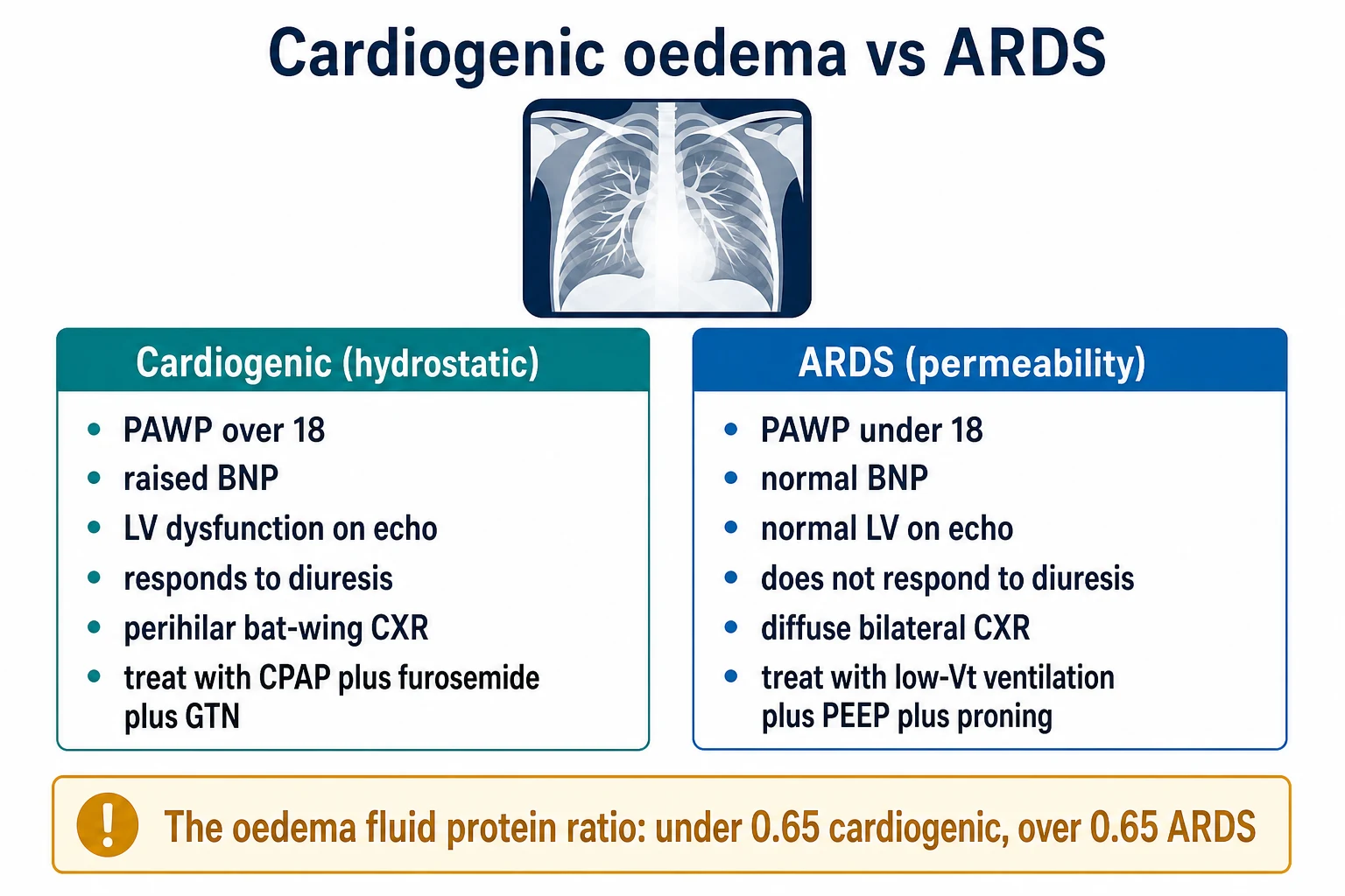
Differentiation toolkit — PCWP, BNP, echo, POCUS
BNP and NT-proBNP
The single best biochemical discriminator. NT-proBNP < 300 pg/mL in the acute setting effectively excludes heart failure as the cause of dyspnoea (high negative predictive value); values > 450 pg/mL in patients < 50, > 900 in those 50-75, and > 1800 in those > 75 support heart failure (age-stratified cutoffs). Confounders: renal failure raises it (reduced clearance), obesity lowers it, AF/PE/sepsis all elevate it by non-HF mechanisms. A rising trend during treatment supports an HF contribution. A low NT-proBNP in a patient with bilateral infiltrates and sepsis favours ARDS.[2]
Echocardiography — the definitive bedside tool
Reduced EF (< 40%) with regional or global hypokinesis → cardiogenic (HFrEF). Normal EF with diastolic dysfunction (E/e' > 14, LA enlargement, LVH) → cardiogenic (HFpEF). Normal EF and normal diastolic function in a patient with bilateral infiltrates and a credible insult → non-cardiogenic (ARDS). Critically, echo also identifies the mechanical causes of cardiogenic oedema with normal EF — acute MR (flail leaflet, eccentric jet, systolic flow reversal in pulmonary veins), severe MS (rheumatic thickening, raised transmitral gradient), prosthetic valve thrombosis, and tamponade.[4]
Point-of-care ultrasound (POCUS) — bilateral B-lines, IVC, focused cardiac
The combination of bilateral diffuse B-lines (comet-tail artefacts from the pleura moving with respiration), a plethoric IVC (> 2 cm with < 50% collapsibility), and reduced LV systolic function on focused cardiac view strongly supports cardiogenic oedema. Bilateral B-lines with a small, collapsing IVC and normal LV favour ARDS. B-lines do NOT distinguish cardiogenic from non-cardiogenic in isolation — both produce them — but the IVC, cardiac, and clinical context do. Lung ultrasound can also reveal focal B-lines (pneumonia, contusion), pleural effusions (anechoic quadrilaterals — large in cardiogenic, small in ARDS), and pneumothorax (absent lung sliding, lung point).[5]
Pulmonary capillary wedge pressure (PCWP)
The historical gold standard. PCWP > 18 mmHg supports hydrostatic (cardiogenic) oedema; PCWP < 18 with bilateral infiltrates and a credible insult supports ARDS. The PA catheter has been displaced by echo and POCUS for routine use (no mortality benefit in routine ICU populations — Connors observational analysis, FACTT sub-study, ESCAPE), but still has a role in the patient with mixed shock, severe pulmonary hypertension, or unexplained oedema where echo windows are poor. The Berlin ARDS definition no longer requires a PA catheter — "oedema not fully explained by cardiac failure or fluid overload" suffices, often adjudicated by echo/POCUS rather than PCWP.[1]
Trial of therapy
If cardiogenic oedema is suspected, a diagnostic–therapeutic trial of CPAP + IV furosemide is justified. Rapid improvement (SpO2 up, RR down, BP stabilises, distress resolves) within 15-30 minutes confirms cardiogenic. No rapid improvement — reconsider ARDS, pneumonia, or a mechanical complication. This trial must not delay definitive imaging/echo in the unstable patient. [1]
The mixed picture
Many ICU patients have BOTH cardiogenic and non-cardiogenic components — sepsis-induced cardiomyopathy (cardiac stunning + ARDS), heart failure with secondary pneumonia, ARDS aggressively fluid-resuscitated (hydrostatic contribution). These patients need BOTH treatments: diurese for the hydrostatic component, lung-protective ventilation and conservative fluids for the ARDS component, and serial echo + lactate + POCUS to track the balance. [1]
POCUS findings — cardiogenic vs non-cardiogenic
| POCUS modality | Cardiogenic | Non-cardiogenic (ARDS) |
|---|---|---|
| Lung — B-lines | Diffuse bilateral, often with effusions | Diffuse bilateral, may be patchy, subpleural consolidations |
| Lung — A-lines | Absent in oedematous zones | May be present in spared zones (non-homogeneous) |
| Pleural effusion | Common — bilateral, right > left | Usually small or absent |
| IVC | Plethoric (> 2 cm, < 50% collapse) — high right atrial pressure | Small/collapsible if hypovolaemic; may be normal |
| Focused cardiac | Reduced LV (HFrEF) or thick/stiff LV with LA enlargement (HFpEF); may show acute MR / MS | Usually normal LV; may show RV dilation in severe ARDS (acute cor pulmonale) |
| LV EF | Reduced OR preserved (HFpEF) | Usually normal |
| Estimated LAP | Raised (E/e' > 14) | Normal |
Bedside approach to bilateral infiltrates + hypoxaemia
- Assess clinically — history (heart failure? sepsis? trauma?), signs (raised JVP, S3 gallop, peripheral oedema = cardiogenic; fever, sepsis source, clear lung on auscultation initially = non-cardiogenic)
- BNP/NT-proBNP — HIGH (>300 NT-proBNP) → cardiogenic likely. LOW → non-cardiogenic likely. INTERMEDIATE → grey zone (renal failure, sepsis, obesity confound)
- Echocardiography (bedside) — reduced EF or wall motion abnormality → cardiogenic. Normal EF → non-cardiogenic (or HFpEF). Also: assess valves (mitral stenosis/regurgitation), pericardial effusion (tamponade)
- Lung ultrasound — B-lines (comet tails): diffuse bilateral = oedema (cardiogenic or non). Focal = consolidation/pneumonia. A-lines (normal): no oedema. Pleural effusion (cardiogenic more common)
- CT chest (if uncertain) — cardiogenic: central, perihilar, ground-glass, pleural effusions, cardiomegaly. Non-cardiogenic: diffuse, peripheral, no effusions (usually), no cardiomegaly
- Trial of therapy — if cardiogenic suspected: CPAP + IV frusemide → RAPID improvement (within 30-60 min) = cardiogenic confirmed. If NO rapid improvement → reconsider (ARDS?)
- Treat accordingly — cardiogenic: diuretics, vasodilators, CPAP/NIV, treat cardiac cause. Non-cardiogenic: treat underlying cause, lung-protective ventilation, prone, conservative fluids
Cardiogenic pulmonary oedema — the first 60 minutes
- POSITION and OXYGENATE — sit upright; oxygen via face mask to target SpO2 92-96% (avoid hyperoxia). Continuous ECG, IV access, arterial line if unstable, urinary catheter
- CLASSIFY the Stevenson phenotype — wet vs dry (congestion) crossed with warm vs cold (perfusion). Most cardiogenic oedema is WARM-WET (BP usually > 110); cold-wet = cardiogenic shock
- START NIV/CPAP EARLY (the wet patient) — CPAP 5-10 cmH2O or BiPAP IPAP 10-15 / EPAP 5-8. Mechanism: drops preload and afterload, recruits alveoli, unloads respiratory muscles. Do NOT wait for exhaustion (3CPO)
- IV LOOP DIURETIC — furosemide IV. Naive patient: 20-40 mg. Established oral loop: 1-2.5x total daily oral dose IV. Reassess at 2 h, double if poor response. Target 3-5 L/day net negative
- VASODILATOR if SBP > 110 — IV nitroglycerin 10-200 mcg/min. Avoid in RV infarct, severe AS, HOCM, recent PDE-5. Nitroprusside for severe hypertension (cyanide toxicity in renal failure)
- COLD-WET (shock) → INOTROPE + VASOPRESSOR — noradrenaline to MAP > 65 (first-line, SOAP-II), dobutamine 2.5-10 mcg/kg/min or milrinone if beta-blocked/RV failure. Reassess for mechanical complication (VSD, papillary muscle rupture)
- MECHANICAL CIRCULATORY SUPPORT if refractory — Impella (active LV unloading) or VA-ECMO (full support). Add Impella to VA-ECMO (ECPELLA) if LV dilating. IABP no longer routine (IABP-SHOCK II)
- FIND AND TREAT THE TRIGGER — ACS (revascularise), AF (rate/rhythm control), MS/MR (valve intervention), prosthetic valve thrombosis (surgery/thrombolysis), pneumonia/sepsis (antibiotics), renal failure (RRT)
Non-cardiogenic (ARDS) — lung-protective ventilation setup
- CONFIRM the diagnosis — bilateral opacities + hypoxaemia within 1 week of insult + oedema NOT fully cardiac/fluid (echo/POCUS) + PCWP < 18 if measured (Berlin definition)
- CALCULATE PREDICTED BODY WEIGHT (PBW) — Male: 50 + 2.3 × (height in inches − 60). Female: 45.5 + 2.3 × (height in inches − 60). Use PBW (NOT actual weight) for Vt — actual weight over-doses the oedematous lung
- SET INITIAL Vt at 8 mL/kg PBW, then REDUCE to 6 mL/kg PBW over 4 h
- TITRATE TO PLATEAU PRESSURE (Pplat) — measure with 0.5-s inspiratory hold. If Pplat > 30 cmH2O, reduce Vt to 4 mL/kg PBW minimum. If Pplat < 25 and there is dual triggering, may increase to 7-8 mL/kg
- SET PEEP/FiO2 using the ARDSNet lower-PEEP/higher-PEEP table; consider higher PEEP table for moderate-severe ARDS (ALVEOLI, EXPRESS)
- DRIVING PRESSURE (Pplat − PEEP) — keep < 15 cmH2O where possible (Amato); the ventilation variable most associated with survival
- RESPIRATORY RATE 20-35/min to keep pH > 7.20 (permissive hypercapnia acceptable). I:E 1:1 or 1:2
- TARGET SpO2 88-95% / PaO2 55-80 mmHg — permissive hypoxaemia; do not chase "normal" by escalating harmful FiO2/PEEP
- CONSIDER PRONE if PaO2/FiO2 < 150 — at least 16 h/day (PROSEVA)
- CONSERVATIVE FLUIDS (FACTT) — fluid-restrict once resuscitated; daily net even or negative; track cumulative balance
- ESCALATE — refractory hypoxaemia (P/F < 80 despite optimisation) → inhaled pulmonary vasodilator, recruitment manoeuvres, ECMO referral
Negative pressure pulmonary oedema (NPPE) — recognition and management
- RECOGNISE the setting — post-extubation laryngospasm, biting on the endotracheal tube, near-drowning, strangulation, severe obstructive sleep apnoea, post-anaesthesia in a young healthy patient, thyroid mass or tumour obstructing the airway
- MECHANISM — forceful inspiration against a closed glottis → very negative intrathoracic pressure (Mueller manoeuvre) → high transcapillary pressure gradient → fluid pulled into interstitium and alveoli. May also injure the capillary (stress failure) giving a mixed hydrostatic + permeability picture
- CLINICAL — pink frothy sputum, hypoxaemia, diffuse crackles, developing within minutes to hours of relief of the obstruction. CXR: bilateral infiltrates
- RELIEVE THE OBSTRUCTION — definitive airway if still obstructed (re-intubate, surgical airway); bite block if biting
- OXYGEN + CPAP/NIV — most cases respond within 24 h; positive pressure reverses the hydrostatic gradient
- DIURETICS — small dose (furosemide 20-40 mg IV) often helps; not the primary therapy
- DIFFERENTIATE — no heart failure, no ARDS-insult (think ARDS if sepsis/pneumonia coexists). Echo usually normal. Self-limiting in 24 h if obstruction is relieved
- REASSURE the team — NPPE in a healthy young patient after anaesthesia typically resolves fully within 24 h with supportive care; rare mortality
Exam practice
SAQ — Acute cardiogenic pulmonary oedema: NIV/CPAP and the 3CPO trial
10 minutes · 10 marks
A 78-year-old man with known HFrEF (EF 28%), prior MI and CKD stage 3 presents with 4 hours of worsening breathlessness, orthopnoea and cough. He is diaphoretic, sitting upright, RR 34, SpO2 86% on 15 L/min NRM, BP 168/96, HR 118 sinus tachycardia, JVP to the earlobes, bilateral crackles two-thirds up and expiratory wheeze. CXR shows perihilar 'bat-wing' infiltrates, Kerley B lines and small bilateral pleural effusions. ABG: pH 7.22, PaCO2 7.1 kPa (54 mmHg), PaO2 7.8 kPa (59 mmHg), lactate 2.8. NT-proBNP 6800 pg/mL. The ED team has started CPAP at 10 cmH2O.
SAQ — Cardiogenic pulmonary oedema with acute kidney injury (cardiorenal syndrome)
10 minutes · 10 marks
A 72-year-old woman with HFrEF (EF 25%), type 2 diabetes and CKD stage 3b (baseline creatinine 160 µmol/L) is admitted with decompensated heart failure — flash pulmonary oedema, JVP to the jaw, sacral and leg oedema, weight gain 6 kg over 10 days. She is on furosemide 80 mg BD orally. BP 96/60, RR 30, SpO2 90% on 10 L/min, warm-cool peripheries, oliguric (15 mL/h). Creatinine 290 µmol/L, K+ 5.8, lactate 3.1, NT-proBNP 12000. Echo: dilated LV, EF 22%, no mechanical valve lesion. She has been started on CPAP and a furosemide infusion.
Clinical pearls
Red flags
Prognosis
3CPO trial (Gray 2008, Lancet) — NIV in cardiogenic pulmonary oedema
RCT: 1,069 patients with cardiogenic pulmonary oedema. CPAP vs NIV (BiPAP) vs standard oxygen.
- Primary outcome (7-day mortality): CPAP 9.8%, NIV 9.8%, standard 9.8% (NO difference)
- Secondary outcomes (intubation, dyspnoea, length of stay): NIV/CPAP reduced dyspnoea faster, reduced intubation vs standard
- CONCLUSION: NIV (CPAP or BiPAP) reduces intubation and improves dyspnoea in cardiogenic oedema. No mortality difference in this trial (but other meta-analyses suggest benefit). CPAP is SIMPLE and EFFECTIVE for cardiogenic oedema. [1]
BNP for diagnosis: high NT-proBNP (>300) → heart failure likely. Low → excluded (NPV 90%). Both CPAP and BiPAP effective for cardiogenic oedema.
ARDSNet ARMA (Acute Respiratory Distress Syndrome Network, NEJM 2000) — the lung-protective ventilation trial
RCT: 861 patients with ALI/ARDS. Vt 6 mL/kg PBW (Pplat < 30) vs Vt 12 mL/kg PBW (Pplat < 50).
- Primary outcome (mortality before hospital discharge): 31% (low Vt) vs 40% (traditional Vt); NNT ~11 — trial stopped early for efficacy
- Ventilator-free days: 12 vs 10 days; barotrauma: similar
- CONCLUSION: Lung-protective ventilation (Vt 6 mL/kg PBW, Pplat < 30) reduces mortality in ARDS by ~9 absolute percentage points — the most influential ARDS trial ever run. Tidal volume calculated from PREDICTED body weight (height-based), not actual.[7]
FACTT (Wiedemann, NEJM 2006) — conservative vs liberal fluid in ALI/ARDS
RCT: 1,000 patients with ALI/ARDS. Conservative (CVP < 4, PCWP < 8) vs liberal (CVP 10-14, PCWP 14-18) fluid strategy, using protocolised diuretics/fluids for 7 days.
- Primary outcome (60-day mortality): 25.5% (conservative) vs 28.4% (liberal) — NOT significant
- Secondary outcomes: conservative gave MORE ventilator-free days (14.5 vs 12.0) and MORE ICU-free days (13.4 vs 11.2); NO increase in renal failure or dialysis
- CONCLUSION: A conservative fluid strategy in established ARDS improves lung function and shortens ventilation and ICU stay without harming the kidneys. Apply once the patient is resuscitated.[8]
LUNG SAFE (Bellani, JAMA 2016) — ARDS epidemiology across 50 countries
Prospective observational: 29,144 ICU admissions in 459 ICUs across 50 countries over 4 winter weeks; 3,022 met ARDS criteria.
- Incidence: 10.4% of ICU admissions; ARDS is common and under-recognised
- Recognition: clinicians recognised ARDS in only ~51% of mild, ~79% of moderate, ~84% of severe cases — substantial under-recognition, especially in mild ARDS
- Mortality (ICU): mild 34.9%, moderate 40.3%, severe 46.1% (Berlin severity correlates with outcome)
- CONCLUSION: ARDS is under-recognised, under-treated (only ~60% received a low Vt), and carries high mortality that scales with severity. The case for systematic low-Vt ventilation and protocolised recognition.[9]
IABP-SHOCK II (Thiele, NEJM 2012) — IABP in MI-related cardiogenic shock
RCT: 600 patients with acute MI and cardiogenic shock, planned early revascularisation. IABP vs no IABP.
- Primary outcome (30-day mortality): 39.7% (IABP) vs 41.3% (no IABP) — NOT significant
- Secondary outcomes (process variables, lactate clearance, organ function, bleeding, stroke): no difference
- CONCLUSION: Routine IABP in MI-related cardiogenic shock provides no mortality benefit. The IABP has lost routine status; percutaneous options now include Impella (active LV unloading) and VA-ECMO (full support), with selection individualised.[13]
CARRESS-HF (Bart, NEJM 2012) — ultrafiltration vs stepped pharmacological therapy in ADHF with cardiorenal syndrome
RCT: 188 patients with ADHF, persistent congestion AND worsening renal function. Venovenous ultrafiltration vs stepped pharmacological therapy (high-dose + sequential diuretics).
- Primary outcome (change in creatinine at 96 h): +0.23 mg/dL (ultrafiltration) vs −0.04 mg/dL (pharmacological); P = 0.003 — ultrafiltration WORSE
- Serious adverse events: 72% (ultrafiltration) vs 57% (pharmacological); P = 0.03 — stopped early for futility/safety
- CONCLUSION: Ultrafiltration is NOT first-line decongestion in ADHF; reserve for refractory volume overload unresponsive to high-dose + sequential diuretics, or during RRT.[12]
PROSEVA (Guérin, NEJM 2013) — prone ventilation in severe ARDS
RCT: 466 patients with ARDS and PaO2/FiO2 < 150, within 36 h of enrolment. Prone ventilation ≥ 16 h/day vs supine.
- Primary outcome (28-day mortality): 16.0% (prone) vs 32.8% (supine); P < 0.001 — large mortality reduction
- 90-day mortality: 23.6% (prone) vs 41.0% (supine); NNT ~6
- CONCLUSION: In severe early ARDS (P/F < 150 within 36 h), prolonged prone ventilation (≥ 16 h/day) dramatically reduces mortality. Proning is safe with a trained team and should be initiated early in eligible patients.
TRALI and TACO — the two transfusion-related lung injuries
TRALI (Transfusion-Related Acute Lung Injury) — non-cardiogenic (permeability) oedema within 6 h of transfusion. Donor anti-HLA/anti-HNA antibodies (or, less commonly, biologically active lipids) activate recipient neutrophils, damaging the pulmonary endothelium. Bilateral infiltrates + hypoxaemia + NORMAL left atrial pressure. Treatment: STOP the transfusion, supportive care (oxygen, lung-protective ventilation if intubated). NO role for diuretics (this is permeability, not hydrostatic). Resolves in 48-72 h. Mortality 5-10%. REPORT to the blood bank; the donor unit should be quarantined and the donor deferred.[14]
TACO (Transfusion-Associated Circulatory Overload) — hydrostatic (cardiogenic) overload from the transfused volume, usually within 6-12 h, in patients with limited cardiac reserve. Bilateral infiltrates + hypertension + raised BNP + raised JVP. Treatment: stop the transfusion, diuretic (furosemide IV), oxygen/CPAP. Differentiate from TRALI by the BNP (high in TACO), the haemodynamics (hypertensive in TACO), and the response to diuretic (rapid in TACO).
Special pulmonary oedema syndromes — when the heart and the lung are NOT the usual suspects
NPPE (Negative Pressure Pulmonary Oedema) — forceful inspiration against a closed glottis (laryngospasm post-extubation, biting, strangulation, near-drowning, severe OSA). Generates very negative intrathoracic pressure (Mueller manoeuvre), raising the transcapillary gradient; may also cause capillary stress failure (mixed hydrostatic + permeability). Presents within minutes-hours with pink frothy sputum and hypoxaemia in an otherwise healthy patient. Treatment: relieve obstruction, CPAP/NIV, small-dose diuretic. Self-limiting in < 24 h.[11]
Re-expansion oedema — rapid re-expansion of a chronically collapsed lung (draining > 1.5 L pleural effusion or applying high suction to a chronic pneumothorax) produces local capillary leak and surfactant loss. Presents within 1-24 h on the DRAINED side. Prevention: limit drainage to < 1.5 L per session; clamp if chest tightness or cough. Treatment: supportive. [1]
Neurogenic oedema — within hours of severe CNS insult (SAH, TBI, status epilepticus). Massive sympathetic surge → catecholamine cardiac stunning (often takotsubo-pattern) → systemic hypertension and capillary stress failure. Echo may show apical ballooning; troponin elevated. Treatment: supportive (oxygen, ventilation); alpha/beta-blockade in selected cases. Often self-limiting over 48-72 h if the primary insult is survivable. [1]
HAPE (High-Altitude Pulmonary Oedema) — at altitude (> 2500 m) from uneven hypoxic pulmonary vasoconstriction — over-perfused capillary segments leak under high pressure. Treatment: DESCEND (single most effective), oxygen, nifedipine (reduces PVR); prevention by gradual ascent, acetazolamide. Echo shows normal LV.
Exam answer — the long-form / viva response
[1]References
- [1]Murray JF, et al. Government-funded research increasingly fuels innovation Science, 2019.PMID 31221848
- [2]Pittman JR, et al. Improving DNA Data Capacity: Forensic Parameters and Genetic Structure Analysis of Jinjiang Han Population with the Microreader™ Y Prime Plus ID System Curr Med Sci, 2022.PMID 35403953
- [3]Gray A, et al. Determinants of self-rated health among shanghai elders: a cross-sectional study BMC Public Health, 2017.PMID 29029627
- [4]Vieillard-Baron A, et al. Can sand nourishment material affect dune vegetation through nutrient addition? Sci Total Environ, 2020.PMID 32278174
- [5]Ferre RM, et al. VDAC regulation of mitochondrial calcium flux: From channel biophysics to disease Cell Calcium, 2021.PMID 33529977
- [6]Malbrain ML, et al. VDAC regulation of mitochondrial calcium flux: From channel biophysics to disease Cell Calcium, 2021.PMID 33529977
- [7]The Acute Respiratory Distress Syndrome Network (ARDSNet). RESPONSE: re: environmental tobacco smoke, genetic susceptibility, and risk of lung cancer in never-smoking women J Natl Cancer Inst, 2000.PMID 10793124
- [8]Wiedemann HP, et al.; National Heart, Lung, and Blood Institute Acute Respiratory Distress Syndrome (ARDS) Clinical Trials Network. Duration of hypotension before initiation of effective antimicrobial therapy is the critical determinant of survival in human septic shock Crit Care Med, 2006.PMID 16625125
- [9]Bellani G, Laffey JG, Pham T, et al.; LUNG SAFE Investigators. Alternative Quantitative Tools in the Assessment of Diabetic Peripheral and Autonomic Neuropathy Int Rev Neurobiol, 2016.PMID 27133153
- [10]Del Sorbo L, et al. Living and coping with cancer: experiences of cancer blog users in Turkey Holist Nurs Pract, 2015.PMID 25882264
- [11]Gueret G, et al. Epidemiology, Patterns of Care, and Mortality for Patients With Acute Respiratory Distress Syndrome in Intensive Care Units in 50 Countries JAMA, 2016.PMID 26903337
- [12]Bart BA, et al.; Heart Failure Clinical Research Network. Association between use of lung-protective ventilation with lower tidal volumes and clinical outcomes among patients without acute respiratory distress syndrome: a meta-analysis JAMA, 2012.PMID 23093163
- [13]Thiele H, et al.; IABP-SHOCK II Trial Investigators. Detection of Coxiella burnetii DNA in inhalable airborne dust samples from goat farms after mandatory culling Appl Environ Microbiol, 2012.PMID 22582072
- [14]Vlaar AP, et al. Phytol is lethal for Amacr-deficient mice Biochim Biophys Acta, 2015.PMID 26248199