ICU · Respiratory
Severe community-acquired pneumonia (CAP)
Also known as Community-acquired pneumonia (CAP) · Severe CAP · CURB-65 · IDSA/ATS criteria · Pneumonia severity index (PSI)
Severe CAP is one of the most common reasons for ICU admission. Defined by IDSA/ATS minor criteria (2+ of: RR=30, PaO2/FiO2<250, multilobar infiltrates, confusion, BUN=20, WBC<4, platelets<100, hypothermia<36, hypotension needing fluids) or major criteria (invasive mechanical ventilation or septic shock). Severity scoring: CURB-65 (Confusion, Urea7, RR=30, BP<90/60, Age=65 — score 3+ = ICU), PSI (Pneumonia Severity Index). Antibiotics within 1 hour: beta-lactam (ceftriaxone/ampicillin) + macrolide (azithromycin) OR fluoroquinolone (moxifloxacin) for severe CAP. Add MRSA and Pseudomonas cover if risk factors. Corticosteroids (hydrocortisone) may reduce mortality in severe CAP (meta-analysis evidence).
On this page & tools
Your progress
Saved locally on this device.
Target exams
Red flags
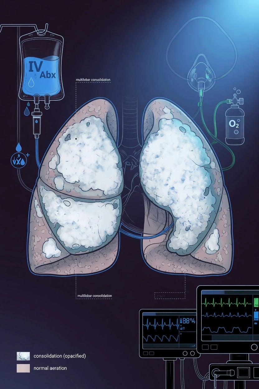
Severity assessment
CURB-65
CURB-65 score (click each)
Severe — ICU
Score >=3 = severe CAP. Consider ICU admission. Inpatient treatment with IV antibiotics. High mortality especially with score 4-5.
IDSA/ATS criteria for severe CAP
Microbiology
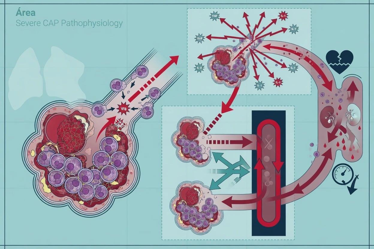
Common pathogens
All CAP
- Streptococcus pneumoniae (#1)
- Haemophilus influenzae
- Mycoplasma pneumoniae
- Chlamydophila pneumoniae
- Legionella pneumophila
- Respiratory viruses (influenza, RSV, COVID-19)
Severe CAP specifics
ICU patients
- S. pneumoniae still #1
- Legionella — disproportionately severe (hyponatraemia, GI symptoms, confusion)
- Staphylococcus aureus — especially post-viral (influenza)
- Gram-negative bacilli — Klebsiella, Pseudomonas (if structural lung disease)
- MRSA — if recent hospitalisation, antibiotics
- Always test for influenza and COVID-19
Antibiotic therapy
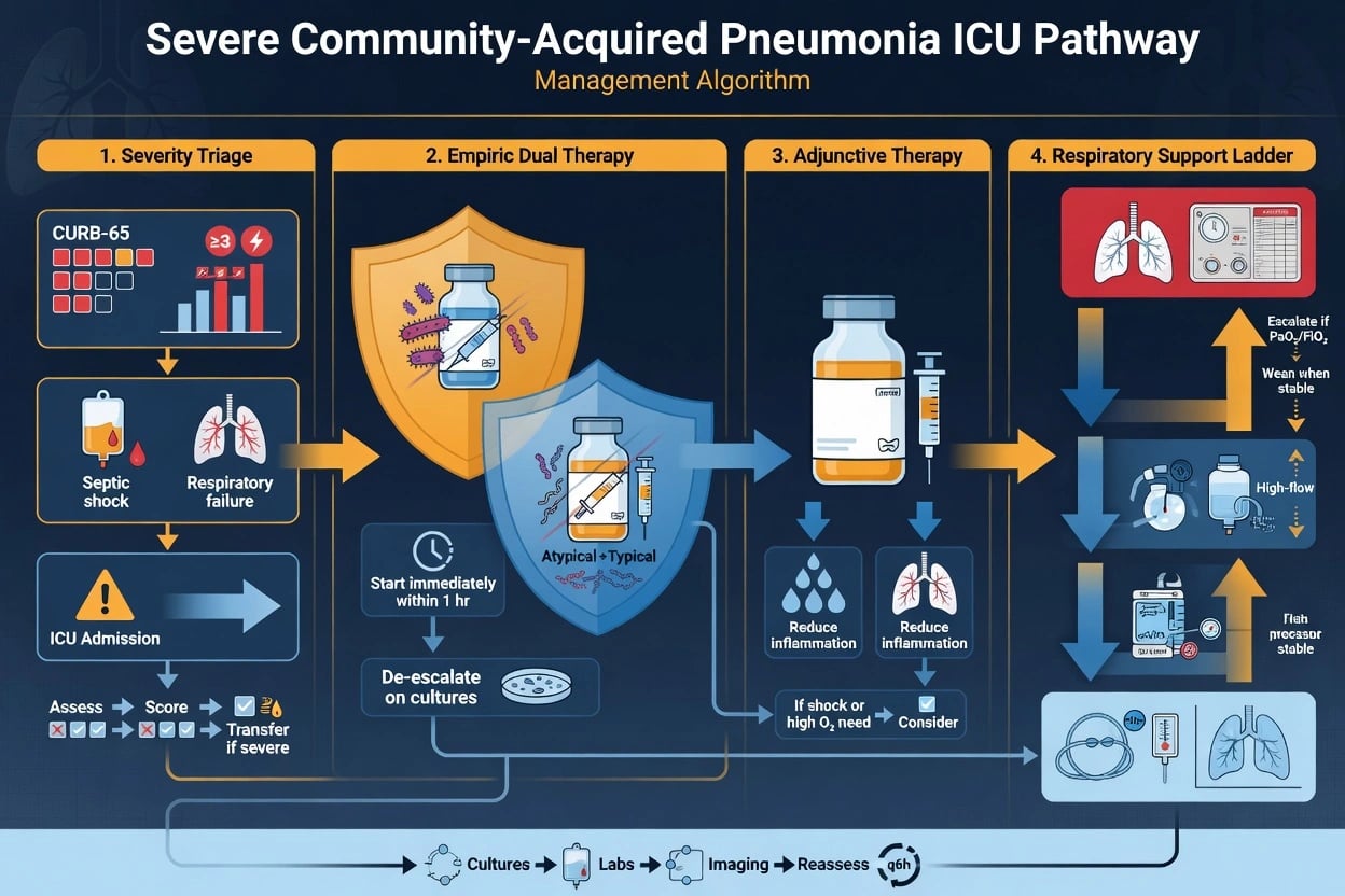
Severe CAP antibiotic protocol
Assess risk factors for resistant organisms
MRSA risk: recent hospitalisation (90 days), IV antibiotics (90 days), nursing home resident, known MRSA colonisation, structural lung disease (bronchiectasis). Pseudomonas risk: bronchiectasis, recent broad-spectrum antibiotics, repeated CAP admissions, structural lung disease.
Standard severe CAP cover (no MRSA/Pseudomonas risk)
Beta-lactam + macrolide: ceftriaxone 2g IV daily + azithromycin 500mg IV daily. OR ampicillin 2g IV Q6H + azithromycin. Alternative: respiratory fluoroquinolone (moxifloxacin 400mg IV daily) — but CAUTION: QT prolongation, tendon rupture, hypoglycaemia.
Add MRSA cover if risk factors
Add vancomycin (loading 25-30 mg/kg, then trough 15-20) OR linezolid 600mg IV BD. Linezolid preferred for pneumonia (better lung penetration, covers MRSA AND penicillin-resistant pneumococcus).
Broaden for Pseudomonas if risk factors
Use anti-pseudomonal beta-lactam: piperacillin-tazobactam 4.5g IV TDS, OR ceftazidime 2g IV TDS, OR meropenem 1g IV TDS. Plus macrolide (azithromycin) for atypical cover. If MRSA risk too: add vancomycin/linezolid.
Test for specific pathogens
Sputum culture + blood cultures (BEFORE antibiotics if possible, but do NOT delay >1 hour). Urine: pneumococcal antigen, Legionella antigen (especially severe CAP). Nasopharyngeal swab: influenza PCR, COVID-19 PCR. Consider procalcitonin (guides antibiotic duration).
Duration: 5-7 days (if clinical improvement)
Standard duration 5-7 days if clinically responding (afebrile 48-72h, improving WBC, stable vitals). Procalcitonin-guided: stop when procalcitonin <0.25 ng/mL or drops >80%. Longer for: S. aureus (14 days), Legionella (14 days), Pseudomonas (7-14 days), complications (empyema, abscess).
Adjunctive corticosteroids
Corticosteroids in severe CAP
Meta-analysis and observational studies
Population: Severe CAP (ICU)
Key finding
Reduced mortality in severe CAP with high inflammatory burden (high CRP, high procalcitonin). Greatest benefit in severe CAP with septic shock or high CURB-65.
Practice change
Consider corticosteroids in severe CAP with high inflammatory burden. CAUTION: increased infection risk with prolonged use.
Pneumonia Severity Index (PSI / PORT score)
The PSI (Pneumonia Severity Index, also called the PORT score) is the most validated severity tool for CAP. Derived from the 1997 Fine / PORT study of >14,000 patients, it uses 20 variables (demographics, comorbidities, physical examination, labs) to estimate 30-day mortality and stratify patients into five risk classes (I–V). It is more sensitive than CURB-65 for identifying low-risk patients but is cumbersome to calculate at the bedside, requires arterial oxygenation (or SpO₂), and under-weights age less aggressively in young patients with single derangements.[9]
[1]PSI risk classes
High — admit
Score 91–130. Inpatient admission. Consider ward level.
SMART-COP / SMRT-CO (ANZ-relevant)
SMART-COP (Charles 2008, derived in Australian patients) predicts the need for intensive respiratory or vasopressor support (IRVS) within the first 72 h of admission — arguably more directly relevant to ICU triage than CURB-65 or PSI which estimate mortality. The ANZ flavour is the SMRT-CO (no A for age, no P for pulse oximetry — both intentional) for simpler use.[10]
[1]Severity score comparison
CURB-65
5 variables — simplest
- Strength: very simple, easy at bedside, mortality-validated
- Weakness: ignores oxygenation, social and comorbidity factors
- Best use: triage decision (home vs hospital), score >=3 → ICU
- ANZ equivalent: CRB-65 (no urea, often used in community)
PSI / PORT
20 variables — most data
- Strength: best sensitivity for low-risk (Class I–II) outpatients
- Weakness: complex, ~30% of Class IV–V do NOT need ICU (over-triage)
- Best use: avoid admission in confidently low-risk patients
- Underestimates young single-derangement patients
IDSA/ATS criteria
9 minor + 2 major
- Strength: identifies SEVERE CAP requiring ICU specifically
- Weakness: retrospective criteria, 3-minor sensitivity only ~50%
- Best use: ICU triage in confirmed CAP
- Replaced older modified ATS criteria; needs 1 major or >=3 minor
SMART-COP
8 variables — predicts IRVS
- Strength: directly predicts need for ventilation/vasopressors
- Weakness: less widely adopted outside ANZ
- Best use: ICU/HDU triage, especially early in course
- Higher sensitivity for severe CAP than IDSA/ATS 3-minor rule
Atypical pathogens in severe CAP
"Atypical" pneumonias are caused by intracellular organisms that are not visible on Gram stain and do not grow on routine sputum culture (require special media or PCR). They cause a syndrome that may differ from "typical" pneumococcal CAP — more prominent dry cough, prodrome, extrapulmonary features, and disproportionate progression to severe disease. Empiric cover is mandatory in severe CAP because no atypical is reliably covered by beta-lactam monotherapy. [1]
Legionella pneumophila
Legionella is the most important atypical pathogen in severe CAP — it accounts for only ~2–8% of all CAP but up to 15–25% of ICU-admitted CAP, and it carries 2–3× the mortality of pneumococcal CAP of similar severity. Legionnaires' disease classically presents with the "LEGIONAIRE'S" cluster: fever, dry cough, hyponatraemia (SIADH), diarrhoea, confusion / encephalopathy, relative bradycardia, renal impairment / rhabdomyolysis, elevated transaminases, haematuria/proteinuria, and high fever with sparse CXR findings. The CXR typically shows patchy peripheral consolidation that progresses rapidly despite antibiotics — radiographic lag behind clinical severity is characteristic. [1]
- Diagnosis: urinary antigen detects L. pneumophila serogroup 1 (~80–90% of clinical disease) with >90% sensitivity/specificity, available within hours. PCR on sputum/NP swab detects other serogroups and species. Serology (4-fold IgG rise) is retrospective. Culture on buffered charcoal yeast extract (BCYE) agar takes 3–5 days.
- Treatment: macrolide (azithromycin 500 mg IV/PO daily) or respiratory fluoroquinolone (moxifloxacin, levofloxacin 750 mg). Fluoroquinolone may be slightly more effective in severe disease. Duration 14 days (21 days if immunocompromised; azithromycin for 5–7 days acceptable as monotherapy in mild disease).
- Public health: Legionnaires' disease is notifiable in nearly all jurisdictions — report for outbreak investigation (cooling towers, warm-water systems, spa pools, decorative fountains, hospital plumbing). Case-fatality 5–10% in immunocompetent, up to 25–50% if untreated. [1]
Mycoplasma pneumoniae
Mycoplasma is the most common atypical pathogen overall (especially in children and young adults, 5–20-year-olds) but rarely causes severe CAP requiring ICU. It produces the "walking pneumonia" syndrome — insidious onset, dry cough, headache, sore throat, low-grade fever. Extracellular features are common and often more prominent than the chest infection itself: [1]
- Extrapulmonary manifestations: erythema multiforme / Stevens-Johnson syndrome, bullous myringitis (ear pain), cold agglutinin haemolytic anaemia, polyarthritis, Guillain-Barré / transverse myelitis / cerebellar ataxia, myocarditis / pericarditis, pancreatitis, renal dysfunction, mucositis.
- Treatment: macrolide (azithromycin 500 mg day 1 then 250 mg, or clarithromycin), doxycycline 100 mg BD, or respiratory fluoroquinolone. Macrolide resistance is rising globally (especially Asia, >90% in some series via 23S rRNA mutations) — doxycycline or fluoroquinolone may be needed. Duration 7–14 days.
- Severe disease: an overactive host immune response (cytokine storm) drives severity rather than organism burden — steroids and IVIG have a role in refractory cases. [1]
Chlamydophila species
- Chlamydophila pneumoniae: endemic atypical pathogen, causes ~5–10% of CAP, often in older adults. Mild atypical syndrome with hoarseness/laryngitis. Treated with macrolide or doxycycline. Rarely severe alone but a common co-pathogen.
- Chlamydophila psittaci (psittacosis/ornithosis): bird exposure (parrots, pigeons, poultry). Splenomegaly, relative bradycardia, Horder's spots, epistaxis, and a severe rapidly progressive illness in >20% — can mimic Legionella. Treat with doxycycline or tetracycline (NOT beta-lactams — these organisms have cell-wall–deficient forms). Tetracycline 500 mg QID × 14 days.
- Chlamydophila abortus: ovine exposure, particularly pregnant women (live stock) — causes abortion/sepsis. [1]
Other intracellular / atypical agents
- Coxiella burnetii (Q fever): abattoir, sheep/cattle exposure. Severe disease — hepatitis, endocarditis, pneumonia. Treat with doxycycline; add hydroxychloroquine for chronic Q fever endocarditis.
- Francisella tularensis (tularemia): tick/animal exposure, ulceroglandular + severe pneumonia; add gentamicin or ciprofloxacin.
- Pneumocystis jirovecii (PJP): in immunosuppressed (HIV/AIDS CD4 <200, transplant, steroids, biologic). Hypoxia out of proportion to CXR; pneumatocele formation; pneumothorax risk. Treat with high-dose TMP-SMX + steroids (PaO₂ <70). BJC prophylaxis if at risk. [1]
Legionella
Most severe atypical
- Hyponatraemia, diarrhoea, confusion, high fever
- Urinary antigen detects serogroup 1 (most cases)
- Cover with macrolide OR fluoroquinolone — 14 days
- NOTIFIABLE disease
Mycoplasma
Commonest, usually mild
- Children/young adults, walking pneumonia
- Extrapulmonary: SJS, GBS, cold agglutinin anaemia, myocarditis
- Macrolide resistance rising — consider doxycycline/FQ
- Rarely ICU — severe disease driven by host response
Chlamydophila
pneumoniae / psittaci / abortus
- C. pneumoniae: hoarseness, mild, common co-pathogen
- C. psittaci: BIRD EXPOSURE, splenomegaly, severe — doxycycline
- C. abortus: pregnant women, sheep exposure
- Beta-lactams DO NOT work (cell-wall deficient)
Coxiella (Q fever)
Abattoir / livestock
- Sheep/cattle exposure, abattoir workers
- Hepatitis + pneumonia + endocarditis triad
- Doxycycline + hydroxychloroquine if chronic
- Notifiable; chronic form causes culture-negative endocarditis
Empiric antibiotic therapy — IDSA/ATS 2019 (Metlay)
The 2019 ATS/IDSA guideline (Metlay) replaced earlier 2007 recommendations and refined empiric therapy based on patient risk stratification and outpatient vs inpatient vs ICU setting, with specific attention to risk factors for MRSA and Pseudomonas aeruginosa.[5]
[1]Empiric therapy ladder — inpatient and ICU severe CAP
Inpatient ward CAP (non-severe, no risk factors)
Beta-lactam + macrolide: ceftriaxone 1–2g IV daily + azithromycin 500mg IV/PO daily. OR respiratory FQ monotherapy (moxifloxacin 400mg IV daily) — caution QT, tendinopathy, dysglycaemia, aortic dissection, hypokalaemia-related arrhythmia.
Severe CAP — ICU (no MRSA/Pseudomonas risk)
Beta-lactam + macrolide: ceftriaxone 2g IV daily + azithromycin 500mg IV daily. Beta-lactam alternatives: ampicillin 2g IV Q6H, cefotaxime 1–2g IV TDS. Continue at least until clinical stability.
Severe CAP with MRSA risk
Add vancomycin 25–30 mg/kg loading then 15–20 mg/kg Q8–12H (target AUC/MIC 400–600, trough 15–20) OR linezolid 600mg IV BD. Linezolid preferred for pneumonia (superior lung penetration, exotoxin suppression, MRSA + drug-resistant pneumococcus). CAUTION: linezolid — serotonin syndrome (MAO-A inhibition), thrombocytopenia >14 days, lactic acidosis, peripheral/optic neuropathy with prolonged use.
Severe CAP with Pseudomonas risk
Switch beta-lactam to anti-pseudomonal agent: piperacillin-tazobactam 4.5g IV TDS/QDS, ceftazidime 2g IV TDS, cefepime 2g IV TDS, or meropenem 1g IV TDS (reserve carbapenem for ESBL/CRE risk). Add macrolide for atypical cover (azithromycin). If both MRSA and Pseudomonas risk: anti-pseudomonal beta-lactam + vancomycin/linezolid + macrolide.
Beta-lactam allergy in severe CAP
Respiratory fluoroquinolone (moxifloxacin 400mg or levofloxacin 750mg) + aztreonam 2g IV TDS for Pseudomonas cover. Avoid cephalosporins if true anaphylaxis history. Skin test / allergy review where possible.
Influenza season — add oseltamivir
Oseltamivir 75mg PO BD within 6 hours of admission for all severe CAP during influenza activity (regardless of rapid test — empiric, stop if PCR negative). Continue 5 days (longer if immunocompromised). IV peramivir is alternative if PO/NG not feasible.
De-escalate at 48–72 hours
Narrow based on cultures / antigen / PCR results. Stop MRSA cover if cultures negative AND low suspicion. Stop anti-pseudomonal cover if no Pseudomonas grown. Continue atypical cover if Legionella confirmed (14 days) or if no pathogen identified (clinical judgment). Standard total duration 5–7 days if clinically stable.
Beta-lactam + macrolide
Standard severe CAP
- Ceftriaxone 2g IV + azithromycin 500mg IV daily
- Synergy + atypical cover + macrolide anti-inflammatory effect
- Recommended first-line by Metlay 2019 for severe CAP
- Azithromycin preferred (long t½, fewer interactions)
Respiratory fluoroquinolone
Monotherapy option
- Moxifloxacin 400mg IV / levofloxacin 750mg IV daily
- Covers pneumococcus (incl. penicillin-resistant), atypicals
- CAUTION: QT prolongation, aortic dissection, tendinopathy, hypoglycaemia
- Avoid if QTc >450, on amiodarone/antiarrhythmic, elderly
- Inadequate Pseudomonas cover (except cipro/levo)
Anti-pseudomonal regimen
If Pseudomonas risk
- Piperacillin-tazobactam / cefepime / ceftazidime / meropenem
- Plus macrolide (azithromycin) for atypicals
- Risk factors: bronchiectasis, repeated CAP, recent broad-spectrum abx
- Tailor to local antibiogram (resistance patterns vary)
MRSA regimen
If MRSA risk
- Vancomycin (AUC-guided) or linezolid 600mg BD
- Linezolid preferred for pneumonia — better ELF penetration
- Risk factors: recent hospitalisation, IV abx 90d, NH, MRSA colonisation
- Add to beta-lactam + macrolide; do NOT use MRSA cover routinely
Antibiotic duration & clinical stability (Halm criteria)
Halm et al. (JAMA 1998) defined time to clinical stability in CAP — the point at which antibiotics can be safely transitioned from IV to oral and discharge can be planned. The landmark study established that most patients achieve stability within 3–7 days and prolonged antibiotic courses offer no benefit in clinically responding patients.[8]
[1]Duration decision flow
Standard course — 5–7 days if clinically stable
Most CAP. Stop at 5 days if afebrile 48–72h AND clinically stable AND no uncomplicated extrapulmonary infection. Halm criteria guide IV-to-PO switch.
Procalcitonin-guided
Procalcitonin (PCT) <0.25 ng/mL OR >80% drop from baseline → stop antibiotics. Repeat PCT day 3, 5, 7. Reduces duration by ~2–3 days without adverse outcomes (PRORATA, ProHOSP trials).
Extended duration — specific pathogens
Legionella: 14 days (21 if immunocompromised). Pseudomonas: 7–14 days. S. aureus (incl. MRSA): 7–14 days (longer if bacteraemia/endocarditis). M. pneumoniae / C. pneumoniae: 7–14 days.
Complications — extend duration
Empyema / parapneumonic effusion requiring drainage: 2–4 weeks. Lung abscess: 4–6 weeks (until cavity resolves). Meningitis/endocarditis with CAP: 4–6 weeks. ARDS from CAP: standard duration unless ongoing infection.
Failing to respond — reassess NOT extend
Day 3 fever or worsening: do NOT empirically prolong. Reassess: resistant organism? empyema? source control? wrong diagnosis? ARDS? secondary infection? Imaging (CT chest), repeat cultures, procalcitonin trend, consider bronchoscopy.
Procalcitonin-guided antibiotic stewardship
Procalcitonin (PCT) is the pro-hormone of calcitonin, produced by extra-thyroid tissues (lung, intestine, liver) in response to systemic bacterial infection via IL-1β/TNF-α signalling. It rises within 3–6 h of bacteraemia, has a t½ of ~24 h, and falls rapidly with effective therapy — making it the best available biomarker to start, monitor and stop antibiotics in CAP. [1]
PCT <0.1 ng/mL
Bacterial infection unlikely
- Strongly discourages antibiotics in non-severe LRTI
- In CAP: still treat initially — severity overrides
- Reassess daily; consider early stop
PCT 0.1–0.25 ng/mL
Unlikely bacterial
- Short course (3 days) reasonable
- Recheck after 24–48h
- Encourage antibiotic stop
PCT 0.25–0.5 ng/mL
Possible bacterial
- Standard duration 5–7 days
- Stop when PCT falls by >80% or <0.25
- Recheck day 3, 5, 7
PCT >0.5 ng/mL
Bacterial infection likely
- Treat as bacterial CAP
- High PCT >2.0 supports severe bacterial infection
- Stop when PCT drops >80% or to <0.25
Limitations
When PCT misleads
- Falsely low in: localized infection, early sepsis, immunosuppression, anti-inflammatory biologics
- Falsely high in: trauma, surgery, cardiogenic shock, malignancy, pancreatitis, ECMO first 24h
- NEVER withhold antibiotics in severe CAP based on initial low PCT
- Trend over time is more useful than absolute value
Adjunctive corticosteroids — multiple trials
Torres 2015 — Dexamethasone in severe CAP with high inflammatory response
Multicentre RCT, single-blind
Population: 120 patients with severe CAP and CRP >150 mg/L at admission
Key finding
Treatment failure 16% vs 36% (RR 0.46, 95% CI 0.22–0.94). Mortality non-significant 5% vs 12%. Median time to clinical stability shortened.
Practice change
Consider 5-day dexamethasone in severe CAP with CRP >150. Hyperglycaemia was the main adverse effect. Established the 'inflammation-targeted' steroid paradigm.
CAPO meta-analysis (Siemieniuk 2015) — Corticosteroids in hospitalized CAP
Systematic review + individual-patient-data meta-analysis of 13 RCTs
Population: ~2000 patients hospitalized with CAP (mixed severity)
Key finding
Mortality RR 0.67 (95% CI 0.45–1.01) overall; **RR 0.39 (0.20–0.78) in severe CAP**. Reduced need for mechanical ventilation (RR 0.45) and ARDS (RR 0.30). Slight increase in hyperglycaemia; no excess infection or GI bleed.
Practice change
Steroids reduce mortality in severe CAP (NNT ~10). Effect concentrated in severe disease and high inflammatory burden. Supports routine consideration in ICU CAP.
RECOVERY-SNAP — Short-course steroids in CAP requiring supplemental oxygen
Multicentre, pragmatic, randomised, open-label platform trial
Population: Hospitalized adults with CAP requiring supplemental oxygen
Key finding
Reduced escalation to intensive respiratory support. No significant increase in secondary infection or hyperglycaemia requiring new insulin. Benefit greatest in SpO₂/FiO₂ <315 and high CRP.
Practice change
Bridges the COVID-era steroid evidence to non-COVID CAP. Supports short-course dexamethasone in hypoxic CAP. Lower dose than Torres 2015 — pragmatic bedside choice.
Failure to respond / non-responding pneumonia
Non-response is defined as persistent fever, hypoxaemia, or clinical/radiographic deterioration despite 48–72 hours of adequate therapy. Reported in 6–15% of CAP (higher in severe CAP), with associated mortality up to 40–50%. A systematic approach is essential — empiric broadening without diagnosis is harmful.[1]
Approach to non-responding CAP
Define the failure pattern
TRUE FAILURE: no improvement by 72h of appropriate therapy. PROGRESSIVE: worsening in first 48h (radiographic spread, septic shock, ARDS). DELAYED: initial improvement then deterioration after day 5 (secondary infection, complication).
Reassess microbiology — resistant organism?
Repeat blood cultures, sputum culture, urinary antigens. Consider MRSA, Pseudomonas, ESBL/CRE, Stenotrophomonas, anaerobes. Send respiratory viral PCR (influenza, RSV, COVID, adenovirus, metapneumovirus). Atypicals: Legionella urinary antigen (re-test), Mycoplasma serology/PCR, Q fever serology. Consider fungal (Aspergillus galactomannan, beta-D-glucan, PJP IF) if immunocompromised or post-influenza.
Search for complication — source control?
CT chest: empyema, lung abscess, cavitation, necrotising pneumonia, pleural collection, mediastinal/hilar nodes (TB, lymphoma), pulmonary embolism (hypercoagulable in sepsis), ARDS pattern. Bronchoscopy: atypical organisms, obstructing lesion, mucus plugging. Echocardiography: endocarditis, emboLic PE, myocardial abscess.
Wrong diagnosis?
Pulmonary embolism (pulmonary infarction mimics CAP), pulmonary oedema / cardiogenic, pulmonary haemorrhage (vasculitis, APLA, Goodpasture), organising pneumonia, eosinophilic pneumonia, vasculitis (GPA, MPA), malignancy (lymphangitis, post-obstructive), drug-induced pneumonitis, radiation pneumonitis (if recent RT), alveolar haemorrhage.
Host factors
Immunocompromise (HIV, neutropenia, transplant, biologic — TNF inhibitor, rituximab), malnutrition, alcohol use disorder, chronic renal failure, diabetes, malignancy. Optimise glycaemia, nutrition, fluid balance, smoking cessation.
Pharmacology / adherence
Drug fever (esp. beta-lactams, sulfonamides). Inadequate dosing (renal dosing not adjusted, low trough vancomycin). Malabsorption, line-related infection (CRBSI masquerading), antibiotic de-escalation too early.
Action plan
Do NOT simply broaden empirically. Targeted escalation based on findings: change antibiotics per sensitivities, drainage of empyema, anticoagulation for PE, immunosuppression for vasculitis, bronchoscopy for diagnosis. Steroids may help inflammatory failure (high CRP, organising pneumonia pattern). Multidisciplinary review with microbiology, radiology, ID.
Resistant organism
~30% of failures
- MRSA, Pseudomonas, ESBL/CRE, Stenotrophomonas, anaerobes
- Resistant pneumococcus (penicillin MIC elevated) — rare but consider
- Always re-culture + sensitivities
- Tailor to local antibiogram
Empyema / parapneumonic effusion
~20%
- Chest CT, ultrasound, diagnostic thoracentesis
- Pleural pH <7.2, LDH >1000, glucose <2.2 = complicated effusion/empyema
- Chest tube drainage + intrapleural tPA/DNase (MIST-2)
- Surgical decortication if organised; VATS first-line
ARDS
~15%
- Worsening hypoxaemia, bilateral infiltrates, P/F <300
- Lung-protective ventilation VT 6 mL/kg PBW, plateau <30
- Prone positioning, conservative fluid strategy
- Consider ECMO if P/F <100 despite optimisation
Secondary infection
~10–15%
- Late deterioration after initial improvement
- Ventilator-associated pneumonia (VAP), candidaemia, C. difficile
- CRBSI, UTI, sinusitis (nasogastric tube)
- Daily surveillance cultures, stewardship
Wrong diagnosis
~10%
- PE with pulmonary infarction
- Vasculitis: GPA, MPA, APLA, Goodpasture
- Malignancy: lymphangitis, post-obstructive
- Eosinophilic pneumonia, organising pneumonia, drug-induced
Parapneumonic effusion
Often missed
- Bedside ultrasound in every non-responding CAP
- Simple: clear, pH >7.2, LDH <1000 — antibiotics alone
- Complicated: pH <7.2 — chest tube + antibiotics
- Empyema: frank pus/positive culture — drain + consider surgery
Viral-bacterial co-infection
Co-infection (virus + bacteria at presentation) and secondary bacterial infection (bacterial superinfection after viral) both substantially worsen CAP outcomes. The classic pattern is post-influenza Staphylococcus aureus pneumonia — historically the dominant killer in 1918 and 2009 H1N1 pandemics.[11]
[1]Co-infection management
Empiric antiviral
Oseltamivir 75 mg PO BD within 6 h of admission during influenza season — empiric, do not wait for PCR. Stop if PCR negative after 24–48 h.
Empiric antibacterial
Standard severe CAP regimen. During peak influenza, consider ADDING MRSA cover (vancomycin/linezolid) regardless of formal risk factors if rapidly progressive / necrotising CXR / high PVL suspicion.
Consider antifungal
If post-influenza with cavitating/nodular infiltrates and negative bacterial cultures, send Aspergillus galactomannan (BAL > serum) + beta-D-glucan. Voriconazole/isavuconazole if influenza-associated pulmonary aspergillosis (IAPA) confirmed.
Source control & support
Lung-protective ventilation if ARDS. Conservative fluid strategy. Vasopressors for septic shock. Steroid if high inflammatory burden — note steroids may INCREASE influenza viral replication; balance against anti-inflammatory benefit (use if refractory shock or severe ARDS).
Parapneumonic effusion and empyema
Light's criteria + management of pleural collection in CAP
Detect
Bedside ultrasound in EVERY CAP — effusion found in 30–50% of hospitalised CAP, 5–10% complicated. CXR underestimates volume; lateral decubitus film or ultrasound better.
Light’s criteria — exudate vs transudate
Exudate if ANY of: pleural protein / serum protein >0.5; pleural LDH / serum LDH >0.6; pleural LDH >2/3 upper limit serum LDH. Almost all parapneumonic effusions are exudates.
Diagnostic thoracentesis if effusion >10 mm on lateral decubitus
Send pleural fluid for: pH (blood gas machine, NOT bedside strip), glucose, LDH, protein, Gram stain, culture, cell count. pH <7.2 or glucose <2.2 mmol/L or LDH >1000 = complicated = needs drainage.
Categorise ACCP — category 1–4
1: minimal, free-flowing, >10 mm — antibiotics alone. 2: 10 mm to <1/2 hemithorax — antibiotics, repeat imaging. 3: >1/2 hemithorax, loculated, pH <7.2 — tube drainage. 4: frank pus (empyema) — tube drainage ± surgery.
Drainage + intrapleural therapy (MIST-2)
Chest tube (pigtail or surgical) for category 3–4. Add **tissue plasminogen activator (tPA) 10 mg + DNase 5 mg** intrapleurally BD × 3 days (MIST-2: reduces length of stay, surgery, and treatment failure). Avoid tPA if recent surgery/bleeding.
Surgery
VATS decortication first-line for organised empyema or failed medical therapy within 7 days. Open decortitation reserved for chronic (stage III) empyema or VATS failure.
Antibiotics
Tailor to culture; cover anaerobes if aspiration risk. Empiric: ceftriaxone + metronidazole, or piperacillin-tazobactam. Duration 2–4 weeks (4–6 weeks for empyema).
Adjunctive & supportive ICU therapy
[1]Prevention
[1]Prognosis
Severe CAP outcomes
Exam practice
SAQ — Severe CAP
10 minutes · 10 marks
A 65-year-old man presents with 3 days of fever, productive cough, and dyspnoea. RR 32, SpO2 88% on room air, BP 88/52 after 2L crystalloid, confusion (oriented to person only). CXR shows right middle and lower lobe consolidation. WBC 3.2, platelets 95, BUN 22, Na 128. Temp 35.5C.
Clinical pearls
Red flags
References
- [1]Martin-Loeches I, Torres A. Severe community-acquired pneumonia Eur Respir Rev, 2022.PMID 36517046
- [2]Lim WS, van der Eerden MM, Laing R, et al. Defining community acquired pneumonia severity on presentation to hospital: an international derivation and validation study Thorax, 2003.PMID 12728155
- [3]Ferrer M, Travierso C, Cilloniz C, et al. Simplification of the IDSA/ATS criteria for severe CAP using meta-analysis and observational data Eur Respir J, 2014.PMID 24114960
- [4]Various authors. Systemic Corticosteroids, Mortality, and Infections in Pneumonia and Acute Respiratory Distress Syndrome : A Systematic Review and Meta-analysis Ann Intern Med, 2026.PMID 41325621
- [5]Metlay JP, Waterer GW, Long AC, et al. Chemically induced herbicide tolerance in rice by the safener metcamifen is associated with a phased stress response J Exp Bot, 2020.PMID 31565749
- [6]Siemieniuk RA, Meade MO, Alonso-Coello P, et al. Quantification of Bufadienolides in Bryophyllum pinnatum Leaves and Manufactured Products by UHPLC-ESIMS/MS Planta Med, 2015.PMID 26132852
- [7]Torres A, Sibila O, Ferrer M, et al. Predictive value of the admissions process and the UK Clinical Aptitude Test in a graduate-entry dental school Br Dent J, 2015.PMID 26114703
- [8]Halm EA, Fine MJ, Marrie TJ, et al. Immunoablative high-dose cyclophosphamide without stem-cell rescue for refractory, severe autoimmune disease Ann Intern Med, 1998.PMID 9867758
- [9]Fine MJ, Auble TE, Yealy DM, et al. Lieberkühn of the crypts of Lieberkühn Gastroenterology, 2003.PMID 12512022
- [10]Charles PGP, Wolfe R, Whitby M, et al. Identifying Human Trafficking Issues in Our Patients J Am Psychiatr Nurses Assoc, 2020.PMID 32066309
- [11]Martin-Loeches I, Torres A, Nagavci B, et al. Author Correction: A stochastic agent-based model of the SARS-CoV-2 epidemic in France Nat Med, 2020.PMID 33067584
- [12]RECOVERY Collaborative Group. Breast Cancer Treatment Delay in SafetyNet Health Systems, Houston Versus Southeast Brazil Oncologist, 2022.PMID 35348756