ICU · Respiratory
Severe viral pneumonia in ICU: influenza, COVID-19, RSV
Also known as Influenza pneumonia · COVID-19 · RSV pneumonia · Viral ARDS · Severe viral respiratory infection
Severe viral pneumonia requiring ICU: INFLUENZA (seasonal A/H1N1/H3N2, pandemic H1N1pdm09, avian H5N1), COVID-19 (SARS-CoV-2), RSV (respiratory syncytial virus), ADENOVIRUS, CMV (immunocompromised), MEASLES, HANTAVIRUS (cardiopulmonary syndrome). Common features: viral ARDS, high oxygen requirement, lymphopenia, secondary bacterial/fungal infection. COVID-19 specific: hypercoagulability (VTE), multisystem inflammation (MIS), cytokine storm. Diagnosis: multiplex respiratory PCR panel; viral vs bacterial differentiation with procalcitonin (low in viral), CRP. Treatment: SUPPORTIVE (oxygen, lung-protective ventilation, prone positioning), ANTIVIRALS (oseltamivir/zanamivir for influenza within 48h; remdesivir for COVID-19; ribavirin for RSV in transplant), IMMUNOMODULATORS (dexamethasone, tocilizumab, baricitinib for COVID-19). Infection prevention: airborne/droplet isolation, PPE. Antiviral resistance (oseltamivir H275Y). Secondary infection (bacterial/fungal — CAPA aspergillosis, staphylococcal) common — monitor.
On this page & tools
Your progress
Saved locally on this device.
Target exams
Red flags
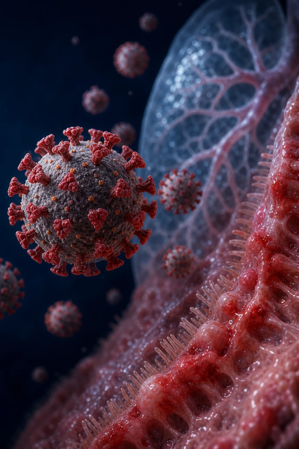
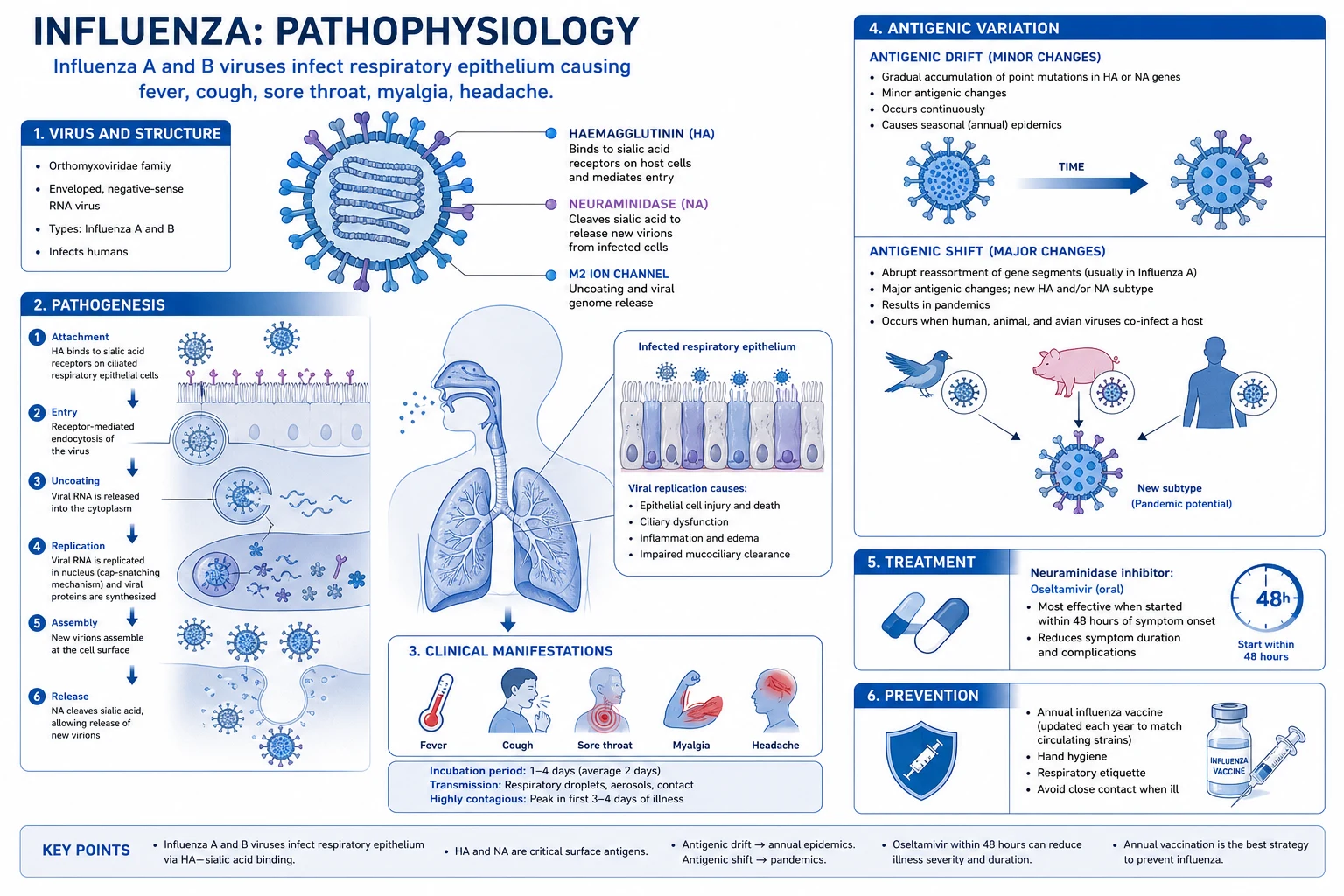
Viral pneumonias comparison
| Feature | Influenza (A/H1N1, H3N2) | COVID-19 (SARS-CoV-2) | RSV |
|---|---|---|---|
| Season | Winter (seasonal) | Year-round (waves) | Winter (paediatric; elderly) |
| Incubation | 1-4 days | 2-14 days | 2-8 days |
| Chest CT | Bilateral GGO, consolidation | Bilateral GGO (peripheral, basal), crazy paving | Bronchial wall thickening, GGO, AT |
| Hallmarks | High fever, myalgia, sore throat, dry cough | Anosmia, ageusia, variable severity | Wheeze, bronchiolitis (children) |
| Lymphopenia | Common | VERY common (marker of severity) | Less common |
| Coagulopathy | Mild | HYPERCOAGULABLE (VTE, microthrombi) | No |
| Antiviral | Oseltamivir (within 48h) | Remdesivir (early), nirmatrelvir/ritonavir (early) | Ribavirin (rare) |
| Steroids | NOT routine (may worsen viral) | YES (dexamethasone — RECOVERY) | NOT routine |
| Mortality (ICU) | 10-20% | 20-40% (early pandemic), lower now | 10-20% (elderly) |
ICU management of severe viral pneumonia
- Recognise — hypoxia (SpO2 <92%), respiratory distress, bilateral infiltrates, viral syndrome (fever, cough, lymphopenia). PCR (nasopharyngeal) for influenza/COVID/RSV
- Oxygen support — escalate as needed: nasal cannula → high-flow nasal cannula (HFNC) → CPAP/NIV → intubation. Target SpO2 92-96%
- Antiviral therapy — INFLUENZA: oseltamivir 75 mg BD (within 48h of symptoms; give even if late in ICU). COVID-19: remdesivir (200 mg day 1, then 100 mg OD × 4-10 days; early/moderate). RSV: ribavirin (rarely used in adults)
- Immunomodulation (COVID-19) — DEXAMETHASONE 6 mg OD (RECOVERY — reduces mortality in oxygen/ventilated). TOCILIZUMAB (IL-6 inhibitor — severe with CRP >75 or rapid escalation). BARICITINIB (JAK inhibitor)
- Ventilation (if needed) — LUNG-PROTECTIVE: tidal volume 4-6 mL/kg, plateau <30, PEEP titrated. PRONE POSITIONING (>12h/day) for moderate-severe ARDS
- VTE prophylaxis — LMWH (COVID-19 hypercoagulable). Therapeutic if confirmed VTE or high D-dimer
- Monitor for secondary infection — bacterial (staph, pneumococcus), fungal (aspergillosis — CAPA/IAPA). BAL galactomannan in severe/ventilated
- ECMO — refractory hypoxaemia (VV-ECMO) despite optimised ventilation
SAQ — Severe COVID-19 pneumonia with refractory hypoxaemia
10 minutes · 10 marks
A 58-year-old man with PCR-confirmed SARS-CoV-2 pneumonia is intubated and ventilated for refractory hypoxaemia (PaO2/FiO2 90 on FiO2 0.8, PEEP 14). CRP is 145, ferritin 1800, D-dimer 3200, lymphocytes 0.6. He is on lung-protective ventilation and has been proned. Outline the evidence-based pharmacological therapy you would add.
SAQ — Influenza A pneumonia with Staphylococcal co-infection
10 minutes · 10 marks
A 42-year-old previously fit man presents in mid-winter with 3 days of fever, myalgia and dry cough, now with worsening dyspnoea, septic shock and bilateral consolidations with cavitation. Rapid PCR is positive for influenza A. Blood cultures grow Staphylococcus aureus. Procalcitonin is 12 ng/mL. Critically appraise the antiviral, antibiotic and supportive management.
Clinical pearls
Red flags
Prognosis
RECOVERY trial — dexamethasone in COVID-19 (2021, NEJM)
RCT: 6,425 hospitalised COVID-19 patients. Dexamethasone 6 mg OD for 10 days vs usual care.
- Mortality — VENTILATED: 29.3% (dexa) vs 41.4% (usual) — RR 0.64, NNT 8
- Mortality — OXYGEN ONLY: 23.3% (dexa) vs 26.2% (usual) — RR 0.82, NNT 25
- Mortality — NO OXYGEN: 17.8% (dexa) vs 14.0% (usual) — RR 1.19 (HARM — do NOT give to non-hypoxic)
- CONCLUSION: Dexamethasone reduces mortality in COVID-19 requiring oxygen/ventilation. Do NOT give to patients not requiring oxygen. [1]
Remdesivir (ACTT-1): reduced recovery time (10 vs 15 days), no clear mortality benefit. Tocilizumab (REMAP-CAP/RECOVERY): reduces mortality in severe COVID-19 with systemic inflammation. Overall COVID-19 ICU mortality: 20-40% (early pandemic), reduced with dexamethasone, prone, antivirals.
Pathogen deep-dive
Influenza — the seasonal and pandemic killer
Influenza viruses are segmented negative-sense single-stranded RNA orthomyxoviruses, classified A, B and C. Influenza A (subtyped by haemagglutinin H and neuraminidase N) causes seasonal disease and all pandemics; influenza B (Victoria and Yamagata lineages) causes seasonal disease only (no pandemics, no animal reservoir). Antigenic drift (point mutations in H/N) drives annual seasonal epidemics and necessitates yearly vaccine reformulation; antigenic shift (reassortment of segmented genomes when two strains co-infect a host — classically pigs as the "mixing vessel") produces novel pandemic strains. The 1918-19 "Spanish flu" (H1N1, ~50-100 million deaths), 1957 Asian (H2N2), 1968 Hong Kong (H3N2) and 2009 pandemic H1N1pdm09 all arose from shift/reassortment.[9] }
Severe influenza pneumonia in ICU presents after a 1-4 day incubation with abrupt high fever, rigors, myalgia (out of proportion — "hit by a truck"), headache, sore throat and a dry cough. Progression to hypoxaemic respiratory failure can be rapid (12-48 h). Classical CT findings are bilateral patchy ground-glass opacities and consolidation, often with a basal and peripheral predominance; lobar or multilobar consolidation suggests bacterial superinfection. The single most dangerous complication is post-influenza bacterial pneumonia, classically caused by Staphylococcus aureus (including MRSA — often necrotising and cavitating), Streptococcus pneumoniae and Haemophilus influenzae — this is what killed most victims in 1918 (bacterial, not viral).[9] }
Antiviral therapy — start oseltamivir immediately, do NOT wait for PCR. Oseltamivir (oral neuraminidase inhibitor) is the workhorse: 75 mg PO BD for 5 days (double to 150 mg BD in critically ill/immunocompromised, longer duration). The IDSA 2018 guidelines recommend treating ALL hospitalised patients with suspected/confirmed influenza empirically, irrespective of symptom duration — the 48-hour "window" applies to outpatient benefit, but ICU patients with ongoing viral replication still derive mortality reduction.[5][9] }
SARS-CoV-2 (COVID-19) — the modern paradigm
Severe acute respiratory syndrome coronavirus 2 is a positive-sense single-stranded RNA betacoronavirus (same genus as SARS-CoV-1 and MERS-CoV) that emerged in late 2019 and caused a global pandemic. The virus binds ACE2 receptors (highly expressed in type II pneumocytes, nasopharynx, intestine, kidney, endothelium), which explains its multi-organ tropism. Severe COVID-19 progresses through two overlapping phases: an early viral replicative phase (days 1-7: fever, cough, anosmia, viraemia) and a later hyperinflammatory phase (days 7-14: cytokine release, endothelial injury, microthrombi, ARDS). This biphasic course underpins the timing of therapy — antivirals early, immunomodulators late.[1] }
Hallmark features distinguishing COVID-19 from other viral pneumonias: profound lymphopenia (the most consistent severity marker), silent/happy hypoxaemia (deep hypoxia with minimal dyspnoea — explained by preserved lung compliance early and loss of hypoxic ventilatory drive), hypercoagulability (D-dimer often >1000; VTE in 20-40% of ICU patients despite prophylaxis; pulmonary microthrombi and strokes), anosmia/ageusia, and the bizarrely discordant chest X-ray (severe CT changes with a relatively normal portable CXR). Typical CT: bilateral, peripheral, basal, rounded ground-glass opacities with a posterior predominance; "crazy-paving" later.[1] }
Specific therapy (the COVID armamentarium). (1) Dexamethasone 6 mg OD for up to 10 days — RECOVERY showed a mortality benefit ONLY in patients on oxygen or ventilation (NNT 8 ventilated, NNT 25 on oxygen); it was harmful in those not needing oxygen. (2) Remdesivir (nucleotide analogue RNA-polymerase inhibitor) 200 mg IV day 1 then 100 mg OD × 4-9 days — ACTT-1 shortened recovery (10 vs 15 days); best given early. (3) Tocilizumab (IL-6 receptor blocker) 8 mg/kg IV (max 800 mg, one repeat) — RECOVERY + REMAP-CAP showed mortality reduction in rapidly escalating or ventilated patients with systemic inflammation (CRP >75). (4) Baricitinib (JAK1/2 inhibitor) — ACTT-2 (with remdesivir) and RECOVERY (monotherapy) both showed mortality benefit in oxygen-requiring COVID-19. Choose ONE immunomodulator (tocilizumab OR baricitinib) plus dexamethasone, not both.[2][10][11][12] }
Respiratory syncytial virus (RSV) — under-recognised in adults
RSV is an enveloped negative-sense ssRNA pneumovirus (now Orthopneumovirus). It is the commonest cause of bronchiolitis in infants (virtually all children are infected by age 2) but is increasingly recognised in adults — severe disease targets three groups: the elderly (especially aged care residents, where RSV rivals influenza for hospitalisation and death), adults with chronic cardiopulmonary disease (COPD, heart failure), and the immunocompromised (haematopoietic stem cell and lung transplant recipients, where mortality reaches 20-80%). There is no proven specific antiviral of clear benefit in immunocompetent adults — management is supportive (oxygen, ventilation). Ribavirin (aerosolised or oral guanosine analogue) is reserved for the immunocompromised transplant patient with proven RSV lower respiratory tract disease — evidence is observational but mortality in untreated HSCT RSV pneumonitis is very high. Palivizumab (monoclonal anti-RSV F-protein) is licensed for prophylaxis in high-risk infants (premature, haemodynamically significant congenital heart disease, BPD) — NOT used in adults. A long-acting monoclonal, nirsevimab, and the first adult RSV vaccines (Arexvy, Abrysvo) have recently been licensed for older adults and pregnancy.[18] }
Adenovirus — community outbreaks and the immunocompromised
Non-enveloped dsDNA viruses (>50 serotypes; types 3, 4, 7, 14, 21 most often cause severe pneumonia). In immunocompetent adults, adenovirus causes a self-limiting upper respiratory illness or pharyngoconjunctival fever, but adenovirus type 7 can cause severe, sometimes fatal, community-acquired pneumonia in young, previously fit adults (military recruits, closed communities). In the immunocompromised (paediatric HSCT, solid organ transplant), disseminated adenovirus causes pneumonia, hepatitis, colitis, encephalitis and haemorrhagic cystitis. Diagnosis is by respiratory PCR (and quantitative blood PCR in immunocompromised — viral load predicts disease). Treatment is largely supportive; cidofovir (with probenecid and hydration to limit nephrotoxicity) or brincidofovir is used for severe/disseminated disease. Strict contact and droplet precautions prevent nosocomial spread (adenovirus resists desiccation and survives on surfaces).[21] }
Cytomegalovirus (CMV) — the transplant pneumonitis
CMV is a betaherpesvirus that establishes lifelong latency; disease is almost exclusively a problem of the immunocompromised — classically the allogeneic haematopoietic stem cell transplant (HSCT) and lung transplant recipient in the first 100 days post-transplant. CMV pneumonitis presents with fever, dyspnoea, hypoxaemia and bilateral interstitial infiltrates; the histological hallmark is an interstitial pneumonitis with characteristic "owl's-eye" intranuclear inclusion bodies. Diagnosis requires quantitative CMV PCR (blood and BAL) plus compatible clinical/radiological syndrome — the 2017 consensus definitions distinguish CMV infection (viraemia/replication) from CMV disease (end-organ damage with symptoms). Treatment is ganciclovir IV (induction 5 mg/kg BD for 2-3 weeks, then maintenance/valganciclovir); foscarnet or cidofovir are alternatives for ganciclovir resistance (UL97 mutation). Prevention is central: letermovir prophylaxis (terminase inhibitor) in HSCT recipients dramatically reduces CMV reactivation and mortality, and pre-emptive therapy guided by PCR surveillance is standard.[18][19] }
Measles — giant-cell pneumonia
Measles (morbillivirus, paramyxovirus) causes giant-cell pneumonia (pathognomonic Warthin-Finkeldey multinucleated giant cells) — most severe in the malnourished, pregnant, and immunocompromised (especially those with cell-mediated defects, in whom measles is often fatal and may present without the rash). Secondary bacterial pneumonia (S. pneumoniae, H. influenzae, staph) and measles inclusion-body encephalitis are major killers. There is no specific antiviral; management is supportive plus vitamin A (two doses, reduces morbidity/mortality — especially in deficiency), aggressive treatment of secondary bacterial infection, and ribavirin (occasionally, for severe immunocompromised pneumonitis, on an unlicensed basis). Prevention via MMR vaccination is the only durable public-health answer. [1]
Hantavirus — cardiopulmonary syndrome (HCPS)
Hantaviruses are bunyaviruses shed in rodent excreta (urine, droppings, saliva); humans inhale aerosolised virus. Two main syndromes: haemorrhagic fever with renal syndrome (HFRS) (Old World hantaviruses — Puumala, Hantaan, Seoul — nephritis and haemorrhage) and hantavirus cardiopulmonary syndrome (HCPS) (New World — Sin Nombre in North America, Andes in South America — a fulminant pulmonary oedema with myocardial depression). HCPS has four phases: febrile prodrome (3-5 days: fever, myalgia, headache, GI symptoms), cardiopulmonary (sudden hypoxaemia, non-cardiogenic pulmonary oedema, myocardial depression with low cardiac output — NOT a septic vasodilatory shock), diuretic (rapid resolution), and convalescent. The diagnostic triad at presentation is thrombocytopenia + hemoconcentration (rising haematocrit) + left-shift neutrophilia with myelocytes plus bilateral infiltrates and a rodent-exposure history. There is no proven antiviral; management is meticulous supportive care — early intubation, lung-protective ventilation, and judicious fluids (over-resuscitation worsens pulmonary oedema; a falling cardiac output is from myocardial failure, not hypovolaemia — so vasopressors/inotropes, not fluid boluses). Extracorporeal support (ECMO) has dramatically improved HCPS survival. Mortality untreated is 35-50%. The Andes virus is unique in showing person-to-person transmission.[17] }
Beyond the "big three" — other viral pneumonias in ICU
| Pathogen | Host / setting | Hallmark features | Diagnosis | Specific therapy |
|---|---|---|---|---|
| Adenovirus (esp. type 7) | Immunocompetent (military recruits), immunocompromised | Pharyngoconjunctival fever, severe CAP, pneumonia + hepatitis/colitis/cystitis | Respiratory + quantitative blood PCR | Supportive; cidofovir/brincidofovir if severe |
| CMV | HSCT, lung/heart transplant (first 100 days) | Bilateral interstitial pneumonitis, "owl-eye" inclusions | Quantitative PCR (blood + BAL); histology | Ganciclovir IV (or foscarnet); letermovir prophylaxis |
| Measles | Unvaccinated, malnourished, immunocompromised | Giant-cell pneumonia, rash (may be absent if immunocompromised) | Serology, PCR, viral culture (urine/throat) | Supportive + vitamin A; ribavirin (rare) |
| Hantavirus (HCPS) | Rodent exposure (Sin Nombre NA, Andes SA) | Thrombocytopenia + hemoconcentration + myeloid left-shift; pulmonary oedema + myocardial depression | Serology (IgM/IgG), PCR | Supportive; minimise fluids; ECMO |
| Varicella (VZV) | Non-immune adults, pregnancy | Pneumonia 1-6 days after rash, miliary nodules on CXR | PCR, DFA of scrapings | IV aciclovir (10 mg/kg TDS) |
| MERS-CoV / SARS-CoV-1 | Middle East travel (MERS); zoonotic | Rapid ARDS, renal failure (MERS) | PCR | Supportive (no proven antiviral) |
| Metapneumovirus | All ages, immunocompromised | RSV-like illness | Multiplex PCR | Supportive; ribavirin (rare) |
Diagnostic workup
Respiratory viral diagnostic pathway in the ICU
- Multiplex respiratory PCR panel — the first-line test. A single nasopharyngeal swab or BAL sample simultaneously detects influenza A/B (+ subtyping), SARS-CoV-2, RSV, adenovirus, parainfluenza 1-4, metapneumovirus, rhinovirus/enterovirus, bocavirus, Mycoplasma, Chlamydia, Bordetella, and often M. pneumoniae and C. pneumoniae. Turnaround 1-4 h. Sensitivity/specificity >95% for most targets. Use EARLY — before antibiotics confound.[21] }
- Send a rapid point-of-care influenza + SARS-CoV-2 + RSV PCR at the bedside — a positive influenza result triggers immediate oseltamivir and a negative one (in the right patient) lets you withhold it. Do NOT wait for the full panel to start oseltamivir if influenza is suspected.[9] }
- BAL / lower-respiratory sample in intubated patients — for viruses (CMV, adenovirus quantitative PCR), bacterial culture, Aspergillus galactomannan, and atypicals. Send BAL galactomannan on EVERY ventilated COVID patient to screen for CAPA.[14] }
- Bloods — distinguish viral from bacterial. Procalcitonin is LOW (<0.1-0.5 ng/mL) in viral infection and rises with bacterial superinfection — a rising procalcitonin on day 3-7 is the earliest clue to secondary bacterial pneumonia. CRP is non-specific (high in both, but very high in bacterial). Ferritin/LDH high in COVID (cytokine storm, severity markers). D-dimer high in COVID (coagulopathy). Lymphocytes low (viral); neutrophilia suggests bacterial.[20] }
- CMV / EBV / BK PCR in any immunocompromised patient (transplant, neutropenia, HIV, biologics) — do NOT miss CMV pneumonitis in the HSCT recipient.[18] }
- Imaging — CXR (bilateral infiltrates), CT chest (ground-glass opacities; look for consolidation suggesting bacterial superinfection, nodules, cavitation or pleural effusion). Ultrasound (B-lines, consolidation, effusion) at the bedside.
- Serology (paired) — acute + convalescent for hantavirus, adenovirus, Mycoplasma, Chlamydia — confirms retrospectively, useful for public health.
- Blood and sputum cultures — sent BEFORE antibiotics if possible; repeat if deteriorating (secondary bacteraemia).
Viral vs bacterial pneumonia — bedside and laboratory differentiation
| Feature | Viral pneumonia | Bacterial pneumonia |
|---|---|---|
| Onset | Gradual (days), prodromal viral illness | Abrupt, single rigor |
| Symptoms | Dry cough, myalgia, sore throat, headache | Purulent/rusty sputum, pleuritic chest pain |
| White cells | Normal/low, lymphopenia | Leucocytosis, neutrophilia |
| Procalcitonin | LOW (<0.1-0.5) — characteristic | HIGH (>0.5, often >2) |
| CRP | Mild-moderately raised | Markedly raised (>100) |
| Chest imaging | Bilateral, interstitial, ground-glass | Lobar/segmental consolidation |
| Sputum Gram stain | Scant, no dominant organism | Neutrophils + organism |
| Response to beta-lactam | None | Good within 48-72 h |
| Hallmark complication | Secondary bacterial infection, viral ARDS | Bacteraemia, empyema, cavitation |
Using procalcitonin to start/stop antibiotics in viral pneumonia
- Admission procalcitonin <0.1 ng/mL — strongly supports viral; withhold antibiotics unless clearly deteriorating or bacterial superinfection suspected.[20] }
- Procalcitonin 0.1-0.5 — borderline; reassess with CRP, cultures and imaging; consider short empiric cover if high suspicion.
- Procalcitonin >0.5 (and especially >2) — bacterial infection likely; start empiric antibiotics (cover typicals + atypicals + MRSA if severe).
- Rising procalcitonin on day 3-7 in a viral pneumonia — early bacterial superinfection; treat.
- Falling procalcitonin >80% from peak — stop antibiotics (antibiotic stewardship, shorter course).
- Caveats — procalcitonin is low in early bacterial sepsis (lag), in some atypicals (Mycoplasma) and in immunosuppressed/elderly; high after major trauma/surgery/cardiac arrest; never override clear clinical deterioration.
Pathogen-specific antiviral therapy
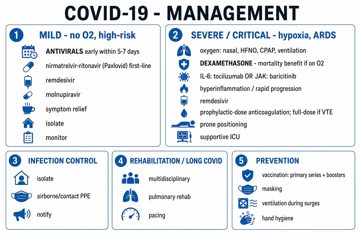
Antivirals for severe viral pneumonia — agents, doses and evidence
| Virus | Drug (class) | Dose (adult, ICU) | Key evidence / note |
|---|---|---|---|
| Influenza A/B | Oseltamivir (neuraminidase inhibitor) | 75 mg PO BD × 5 d (150 mg BD, longer course if severe/immunocompromised) | Mortality benefit even when started >48h in ICU; do NOT wait for PCR[5][9] } |
| Influenza (oseltamivir-resistant, H275Y) | Zanamivir (inhaled neuraminidase inhibitor) | 10 mg inhaled BD × 5 d | Use when H275Y resistance proven/suspected (no renal dose adjust; avoid in severe bronchospasm) |
| Influenza (severe, ICU) | Peramivir (IV neuraminidase inhibitor) | 600 mg IV single dose (repeat if critical) | IV option when PO/inhaled not feasible |
| Influenza A (baloxavir) | Baloxavir (cap-dependent endonuclease inhibitor) | Single dose by weight | Mostly outpatient; emerging resistance (PA I38T) limits ICU role |
| SARS-CoV-2 | Remdesivir (RNA-polymerase inhibitor) | 200 mg IV d1, then 100 mg OD × 4-9 d | ACTT-1: shorter recovery; best early; renal dosing[3] } |
| SARS-CoV-2 | Baricitinib (JAK1/2 inhibitor) | 4 mg PO OD × 14 d (2 mg if eGFR 30-60) | ACTT-2 + RECOVERY: mortality benefit; choose vs tocilizumab[10][11] } |
| SARS-CoV-2 | Tocilizumab (IL-6R blocker) | 8 mg/kg IV (max 800 mg) × 1, one repeat | REMAP-CAP/RECOVERY: severe/escalating/ventilated + CRP>75[4][12] } |
| SARS-CoV-2 | Dexamethasone (glucocorticoid) | 6 mg PO/IV OD × up to 10 d | RECOVERY + WHO REACT: mortality benefit if on O2/ventilated[2][13] } |
| RSV (immunocompromised) | Ribavirin (inhaled/oral guanosine analogue) | Aerosolised 6 g/300 mL × 18 h/day, or oral weight-based | Observational benefit in HSCT/lung transplant RSV pneumonitis; teratogenic |
| CMV | Ganciclovir (IV) → valganciclovir | 5 mg/kg IV BD × 2-3 wk induction | UL97 resistance → foscarnet/cidofovir; letermovir prophylaxis[18][19] } |
| Adenovirus | Cidofovir (+ probenecid/hydration) | 5 mg/kg IV weekly × 2, then fortnightly | Nephrotoxic — reserve for severe/disseminated; brincidofovir alternative |
| VZV / HSV | Aciclovir (IV) | 10 mg/kg IV TDS × 7-10 d | Reduce dose in renal failure |
Viral ARDS — ventilation and adjuncts
Ventilation strategy for viral ARDS (influenza, COVID-19)
- Diagnose ARDS — Berlin criteria: acute (<1 week) bilateral infiltrates not fully explained by cardiac failure/fluid overload, with PaO2/FiO2 ≤300 (mild ≤300, moderate ≤200, severe ≤100). Viral ARDS (especially COVID-19) is often a high-compliance, low-recruitability phenotype early — true "stiff lung" appears later.[7] }
- Lung-protective ventilation (the ARDSnet standard). Tidal volume 4-6 mL/kg predicted body weight, plateau pressure <30 cmH2O, driving pressure <15, permissive hypercapnia (pH ≥7.20), PEEP titrated on a PEEP/FiO2 table. This single intervention reduced mortality from 40% to 31% in the original ARDSnet trial.[7] }
- Prone positioning (PROSEVA). For moderate-severe ARDS (PaO2/FiO2 <150): prone >16 h/day (continuous sessions). PROSEVA reduced 90-day mortality from 32.8% to 16.0%. In COVID-19, consider early "awake proning" for non-intubated patients on HFNC.[6] }
- Fluid conservative — target a negative fluid balance / euvolaemia once shock resolves; reduces time ventilated. Avoid fluid overload (worsens pulmonary oedema).
- Pulmonary vasodilator — inhaled nitric oxide or epoprostenol as a rescue for refractory hypoxaemia (improves oxygenation but NO proven mortality benefit).
- Neuromuscular blockade — short continuous infusion (48 h) for severe ARDS to facilitate lung-protective ventilation and reduce dyssynchrony (ACURASYS showed benefit; ROSE did not — current practice is selective).
- VV-ECMO — for refractory hypoxaemia (PaO2/FiO2 <80) or refractory hypercapnia/acidosis despite optimised ventilation + prone. EOLIA defined modern criteria; survival to recovery ~50-60% in experienced centres.[8] }
- Reassess daily — driving pressure trend, recruitability, secondary infection, DVT, ICU-acquired weakness.
Non-COVID vs COVID-19 ARDS — what is different?
| Feature | Non-COVID viral/bacterial ARDS | COVID-19 ARDS (early) |
|---|---|---|
| Compliance | Severely reduced (stiff lung) | Often PRESERVED (high compliance) early |
| Mechanism of hypoxaemia | Shunt, low V/Q | Shunt + loss of hypoxic pulmonary vasoconstriction + microthrombi |
| Ventilatory drive | Tachypnoea, distress | Often reduced ("happy/silent hypoxia") |
| D-dimer / coagulopathy | Mild-moderate | Marked; microthrombi, high VTE |
| Prone response | Moderate (recruits dorsal lung) | Often dramatic |
| Response to steroids | Routine steroids NOT recommended (possible harm in influenza) | Dexamethasone REDUCES mortality (RECOVERY)[2] } |
| Fungal superinfection | Uncommon | CAPA in 5-30% of ventilated[14] } |
Secondary bacterial and fungal superinfection
Secondary infection is the rule rather than the exception in severe viral pneumonia and is a major driver of late mortality. Distinguish co-infection (present at admission, day 0-2) from superinfection (arising days 5-14). The classic pathogens: [1]
- Influenza → Staphylococcus aureus (including MRSA) is the most feared — often a necrotising, cavitating pneumonia with rapid progression and septic shock; viral neuraminidase exposes bacterial adherence receptors on respiratory epithelium. Also S. pneumoniae and H. influenzae. Combined influenza + S. aureus mortality can reach 30-50%.
- COVID-19 → Aspergillus (CAPA). The combination of severe lung injury, dexamethasone, broad-spectrum antibiotics and tocilizumab creates the perfect storm for invasive aspergillosis. Reported in 5-30% of ventilated COVID-19 patients; mortality 40-60%. Diagnosis (ECMM/ISHAM consensus): BAL galactomannan >1.0 (serum less useful in CAPA), Aspergillus culture/PCR, new pulmonary infiltrates/nodules. Treat with voriconazole or isavuconazole (isavuconazole has fewer drug interactions and a survival signal in some series). Screen BAL galactomannan routinely in the ventilated COVID-19 patient.[14][15] }
- Influenza → Aspergillus (IAPA). Influenza-associated pulmonary aspergillosis is now recognised in immunocompetent hosts with severe influenza (rate 7-19% in ICU cohorts) — diagnose and treat as for CAPA.
- General bacterial superinfection — Pseudomonas, Klebsiella, Enterobacterales, and Staph in the ventilated patient. Overall bacterial co/superinfection in COVID-19 ICU cohorts is ~14% (Lansbury meta-analysis).[16] }
Detecting and treating secondary infection in viral pneumonia
- Baseline cultures + procalcitonin on admission — to compare against later.
- Daily clinical review — new fever, rising secretions/purulence, worsening oxygenation, rising lactate or new sepsis suggests secondary infection.
- Repeat procalcitonin and CRP — a rising procalcitonin from a low baseline is the earliest bacterial clue.[20] }
- Repeat imaging — new lobar consolidation, cavitation, pleural effusion, or nodules suggests bacterial/fungal superinfection (vs the diffuse viral pattern).
- Send BAL galactomannan + culture in the ventilated COVID-19/influenza patient (screen for CAPA/IAPA).[14] }
- Start empiric antibiotics if bacterial superinfection suspected — cover typicals + atypicals + MRSA (vancomycin/linezolid) if severe/known colonised, and Pseudomonas if late/ventilated. Use procalcitonin-guided stopping.[20] }
- Antifungal therapy if CAPA/IAPA confirmed or probable — voriconazole or isavuconazole; therapeutic drug monitoring (voriconazole trough 1-5.5 mg/L).[15] }
- De-escalate aggressively as cultures and procalcitonin resolve — antifungal/antibiotic stewardship limits resistance and further CAPA risk.
Infection prevention and control (IPC)
IPC for the patient with severe viral pneumonia
- Isolation at triage — apply source control immediately on suspicion: surgical mask on the patient, single room or cohort bay. Do NOT wait for PCR confirmation.
- Choose the route of transmission. Airborne (negative-pressure single room, N95/FFP3/P2 respirator): SARS-CoV-2 (during aerosol-generating procedures — AGPs), influenza (AGPs), measles (always airborne), adenovirus (severe), varicella (always airborne — also requires negative pressure and immunity check). Droplet (single room or cohort; surgical mask, eye protection within 1 m): influenza/RSV/SARS-CoV-2 for routine care. Contact (gloves, gown): RSV, adenovirus, all enteric viruses.
- Personal protective equipment (PPE) — for AGPs (intubation, NIV, HFNC, nebulisers, suction, bronchoscopy, CPR) use full airborne PPE: fitted N95/FFP3/P2 respirator, eye protection (goggles/face shield), long-sleeved fluid-resistant gown, gloves. Don and doff carefully — a trained "buddy" observer at doffing prevents self-contamination.
- AGP risk — HFNC and NIV are AGPs (generate aerosol); use them with airborne precautions in COVID-19/influenza, a closed circuit, viral/bacterial filter on the exhalation port, and adequate room ventilation.
- Hand hygiene — the single most effective IPC measure; alcohol-based hand rub before/after every patient contact and PPE change.
- Environmental cleaning — adenovirus and some viruses survive on surfaces (adenovirus resists desiccation); use a hospital-grade detergent/disinfectant with proven virucidal activity; dedicate equipment (stethoscope, BP cuff, thermometer).
- Staff protection — annual influenza vaccination (mandatory in many ICU policies); COVID-19 vaccination; measles/MMR immunity documentation; varicella immunity; fit-testing of respirators.
- Outbreak management — notify public health; cohort cases; restrict symptomatic staff; visitor screening; rapid molecular diagnostics; daily surveillance of exposed patients and staff.
- Duration of isolation — pathogen-specific: influenza until 24 h fever-free + improving + (immunocompromised) negative PCR; RSV for the duration of symptoms; SARS-CoV-2 per local policy (symptom- or test-based); measles for 4 days after rash onset (longer if immunocompromised); adenovirus until stool PCR negative in immunocompromised.
Antiviral resistance
- Oseltamivir-resistant influenza (H275Y / H274Y in N1 numbering). A single histidine-to-tyrosine substitution at neuraminidase position 275 confers high-level oseltamivir resistance while retaining susceptibility to zanamivir (and peramivir). The classic scare was the 2007-2009 seasonal H1N1 (A/Brisbane/59) which circulated with ~100% H275Y — patients were untreated by oseltamivir. Risk factors: prolonged viral replication in severely immunocompromised (HSCT, haematological malignancy) patients treated with oseltamivir (selection pressure over weeks of shedding). Suspect H275Y in an immunocompromised patient with persistent influenza shedding or clinical deterioration despite oseltamivir — send resistance testing (genotypic Sanger/next-generation sequencing of NA) and switch to inhaled zanamivir (10 mg BD).[9] }
- Baloxavir resistance (PA I38T/M/F substitution). Emerges readily in immunocompromised patients and within households; does NOT affect susceptibility to oseltamivir/zanamivir. Limits baloxavir's ICU role.
- Remdesivir resistance. Rare but described (nsp12 RdRp mutations) in immunocompromised patients with prolonged SARS-CoV-2 replication; clinically uncommon.
- Ganciclovir-resistant CMV (UL97 kinase > UL54 polymerase mutations). Suspect in a transplant recipient with rising CMV viral load despite ≥2 weeks of full-dose ganciclovir; confirm by genotypic assay; switch to foscarnet (or cidofovir); letermovir has no role as treatment of established disease.[18] }
- Cidofovir-resistant adenovirus — rare; brincidofovir alternative.
Extended clinical pearls (second set)
Extended red flags
Evidence and prognosis (extended)
ACTT-1 — Remdesivir for COVID-19 (Beigel 2020, NEJM)
RCT, 1062 hospitalised COVID-19 patients. Remdesivir (200 mg IV d1, then 100 mg OD × 9 d) vs placebo.
- Primary outcome — median recovery time: 10 days (remdesivir) vs 15 days (placebo) — rate ratio for recovery 1.29, p<0.001.
- Mortality: 11.4% vs 15.7% by day 15 (not statistically significant; trend favouring remdesivir in oxygen-requiring).
- Best effect: in patients on LOW-FLOW oxygen (recovery 5 days shorter). Minimal benefit in high-flow/ventilated or no-oxygen groups.
- CONCLUSION: Remdesivir shortens recovery in hospitalised COVID-19, especially in patients on low-flow oxygen. Now WHO "living guidelines" recommend remdesivir for severe COVID-19 (with caveats); most benefit when given early (<10 days of symptoms).[3] }
ACTT-2 — Baricitinib + Remdesivir (Kalil 2021, NEJM)
RCT, 1033 hospitalised COVID-19 patients on remdesivir. Baricitinib 4 mg OD × 14 d vs placebo.
- Primary — median recovery: 7 days (baricitinib) vs 8 days (placebo) — RR 1.16.
- Day-29 mortality: 5.1% (baricitinib) vs 7.8% (placebo) — OR 0.65, particularly in the high-flow oxygen/ventilated subgroup.
- CONCLUSION: Baricitinib + remdesivir superior to remdesivir alone in hospitalised COVID-19, with a mortality signal in the sickest patients. RECOVERY later confirmed baricitinib mortality benefit as monotherapy vs usual care. Choose baricitinib OR tocilizumab — not both — added to dexamethasone.[10][11] }
REMAP-CAP — IL-6 receptor antagonists (Gordon 2021, NEJM)
RCT (adaptive platform), 802 critically ill COVID-19 patients. Tocilizumab or sarilumab vs control.
- 90-day survival: tocilizumab 64%, sarilumab 60.8%, control 59.0% — both drugs superior to control (combined OR for death 0.77).
- Median organ-support-free days: tocilizumab 10, sarilumab 11, control 0 (i.e. still on organ support at day 21).
- Effect largest in patients NOT yet on invasive ventilation at randomisation (i.e. give BEFORE they are intubated).
- CONCLUSION: IL-6 receptor blockade improves survival and organ-support-free days in critically ill COVID-19 — give to the rapidly escalating or newly ventilated patient with systemic inflammation (CRP >75).[12][4] }
WHO REACT meta-analysis — corticosteroids in COVID-19 (2020, JAMA)
Individual-patient-data meta-analysis, 7 RCTs, 1703 critically ill COVID-19 patients (incl. RECOVERY dexamethasone).
- 28-day all-cause mortality: steroids 32.4% vs usual care 39.6% — OR 0.66.
- Effect consistent across dexamethasone, hydrocortisone and methylprednisolone.
- Greatest benefit in ventilated (OR 0.59) and oxygen-requiring (OR 0.75) patients; possible harm in those not on oxygen.
- CONCLUSION: Systemic corticosteroids reduce mortality in critically ill COVID-19. Dexamethasone 6 mg IV/PO OD for up to 10 days is the standard. Do NOT give steroids to non-hypoxic COVID-19 (no benefit/harm) OR to non-COVID viral pneumonia routinely (corticosteroids may increase influenza mortality — test for influenza first).[13] }
ARDSnet & EOLIA — ventilation & ECMO for refractory viral ARDS
- ARDSnet (2000, NEJM): 861 ARDS patients. Low tidal volume (6 mL/kg PBW, plateau <30) vs traditional (12 mL/kg). Mortality 31% vs 40% — NNT 11, the foundational ARDS trial. Apply to ALL ARDS, including viral.[7] }
- PROSEVA (2013, NEJM): 466 severe ARDS (PaO2/FiO2 <150). Prone ≥16 h/day vs supine. 90-day mortality 16.0% vs 32.8% — NNT 6, the strongest single ARDS-mortality intervention after low Vt.[6] }
- EOLIA (2018, NEJM): 249 very severe ARDS (PaO2/FiO2 <50). VV-ECMO vs conventional. Trial stopped early for futility on the primary endpoint, but post-hoc Bayesian reanalysis suggests benefit; current practice: consider ECMO for refractory hypoxaemia (PaO2/FiO2 <80) or refractory hypercapnia/acidosis despite optimised lung-protective ventilation + prone, in an experienced centre.[8] }
Muthuri 2014 — neuraminidase inhibitors in severe influenza (Lancet Respir Med)
Individual-patient-data meta-analysis, 29 234 patients hospitalised with pandemic H1N1pdm09 across 78 studies.
- Mortality — overall: neuraminidase inhibitors reduced mortality (adjusted OR ~0.75-0.81).
- Timing: greatest benefit when started <48 h of symptom onset (OR 0.48), but mortality benefit persisted even when started late (>5 days) in critically ill patients (OR ~0.65) — supporting oseltamivir in ICU regardless of symptom duration.
- CONCLUSION: Neuraminidase inhibitors reduce mortality in hospitalised influenza; treat every ICU influenza patient empirically.[5] }
CAPA — COVID-associated pulmonary aspergillosis (Bartoletti 2021; ECMM/ISHAM)
- Incidence: 5-30% of ventilated COVID-19 patients (cohort-dependent); IAPA (influenza-associated) similar in severe influenza.
- Risk factors: ventilated, dexamethasone, tocilizumab/baricitinib, broad-spectrum antibiotics, structural lung disease, immunosuppression.
- Diagnosis (ECMM/ISHAM consensus): BAL galactomannan ≥1.0 (optical index), Aspergillus PCR/culture from BAL, new pulmonary infiltrates/nodules/cavitation on imaging. Serum galactomannan is often negative in CAPA (angioinvasion is unusual).
- Treatment: voriconazole (TDM 1-5.5 mg/L) or isavuconazole (fewer interactions) — start as soon as CAPA is probable.
- Mortality: 40-60%. Screen BAL galactomannan routinely in ventilated COVID-19.[14][15] }
Severity and prognosis — at-a-glance
Severe viral pneumonia — severity, complications and prognosis
| Pathogen | Typical ICU severity | Hallmark complication | ICU mortality (treated) | Specific therapy impact |
|---|---|---|---|---|
| Influenza A/B | Moderate-severe | Staphylococcal superinfection, viral ARDS | 10-25% | Oseltamivir reduces mortality[5] } |
| SARS-CoV-2 (COVID-19) | Moderate-severe | Hypercoagulability, CAPA, MIS | 20-40% (early) → <20% | Dexa/tocilizumab/baricitinib/remdesivir reduce mortality[2] } |
| RSV (adult) | Mild→severe (immunocomp.) | Bronchiolitis obliterans (transplant) | 10-20% elderly; up to 80% HSCT | Ribavirin in transplant[18] } |
| Adenovirus | Severe in type 7/immunocomp. | Dissemination (hepatitis, cystitis) | Variable | Cidofovir if severe[21] } |
| CMV (transplant) | Severe | Interstitial pneumonitis, dissemination | 30-50% | Ganciclovir; letermovir prophylaxis[18][19] } |
| Measles | Severe in immunocomp./malnourished | Giant-cell pneumonia, encephalitis | High if untreated | Vitamin A; supportive |
| Hantavirus (HCPS) | Very severe | Cardiogenic shock, pulmonary oedema | 35-50% (lower with ECMO) | Supportive + ECMO[17] } |
Mnemonics and memory aids
OSCE-V — when to think "this is viral" in the ICU
COVID-19 specific therapy — "D-R-T-B" ladder
HCPS bedside triad — "P-H-A-N-T-O-M" clues
References
- [1]Grasselli G, Zangrillo A, Zanella A, et al. Baseline Characteristics and Outcomes of 1591 Patients Infected With SARS-CoV-2 Admitted to ICUs of the Lombardy Region, Italy JAMA, 2020.PMID 32250385
- [2]RECOVERY Collaborative Group. Dexamethasone in Hospitalized Patients with Covid-19 N Engl J Med, 2021.PMID 32678530
- [3]Beigel JH, Tomashek KM, Dodd LE, et al. Remdesivir for the Treatment of Covid-19 - Final Report N Engl J Med, 2020.PMID 32445440
- [4]RECOVERY Collaborative Group. Tocilizumab in patients admitted to hospital with COVID-19 (RECOVERY): a randomised, controlled, open-label, platform trial Lancet, 2021.PMID 33933206
- [5]Muthuri SG, Venkatesan S, Myles PR, et al. Effectiveness of neuraminidase inhibitors in reducing mortality in patients admitted to hospital with influenza A H1N1pdm09 virus infection: a meta-analysis of individual participant data Lancet Respir Med, 2014.PMID 24815805
- [6]Guérin C, Reignier J, Richard JC, et al. Prone positioning in severe acute respiratory distress syndrome N Engl J Med, 2013.PMID 23688302
- [7]Acute Respiratory Distress Syndrome Network (ARDSnet). Ventilation with lower tidal volumes as compared with traditional tidal volumes for acute lung injury and the acute respiratory distress syndrome N Engl J Med, 2000.PMID 10793162
- [8]Combes A, Hajage D, Capellier G, et al. Extracorporeal Membrane Oxygenation for Severe Acute Respiratory Distress Syndrome N Engl J Med, 2018.PMID 29791822
- [9]Uyeki TM, Bernstein HH, Bradley JS, et al. Clinical Practice Guidelines by the Infectious Diseases Society of America: 2018 Update on Diagnosis, Treatment, Chemoprophylaxis, and Institutional Outbreak Management of Seasonal Influenzaa Clin Infect Dis, 2019.PMID 30566567
- [10]Kalil AC, Patterson TF, Mehta AK, et al. Baricitinib plus Remdesivir for Hospitalized Adults with Covid-19 N Engl J Med, 2021.PMID 33306283
- [11]RECOVERY Collaborative Group. Baricitinib in patients admitted to hospital with COVID-19 (RECOVERY): a randomised, controlled, open-label, platform trial and updated meta-analysis Lancet, 2022.PMID 35908569
- [12]REMAP-CAP Investigators, Gordon AC, Mouncey PR, et al. Interleukin-6 Receptor Antagonists in Critically Ill Patients with Covid-19 N Engl J Med, 2021.PMID 33631065
- [13]WHO Rapid Evidence Appraisal for COVID-19 Therapies (REACT) Working Group. Association Between Administration of Systemic Corticosteroids and Mortality Among Critically Ill Patients With COVID-19: A Meta-analysis JAMA, 2020.PMID 32876694
- [14]Bartoletti M, Pascale R, Cricca M, et al. Epidemiology of Invasive Pulmonary Aspergillosis Among Intubated Patients With COVID-19: A Prospective Study Clin Infect Dis, 2021.PMID 32719848
- [15]Salmanton-García J, Giacobbe DR, Pescarini L, et al. COVID-19-Associated Pulmonary Aspergillosis, March-August 2020 Emerg Infect Dis, 2021.PMID 33539721
- [16]Lansbury L, Lim B, Baskaran V, Lim WS. Co-infections in people with COVID-19: a systematic review and meta-analysis J Infect, 2020.PMID 32473235
- [17]Jonsson CB, Figueiredo LT, Vapalahti O. A global perspective on hantavirus ecology, epidemiology, and disease Clin Microbiol Rev, 2010.PMID 20375360
- [18]Ljungman P, Boeckh M, Hirsch HH, et al. Guidelines for the management of cytomegalovirus infection in patients with haematological malignancies and after stem cell transplantation from the 2017 European Conference on Infections in Leukaemia (ECIL 7) Lancet Infect Dis, 2019.PMID 31153807
- [19]Marty FM, Ljungman P, Chemaly RF, et al. Letermovir Prophylaxis for Cytomegalovirus in Hematopoietic-Cell Transplantation N Engl J Med, 2017.PMID 29211658
- [20]Schuetz P, Müller B, Christ-Crain M, et al. Procalcitonin to initiate or discontinue antibiotics in acute respiratory tract infections Evid Based Child Health, 2013.PMID 23877944
- [21]Wingfield T, Hine P, Bates M, et al. Adenovirus Type 7 causing severe lower respiratory tract infection in immunocompetent adults: a comparison of two contrasting cases from an intensive care unit in North West England Clin Infect Pract, 2019.PMID 31886457