ICU · Respiratory
Acute severe asthma in the ICU (status asthmaticus)
Also known as Status asthmaticus · Near-fatal asthma · Severe acute asthma · Magnesium sulphate for asthma · Volatile anaesthetic agents for asthma · Permissive hypercapnia · Dynamic hyperinflation / breath-stacking · Heliox in asthma
Status asthmaticus is severe acute asthma unresponsive to standard bronchodilator therapy. Features of life-threatening asthma: silent chest (no wheeze = no air movement), exhaustion, cyanosis, bradycardia, hypotension, altered consciousness, SpO2 <92%, normal/rising PaCO2. Management: high-flow oxygen, continuous nebulised salbutamol + ipratropium, IV magnesium sulphate (2g over 20min), systemic corticosteroids (hydrocortisone 100mg IV or prednisolone 40-60mg PO), IV aminophylline (if refractory). Mechanical ventilation: if required, use PERMISSIVE HYPERCAPNIA (small tidal volumes, low respiratory rate, long expiratory time) to avoid dynamic hyperinflation. Volatile anaesthetics (sevoflurane, isoflurane) as last-line bronchodilators. VV-ECMO for near-fatal refractory asthma achieves 80-90% survival (asthma is reversible).
On this page & tools
Your progress
Saved locally on this device.
Target exams
Red flags
Severity assessment
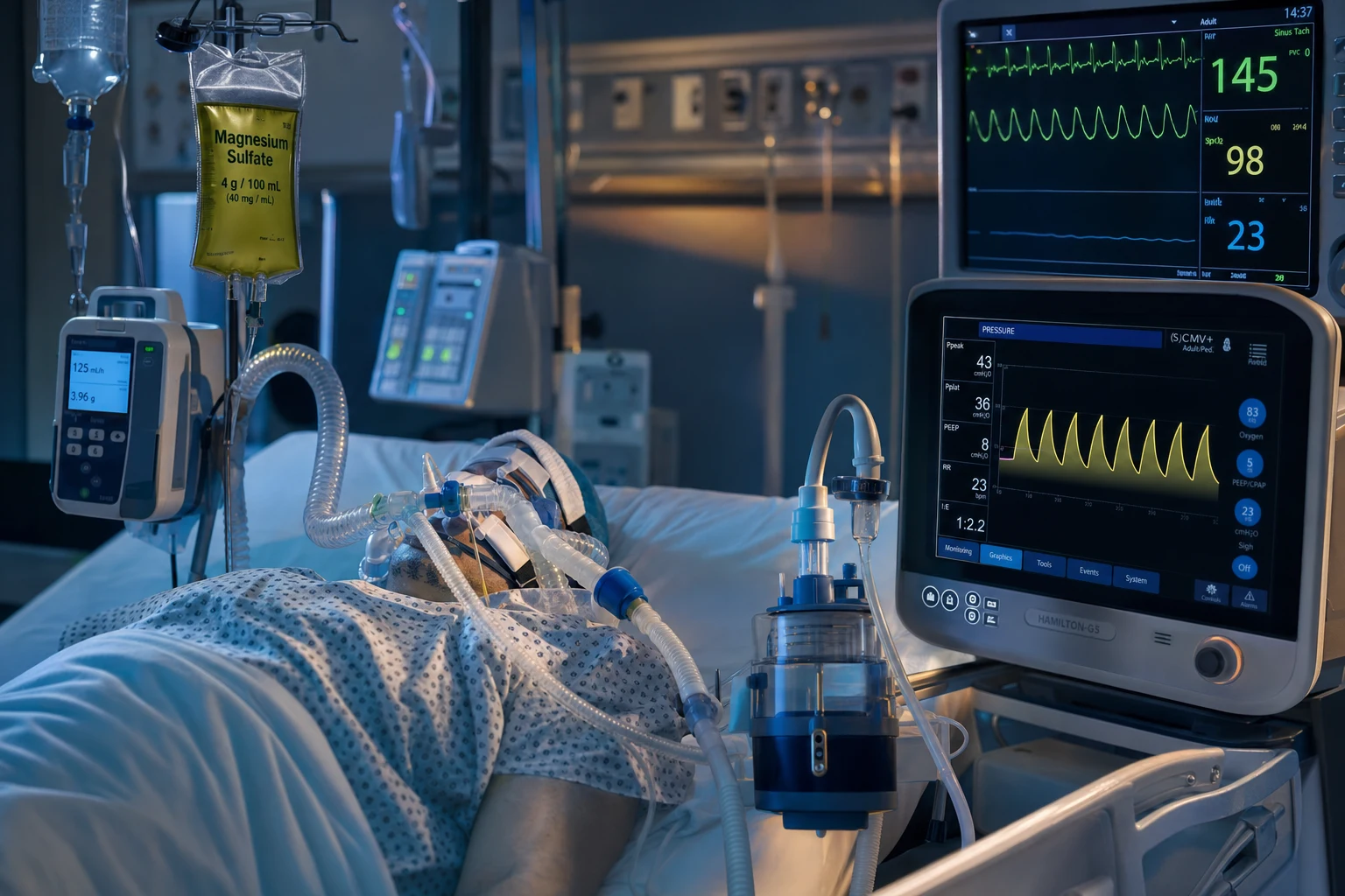
Accurate stratification drives disposition and the decision to intubate. The principle that unifies every marker below is simple: asthma normally causes HYPOcapnia and respiratory alkalosis (the patient hyperventilates to compensate for hypoxaemia and the hypoxic ventilatory drive). Loss of that hypocapnia — a "normal" PaCO2 — means the patient is fatiguing, and a rising PaCO2 means imminent respiratory arrest. This single gas observation is the most heavily examined concept in acute asthma.[1]
Clinical signs by severity tier
Acute asthma severity (BTS/SIGN) — full clinical profile
| Parameter | Moderate | Acute severe | Life-threatening | Near-fatal |
|---|---|---|---|---|
| PEF (% predicted/best) | 50-75% | 33-50% | <33% | — (intubated) |
| Speech | Full sentences | Cannot complete sentences | Words / cannot speak | — |
| Respiratory rate | <25 | >25 | Exhaustion / slow | — |
| Heart rate | <110 | >110 | Tachy → bradycardia | Variable |
| SpO2 | 92-94% | <92% | <92% on O2 | — |
| PaCO2 | Low (<4.5 kPa) | Low (hyperventilating) | NORMAL or RISING | RISING (permitted) |
| Chest auscultation | Wheeze ± accessory use | Wheeze, accessory muscles | SILENT CHEST (no air movement) | — |
| Other | — | — | Cyanosis, altered GCS, exhaustion, hypotension | Rising PaCO2 needing ventilation |
| Disposition | Ward / discharge | HDU / resus | ICU — prepare for intubation | ICU ventilated |
Asthma severity in ICU (click each)
Any ONE: SpO2 <92%, silent chest, cyanosis, exhaustion, bradycardia, hypotension, altered GCS, normal/rising PaCO2, PF <33%
MEDICAL EMERGENCY. Continuous nebulised bronchodilators, IV magnesium 2g, IV steroids, high-flow oxygen. Prepare for intubation if deteriorating. ICU admission.
Key assessment modalities
How to assess the deteriorating asthmatic
Peak expiratory flow (PEF)
Most useful single objective measure if the patient can perform it. Compare to the patient's PERSONAL BEST (not just predicted — best captures their baseline). <33% best/predicted = life-threatening. Re-check after each bronchodilator cycle to chart trajectory. CANNOT perform PEF in the exhausted patient = itself a danger sign.
Oxygen saturation + arterial blood gas
SpO2 <92% on air = severe; <92% on supplemental O2 = life-threatening. ALWAYS take an arterial gas in acute severe asthma — the PaCO2 trajectory is the key. Low PaCO2 (respiratory alkalosis) = reassuring; normal PaCO2 = fatigue; rising PaCO2 = pre-arrest. Expect initial respiratory alkalosis; a NORMAL pH with normal/high CO2 is the warning.
Speech and posture
Ability to count to ten on one breath (~complete a sentence) tracks airflow. Words only = life-threatening. Sitting upright, leaning forward on arms (tripod), accessory muscle use, and intercostal recession indicate high work of breathing. Agitation and diaphoresis reflect hypoxaemia and hypercapnia.
Examination: wheeze is NOT severity
Wheeze reflects turbulent flow through narrowed airways — its LOUDNESS does not correlate with severity. The DANGER is the SILENT CHEST (no wheeze = no air movement). Reassessing wheeze that then disappears may mean improvement OR terminal fatigue — use PEF/gas, not auscultation alone, to distinguish.
Chest X-ray (after stabilisation)
Not routine — only to exclude differential/complications: pneumothorax (asymmetric hyperresonance, sudden deterioration), mucus plugging with lobar collapse, pneumomediastinum (subcutaneous emphysema), aspiration, or pneumonia. Do not delay treatment for a CXR.
Risk factors for fatal asthma (recognise and flag early)
Prior events
Strongest predictors
- Previous near-fatal asthma (ICU/intubation/arest)
- ≥2 ED visits or ≥1 admission in the last 12 months
- Sudden-onset (Type 2) rather than slow-onset attacks
Treatment & adherence
Modifiable
- Over-reliance on SABA (≥3 canisters/year) — marker of poor control
- Poor ICS adherence or no maintenance preventer
- Non-attendance at asthma review
- Food allergy (anaphylaxis–asthma overlap)
Psychosocial
Often missed
- Psychiatric illness, depression, substance misuse
- Low socioeconomic status, employment/housing instability
- Adolescence / young adult — risk-taking, denial
- Learning difficulty or poor health literacy
Pathophysiology
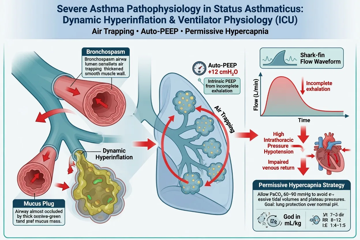
Status asthmaticus is the end-result of three overlapping processes that, unlike a routine exacerbation, become self-sustaining: [1]
- Bronchial smooth muscle spasm — the acute, reversible component; the target of bronchodilators.
- Airway inflammation — mucosal oedema, eosinophilic/neutrophilic infiltrate, epithelial denudation; the target of corticosteroids (hours to work).
- Mucus plugging — tenacious, inspissated secretions and Casts occluding small and medium airways; largely refractory to bronchodilators and the dominant finding at autopsy in fatal asthma. [1]
The dangerous mechanical consequence is critical small-airway narrowing producing a high respiratory time constant (resistance × compliance). Expiration cannot complete before the next inspiration arrives, so each breath stacks on the last (dynamic hyperinflation). Alveolar pressure rises, the lungs over-distend, and the intrathoracic pressure transmitted to the heart and great vessels obstructs venous return and compresses the right ventricle — producing the characteristic preload-dependent hypotension that resolves the instant the ventilator is disconnected.[2]
Two pathophysiological phenotypes behave very differently and the distinction is examinable: [1]
Asthma phenotypes in status asthmaticus
| Feature | Type 1 — slow-onset (eosinophilic) | Type 2 — sudden-onset (neutrophilic / asphyxic) |
|---|---|---|
| Onset | Hours-days of worsening | Minutes-hours |
| Trigger | Viral infection, allergen drift, non-adherence | Allergen/irritant bolus, exercise, stress, anaphylaxis |
| Dominant mechanism | Inflammation + oedema + mucus | Massive bronchospasm |
| Inflammatory cell | Eosinophil (T2-high) | Neutrophil (T2-low) |
| Demographic | Adults, chronic severe asthma | Younger, sensitised patients |
| Gas at presentation | Often already hypoxaemic/hypercapnic | Often near-arrest at first contact |
| Response to steroids | Good | Limited (steroids need hours) |
| Mortality pattern | Slow deterioration, arrest if untreated | Rapid asphyxia before therapy can act |
Status asthmaticus — why the numbers matter
ICU management — the full ladder
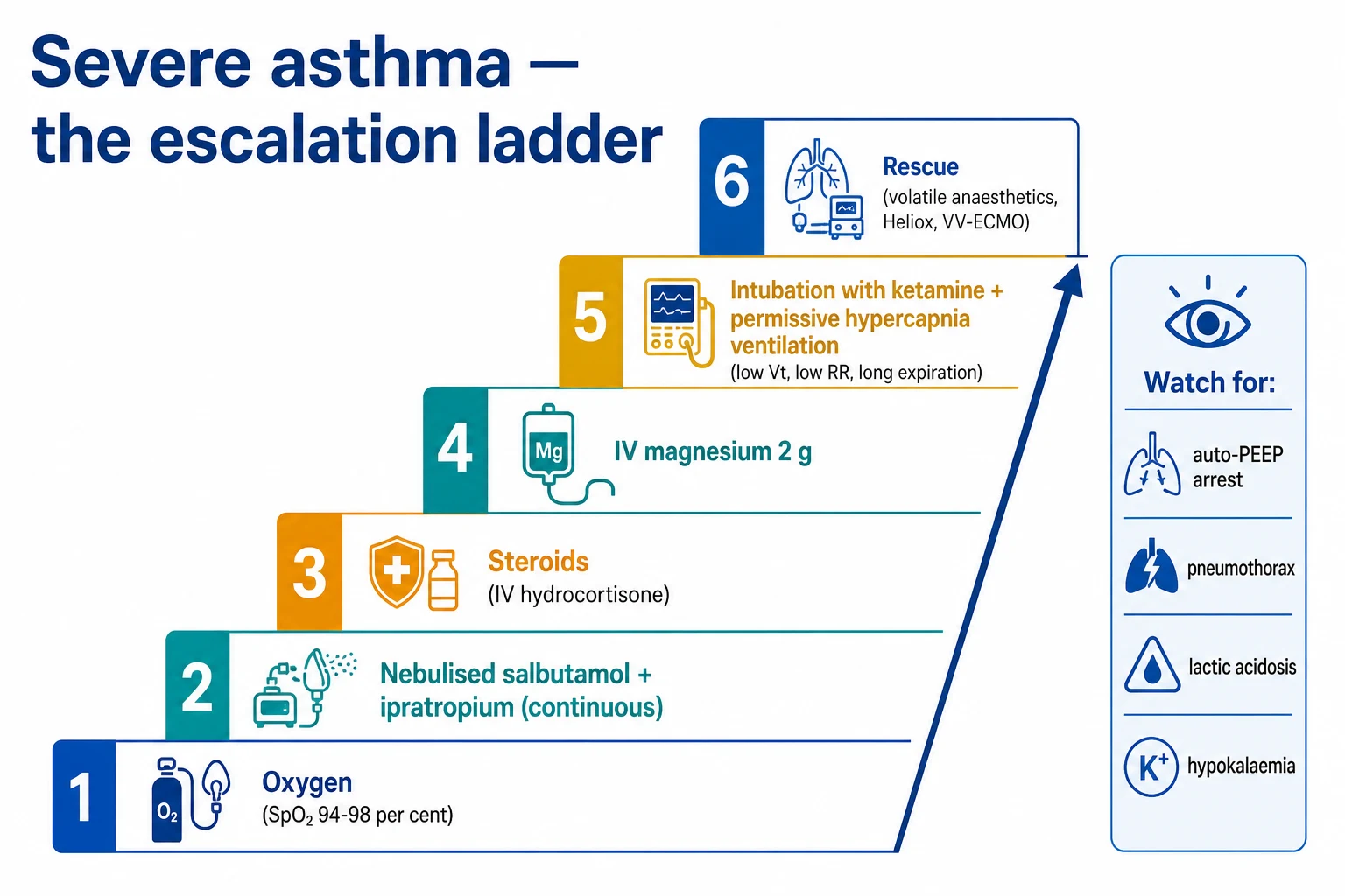
Initial resuscitation and first-line therapy
Status asthmaticus ICU protocol
Oxygen + continuous bronchodilators
High-flow oxygen (target SpO2 94-98%). Continuous nebulised salbutamol 5-10 mg/h + ipratropium 500 mcg every 4-6h. Do NOT stop salbutamol for heart rate alone (tachycardia is expected and tolerated). Monitor K+ (salbutamol causes hypokalaemia).
IV magnesium sulphate
Magnesium sulphate 2 g IV over 20 min (not bolus — can cause hypotension). Mechanism: inhibits calcium influx into bronchial smooth muscle → bronchodilation. Effective in acute severe asthma. Repeat dose may be given. Monitor for hypotension, areflexia (magnesium toxicity).
Systemic corticosteroids
Hydrocortisone 100 mg IV (or prednisolone 40-60 mg PO if tolerating oral). Take 6-12h to work (gene transcription effect). Give early — do NOT wait to see if bronchodilators alone work. Continue for 5 days. Equivalent oral/IV efficacy — use IV if cannot tolerate oral.
IV bronchodilators (if refractory)
IV salbutamol infusion (5-15 mcg/min — monitor for tachycardia, hypokalaemia, lactic acidosis). IV aminophylline (5 mg/kg loading over 20 min, then 0.5 mg/kg/h infusion — monitor levels, side effects: arrhythmia, seizures, nausea). IV magnesium already given. Consider IV ketamine (bronchodilator + anaesthetic — useful for intubation).
Ventilation strategy (if intubation required)
PERMISSIVE HYPERCAPNIA. Ketamine induction (bronchodilator). Rocuronium (NOT suxamethonium — risk of bradycardia/asystole with high-dose beta-2 agonists). Settings: small tidal volume 6 mL/kg PBW, LOW respiratory rate 10-12/min, LONG expiratory time (I:E 1:3 or 1:4), PEEP 0-5 (minimise — high PEEP worsens hyperinflation). Accept PaCO2 up to 80-100 mmHg if pH >7.15.
Avoid dynamic hyperinflation
Dynamic hyperinflation = trapped air cannot be exhaled before next breath → lungs overinflate → compresses heart and great vessels → hypotension → cardiac arrest. SIGNS: hypotension at end-inspiration, improvement when ventilator disconnected (allowing exhalation). PREVENT: low RR, small Vt, long expiration. If hypotension with ventilation: DISCONNECT briefly, let patient exhale, then resume at lower RR/Vt.
Last-line therapies
Volatile anaesthetics (sevoflurane, isoflurane via anaesthetic machine) — potent bronchodilators. ECMO (VV-ECMO) for refractory cases as bridge to recovery. Heliox (helium-oxygen mixture — lower density, reduces turbulent flow and work of breathing) — may help before intubation. Bronchoscopy to remove mucus plugs.
The pharmacological ladder (exam-must-know depth)
Bronchodilators treat the spasm; corticosteroids treat the inflammation; neither addresses mucus plugging (which is why ventilation may still be needed). Steroids take 6-12 h to act — give them early and in parallel with bronchodilators, never sequentially. [1]
Pharmacological ladder for status asthmaticus
| Step | Drug | Dose / route | Mechanism | Key cautions |
|---|---|---|---|---|
| 1 | Oxygen | High-flow, titrate SpO2 94-98% | Treats hypoxaemia; ↓hypoxic drive | Don't hyperoxia — CO2 retention rare in asthma vs COPD |
| 2a | Salbutamol (SABA) | 5 mg NEB q15-20 min or continuous 5-10 mg/h | β2-agonist → ↑cAMP → bronchial SM relaxation | Hypokalaemia (shifts K⁺ into cells), lactic acidosis (↑glycolysis), tremor, tachycardia — do NOT stop for HR alone |
| 2b | Ipratropium bromide (SAMA) | 500 mcg NEB q4-6h (combine with salbutamol) | Muscarinic antagonist → ↓cGMP → bronchodilation; additive with β2 | Less effective alone; benefit is in combination. Dry mouth, urinary retention in elderly |
| 3 | Systemic corticosteroid | Prednisolone 40-60 mg PO or hydrocortisone 100 mg IV q6h. Oral = IV efficacy | Genome effects: ↑anti-inflammatory genes, ↓cytokines/transcription. 6-12 h onset | Give EARLY. Hyperglycaemia, mood effects. 5-7 day course, no taper if <14 days |
| 4 | IV magnesium sulphate | 2 g (8 mmol) over 20 min; may repeat q4-6h | Ca²⁺ antagonist on bronchial SM; inhibits ACh release; mast-cell stabilisation | Hypotension, flushing, hyporeflexia. Not as a bolus. Watch if pre-existing heart block |
| 5 | IV salbutamol infusion | 250 mcg over 10 min load → 5-20 mcg/min | As above but systemic delivery reaches obstructed airways | Tachyarrhythmia, severe hypokalaemia, lactate ↑. Reserve for poor inhaled delivery |
| 5b | IV ketamine | 1-2 mg/kg bolus, then 0.5-1 mg/kg/h infusion (if intubated) | NMDA antagonist; ↑sympathetic tone → bronchodilation; ideal induction + sedation agent | Hypertension, tachycardia, emergence phenomena, hypersalivation (co-administer antisialagogue) |
| 6 | Aminophylline (methylxanthine) | 5 mg/kg load over 20 min → 0.5 mg/kg/h | PDE inhibitor → ↑cAMP; weak bronchodilator | Narrow therapeutic window: arrhythmia, seizures, nausea. Check levels. CYP1A2 interactions. Many guidelines now advise against starting acutely |
| 7 | Volatile anaesthetics | Sevoflurane / isoflurane 1-2% via anaesthetic machine | Direct bronchial SM relaxation (β2-independent); blunt airway reflexes; sedation | Hypotension, arrhythmia, need scavenging, malignant hyperthermia risk. Needs anaesthetic machine/vaporiser |
| 8 | Heliox | 70:30 or 80:20 He:O2 by facemask | ↓Density → laminar flow → ↓resistance & work of breathing | Max FiO2 only 30-40% — fails if severe hypoxaemia; most ICU ventilators uncalibrated for He |
| 9 | VV-ECMO | Cannula → membrane oxygenator → return | Bypasses lungs → rest the lung → no hyperinflation | Bleeding, thrombosis, resource-intensive. Survival 80-90% in asthma |
Steroids — oral vs IV, and timing
- Oral prednisolone 40-60 mg is equivalent in efficacy to IV hydrocortisone provided the patient can swallow and absorb. Use IV if vomiting, intubated, or in extremis.[1]
- Onset is 6-12 hours (genomic mechanism); effect peaks at 24-48 h. Give immediately alongside the first bronchodilator — do not wait to "see if salbutamol works".
- 5-7 day course; no taper needed for courses <14 days. Steroids reduce relapse after the acute attack.
- Consider adding ICS (inhaled corticosteroid) early in recovery to rebuild preventer therapy and reduce re-admission.
IV magnesium — mechanism, evidence, dosing
Mechanism: Mg²⁺ is a calcium-channel antagonist at bronchial smooth muscle (competitively inhibits Ca²⁺ influx), inhibits acetylcholine release at the neuromuscular junction, and stabilises mast cells — collectively producing bronchodilation independent of the β2 pathway.[1]
Dose: 2 g (8 mmol) in 100 mL over 20 minutes (faster causes hypotension; a true bolus is contraindicated). Repeat doses every 4-6 h are reasonable in refractory cases; monitor reflexes. [1]
Evidence — the MAGNETIC and 3Mg trials are the landmark studies: [1]
MAGNETIC
Lancet Respir Med 2013
508 children (2-16 y), severe asthma — nebulised MgSO4 vs placebo, added to salbutamol+ipratropium
Key finding
Nebulised Mg improved Yung Asthma Severity Score only in the more severe subgroup; modest benefit overall, no significant harm
Practice change
Nebulised Mg considered in children with severe attacks not responding to first-line therapy
3Mg
Lancet Respir Med 2013
1109 adults with severe acute asthma — IV MgSO4 vs nebulised MgSO4 vs placebo, added to standard care
Key finding
IV Mg did NOT significantly reduce admissions overall (trend to benefit); nebulised Mg had no clear role in adults. Benefit concentrated in the most severe subset.
Practice change
IV Mg reserved for severe attacks failing standard therapy; not routine in all acute asthma
Magnesium in acute asthma — pragmatic use
Aminophylline / methylxanthines — downgraded
GINA and BTS/SIGN do not recommend starting aminophylline or theophylline routinely in acute asthma. The Cochrane meta-analysis found no clear benefit over β2-agonists, with a narrow therapeutic window (toxicity: nausea, seizures, supraventricular and ventricular arrhythmia) and significant CYP1A2 drug interactions. Exceptions: a patient already on maintenance theophylline (check level — may be subtherapeutic) and the rare refractory case under specialist guidance. Do not start acutely as a reflex.[1]
Non-invasive ventilation (BiPAP) in asthma
NIV in acute asthma is controversial and adjunctive, not standard. Unlike COPD (where BiPAP is first-line for hypercapnic exacerbation), asthma is fundamentally a low-CO2 / high-drive disease — the patient who becomes hypercapnic is near-arrest and usually needs intubation, not a mask. NIV's theoretical role is to reduce the work of breathing and improve aerosolised drug delivery in the tiring but not yet failing patient, buying time for steroids to act.[8]
When to consider NIV
- Acute severe asthma, rising work of breathing, paCO2 still normal or low (not yet hypercapnic).
- Patient cooperative and able to protect airway.
- As a bridge while waiting for steroids/Mg to work — not as a substitute for intubation in the clearly failing patient.
- Contraindicated if: altered consciousness, inability to clear secretions, haemodynamic instability, vomiting/bowel obstruction, facial trauma, or a rising PaCO2 with fatigue (these mandate intubation). [1]
Practical BiPAP settings in asthma
BiPAP setup for acute asthma (if used)
Mode and interface
BiPAP (spontaneous/timed). Full face mask initially (least leak, best in respiratory distress); switch to nasal once improving. Continuous SpO2, ECG, and ideally serial gases (NIV + asthma can mask deterioration).
Pressure settings
Start LOW to avoid hyperinflation: IPAP 8-10 cmH2O, EPAP 4-5 cmH2O (ΔP 4-6 cmH2O to augment tidal volume). Titrate IPAP up slowly to a max ~15 cmH2O guided by effort and gas exchange. Keep EPAP LOW — extrinsic PEEP stacks on intrinsic PEEP and worsens hyperinflation.
FiO2
Titrate to SpO2 94-98% (attach O2 to mask port). Lower density of He-O2 not reliably delivered via standard BiPAP circuits.
Nebulised bronchodilators through NIV
Place the nebuliser in the circuit (between the Y-piece and mask) — ventilatory airflow actually IMPROVES drug delivery to obstructed airways. Continue salbutamol 5 mg q1h + ipratropium 500 mcg q6h.
Re-assess at 30-60 min — strict stop criteria
IMPROVING (↓RR, ↓accessory use, ↓PaCO2 if it was raised): continue. STATIC or WORSENING (rising PaCO2, falling pH, exhaustion, silent chest): STOP NIV, intubate. Do not let NIV delay definitive airway management — that delay is the trap.
Mechanical ventilation strategy
The single governing principle: the ventilator cannot cure asthma; it can only keep the patient alive while bronchodilators and steroids act. Your job is to avoid dynamic hyperinflation at all costs — even at the price of severe hypercapnia. Over-ventilating to "normalise" CO2 is the classic and fatal mistake.[2][5]
Indications for intubation
Absolute
Intubate now
- Cardiac or respiratory arrest
- Reduced GCS / inability to protect airway (CO2 narcosis)
- Apnoea / agonal breathing
Relative
Intubate early
- Exhaustion (paradoxical breathing, silent chest)
- Rising PaCO2 with falling pH despite maximal therapy
- Refractory hypoxaemia (SpO2 <90% on high-flow O2)
- Haemodynamic instability (hypotension, bradycardia)
- Failure of NIV trial
Intubation pharmacology
Drugs and technique for the asthmatic intubation
Preoxygenation
100% O2 for 3 min (or 8 vital-capacity breaths). Asthmatics desaturate FAST — low FRC, high O2 consumption from the work of breathing, and V/Q mismatch. Have a backup airway plan and a senior operator.
Induction — KETAMINE preferred
Ketamine 1-2 mg/kg IV. It is a BRONCHODILATOR (increases catecholamine release → β2 effect on airway SM) AND maintains BP (sympathetic stimulation). AVOID propofol (vasodilation + histamine → bronchospasm + hypotension) and thiopental (histamine → bronchospasm).
Paralysis — rocuronium
Rocuronium 1.2 mg/kg (intubating dose). Cautions around suxamethonium: rare reports of bradycardia/asystole in patients loaded with β2-agonists; sux is acceptable but rocuronium avoids this and provides a longer window. Avoid prolonged paralysis (ICU-acquired weakness; combine with myopathy in asthma).
Largest practical ETT
Use the largest ETT that fits (8.0-8.5 in adults). Narrower tubes increase resistance (Poiseuille: resistance ∝ 1/radius⁴), which matters because you will deliver bronchodilators through the tube and will need to suction thick secretions.
Avoid awake / fibreoptic intubation
Stimulation of an already-reactive airway can trigger further bronchospasm. RSI is standard. If anticipated difficult airway, have an experienced operator and a supraglottic-airway/surgical backup.
Initial ventilator settings — "low and slow, long expiratory"
Initial ventilator settings for ventilated status asthmaticus
| Setting | Recommended | Rationale |
|---|---|---|
| Mode | Volume-controlled (VC) preferred | Guarantees delivered Vt; easier to monitor plateau pressure and detect hyperinflation |
| Tidal volume | 6 mL/kg PBW (down to 4 if needed) | Less volume to exhale → less hyperinflation. Use predicted/ideal, not actual weight |
| Respiratory rate | 10-12 /min (down to 8) | Slow rate → long expiratory time → complete emptying |
| Inspiratory flow | High (60-100 L/min) with square (decelerating OFF) waveform | Short inspiratory time → maximises expiratory time |
| I:E ratio | 1:3 to 1:4 (1:5 in severe) | Long expiration is the safety mechanism |
| PEEP | 0-5 cmH2O (often 0 initially) | Minimise — extrinsic PEEP adds to intrinsic PEEP. Set ≈ 80% of measured intrinsic PEEP if you must use it |
| FiO2 | 100% initially, titrate to SpO2 94-98% | Wean as gas exchange improves |
| Plateau pressure | <30 cmH2O (an end-inspiratory hold) | Surrogate for dynamic hyperinflation; >30 = reduce Vt/RR |
Setting up the ventilator — step by step
Calculate PBW
Male: 50 + 0.91 × (height cm − 152.4). Female: 45.5 + 0.91 × (height cm − 152.4). Never use actual body weight — Vt is a proxy for lung size which tracks with height.
Set low Vt and low RR
Vt 6 mL/kg PBW. RR 10-12 (start 10 if very severe). The product — minute ventilation — is deliberately LOW; this is what permits long expiration.
Shorten inspiratory time
High inspiratory flow (60-100 L/min) with a constant-flow (square) waveform keeps Ti short. The ventilator's expiratory time is then maximised (Ti short + low rate = long Te).
Target I:E 1:3 or longer
Most modern ventilators display I:E. If it is 1:2 or shorter, your RR is too high or inspiratory flow too low — fix that before doing anything else.
Set low PEEP
PEEP 0 (or up to 5). The obstructed airway already has high INTRINSIC PEEP (auto-PEEP) — adding extrinsic PEEP can be additive and worsen hyperinflation. (In COPD you match PEEPi to splint airways; in asthma the trapped gas is the problem — be more conservative.)
Sedate deeply ± paralyse (early)
Patient-ventilator asynchrony drives up minute ventilation and worsens bronchospasm. Deep sedation (propofol ± opioid, or a KETAMINE infusion for its bronchodilator effect) is essential. Short-acting cisatracurium for the first 24-48 h if dyssynchronous — but watch for critical-illness myopathy, especially with concomitant steroids.
Monitoring for dynamic hyperinflation (auto-PEEP) — the killer
The defining danger of ventilated asthma. Auto-PEEP (intrinsic PEEP) is the positive end-expiratory pressure generated by trapped gas that has not fully exited before the next breath.[2]
How to measure intrinsic (auto-)PEEP:
- End-expiratory hold (expiratory pause): occlude the expiratory port at end-expiration; the pressure equilibrates to the trapped-gas pressure = intrinsic PEEP. Normal is ~0; >5 cmH2O is significant, >10 is dangerous.
- Plateau pressure (end-inspiratory hold): 0.5 s inspiratory pause; reflects peak alveolar pressure. Keep <30 cmH2O. A high Pplat in asthma = hyperinflation, NOT a target to chase. [1]
Clinical signs of dynamic hyperinflation:
- Hypotension that worsens with each inspiration and improves dramatically when the ventilator is disconnected (the pathognomonic test).
- High Pplat (>30), high auto-PEEP, slow expiratory flow that does not return to baseline before the next breath.
- Rising CVP with inspiration (transmitted intrathoracic pressure). [1]
Treatment — the disconnection manoeuvre: [1]
Dynamic hyperinflation emergency response
Recognise
New hypotension shortly after starting/increasing ventilation, especially with high Pplat or auto-PEEP, OR unexplained hypotension/tachycardia in a ventilated asthmatic.
DISCONNECT the circuit
Manually allow the patient to fully exhale (squeeze the chest gently if needed). Listen for a prolonged whoosh of escaping gas. Watch the blood pressure rise over 10-30 seconds — confirmation of dynamic hyperinflation.
Reduce minute ventilation
Reconnect at a LOWER respiratory rate (8-10), LOWER Vt (4-5 mL/kg if needed), and ensure a LONGER expiratory time (higher inspiratory flow, I:E 1:4-1:5).
Deepen sedation ± paralyse
Eliminate patient-ventilator dyssynchrony (double-triggering, breath-stacking) which is often the precipitant.
Escalate bronchodilation
Continuous nebulised salbutamol through the circuit, ± IV salbutamol, ± repeat IV magnesium. Consider ketamine infusion (bronchodilation + sedation). If still refractory → volatile anaesthetic / VV-ECMO.
The historical basis for permissive hypercapnia is Darioli & Perret (1984), who reported zero mortality in 26 mechanically ventilated status asthmaticus patients using controlled hypoventilation — a paradigm shift away from the aggressive normocapnic ventilation that had produced mortality of 10-30%. The principle survives unchanged.[5]
Severe cases may need helium-oxygen (Heliox)
Heliox is a helium-oxygen mixture (usually 70:30 or 80:20). Helium is less dense than nitrogen, so for any given airway geometry the flow is less turbulent (lower Reynolds number) — improving flow and reducing the work of breathing, particularly through the large/medium airways where turbulence dominates.[9]
Practical role in asthma:
- Most useful pre-intubation in the spontaneously breathing patient with high work of breathing, to buy time for bronchodilators/steroids.
- Theoretical role in ventilated severe asthma — but most ICU ventilators are not calibrated for helium, so delivered Vt and flows are inaccurate (a serious safety issue). Only use on a ventilator validated for Heliox with experienced staff.
- Limitation: maximum FiO2 is only 30-40% — fails in severe hypoxaemia.
- Evidence: Cochrane systematic review shows no consistent outcome benefit; use as a temporising adjunct, not a standard therapy. [1]
Refractory bronchospasm — escalation beyond standard ventilation
Volatile anaesthetic agents
Inhaled sevoflurane, isoflurane, and desflurane are potent bronchodilators — they relax airway smooth muscle by a mechanism independent of β2 receptors (direct effect on smooth muscle, and suppression of airway reflexes). In refractory status asthmaticus unresponsive to continuous β2-agonists, magnesium, and steroids, volatiles can break bronchospasm within minutes.[2]
Practical issues (these are the exam answers):
- Require an anaesthetic machine with a vaporiser — most ICU ventilators do not deliver inhaled anaesthetics (dedicated ICU anaesthetic devices now exist; check local availability).
- Need gas scavenging for staff and environmental safety (occupational exposure limits).
- Hypotension from vasodilation is common — may need vasopressor support.
- Sensitise the myocardium to catecholamines (arrhythmia risk).
- Contraindicated in malignant hyperthermia susceptibility.
- Evidence is case series/reports only (no RCTs feasible) but the effect is dramatic and widely accepted as a last-line salvage therapy. [1]
VV-ECMO for near-fatal asthma
VV-ECMO is the ultimate rescue. The rationale is powerful: asthma is reversible — bronchospasm and inflammation resolve over hours-days with steroids — so ECMO needs only to bridge the patient to recovery, allowing the ventilator to be set to complete "rest" (very low rate, very low Vt, eliminating hyperinflation entirely).[6]
Indications (case-by-case at an ECMO centre):
- Refractory hypoxaemia or life-threatening acidosis (pH <7.1) despite optimised ventilation, volatile anaesthetics, and full bronchodilation.
- Inability to control dynamic hyperinflation/pneumothorax with conventional ventilation.
- Early cannulation — do not wait for cardiac arrest (then consider VA-ECMO/ECPR). [1]
Outcomes — striking: [1]
VV-ECMO in near-fatal asthma — outcomes
The 80-90% survival figure comes from the ESO/ELSO registry and case series; asthma has the best ECMO survival of any adult indication precisely because the disease is self-limiting.[6]
Other adjuncts
- Bronchoscopy: for radiographic lobar collapse from mucus plugging — removes tenacious casts unresponsive to bronchodilation. Use cautiously in a tenuous ventilated patient.
- Mucolytics (N-acetylcysteine, rhDNAse): limited evidence; may help mucus plugging but can provoke bronchospasm — use cautiously.
- Treat the trigger: antibiotics only if bacterial infection (most exacerbations are viral); stop culprit drugs (β-blockers, NSAIDs in sensitive patients, cholinesterase inhibitors). [1]
Complications
Complications of severe asthma and its ventilation
| Complication | Mechanism | Recognition | Action |
|---|---|---|---|
| Dynamic hyperinflation → arrest | Breath-stacking compresses great vessels | Hypotension ↑with inspiration, ↓on disconnection | DISCONNECT, ↓RR, ↓Vt, deepen sedation |
| Tension pneumothorax | Alveolar rupture from high pressure | Unilateral signs + hypotension, ↑Pplat | Immediate needle decompression → chest drain |
| Pneumomediastinum / SQ emphysema | Alveolar rupture tracking centrally | Subcutaneous crepitus, CXR | Usually self-limiting; exclude pneumothorax |
| Critical-illness myopathy (CIM) | Steroids + NMB + sepsis | Flaccid weakness, failed wean | Minimise NMB duration; early PT |
| Hypokalaemia | β2 agonists shift K⁺ into cells | ECG (T-wave changes, ectopics) | Replace K⁺; monitor with continuous nebs |
| Lactic acidosis | β2-driven aerobic glycolysis | ↑Lactate, normal ScvO2 | Do not over-treat; ↓bronchodilator once improving |
| Atelectasis / mucus plugging | Tenacious secretions | Lobar collapse on CXR | Physiotherapy, bronchoscopy, hydration |
| Aspiration | Reduced GCS / post-extubation | New infiltrate, fever | Antibiotics, lung-protective ventilation |
Prognosis
Outcomes in status asthmaticus
Prognosis is driven by avoiding the preventable killers: dynamic hyperinflation (the dominant cause of ventilated-asthma death), pneumothorax, and delayed intubation. With permissive hypercapnia, early bronchodilation, and ECMO for the truly refractory, mortality has fallen from the 10-30% of the normocapnic era to single digits. Long-term, survivors need a written asthma action plan, preventer therapy optimisation (ICS ± biologic for T2-severe asthma), trigger avoidance, and follow-up to prevent recurrence — a near-fatal attack is the strongest predictor of a future fatal one.[1]
Exam practice
SAQ — Near-fatal asthma ventilation
10 minutes · 10 marks
A 28-year-old woman with known asthma presents with 2 days of worsening wheeze and breathlessness despite her usual inhalers. She is agitated, speaking single words, RR 32, SpO2 88% on 15 L/min via NRM, HR 132, BP 148/92, chest almost silent on auscultation. ABG: pH 7.18, PaCO2 7.8 kPa (59 mmHg), PaO2 7.5 kPa (56 mmHg), HCO3 24, lactate 3.4. PEF 90 L/min (best 480).
Clinical pearls
Red flags
Evidence and landmark trials summary
MAGNETIC
Lancet Respir Med 2013
508 children (2-16 y) with severe asthma — nebulised MgSO4 vs placebo + standard care
Key finding
Modest benefit overall; benefit concentrated in the most severe subgroup. No significant harm.
Practice change
Nebulised Mg a reasonable adjunct in severe paediatric attacks
3Mg
Lancet Respir Med 2013
1109 adults with severe acute asthma — IV vs nebulised MgSO4 vs placebo
Key finding
IV Mg did NOT significantly reduce admissions overall; nebulised Mg had no clear role in adults
Practice change
IV Mg reserved for severe attacks failing standard therapy; not routine
Darioli & Perret
Intensive Care Med 1984
Case series of 26 mechanically ventilated status asthmaticus patients using controlled hypoventilation
Key finding
Zero mortality with permissive hypercapnia vs 10-30% historical mortality
Practice change
Established controlled hypoventilation / permissive hypercapnia as the ventilator philosophy
Soroksky (BiPAP)
Chest 2003
Pilot RCT — BiPAP vs sham in acute severe asthma
Key finding
BiPAP improved lung function faster; small study, short-term
Practice change
NIV a reasonable adjunct in carefully selected, monitored asthmatics
Brenner (ECMO)
Intensive Care Med 2014
Case report + literature review of VV-ECMO for refractory status asthmaticus
Key finding
Survival 80-90% in pooled case series — best of any adult ECMO indication
Practice change
VV-ECMO is the rescue of choice for near-fatal refractory asthma
References
- [1]British Thoracic Society/SIGN. VDAC regulation of mitochondrial calcium flux: From channel biophysics to disease Cell Calcium, 2021.PMID 33529977
- [2]Louie S, et al. Notum palmitoleoyl-protein carboxylesterase regulates Fas cell surface death receptor-mediated apoptosis via the Wnt signaling pathway in colon adenocarcinoma Bioengineered, 2021.PMID 34402722
- [3]Goodacre S, Cohen J, Bradburn M, et al. Neurosyphilitics and madmen: The French fin-de-siècle fictions of Huysmans, Lermina, and Maupassant Prog Brain Res, 2013.PMID 24290477
- [4]Powell C, Kolamunnage-Dona R, Lowe J, et al. Hospital-based school for children with chronic illness in Taiwan J Formos Med Assoc, 2015.PMID 24461878
- [5]Darioli R, Perret C. Pentane formation during the anaerobic reactions of reticulocyte lipoxygenase. Comparison with lipoxygenases from soybeans and green pea seeds Biochim Biophys Acta, 1984.PMID 6433987
- [6]Brenner K, Ryu J, Fissette P, et al. Advanced hybrid supercapacitor based on a mesoporous niobium pentoxide/carbon as high-performance anode ACS Nano, 2014.PMID 25137384
- [7]Brand PL, Luz Filho A, Vaz Fragoso CA. The elite and stochastic model for iPS cell generation: multilineage-differentiating stress enduring (Muse) cells are readily reprogrammable into iPS cells Cytometry A, 2013.PMID 22693162
- [8]Soroksky A, Stav D, Shpirer I. Left ventricular diastolic function in patients with advanced cystic fibrosis Chest, 2003.PMID 12740265
- [9]Rodrigo G, Pollack C, Rodrigo C, Rowe BH. Measuring successful treatment of irritable bowel syndrome: is satisfactory relief enough? Am J Gastroenterol, 2006.PMID 16696786