ICU · Respiratory
Tracheostomy in the ICU
Also known as Percutaneous tracheostomy · Surgical tracheostomy · Percutaneous dilatational tracheostomy (PDT) · Trach · Early vs late tracheostomy · Tracheostomy weaning · Ciaglia Blue Rhino · Speaking valve (Passy-Muir)
Tracheostomy provides a secure, long-term artificial airway placed through the anterior tracheal wall (typically between the 2nd and 3rd, or 3rd and 4th, tracheal rings) for prolonged mechanical ventilation, airway protection, or upper airway obstruction. Indications: prolonged ventilation (usually considered after 7-10 days of ventilation, or when 10-14 days of ventilation are anticipated), failed extubation, inability to protect the airway (neurological injury, neuromuscular disease), excessive secretions needing frequent suctioning, and upper airway obstruction (tumour, trauma, infection, bilateral vocal cord palsy). Percutaneous dilatational tracheostomy (PDT) is preferred in the ICU (bedside, bronchoscopy-guided, Seldinger technique) using techniques such as the Ciaglia Blue Rhino (single tapered dilator), Ciaglia Blue Dolphin (radially-expanding balloon), or Griggs guidewire dilating forceps. Surgical tracheostomy is reserved for complex anatomy, paediatric patients, coagulopathy, or failed PDT. Timing: the TracMan trial (Young 2013, NEJM) showed no mortality benefit of early (day 1-4) versus late (day 10+) tracheostomy, though early tracheostomy modestly reduced sedation exposure. Benefits of tracheostomy: reduced sedation, easier weaning, improved patient comfort and communication (speaking valve), better oral hygiene, easier pulmonary toilet, and facilitated transfer from ICU. Complications: early (0-7 days) — bleeding (thyroid isthmus, anterior jugular veins), infection, stomal cellulitis, tube displacement (airway emergency if <7 days as the tract is not yet formed), pneumothorax (apex of pleura extends above the clavicle into the neck), subcutaneous emphysema; late (7 days) — tracheal stenosis, tracheomalacia, tracheo-oesophageal fistula, tracheo-innominate artery fistula (catastrophic bleeding, surgical emergency), tracheocutaneous fistula. Decannulation follows a protocolised pathway: cuff deflation tolerance, cuff leak test to assess upper airway patency, progressive downsizing, capping trials, endoscopic assessment, and tube removal with occlusive stoma dressing.
On this page & tools
Your progress
Saved locally on this device.
Target exams
Red flags
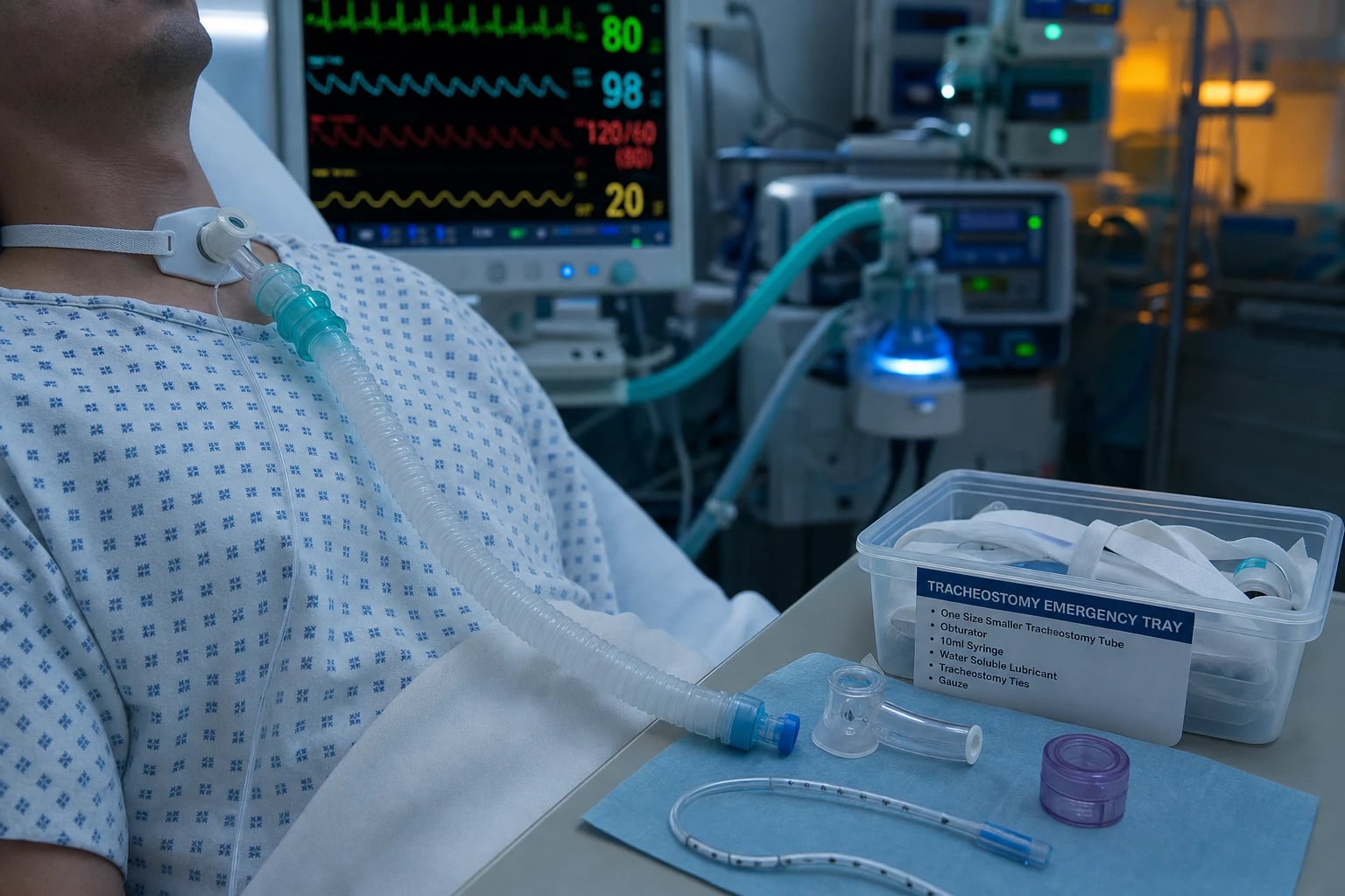
Why a tracheostomy? — the physiological rationale
Advantages of tracheostomy
Over translaryngeal intubation
- Reduced dead space (~50-100 mL) and lower work of breathing — easier weaning
- Reduced laryngeal injury and risk of post-extubation stridor/glottic stenosis
- Less sedation required (cuff can be deflated, patient can communicate via speaking valve)
- Improved patient comfort, ability to eat orally, and psychological wellbeing
- Better oral hygiene and easier pulmonary toilet (suctioning)
- More secure airway (less risk of accidental extubation) — tube is sutured and held by the tract
- Facilitates transfer out of ICU and long-term rehabilitation
Disadvantages / risks
Procedure and long-term
- Procedural risks: bleeding (thyroid isthmus, anterior jugular veins), pneumothorax, infection
- Tube displacement in first week = airway emergency (no formed tract → false passage risk)
- Late complications: tracheal stenosis, tracheomalacia, tracheo-oesophageal fistula
- Tracheo-innominate artery fistula — catastrophic haemorrhage (mortality 50-80%)
- Loss of upper airway humidification, filtration, and warming → requires heated humidification
- Scarring, persistent tracheocutaneous fistula after decannulation
- Reduced cough effectiveness (bypasses glottis) and impaired swallow initially
Indications
The decision to place a tracheostomy balances the expected duration of ventilation, the underlying disease trajectory, airway protection needs, and secretion management. The major categories are summarised below. [1]
Prolonged ventilation
The commonest indication
- Expected to need mechanical ventilation >10-14 days (or already ventilated >7-10 days with no prospect of imminent extubation)
- Typically considered around day 7-10 of ventilation once the need for prolonged support becomes clear
- Reduces cumulative laryngeal injury, sedation, and facilitates ventilator weaning
- Individualise: a patient rapidly recovering from a reversible cause (e.g. drug overdose) does not need a trach
Failed extubation / weaning
Repeated reintubation
- Failed extubation (reintubation within 48-72h) — especially in cardiac, respiratory, or older patients
- Repeated failed spontaneous breathing trials (SBT) despite treating reversible causes
- Difficult airway — predicted difficult reintubation (anatomical, prior difficulty)
- Severe ICU-acquired weakness (CIP/CIM) preventing effective spontaneous ventilation
Airway protection
Neurological impairment
- Severe traumatic brain injury with absent or poor cough/gag reflexes
- Bulbar dysfunction (stroke, motor neuron disease, Guillain-Barré, myasthenia)
- Inability to manage secretions — aspiration risk despite NIV/tracheal suctioning
- Allows ongoing mechanical ventilation while protecting against aspiration
Upper airway obstruction
Mechanical bypass
- Head and neck tumours (laryngeal, pharyngeal) — often planned (surgical trach in theatre)
- Maxillofacial trauma, laryngeal fracture, inhalation thermal injury with airway oedema
- Upper airway infection — deep neck space infection, epiglottitis, Ludwig angina
- Bilateral vocal cord palsy, fixed laryngeal stenosis
- Sleep-disordered breathing / severe obstructive sleep apnoea (rarely, for chronic respiratory failure)
Secretion management
Pulmonary toilet
- Excessive secretions requiring frequent, deep suctioning (e.g. neuromuscular disease, pneumonia)
- Inadequate cough to clear secretions despite aggressive physiotherapy
- Facilitates direct tracheal suctioning and bronchoscopy for toilet
Timing of tracheostomy — early vs late
The optimal timing of tracheostomy has been the most debated question in the field. The consensus, grounded in the TracMan trial, is that early tracheostomy does not improve survival but may modestly reduce sedation and is a patient-centred decision. [1]
The TracMan trial — the definitive timing study
TracMan — Young et al. 2013, NEJM (PMID 20215379)
Design
Multicentre randomised controlled trial, 909 critically ill patients expected to need at least 7 days of ventilation
Intervention
Early tracheostomy (within 4 days of intubation) vs late tracheostomy (day 10 or later, if still needed)
Primary outcome
30-day mortality: 30.8% (early) vs 31.6% (late) — NO significant difference
Secondary outcomes
Early group had more tracheostomies performed (91.5% vs 45.5%), and a small reduction in days of intravenous sedation, but no difference in ICU stay, ICU mortality, antibiotics, or infections
Bottom line
Early (day 1-4) tracheostomy does NOT improve mortality. The decision should be individualised and patient-centred (sedation, comfort, communication) rather than driven by a fixed timepoint
Other timing evidence
Rumbak et al. 2004, Critical Care Medicine (PMID 14758164) — early vs late in medical ICU
Design
Single-centre RCT, 112 long-stay medical ICU patients expected to need ventilation >14 days
Intervention
Early percutaneous tracheostomy (within 48 h) vs prolonged intubation with delayed tracheostomy (day 14-16)
Primary outcome
Mortality: 31.7% (early) vs 61.7% (delayed), p=0.005; early group also had less pneumonia, shorter ICU stay, shorter ventilation
Bottom line
Suggested a dramatic mortality benefit of early trach in a SELECTED medical long-stay cohort. BUT: small, single-centre, and the selected population (all truly needed >14 days ventilation) differs from the general ICU population. Findings NOT reproduced in the much larger TracMan trial, which is the definitive study
Trouillet (SITARE) 2011, Annals of Internal Medicine (PMID 21505135) — early trach after cardiac surgery
Design
Multicentre RCT, 220 post-cardiac-surgery patients ventilated >48 h
Intervention
Early percutaneous tracheostomy (day 5) vs prolonged intubation (trach if still ventilated at day 15)
Primary outcome
No difference in cumulative antibiotic-free days (the primary endpoint), mortality, ICU stay, or nosocomial pneumonia
Bottom line
In the cardiac-surgery population, early trach did not improve outcomes. Adds to TracMan's conclusion that early trach is not superior
Practical timing approach
A practical, individualised approach to tracheostomy timing
Day 1-3: predict and observe
Identify patients likely to need prolonged ventilation (severe ARDS, severe brain injury, neuromuscular failure). Do NOT rush an early trach on day 1-4 — TracMan shows no survival benefit. Continue treating the underlying cause and reassess daily.
Day 3-7: reassess daily
Is the underlying cause improving? Is the patient on reducing ventilator settings? Consider tracheostomy if the patient is failing to wean despite treating reversible causes, or has poor airway protection (neurological injury) that will not resolve quickly.
Day 7-10: the decision point
The traditional and pragmatic time to place a tracheostomy if the patient is clearly going to need prolonged ventilation or has failed extubation. By this point the tract will form safely (7-10 days) and the patient has had a fair trial of translaryngeal intubation.
After day 10: reassess need
If the patient remains ventilator-dependent beyond 10-14 days, a tracheostomy is generally indicated for comfort, weaning, secretion management, and ICU transfer — unless the trajectory is clearly towards death or recovery.
Percutaneous vs surgical tracheostomy
Percutaneous (PDT)
Bedside procedure
- Performed in the ICU at the bedside — no transfer to theatre required
- Bronchoscopy-guided (visualise needle entry, guidewire passage, dilatation, and tube placement)
- Seldinger technique: needle → guidewire → dilators → tracheostomy tube
- Techniques: Ciaglia Blue Rhino (single tapered dilator, most common), Ciaglia Blue Dolphin (radially-expanding balloon), Griggs (guidewire dilating forceps), Fantoni (translaryngeal, retrograde)
- Advantages: no theatre transfer, faster, less scarring, lower cost, can be done out of hours
- Meta-analyses: PDT is as safe as surgical trach, with similar overall complication rates and less stomal bleeding and infection
- Contraindications: unstable cervical spine, uncorrected coagulopathy (INR >1.5, platelets <50), neck infection, abnormal/unknowable anatomy, marked obesity with short neck (relative), children (relative — surgical usually preferred), high PEEP/ FiO2 requirements (risk of derecruitment and desaturation)
Surgical
Operating theatre
- Formal open surgical procedure (horizontal skin incision, division/ligation of thyroid isthmus, vertical incision between tracheal rings, stay sutures, tracheal window or flap)
- Indicated when: PDT contraindicated or has failed, complex/abnormal anatomy, children, need for a precise tracheal window, prior neck surgery or trach, marked obesity with unfavourable anatomy
- Advantages: direct visualisation, controlled haemostasis, ability to create a formal stoma (Bjork flap) that eases reinsertion, can be done emergently for upper airway obstruction
- Disadvantages: requires transfer to theatre (with associated risks), longer, more scarring, higher cost, longer procedure time
- Higher rates of stomal bleeding and infection than PDT in meta-analyses, but lower risk of posterior tracheal wall injury and false passage
Freeman 2000 meta-analysis, Chest (PMID 10893366) — PDT vs surgical tracheostomy
Design
Meta-analysis of 5 prospective randomised studies comparing percutaneous and surgical tracheostomy
Finding
Overall complication rates were NOT significantly different between PDT and surgical trach. PDT showed trends towards less stomal bleeding and infection; surgical trach had a lower rate of decannulation problems and tube obstruction
Bottom line
PDT is as safe as surgical tracheostomy in appropriately selected ICU patients. PDT is now first-line in the ICU for patients without contraindications; surgical trach is reserved for complex anatomy, contraindications, or failed PDT
The percutaneous tracheostomy procedure — step by step
Percutaneous dilatational tracheostomy (Ciaglia Blue Rhino) — the procedure
1. Preparation and consent
Confirm indications, check coagulation (INR <1.5, platelets >50), review anatomy and prior imaging. Optimise FiO2 to 1.0 and ensure adequate ventilation. Position patient: supine, neck extended with a shoulder roll (expose the trachea). Pre-oxygenase. Have emergency airway equipment ready.
2. Team and setup
Two operators: one performs the tracheostomy; the second (anaesthetist/ intensivist) manages the airway, withdraws the ETT, and performs bronchoscopy. Continuous monitoring (SpO2, ETCO2, BP, HR). Bronchoscope connected and white-balanced. Tracheostomy set, guidewire, syringe, dilator, trach tube (typically size 8, with a size 7 standby) checked.
3. Identify landmarks and entry point
Palpate cricoid cartilage and sternal notch. Entry site: between the 2nd and 3rd (or 3rd and 4th) tracheal rings — below the cricoid and above the isthmus of the thyroid. Ultrasound the neck pre-procedure to identify overlying vessels (anterior jugular veins, aberrant thyroid ima artery, high-riding innominate artery) and to confirm tracheal midline.
4. Local anaesthetic and skin incision
Infiltrate skin and subcutaneous tissues with 1% lidocaine with adrenaline. Make a 1.5-2 cm vertical (or horizontal) skin incision at the chosen level. Bluntly dissect subcutaneous tissue to expose the trachea.
5. Withdraw the endotracheal tube
Under bronchoscopic vision, the anaesthetist withdraws the ETT cuff to just below the vocal cords (cuff still inflated, tip above the puncture site). This prevents the needle from piercing the ETT and keeps the airway secure. Bronchoscope remains in the trachea.
6. Needle puncture and guidewire
Insert the needle with saline-filled syringe in the midline, directed caudally at 45 degrees, between the chosen tracheal rings. Aspirate air freely (confirms intratracheal position) and confirm by bronchoscopic visualisation of the needle tip. Pass the guidewire through the needle into the trachea, directed caudally. Remove the needle, leaving the guidewire in situ.
7. Dilatation
Make a small skin incision over the guidewire. Pass the 14-French stiffening cannula, then the Blue Rhino single tapered dilator (lubricated) over the guidewire, advancing into the trachea with steady pressure to dilate the tract. Withdraw the dilator. Maintain control of the guidewire at all times.
8. Tube insertion
Load the tracheostomy tube (size 8) onto its introducer/obturator. Pass the assembly over the guidewire into the trachea. Remove the guidewire and obturator. Inflate the cuff. Connect to the ventilator.
9. Confirm position
Bronchoscopically confirm the trach tube sits in the trachea above the carina, with the tip a few centimetres proximal to the carina. Confirm bilateral chest rise, ETCO2 trace, and adequate ventilation. Secure the tube with sutures and a tracheostomy tape (tie at nape of neck, one finger space).
10. Post-procedure
Chest X-ray to exclude pneumothorax and confirm tube position (though bronchoscopy makes CXR less essential). Document the tube size, level, technique, and any complications. Ensure heated humidification is commenced. Bedside emergency equipment at all times: obturator, spare trach tube (same size + one smaller), tracheal dilators, Yankauer, bag-valve-mask.
Bedside emergency equipment — the non-negotiables
[1]Early complications (0-7 days)
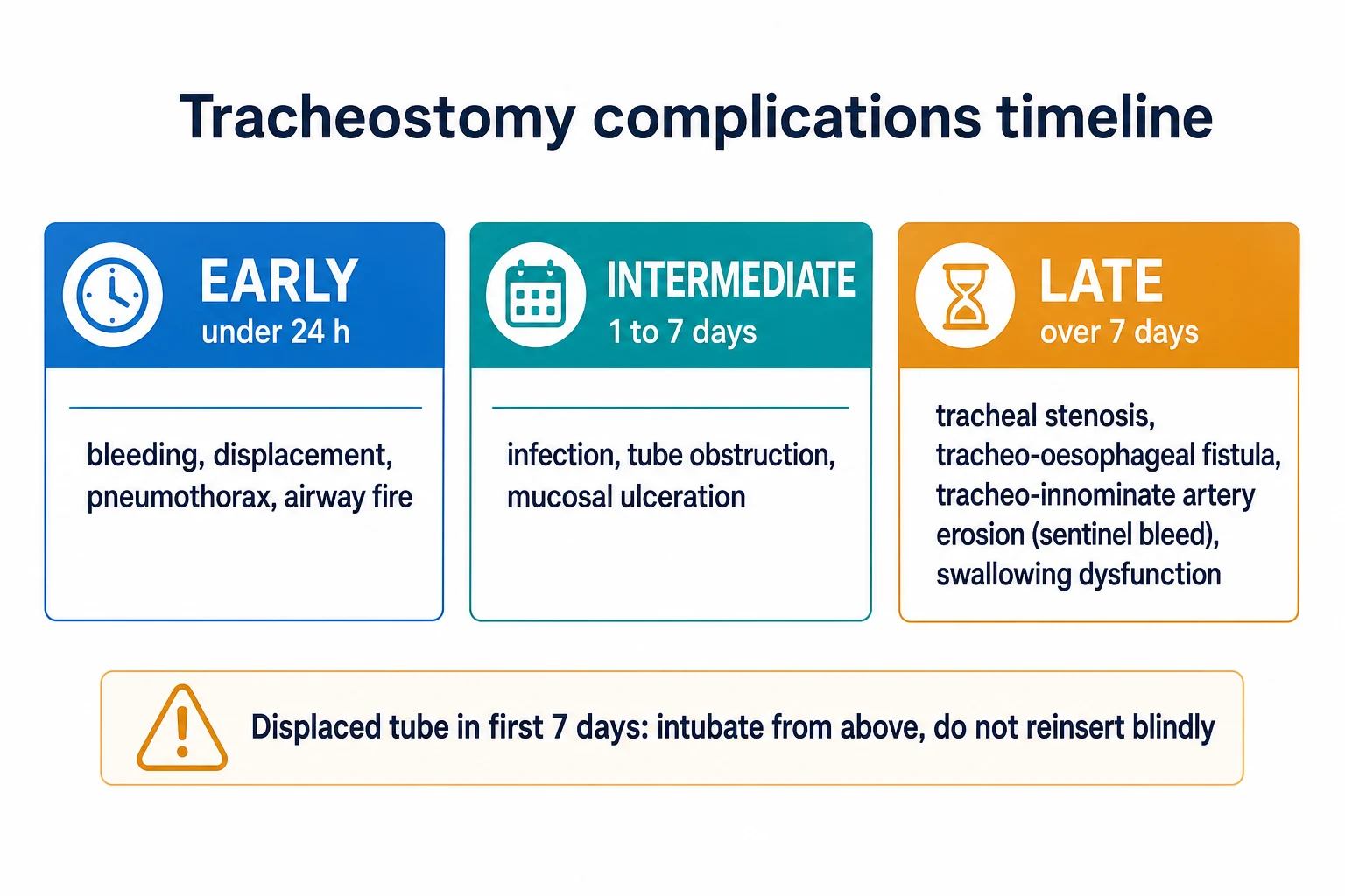
Early (0-7 day) complications and management
Bleeding
The most common early complication. Minor oozing from the skin edge usually settles. Significant bleeding typically from the thyroid isthmus or anterior jugular veins. Manage with direct pressure, adrenaline-soaked packs, and, if persistent, surgical exploration/ligation or diathermy. Severe bleeding may require conversion to surgical tracheostomy.
Tube displacement
Accidental decannulation is the most feared early complication. If displacement occurs BEFORE day 7 (tract not yet formed): do NOT attempt blind reinsertion — risk of creating a false passage into the mediastinum with catastrophic consequences. Call for help, mask-ventilate with bag-valve-mask, and if ventilation is inadequate, perform orotracheal intubation (the ETT passes the trach site). Re-insert the trach electively once the airway is secure.
Pneumothorax
The apex of the pleura extends above the clavicles into the neck — a needle or dilator directed too laterally or too cephalad can puncture the pleura. Suspect with worsening oxygenation, raised airway pressures, or subcutaneous emphysema. Confirm with ultrasound/chest X-ray. Treat with chest drain if symptomatic.
Subcutaneous / mediastinal emphysema
Air tracks through tissue planes from a tract leak or posterior wall injury. Palpate for surgical emphysema. Check tube position bronchoscopically (exclude posterior tracheal wall tear). If the tube is malpositioned (paratracheal), it must be repositioned.
Posterior tracheal wall injury
Excessive force during needle puncture or dilatation (especially with Griggs forceps) can lacerate the posterior membranous tracheal wall and oesophagus, producing an acute tracheo-oesophageal fistula or pneumomediastinum. Suspected with subcutaneous emphysema, difficulty ventilating, or bloody aspirate. Bronchoscopic confirmation; surgical repair if significant.
Infection (stomal cellulitis)
Stoma site infection presents with erythema, purulent discharge, and systemic signs. Treat with local care, antiseptic dressings, and antibiotics (cover skin flora and Gram-negatives). Debride if abscess forms. A tracheostomy is a clean-contaminated procedure — routine antibiotic prophylaxis is NOT recommended.
Tube blockage
Blood clot, mucus plug, or kinking can occlude the tube. Suspect with rising airway pressures, difficulty passing a suction catheter, or desaturation. Manage with immediate suctioning, inline suction, removal and cleaning of the inner cannula (if double-lumen tube), or tube exchange if blocked. Heated humidification prevents most blockages.
Late complications (>7 days)
Tracheal stenosis
Most common late complication
- Granulation tissue and fibrosis at the stoma site, cuff site, or tube tip
- Caused by pressure necrosis from a high cuff pressure (>30 cmH2O), infection, or repeated trauma
- Presents weeks to months AFTER decannulation with worsening dyspnoea, stridor, or difficulty clearing secretions
- Diagnosis: CT neck/bronchoscopy. Treatment: endoscopic dilation/laser for short segments, surgical resection (tracheal segment resection with end-to-end anastomosis) for longer stenoses
- Prevention: maintain cuff pressure 20-30 cmH2O, avoid overinflation, use appropriately sized tube, prevent infection
Tracheomalacia
Wall weakening
- Softening and collapse of the tracheal cartilage due to chronic pressure necrosis — typically at the cuff site
- The tracheal wall collapses on inspiration (dynamic obstruction), causing stridor and airflow limitation
- Diagnosed by bronchoscopy (shows anterior wall collapse on inspiration/ dynamic airway) or CT with inspiratory-expiratory comparison
- Treatment: conservative (humidification, secretion clearance), stenting, or surgical resection
Tracheo-oesophageal fistula (TEF)
Cuff erosion
- Erosion of the posterior tracheal wall into the oesophagus, almost always from an overinflated cuff pressing against an in-situ nasogastric tube
- Presents with copious secretions, recurrent aspiration pneumonia, abdominal distension (air enters stomach), or food/NG contents suctioned from the trachea
- Diagnosis: bronchoscopy/oesophagoscopy, contrast swallow. Prevent by minimising cuff pressure and avoiding a co-located rigid NG tube where possible
- Treatment: surgical repair (often complex), or conservative if prognosis poor
Tracheo-innominate artery fistula (TIF)
CATASTROPHIC
- Erosion of the tube tip or cuff into the innominate (brachiocephalic) artery, which crosses the anterior trachea at ~6th-7th rings
- Typically presents day 2-14 post-placement, often with a small "sentinel" herald bleed hours before catastrophic haemorrhage
- Mortality 50-80%. IMMEDIATE management: (1) hyperinflate the cuff to tamponade; (2) if that fails, slowly withdraw the tube and apply firm digital pressure against the anterior trachea/ sternum via the stoma (over-inflation of the cuff or a finger through the stoma to compress the artery against the sternum); (3) call for immediate vascular/thoracic surgery
- Definitive: surgical ligation/ resection of the innominate artery
- Prevention: correct tube level (avoid too low), cuff pressure 20-30 cmH2O, appropriate tube size
Tracheocutaneous fistula
Persistent stoma
- Failure of the stoma to close after decannulation — a persistent epithelialised tract
- Risk factors: long duration of cannulation (>3-6 months), obesity, steroid use, infection
- Presents with persistent air leak/ mucus from the stoma weeks after decannulation
- Most close spontaneously within days-weeks with occlusive dressings. Persistent (>2 weeks): surgical closure with excision of the tract
Stoma granuloma
Local tissue
- Exuberant granulation tissue around the stoma causing bleeding and cosmetic problems, occasionally obstructing the tube
- Treated with silver nitrate cautery, topical steroids, or excision
Cuff pressure management
Humidification — the upper airway is bypassed
Speaking valve and swallow assessment
A tracheostomy does not have to mean silence. With the cuff deflated and a one-way speaking valve, the patient can redirect exhaled gas through the vocal cords and speak. [1]
Speaking valve trial and swallow assessment
Cuff deflation tolerance
Before any speaking trial, assess tolerance of cuff deflation. Deflate the cuff slowly. Tolerance requires: adequate oxygenation, ability to manage secretions (cough), no risk of gross aspiration (assess swallow reflex). Patients with high PEEP or poor cough may not tolerate cuff deflation — the air leak reduces delivered tidal volume. Suction above the cuff before deflating (clears pooled secretions that would otherwise flood the airway).
Speaking valve (e.g. Passy-Muir)
A one-way valve that fits onto the hub of the trach tube. On inspiration, gas flows through the trach into the lungs. On expiration, the valve closes, redirecting exhaled gas UP through the trach, around the deflated cuff, and THROUGH THE VOCAL CORDS and upper airway — enabling phonation. The cuff MUST be fully deflated before placing the valve (a closed valve + inflated cuff = complete airway obstruction and asphyxiation). Start with short periods (5-15 min) and build up.
Contraindications to speaking valve
Inability to tolerate cuff deflation (unstable ventilation, copious frothy secretions, severe aspiration risk), unconscious patient, marked airway obstruction above the trach (severe laryngeal stenosis/ glottic oedema), or a foam-cuff (low-pressure) tube that does not allow full deflation. Foam-cuff tubes cannot be used with a speaking valve.
Swallow assessment
Tracheostomy and cuff inflation impair swallow (the inflated cuff mechanically tethers the larynx and reduces laryngeal elevation; reduced subglottic air pressure impairs the swallow reflex). Before oral intake: perform a formal swallow assessment (speech-language therapy), ideally with the cuff deflated and a speaking valve in place (restores subglottic pressure). Bedside swallow screen with thickened fluids; proceed to fibreoptic endoscopic evaluation of swallowing (FEES) or videofluoroscopy if aspiration suspected.
Aspiration risk
Aspiration is common in tracheostomised patients and often SILENT (the cuff masks the cough). All tracheostomised patients are at high aspiration risk until swallow is formally assessed. Keep NPO until cleared. Blue-dye test is insensitive and no longer recommended — use FEES.
Decannulation protocol
Decannulation is the planned removal of the tracheostomy tube once the original indication has resolved. It follows a structured pathway to confirm the patient can protect and maintain their own airway. [1]
Tracheostomy decannulation process
1. Assess readiness
Pre-requisites: original indication resolved (no prolonged ventilation needed, no upper airway obstruction); patient off the ventilator or on minimal support; adequate spontaneous ventilation and cough; manageable secretions; stable and cooperative mental status (able to protect airway); swallowing assessed and safe (or NPO with plan); no ongoing need for frequent deep suctioning.
2. Cuff deflation tolerance
Deflate the cuff. Confirm the patient tolerates deflation for at least 24 h with no aspiration, no respiratory distress, and adequate ventilation. Suction above the cuff BEFORE deflating to clear pooled secretions.
3. Cuff leak test (assess upper airway patency)
With cuff deflated, occlude the trach tube (or cap it). Assess whether the patient can move air around the deflated tube and through the upper airway (listen/feel at the nose and mouth). If airflow is inadequate (tube too large relative to tracheal lumen), downsize the tube (e.g. 8 → 6 → 4) and reassess. A patient with laryngeal oedema or undiagnosed upper airway obstruction will not pass and must be investigated before decannulation.
4. Downsizing and capping (blocking) trials
Progressively downsize to a smaller uncuffed tube (e.g. size 4). Insert a decannulation cap (plug) so the patient breathes ENTIRELY through their own upper airway with the trach tube capped. Monitor closely for 24 h (RR, SpO2, ability to cough and clear secretions, comfort). A speaking valve can be used as an intermediate step before full capping (it allows inspiration through the trach but forces expiration through the upper airway).
5. Endoscopic assessment (optional but recommended)
Prior to decannulation (especially after prolonged intubation/trach), perform a bedside endoscopic/ bronchoscopic assessment via the trach to inspect the upper airway, vocal cords, and subglottic region — exclude laryngeal oedema, granulation, vocal cord palsy, or stenosis that would obstruct the airway after decannulation.
6. Tube removal and stoma care
Once the patient has tolerated capping for 24 h with no deterioration, remove the tube. Apply an occlusive (airtight) dressing over the stoma. Instruct the patient to press on the dressing when coughing or speaking (this supports stoma closure). Most stomas close spontaneously within days to weeks. A persistent stoma (>2 weeks) may need surgical closure.
7. Post-decannulation monitoring
Observe for 24-48 h for stridor, respiratory distress, or secretion retention. Have the trach bedside equipment immediately available. Arrange follow-up to assess for late tracheal stenosis (presents weeks-months later with exertional dyspnoea/ stridor).
Pass — proceed to decannulation
All of the following
- Tolerates cuff deflation for 24 h with no distress or aspiration
- Able to cough and clear secretions effectively
- Upper airway patent on cuff leak (air moves around the tube)
- Tolerates capping for 24 h with stable RR, SpO2, and comfort
- Endoscopy excludes laryngeal/ subglottic obstruction
Fail — do NOT decannulate
Any of the following
- Respiratory distress, rising RR, or falling SpO2 on cuff deflation or capping
- Inability to clear secretions (weak cough, copious secretions)
- No cuff leak (tube too big or upper airway obstructed) — downsize and reassess; investigate obstruction
- Ongoing need for ventilation or frequent suctioning
- Reduced conscious state unable to protect airway
Special situations
Tracheostomy in the severely brain-injured patient
Why brain injury needs a trach early
Neurological indications
- Poor airway protection (reduced gag/ cough, bulbar dysfunction) → aspiration risk
- Prolonged ventilation for raised ICP management (to control PaCO2) and pulmonary complications
- Facilitates early mobilisation and rehabilitation, and transfer to a neurorehabilitation facility
- Some neuro-ICU units consider trach earlier (day 5-7) in severe TBI, as duration of ventilation is often predictable
- Note: tracheostomy does NOT improve mortality in brain-injured patients (meta-analyses neutral); it is a logistics/ comfort/ rehabilitation decision
Tracheostomy in children
Paediatric tracheostomy
Different rules apply
- Surgical tracheostomy is PREFERRED over percutaneous in children (small airway, difficult anatomy, higher risk)
- Common indications: prolonged ventilation (neuromuscular disease, bronchopulmonary dysplasia), upper airway obstruction (subglottic stenosis, laryngomalacia, bilateral vocal cord palsy)
- Higher risk of tube displacement (smaller airway, harder to reinsert); uncuffed tubes often used in younger children
- Decannulation is more cautious; structured, often inpatient, with endoscopic airway assessment first
Tracheostomy emergencies — the bedside response
The intubated or tracheostomised patient who deteriorates needs a structured approach. The UK National Tracheostomy Safety Project algorithm is the exam-standard framework. [1]
Tracheostomy emergency algorithm — the patient with a blocked/ displaced trach
1. Call for help and assess
Recognise the deteriorating tracheostomy patient: desaturation, rising RR/ airway pressures, distress, absent ETCO2, or unable to pass a suction catheter. Call for senior/ anaesthetic help immediately. Communicate clearly: "tracheostomy emergency".
2. Oxygenate from BOTH ends
Apply high-flow oxygen to BOTH the tracheostomy (via the trach hub) AND the face/ nose (via a face mask). This is the single most important initial action — oxygenate the upper and lower airway simultaneously in case the trach is malpositioned.
3. Is the trach patent? (suction test)
Pass a suction catheter through the trach. If it passes EASILY and you can aspirate secretions → the trach is patent; the problem is elsewhere (e.g. lung, sedation). If the catheter does NOT pass → the trach is blocked or displaced.
4a. If trach is patent but patient deteriorating
The tube is in the trachea but the patient is failing — ventilate via the trach with a bag-valve. Investigate lung pathology (pneumothorax, mucus plug, pulmonary oedema, bronchospasm).
4b. If trach blocked or displaced (catheter will not pass)
Deflate the cuff. If ventilation does not improve: remove the trach tube entirely (cut the ties). Attempt bag-valve-mask ventilation via the UPPER AIRWAY (face mask + oral airway). If the upper airway is patent and ventilation succeeds → keep mask-ventilating and arrange elective re-insertion. If upper airway ventilation FAILS → orotracheal intubation (the ETT passes the stoma). Only attempt blind trach reinsertion if the tract is mature (>7-10 days) and you have a spare tube + dilators.
5. Confirm any replacement tube position
After any re-insertion or intubation, confirm position with ETCO2, bilateral chest rise, and bronchoscopy. Obtain a chest X-ray.
Evidence and landmark trials
TracMan
NEJM 2013
909 critically ill pts expected to need >=7 days ventilation — early trach (day 1-4) vs late (day 10+)
Key finding
30-day mortality 30.8% early vs 31.6% late (no difference). Early group had modestly less IV sedation but no difference in ICU stay, mortality, or infection. 45% of the late group never needed a trach
Practice change
Early trach does NOT improve survival — decision is individualised and patient-centred
Rumbak
Crit Care Med 2004
112 medical ICU pts expected to need >=14 days ventilation — early PDT (within 48 h) vs delayed trach (day 14-16)
Key finding
Mortality 31.7% early vs 61.7% late (p=0.005); less pneumonia, shorter ICU stay with early
Practice change
Suggested a survival benefit in a highly selected long-stay medical cohort — but single-centre and not reproduced by TracMan. Defines the "selected patient" who may benefit
Trouillet (SITARE)
Ann Intern Med 2011
220 post-cardiac-surgery pts ventilated >48 h — early PDT (day 5) vs prolonged intubation (trach day 15)
Key finding
No difference in antibiotic-free days (primary), mortality, ICU stay, or nosocomial pneumonia
Practice change
Confirms no benefit of early trach after cardiac surgery — adds to TracMan conclusion
Díaz-Regañón
Critical Care 2008
Prospective cohort, 800 PDT procedures — safety and complication profile of percutaneous dilatational tracheostomy
Key finding
Overall complication rate ~6-8% (mostly minor bleeding); serious complications rare; major bleeding <1%, pneumothorax <1%. Confirms PDT is safe when performed by trained operators with bronchoscopic guidance
Practice change
Established PDT as a safe, reproducible bedside procedure in the ICU
Freeman meta-analysis
Chest 2000
Meta-analysis of 5 RCTs comparing percutaneous vs surgical tracheostomy
Key finding
Overall complication rates NOT significantly different. PDT trended towards less stomal bleeding/ infection; surgical trach trended towards fewer decannulation/ obstruction problems
Practice change
PDT is as safe as surgical trach in selected patients; PDT became first-line in the ICU
Marynissen meta-analysis
Critical Care 2021
Updated systematic review and meta-analysis — percutaneous vs surgical tracheostomy
Key finding
No significant difference in mortality, serious bleeding, infection, or overall complications. PDT favoured for bedside convenience and cost; surgical for specific anatomical/ contraindication contexts
Practice change
Confirms modern practice: PDT first-line in ICU, surgical reserved for specific indications
Prognosis and outcomes
Tracheostomy outcomes and procedure statistics
- Procedural mortality of PDT is very low (<0.5%) when performed by trained operators with bronchoscopic guidance.[5]
- Long-term: up to 30-45% of tracheostomy patients have some degree of tracheal stenosis on imaging, but only ~1-5% become symptomatic (stridor, exertional dyspnoea) and require intervention.[2]
- Quality of life: tracheostomy improves comfort, communication, and the ability to participate in rehabilitation compared with prolonged translaryngeal intubation — a patient-centred benefit even in the absence of a mortality effect.
SAQ — Early versus late tracheostomy: appraising the evidence
10 minutes · 10 marks
A 70-year-old man is admitted to ICU with severe community-acquired pneumonia and type 1 respiratory failure requiring mechanical ventilation. On day 3 his FiO2 remains 0.5 with PEEP 8. The registrar asks whether an early tracheostomy within the next 48 hours would improve his outcomes.
SAQ — Decannulation failure and the cuff leak test
10 minutes · 10 marks
A 66-year-old man, day 28 after tracheostomy for prolonged ventilation following a severe COPD exacerbation, is being assessed for decannulation. He is off the ventilator, has an effective cough, and is neurologically intact. On cuff deflation and capping he develops stridor and desaturation within 30 minutes.
Clinical pearls
Red flags
References
- [1]Young D, Harrison DA, Cuthbertson BH, Rowan K, TracMan Collaborators. Atypical mycobacterial keratitis and canaliculitis in a patient with an indwelling SmartPLUG Br J Ophthalmol, 2010.PMID 20215379
- [2]Marynissen J, Lizarrondo L, De Smet K, et al. Notum palmitoleoyl-protein carboxylesterase regulates Fas cell surface death receptor-mediated apoptosis via the Wnt signaling pathway in colon adenocarcinoma Bioengineered, 2021.PMID 34402722
- [3]Rumbak MJ, Newton M, Truncale T, Schwartz SW, Adams JW, Hazard PB. Rosiglitazone, a ligand of the peroxisome proliferator-activated receptor-gamma, reduces the development of nonseptic shock induced by zymosan in mice Crit Care Med, 2004.PMID 14758164
- [4]Trouillet JL, Luyt CE, Guiguet M, et al. Identification of a novel TP53 cancer susceptibility mutation through whole-genome sequencing of a patient with therapy-related AML JAMA, 2011.PMID 21505135
- [5]Díaz-Regañón G, Miñambres E, Ruiz A, González-Herrera S, Holanda-Peña M, López-Espadas F. The effects of involving a nurse practitioner in primary care for adult patients with urinary incontinence: the PromoCon study (Promoting Continence) BMC Health Serv Res, 2008.PMID 18412964
- [6]Freeman BD, Isabella K, Lin N, Buchman TG. Variability in performance of timed walk tests in pulmonary rehabilitation programs Chest, 2000.PMID 10893366