ICU · Respiratory / ventilation
VA-ECMO for Cardiogenic Shock & Extracorporeal CPR (ECPR)
Also known as VA-ECMO · Veno-arterial ECMO · Extracorporeal life support · ECLS · Extracorporeal cardiopulmonary resuscitation · ECPR · Femoro-femoral ECMO · Harlequin syndrome · Differential hypoxaemia · LV distension · Bridge to recovery · SAVE score · SCAI shock stages · Distal perfusion cannula
Veno-arterial ECMO (VA-ECMO) drains venous blood, oxygenates and decarboxylates it, and returns it to the arterial system — providing both circulatory and respiratory support for refractory cardiogenic shock (a bridge to recovery, decision, transplant, or durable LVAD) and for refractory cardiac arrest (extracorporeal CPR, ECPR). Cannulation is peripheral (femoro-femoral, rapid, for shock and ECPR) or central. The complications include bleeding (the largest), limb ischaemia (mitigated by a distal perfusion cannula), left-ventricular distension and pulmonary oedema (the failing LV cannot eject against the retrograde aortic flow — may need an Impella or vent), differential hypoxaemia (the Harlequin or north-south syndrome), thrombosis, haemolysis, and infection. ECPR outcomes are best in selected, rapidly-cannulatable patients. Severity and reversibility are graded by the SCAI A-E stages and the SAVE score; weaning is by staged flow reduction with echocardiographic and pulse-pressure recovery assessment.
On this page & tools
Your progress
Saved locally on this device.
Target exams
Overview & definition
Veno-arterial ECMO (VA-ECMO) drains venous blood, oxygenates and removes CO2 in an external oxygenator, and returns it to the arterial system — thereby bypassing both the heart and the lungs to provide combined circulatory and respiratory support. It is used for refractory cardiogenic shock (a bridge to recovery, decision, transplant, or durable LVAD) and for extracorporeal cardiopulmonary resuscitation (ECPR) in refractory cardiac arrest.[1][1]
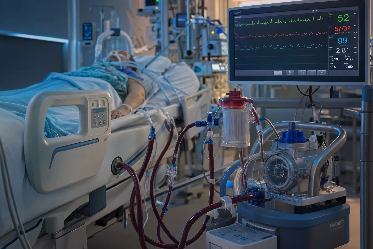
The circuit is a drainage (venous) cannula → centrifugal pump → membrane oxygenator (with gas blender and heat exchanger) → return (arterial) cannula. Flow up to 4–6 L/min is achievable with femoro-femoral cannulation (roughly 60 mL/kg/min). A pumpless arteriovenous configuration is not used for circulatory support — VA-ECMO is, by definition, a pumped circuit that unloads the right heart and provides systemic cardiac output, in contrast to VV-ECMO, which provides only gas exchange. [1]
Indications
- Refractory cardiogenic shock despite optimal medical therapy (inotropes, vasopressors, and an intra-aortic balloon pump) — from a massive myocardial infarction, fulminant myocarditis, a decompensated cardiomyopathy, post-cardiotomy shock, drug toxicity, or massive pulmonary embolism. It is a bridge to recovery, decision, transplant, or durable LVAD.[1][1]
- Extracorporeal CPR (ECPR) — refractory in-hospital or out-of-hospital cardiac arrest not responding to conventional CPR, in selected patients (a witnessed arrest, a reversible cause, and rapid cannulation).[1]
- Refractory hypoxaemia with a cardiac component, when VV-ECMO is insufficient.[1]
Indications by aetiology — the "salvageable" cardiogenic shocks
The decision to cannulate turns on reversibility and the bridge plan. The classic salvageable aetiologies are: [1]
- AMI-cardiogenic shock (AMI-CS) — the largest single indication (around 40 per cent of adult VA-ECMO runs). Used as bridge to revascularisation recovery (after primary PCI for a large infarct with persistent low output), or as a bridge to durable LVAD/ transplant in the irrecoverable but transplant-eligible patient. The IABP-SHOCK II trial showed an IABP does not improve mortality in AMI-CS, which has driven escalation straight to percutaneous MCS (Impella/VA-ECMO) in deteriorating patients.[5][1]
- Fulminant myocarditis — particularly giant-cell and eosinophilic myocarditis and paediatric parvovirus/B19 myocarditis, where the myocardium is inflamed but potentially recoverable over days to weeks. VA-ECMO (often with a short-term BiVAD or CentriMag) buys time for biopsy, immunosuppression (giant-cell), and recovery; survival to discharge approaches 70 per cent in expert series.
- Post-cardiotomy shock — failure to wean from cardiopulmonary bypass, or post-cardiotomy low-output syndrome. Central cannulation is typical. Outcomes are poorer (30–40 per cent survival) than medical cardiogenic shock because of the comorbid surgical insult and the coagulopathy of bypass.
- Drug toxicity / poisoning — the potentially reversible toxidromes where the toxin has a finite circulation time and the heart is expected to recover once it clears: beta-blocker overdose, calcium-channel blocker overdose (verapamil/diltiazem are the most lethal), bupivacaine toxicity (lipid + ECMO is the rescue), tricyclic antidepressant cardiotoxicity, digoxin/severe hyperkalaemia (the calcium chloride debate notwithstanding), and methamphetamine/cocaine-mediated cardiomyopathy. The credo is "bridge to toxin clearance and receptor recovery."
- Peripartum cardiomyopathy and fulminant decompensated cardiomyopathy in the transplant-eligible younger patient — often bridged with VA-ECMO ± Impella as a bridge to decision/recovery/transplant.
- Massive pulmonary embolism with shock — VA-ECMO supports the failing right ventricle and oxygenation; it can be a bridge to catheter-directed or surgical embolectomy, or to clot lysis/recovery.
- Malignant arrhythmia storms (refractory VF/pVT not controlled by antiarrhythmics or ablation) — temporary VA-ECMO/MCS for haemodynamic support during ablation or while drug therapy takes effect.
SCAI classification of cardiogenic shock (stages A–E)
The Society for Cardiovascular Angiography and Interventions (SCAI) stages cardiogenic shock from A (at risk) to E (extremis) — the framework that defines who is sick enough to warrant VA-ECMO and how the patient is decompensating. Mortality rises sharply across the stages (roughly <5 per cent at A, 10–15 per cent at B, 20–30 per cent at C, 40–50 per cent at D, and >75 per cent at E).[4]
| Stage | Definition | Clinical picture | Typical action |
|---|---|---|---|
| A — At risk | Risk factors for shock; not yet shocked | AMI, acute HF; cool, normal BP, normal lactate | Treat the cause; monitor |
| B — Beginning | Hypoperfusion beginning; compensated | Tachy, anxious; oliguria; lactate rising; BP maintained by vasoconstriction | Early inotrope/vasopressor, MCS planning |
| C — Classic | Hypoperfusion + hypotension | SBP <90 (or >30 mmHg below baseline), cold, oliguric, confused, raised lactate | Inotropes/vasopressors; consider MCS (VA-ECMO) |
| D — Deteriorating | Worsening despite escalating therapy | Multi-pressor, worsening lactate/acidosis, organ failure | Initiate VA-ECMO / escalation to MCS; definitive therapy |
| E — Extremis | Circulatory collapse | Cardiac arrest / refractory VF / PEA; CPR in progress | ECPR if eligible; otherwise futile |
The SCAI stage is dynamic — re-stage hourly. The thresholds for VA-ECMO are typically persistent stage C/D (lactate rising despite two inotropes/pressors, or a cardiac index <1.8 L/min/m², or mixed venous saturation <60 per cent despite escalation). The aim is to cannulate before organ failure is fixed (the liver is failing, the kidneys are anuric, the bowel is ischaemic) — late cannulation in stage E without a reversible driver is the commonest reason for futile runs.[4][1]
Cannulation
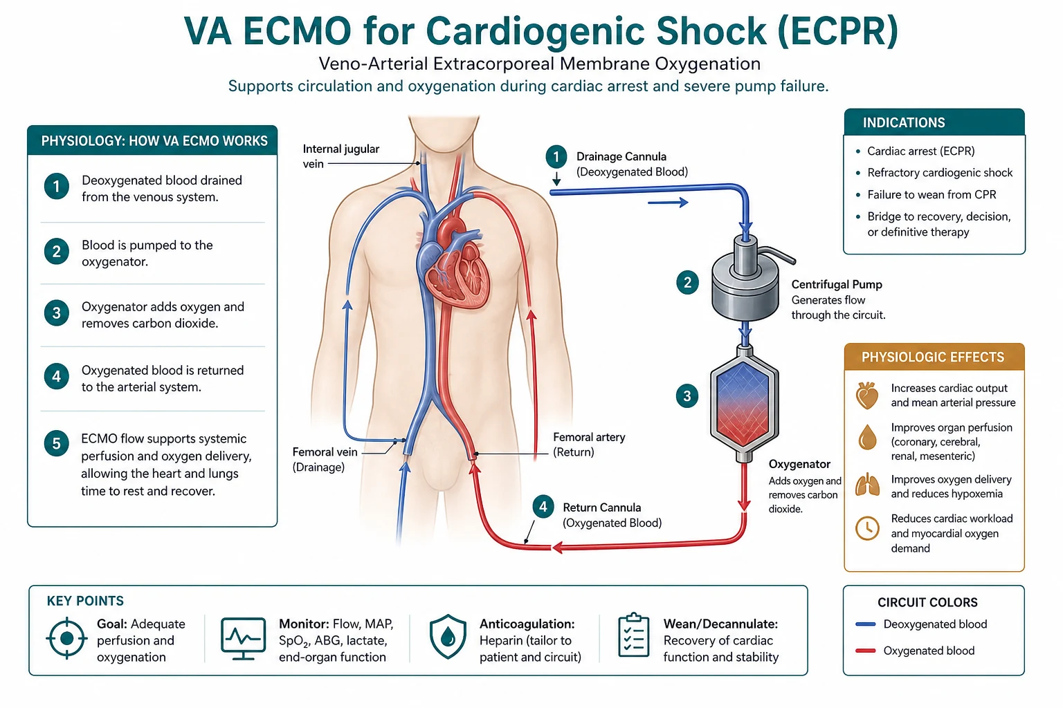
- Peripheral (femoro-femoral) — drains the femoral vein to the right atrium, returns to the femoral artery. It is rapid and bedside-achievable, the choice for cardiogenic shock and ECPR.[1]
- Central — right atrium to aorta, in theatre (typically post-cardiotomy). It avoids the limb and Harlequin issues.[1]
- The distal perfusion cannula (DPC) — the limb distal to the femoral arterial cannula is at ischaemia risk; a small cannula in the superficial femoral artery perfuses the leg and prevents ischaemia.[1]
Cannulation in depth — choosing the configuration
| Configuration | Drainage | Return | When to use | Key advantage | Key drawback |
|---|---|---|---|---|---|
| Peripheral femoro-femoral | Femoral vein → RA | Femoral artery | Cardiogenic shock, ECPR, rapid bedside | Fast, bedside, reversible, surgical-light | Limb ischaemia, Harlequin, retrograde dissection |
| Central | RA (right atriotomy) | Ascending aorta | Post-cardiotomy, cannot wean CPB; peripheral unsuitable | High flow, no limb issue, anterograde arch flow (no Harlequin) | Sternotomy, bleeding, theatre-bound |
| Axillary/subclavian | Femoral/RA | Axillary artery (graft) | Bridge to transplant/LVAD, ambulatory, long support | Anterograde flow, no Harlequin, allows mobilisation | Surgical, slower, graft needed |
| Veno-arterial-venous (VAV) | Femoral vein | Femoral artery + IJ to SVC | Peripheral VA-ECMO + Harlequin (differential hypoxaemia) | Resolves Harlequin | More cannulae, more bleeding |
| Peripheral + LV vent (Impella/atrial septostomy) | Femoral vein | Femoral artery | LV distension / pulmonary oedema | Unloads LV | Second device, vascular access |
The distal perfusion cannula (DPC), also called a reperfusion cannula, is the single most important limb-salvage manoeuvre in peripheral VA-ECMO. Placed at the time of cannulation into the superficial femoral artery (or as a side-arm off the arterial return line), it shunts oxygenated blood distally and reduces critical limb ischaemia from roughly 10–20 per cent down to 2–5 per cent. Limb surveillance (Doppler signals, perfusion, capillary refill, and clinical examination every hour for the first 6 hours then 4-hourly) is mandatory.[1][1]
What VA-ECMO does (and the haemodynamic pitfalls)
- It unloads the failing heart by reducing the preload it must handle and providing the systemic flow.
- It rests the myocardium (reduced workload), allowing recovery.
- It provides oxygenation.
- The LV distension pitfall — the retrograde femoral return increases the aortic pressure against which the failing LV must eject; a severely failing LV cannot eject, distends, and backs up into the lungs (pulmonary oedema). It may need an Impella or a surgical vent to unload the LV.[1]
- The Harlequin (north-south) syndrome — in peripheral VA-ECMO with concurrent lung failure, the native (failing) heart ejects poorly-oxygenated blood to the arch, coronaries, and brain, while the ECMO returns oxygenated blood to the lower body. The upper body (brain, heart) can be hypoxic despite a normal lower-body saturation. Detect it with a right-radial arterial line; treat it by adding a venous return to the upper-body circulation (a veno-arterial-venous, VAV, configuration) or by improving the native lung oxygenation.[1]
A subtlety of VA-ECMO physiology: it does not directly unload the left ventricle — the venous drainage reduces right-sided return to the lungs, but the failing LV still has to eject against an elevated afterload from the retrograde arterial flow. If native LV ejection is poor, blood "stacks up" behind the aortic valve → LV distension → raised LA and pulmonary capillary pressure → pulmonary oedema and, in extreme cases, intracardiac thrombus from stasis. The markers are a rising pulmonary artery pressure, a rising LA pressure, pulmonary oedema on imaging, and a non-pulsatile aortic root on echo (the aortic valve is not opening). Management: reduce ECMO flow, add inotropy (milrinone, dobutamine, isoproterenol) to encourage LV ejection, and — if refractory — vent the LV (percutaneous Impella, IABP, pulmonary artery vent, atrial septostomy, or surgical apical vent).[1]
Complications
- Bleeding — the largest complication (anticoagulation plus large cannulae): cannulation-site bleeding, gastrointestinal and intracranial haemorrhage.
- Thrombosis and embolism (clot in the circuit).
- Infection.
- Limb ischaemia (femoral cannulation — mitigated by a distal perfusion cannula).
- LV distension and pulmonary oedema (the failing LV cannot eject against the retrograde flow).
- Differential hypoxaemia / Harlequin syndrome (peripheral VA-ECMO with lung failure).
- Haemolysis (the centrifugal pump).
- Air embolism, AKI, stroke, and vascular injury.[1][1]
Complication rates (ELSO registry and meta-analytic estimates)
| Complication | Approximate incidence | Notes / mitigation |
|---|---|---|
| Bleeding (any) | 30–50% | Surgical sites, GI, intracranial (~5%). Lowest-effective anticoagulation; targeted transfusion |
| Cannulation-site bleeding | 15–25% | Surgical revision; ultrasound-guided percutaneous insertion |
| Thrombosis (circuit/patient) | 10–20% | Anticoagulation; daily circuit inspection; D-dimer trend |
| Haemolysis (plasma-free Hb >50 mg/dL) | 10–20% | Centrifugal pump; check pump head, flows, kinks |
| Acute kidney injury / RRT | 20–40% | Worse with shock and haemolysis; often recovers |
| Limb ischaemia | 10–20% (no DPC) → 2–5% (with DPC) | Distal perfusion cannula at cannulation |
| Infection (catheter/bloodstream) | 10–30% | Aseptic technique; daily line review; narrow-spectrum if culture-positive |
| Stroke (any) | 5–15% | Both ischaemic and haemorrhagic; anticoagulation balance |
| LV distension / pulmonary oedema | 10–30% | Echo surveillance; LV vent (Impella/IABP/septostomy) |
The single greatest mortality modifier on VA-ECMO is bleeding — particularly intracranial haemorrhage — and the single greatest preventable morbidity is limb loss from a femoral arterial cannula without a distal perfusion cannula. A haemolysis + thrombocytopenia + rising anticoagulant requirement triad signals a clotting circuit and mandates a circuit or oxygenator change.[1]
Anticoagulation, circuit monitoring, and transfusion
- Anticoagulation — unfractionated heparin is standard, targeting an ACT 1.5× baseline (~150–180 s) or an anti-Xa 0.3–0.5 IU/mL (aPTT 50–80 s where anti-Xa unavailable). In bleeding, target the lowest effective range, or hold heparin entirely (e.g. after neurosurgery/bleeding) accepting higher thrombosis risk — direct thrombin inhibitors (bivalirudin, argatroban) are used in heparin-induced thrombocytopenia.
- Circuit surveillance — hourly pump flow, RPM, sweep gas, pre/post-membrane pressures (a rising transmembrane pressure gradient signals oxygenator clotting), and circuit inspection for clots. Daily plasma-free haemoglobin, LDH, and haptoglobin for haemolysis; fibrinogen, antithrombin, and platelet count for consumptive coagulopathy.
- Transfusion thresholds — keep haemoglobin >70–80 g/L, platelets >50 × 10⁹/L (higher, >100, if bleeding or post-neurosurgery), and fibrinogen >1.5–2.0 g/L. Avoid over-transfusion (donor exposure, volume, TRALI). Have a written massive transfusion / ECMO-bleeding protocol — the unifying algorithm for circuit, surgical, and coagulopathic bleeding. [1]
ECPR — extracorporeal cardiopulmonary resuscitation
ECPR is the rapid deployment of VA-ECMO during ongoing CPR for refractory cardiac arrest. It does not treat the cause of the arrest; it provides coronary and cerebral perfusion while the cause is addressed (PCI for AMI, embolectomy for PE, rewarming for hypothermia, toxin clearance for poisoning). The two landmarks for ECPR are: [1]
- A witnessed arrest with a shockable rhythm refractory to conventional ACLS (typically refractory VF/pVT, but also selected asystole/PEA with a reversible cause).
- Rapid cannulation — time from arrest (or from refractory-arrest recognition) to ECMO flow <60 minutes is the dominant survival determinant. Beyond 60 minutes of low-flow (effective CPR) time, survival collapses.
- Prognostication at 24–48 hours — by this window the cause is identified and treated, end-organ injury declares itself, and the question of futility vs bridge-to-recovery/decision can be addressed. Absence of brainstem reflexes, a non-reactive EEG, an uncorrectable lactate/acidosis, or an irreversible cause at 48 h support withdrawal; a recovering myocardium (rising pulse pressure, falling inotrope/vasopressor need) supports continuation toward weaning.[1][2]
ECPR evidence — the ARREST trial
The ARREST trial (Advanced Reperfusion Strategies for Refractory VF; Yannopoulos, Bartos et al., Lancet 2020) randomised 30 patients with refractory VF OHCA to ECPR + standard ACLS versus standard ACLS alone. The trial was stopped early for efficacy: survival to discharge was 43 per cent (6/14) with ECPR vs 7 per cent (1/15) with standard care (relative risk 6.0; absolute risk difference +36 per cent). The result, although small and single-centre, established ECPR as a credible therapy in selected, rapidly-cannulatable OHCA — and the trajectory of survival (sustained neurological recovery in the survivors) supported a structured ECPR programme rather than ad-hoc cannulation.[2]
The subsequent randomised trials have been mixed: Prague-OHCA (Belohlavek et al., NEJM 2024) and the INNOVATE-ECMO trial showed that, applied broadly to OHCA, ECPR did not improve survival with good neurological outcome — benefit concentrated in selected, rapidly-cannulatable, witnessed, shockable-rhythm patients with short no-flow/low-flow times. The modern consensus: ECPR is a programme, not a procedure — outcomes depend on arrest-to-flow time, team expertise, and patient selection, not on the cannulation itself.[1][1]
Patient selection and prognostic scoring — the SAVE score
The SAVE (Survival After Veno-arterial ECMO) score, derived from the Australian and New Zealand Intensive Care (ANZ) ECMO registry, is the most-used pre-cannulation mortality predictor for medical cardiogenic shock on VA-ECMO. It uses age, acute diagnosis (with fulminant myocarditis, drug intoxication, and cardiomyopathy scoring best; post-cardiotomy worst), chronic organ dysfunction, pre-ECMO organ failure (renal, liver, CNS, etc.), arrest, ventilation, and bicarbonate. The score stratifies survival from >75 per cent (class I) down to <10 per cent (class V).[3]
SAVE score — survival classes
| SAVE class | Predicted survival | Typical patient |
|---|---|---|
| I | >75% | Young, fulminant myocarditis / drug toxicity, no chronic disease |
| II | 50–75% | AMI-CS, single-organ failure, no arrest |
| III | 30–50% | Cardiomyopathy with mild MODS |
| IV | 10–30% | Significant multi-organ failure or chronic disease |
| V | <10% | Post-cardiotomy in the elderly, arrested, MODS |
A SAVE class I–III patient with a reversible driver is a clear go for VA-ECMO; a SAVE class V patient without a bridge strategy is a clear futility discussion. The score is a decision-support tool, not an absolute — and is best paired with the dynamic SCAI stage (a deteriorating stage C with good SAVE class is the ideal window).[3][4]
Bridge strategy — decision, recovery, transplant, durable LVAD
The bridge strategy is the second decision after "should we cannulate?" — every VA-ECMO run must declare one of four end-points: [1]
- Bridge to recovery (BTR) — the myocardium is expected to recover (myocarditis, drug toxicity, AMI with successful reperfusion, post-arrhythmic stunning). Aim: myocardial rest, treat the cause, wean in days–2 weeks.
- Bridge to decision (BTD) — the cause/reversibility is unclear at cannulation (often the case in ECPR and unknown shock). Aim: stabilise, investigate (biopsy, echo, coronary angiography), and within 5–7 days commit to recovery, transplant/LVAD, or withdrawal.
- Bridge to transplant (BTT) — transplant-eligible patient with irreversible myocardial failure but preserved end-organs. Aim: maintain end-organ perfusion, ambulation, and rehabilitation while awaiting a donor organ.
- Bridge to durable LVAD (BTT-LVAD/BiVAD) — destination-therapy or transplant-bridge LVAD candidate; VA-ECMO supports while LVAD work-up (psychosocial, financial, infection clearance) is completed, then converts to a durable device. [1]
A fifth, often unstated, end-point is bridge to withdrawal (futility) — when irreversibility (irreversible anoxic brain injury, fixed MODS, non-recoverable myocardium with no LVAD/transplant pathway) declares itself, the VA-ECMO run is ethically a withdrawal discussion rather than a continuation. Declaring the bridge strategy up front avoids open-ended runs that accumulate complications without a destination.[1][1]
Weaning from VA-ECMO
Weaning is a staged, deliberate trial of myocardial recovery, not a passive flow reduction. The prerequisites are: resolution of the cause (revascularised, recovering myocarditis, cleared toxin), minimal inotrope/vasopressor need, stable/normalising lactate and acid-base, no ongoing sepsis, and adequate end-organ function. [1]
Weaning protocol
- Optimise the myocardium — ensure normovolaemia, normalise electrolytes (K⁺, Mg²⁺, Ca²⁺), titrate inotropes (milrinone ± low-dose adrenaline/levosimendan), and treat any residual arrhythmia/ischaemia.
- Staged flow reduction — reduce flow in 0.5 L/min steps every 1–6 hours, down to a minimum safe flow of ~1–1.5 L/min (below which circuit clotting and stasis rise sharply). Monitor MAP, lactate, mixed venous saturation, urine output, and ECG at each step.
- Echo assessment — a transthoracic or transoesophageal echo at low flow assesses LV ejection (LVEF >20–25 per cent), RV function, aortic valve opening, LVOT VTI, and no pericardial effusion. An LVEF <20 per cent, a falling LVOT VTI, or a rising LA pressure fails the trial.
- Pulse pressure recovery — a native pulse pressure >10–15 mmHg at minimal ECMO flow indicates the native heart is ejecting and the aortic valve is opening; an absent pulse pressure suggests inadequate recovery.
- Haemodynamic targets at the lowest tolerated flow — MAP >60 mmHg, lactate stable or falling, mixed venous saturation >65 per cent, cardiac index >2.0–2.2 L/min/m², LVEDP/PCWP <18 mmHg, SVR normalised.
- Trial off (circuit clamped, flush-running) — if flow wean is tolerated, a brief off-trial with the circuit on flush (heparinised saline) confirms readiness before decannulation. This is the final go/no-go.
- Decannulation and site repair — surgical or percutaneous, with reversal of anticoagulation, vascular repair, and distal limb reassessment. [1]
A failed wean (rising lactate, falling MAP/SvO₂, pulmonary oedema, or echo deterioration) is not a failure of strategy — it is information: re-establish full flow, re-diagnose (is the cause really resolved? is there a new ischaemia/arrhythmia/tamponade?), and consider escalation to a durable LVAD or transplant pathway, or, if irreversible, a withdrawal discussion.[1][1]
Comparison tables — VA-ECMO vs the alternatives
VA-ECMO vs VV-ECMO
| Feature | VA-ECMO | VV-ECMO |
|---|---|---|
| Indication | Cardiac ± respiratory failure | Pure respiratory failure (severe ARDS) |
| Cannulation | Vein → artery (femoro-femoral/central) | Vein → vein (femoro-jugular, double-lumen) |
| Provides | Cardiac output + oxygenation | Oxygenation only (no haemodynamic support) |
| LV unloading | Indirect (can distend LV) | N/A |
| Harlequin risk | Yes (peripheral + lung failure) | No |
| Limb ischaemia | Yes (arterial cannula) | No |
| Pulse pressure | Often reduced (non-pulsatile) | Preserved |
| Anticoagulation | Yes | Yes |
Temporary mechanical circulatory support options
| Device | Flow | What it supports | LV unloading | Practical notes |
|---|---|---|---|---|
| VA-ECMO | Up to 5–6 L/min | Biventricular + lungs | Poor (may distend LV) | Bedside, rapid; bleeding, limb, Harlequin |
| IABP | Modest (≈0.5–1 L/min) | LV (afterload reduction, coronary flow) | Yes (mild) | Easy, cheap; no mortality benefit in IABP-SHOCK II |
| Impella (2.5/CP/5.0/5.5) | 2.5–5.5 L/min | LV (antegrade trans-aortic) | Yes (excellent) | Requires competent AV; haemolysis, limb |
| Impella RP | Up to 4 L/min | RV | N/A | RV failure, post-VAD |
| TandemHeart | Up to 4–5 L/min | LV (LA → femoral artery) | Yes | Large venous cannula; femoral arterial |
| CentriMag / BioMedicus (surgical) | Up to 10 L/min (BiVAD capable) | BiVentricular | VAD-dependent | Theatre, central; for post-cardiotomy, myocarditis |
| ECPR (VA-ECMO in arrest) | Up to 5–6 L/min | Biventricular + lungs | Poor | For refractory VF OHCA if <60 min |
The choice between VA-ECMO, an Impella (percutaneous LV unloading), and a CentriMag (surgical BiVAD) depends on which ventricle is failing, whether the lungs are failing, the reversibility window, and the bridge plan. Combined ECMO + Impella ("ECPELLA") is increasingly used in AMI-CS to provide systemic flow (ECMO) plus direct LV unloading (Impella), addressing the LV-distension pitfall — though outcome evidence is observational.[1]
Initiating VA-ECMO for cardiogenic shock
- Recognise deteriorating shock — escalating inotropes/vasopressors, rising lactate, falling SvO₂, oliguria, cool peripheries (SCAI stage C/D).
- Declare a bridge strategy — recovery / decision / transplant / LVAD / withdrawal, with the team and family.
- Assess contraindications — irreversible MODS, aortic regurgitation (relative — worsens LV distension), severe PVD (consider central/axillary), uncontrolled bleeding, futility.
- Vascular access and imaging — ultrasound the femoral vessels, size the artery, plan the DPC.
- Cannulate — large-bore venous (drainage) + arterial (return) cannulae + distal perfusion cannula, with fluoroscopy/TOE where possible.
- Initiate flow — start at 1.5 L/min and titrate up to target (~60 mL/kg/min, MAP >65, SvO₂ >65, lactate falling).
- Anticoagulate — heparin bolus + infusion to ACT 1.5× baseline / anti-Xa 0.3–0.5.
- Set up surveillance — right-radial arterial line, limb Doppler, hourly circuit checks, daily bloods + haemolysis panel, echo within 24 h.
- Manage pitfalls — treat the cause (PCI, biopsy, toxin clearance), watch for LV distension and Harlequin.
Weaning from VA-ECMO
- Confirm cause resolution — revascularised, recovered myocardium, normalising lactate, minimal pressors.
- Optimise myocardium — volume status, electrolytes, inotrope (milrinone ± levosimendan), no ischaemia/arrhythmia.
- Reduce flow in 0.5 L/min steps to ~1–1.5 L/min, monitoring MAP/SvO₂/lactate/urine at each step.
- Echo at low flow — LVEF >20–25%, AV opening, LVOT VTI, RV function, no effusion.
- Pulse pressure check — native PP >10–15 mmHg at low flow.
- Trial off (flush) — circuit on flush, brief off-trial; final go/no-go.
- Decannulate — reverse anticoagulation, vascular repair, reassess limb.
Outcomes and evidence
- Cardiogenic shock: survival to discharge is about 40-50 per cent in selected cohorts, with the best outcomes as a bridge to recovery or to a durable therapy (transplant or LVAD).[1]
- ECPR: outcomes are best in selected, rapidly-cannulatable patients (witnessed, in-hospital, or out-of-hospital with short low-flow time). The evidence is largely from the ELSO registry and observational series; randomised trials (Prague-OHAC, INNOVATE) have shown mixed results, with benefit concentrated in selected patients, so ECPR is reserved for the carefully chosen, rapidly-cannulatable patient rather than applied broadly.[1]
- The IABP-SHOCK II trial (Thiele et al., NEJM 2012) is the key negative trial underpinning the move from IABP to percutaneous MCS (VA-ECMO, Impella) in deteriorating AMI-CS — IABP did not reduce 30-day mortality.[5]
- ARREST trial (Yannopoulos, Bartos et al., Lancet 2020) — ECPR for refractory VF OHCA improved survival to discharge (43% vs 7%) in a small, single-centre, early-stopped trial; subsequent broader trials (Prague-OHCA, INNOVATE) tempered the benefit, confining it to selected, rapidly-cannulatable patients.[2]
TrialCards — the evidence base
ARREST — ECPR for refractory VF OHCA
Lancet 2020;396:1807-1816
PMID 33197396
Phase 3, randomised, open-label, single-centre (US); n=30; stopped early for efficacy.
Population: Refractory VF out-of-hospital cardiac arrest (failed ≥3 shocks) — adults.
Key finding
Survival 43% (6/14) with ECPR vs 7% (1/15) with standard care; RR ~6.0; absolute risk difference +36%.
SAVE score — predicting survival after VA-ECMO
Eur Heart J Acute Cardiovasc Care 2015;4:37-46
PMID 26033984
Retrospective derivation + validation cohort (ANZ ECMO registry); n=386.
Population: Adults on VA-ECMO for medical cardiogenic shock.
Key finding
Five survival classes (I >75% to V <10%); c-statistic ~0.78.
SCAI consensus — classification of cardiogenic shock (A–E)
Catheter Cardiovasc Interv 2019;94:8-13
PMID 31104355
Clinical expert consensus statement.
Population: Adults with, or at risk of, cardiogenic shock.
Key finding
Stage-specific mortality rises steeply across A–E; widely adopted and validated.
IABP-SHOCK II — IABP in AMI-cardiogenic shock
N Engl J Med 2012;367:1287-1296
PMID 22920912
Multicentre, randomised, open-label; n=600.
Population: AMI with cardiogenic shock, planned early revascularisation.
Key finding
No difference (39.7% IABP vs 41.3% no IABP; P=0.69).
ClinicalPearls — the high-yield exam and bedside pearls
[1] [1] [1] [1] [1] [1] [1] [1] [1] [1] [1] [1] [1] [1] [1]Comparison — key distinctions at a glance
| Question | Answer |
|---|---|
| Which vein/artery in peripheral VA-ECMO? | Femoral vein (drainage to RA) → femoral artery (return) + DPC |
| Where to sample for Harlequin surveillance? | Right radial artery (reflects coronaries and brain) |
| Which monitor shows LV recovery? | Pulse pressure + echo (AV opening, LVOT VTI, LVEF) |
| Largest complication? | Bleeding (anticoagulation + large cannulae) |
| Most preventable complication? | Limb ischaemia — use a distal perfusion cannula |
| Time window for ECPR? | Arrest-to-flow <60 minutes |
| Weaning minimum flow? | ~1–1.5 L/min (stasis/clot risk below) |
| Best shock for VA-ECMO? | SCAI C/D, reversible driver, SAVE class I–III |
| Best ECPR rhythm? | Refractory VF/pVT (shockable, witnessed) |
| Anticoagulation target? | ACT 1.5× baseline / anti-Xa 0.3–0.5 IU/mL |
SAQ — Refractory cardiogenic shock from fulminant myocarditis
10 minutes · 10 marks
A 28-year-old previously well woman presents with 3 days of viral prodrome and now chest pain, pulmonary oedema and a lactate of 6 mmol/L on escalating noradrenaline, adrenaline and dobutamine. Echocardiography shows a globally hypokinetic left ventricle with an ejection fraction of 12%, no valvular lesion. The cardiac team is preparing for peripheral VA-ECMO. Critically discuss the indications, cannulation strategy, and the two most important technical complications you must actively prevent.
SAQ — Extracorporeal CPR (ECPR) for refractory VF cardiac arrest
10 minutes · 10 marks
A 45-year-old man has a witnessed out-of-hospital cardiac arrest in refractory ventricular fibrillation. Bystander CPR was immediate; the ambulance arrived at 8 minutes. He has received 4 shocks, adrenaline, and amiodarone per ACLS with persistent VF at 25 minutes. Your centre has an ECPR programme. Discuss the evidence base, the selection criteria, and the prognostic milestones.
Red flags
References
- [1]Choubey U, Mehta K, et al. Extracorporeal membrane oxygenation in cardiogenic shock: evidence, limitations, and patient selection in the contemporary era Postgrad Med, 2026.PMID 42178728
- [2]Yannopoulos D, Bartos J, Raveendran G, et al. (ARREST trial) Advanced reperfusion strategies for patients with out-of-hospital cardiac arrest and refractory ventricular fibrillation (ARREST): a phase 2, single centre, open-label, randomised controlled trial Lancet, 2020.PMID 33197396
- [3]Schmidt M, Burrell A, Roberts L, et al. Predicting survival after ECMO for refractory cardiogenic shock: the survival after veno-arterial-ECMO (SAVE)-score Eur Heart J, 2015.PMID 26033984
- [4]Baran DA, Grines CL, Bailey S, et al. (SCAI Clinical Expert Consensus) SCAI clinical expert consensus statement on the classification of cardiogenic shock: This document was endorsed by the American College of Cardiology (ACC), the American Heart Association (AHA), the Society of Critical Care Medicine (SCCM), and the Society of Thoracic Surgeons (STS) in April 2019 Catheter Cardiovasc Interv, 2019.PMID 31104355
- [5]Thiele H, Zeymer U, Neumann F-J, et al. (IABP-SHOCK II trial) Intraaortic balloon support for myocardial infarction with cardiogenic shock N Engl J Med, 2012.PMID 22920912