ICU · Respiratory / ventilation
Acute Severe Ventilation Weaning and Liberation from Mechanical Ventilation — Comprehensive ICU Management
Also known as Ventilator weaning · Liberation from mechanical ventilation · Spontaneous breathing trial · SBT · ABC trial · Wake up and breathe · Rapid shallow breathing index · Cuff leak test · Post-extubation stridor · ICU-acquired weakness · Weaning-induced pulmonary oedema · Difficult weaning · Protocol-directed weaning
Weaning (liberation) from mechanical ventilation is the graded withdrawal of ventilatory support once the precipitating cause of respiratory failure has resolved. Readiness is assessed daily against objective criteria — resolution of the cause, FiO2 <0.4, PEEP <8 cmH2O, haemodynamic stability, adequate cough and gag, manageable secretions, and an awake, triggering patient — followed by a spontaneous breathing trial (SBT) delivered as pressure support 5-7 cmH2O with PEEP 5, or a T-piece, for 30-120 minutes. The patient passes if respiratory rate is <35, SpO2 >90 per cent, heart rate <140, with no agitation, diaphoresis, or distress. The ABC trial (Girard, Lancet 2008) proved that pairing a daily spontaneous awakening trial (SAT, daily sedation interruption) with a daily SBT — the 'wake up and breathe' protocol — shortens ventilation time and ICU stay and saves one life for every seven patients treated. Protocol-directed (nurse or respiratory-therapist driven) weaning outperforms physician-directed weaning. Weaning is classified as simple, difficult (more than three SBT attempts or more than seven days), or prolonged (more than fourteen days); difficult and prolonged weaning demands a systematic hunt for the cause — cardiac (weaning-induced pulmonary oedema from left ventricular dysfunction unmasked by the switch to negative-pressure breathing), respiratory-muscle weakness (ICU-acquired weakness), residual respiratory load (auto-PEEP, secretions), electrolyte derangement, or delirium and agitation. The cuff leak test (leak <110 mL or <24 per cent of tidal volume) and laryngeal ultrasound identify patients at high risk of post-extubation stridor, who benefit from prophylactic steroids. Prophylactic non-invasive ventilation (NIV) immediately after extubation prevents reintubation in high-risk patients (COPD, hypercapnia, cardiac failure); NIV used as rescue after established extubation failure is harmful. Tracheostomy facilitates prolonged weaning, but the TracMan trial (Young, JAMA 2013) showed no mortality benefit to early (day 4) over late (day 10) placement.
On this page & tools
Your progress
Saved locally on this device.
4 MCQs with explanations
Target exams
Red flags
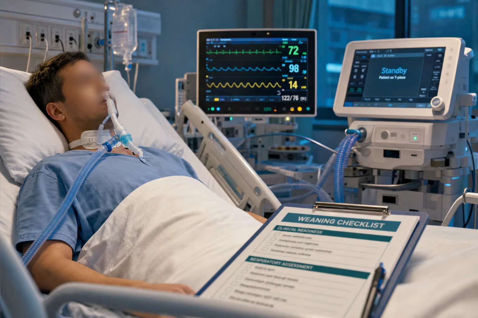
What weaning actually means
Weaning (more accurately termed liberation or discontinuation) is the graded process of withdrawing ventilatory support once the precipitating cause of respiratory failure has resolved. It is emphatically not the abrupt removal of the tube. Two conceptually separate decisions sit inside the word "weaning": [1]
- Separation from the ventilator — the progressive reduction of mechanical support (via a spontaneous breathing trial) until the patient can sustain unassisted breathing through the endotracheal tube.
- Extubation — the removal of the artificial airway once the patient can protect their airway, clear secretions, and breathe unassisted. [1]
A patient may pass a breathing trial but still fail extubation because of upper-airway obstruction (post-extubation stridor), inadequate cough, or impaired consciousness. Conversely, a patient with a tracheostomy can be separated from the ventilator without any extubation event. The fellowship-level point is that these are two distinct assessments and conflating them is a common source of error. [1]
The international consensus conference of the ERS, ATS, ESICM, SCCM, and SRLF (the Boles consensus, 2007) framed weaning around the balance between respiratory load and neuromuscular capacity: liberation succeeds when capacity exceeds load, and fails when load outstrips capacity (because of unresolved disease, increased load, reduced capacity, or an excessive psychological/medication burden). Every step of the weaning pathway — the readiness screen, the breathing trial, the extubation check, and the investigation of failure — is an interrogation of that balance. [1]
The three categories — simple, difficult, and prolonged weaning
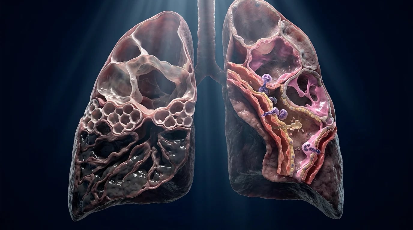
Not every liberation is straightforward. The Boles consensus stratified weaning into three groups by how quickly the patient separates from the ventilator, and this classification predicts outcome and directs the intensity of the workup: [1]
Weaning categories (click each)
~10% of patients
More than three failed SBTs, or more than seven days (often framed as beyond 14 days) of weaning effort. Accounts for about 10 per cent. Carries the highest mortality and resource use; consider tracheostomy, rehabilitation, and a structured prolonged-weaning programme.
A second framework — the WIND (Weaning according to a New Definition) classification — groups patients by the number of days from separation attempt to successful extubation, but the Boles categories remain the exam-friendly and clinically intuitive standard. The key message: difficult and prolonged weaning are not failures of technique but signals of an unaddressed problem that must be diagnosed and treated. [1]
Assessing readiness — the daily screen
The single most important weaning intervention is the daily screen for readiness, because the patient who is ready but not tested stays on the ventilator longer than necessary — accumulating sedation, weakness, and infection. Every intubated patient should be screened at least once a day (usually on the morning ward round) once the precipitating insult is improving.[1][4]
Cause resolved
The precondition
- The precipitating cause of respiratory failure (sepsis, pneumonia, pulmonary oedema, COPD exacerbation) is reversing
- There is no new, untreated problem driving ventilatory demand
- Without this, weaning is premature regardless of the numbers
Oxygenation
FiO2 and PEEP
- FiO2 <0.4 (40 per cent) — able to oxygenate on near-ambient oxygen
- PEEP <5-8 cmH2O
- PaO2/FiO2 >150-200 mmHg
- No escalating oxygen requirement
Respiratory drive
Triggering
- Patient triggers spontaneous breaths (no full mandatory control)
- Adequate cough and gag reflexes (airway protection)
- Minimal or manageable secretions
Stability
Circulation and brain
- Haemodynamically stable — no or low-dose vasopressors, no active myocardial ischaemia, no uncontrolled arrhythmia
- Awake and cooperative or easily arousable — minimal sedation (ideally a daily SAT has been passed)
- No significant metabolic acidosis; acceptable electrolytes (K+, Mg2+, PO4^3-)
- Afebrile or settling; adequate haemoglobin
The spontaneous breathing trial (SBT)
Once a patient passes the readiness screen, the next step is the spontaneous breathing trial (SBT) — a period of breathing with little or no ventilatory support, conducted through the endotracheal tube, to determine whether the patient can sustain unassisted ventilation. The SBT is the cornerstone of the liberation decision: a patient who passes an SBT has roughly an 80 per cent chance of successful extubation.[3]
Conducting an SBT — the pathway
Confirm readiness and safety
Confirm the patient passed the daily readiness screen and meets SBT safety criteria: no active myocardial ischaemia, no significant vasopressor escalation, no copious secretions requiring frequent suctioning, SpO2 acceptable on FiO2 <0.4. Do not run an SBT in an unstable patient.
Choose the modality and set support
Two equivalent options: (a) low level of pressure support — PS 5-7 cmH2O with PEEP 5 cmH2O; or (b) T-piece (zero ventilatory support, humidified oxygen). Pressure support offsets the resistive work of the endotracheal tube; the T-piece is a purer test of capacity. A brief (5 min) low-PS "warm-up" before a T-piece is sometimes used. Both modalities give equivalent extubation outcomes.
Run for 30-120 minutes
Duration is typically 30 minutes (a 30-minute trial is as predictive as a 120-minute trial). Observe continuously. A 120-minute trial is reasonable in higher-risk patients or when cardiac failure is suspected.
Monitor pass/fail criteria in real time
Watch for failure signs throughout: RR rising toward 35, SpO2 falling below 90 per cent, HR rising above 140 or new arrhythmia, agitation, anxiety, diaphoresis, accessory-muscle use, altered mental status, or a falling tidal volume (shallow breathing).
If the patient PASSES
RR <35, SpO2 >90 per cent, HR <140, no agitation/diaphoresis/distress, stable haemodynamics, adequate tidal volume — proceed to extubation assessment (cough, secretions, cuff leak if high-risk, mental status) and extubate.
If the patient FAILS
Terminate the trial at once and restore comfortable ventilatory support. Do not push through distress. Document WHY it failed (respiratory vs cardiac vs neurological) and hunt the cause. Re-attempt daily only when the reversible cause has been addressed.
SBT pass and fail criteria — the numbers to know
SBT — pass criteria (all must hold)
SBT — fail criteria (any one mandates stopping)
The "wake up and breathe" protocol — pairing the SAT with the SBT (ABC trial)
The most influential advance in ventilator liberation was the recognition that sedation and weaning are inseparable: a patient cannot breathe spontaneously while deeply sedated, and accumulated sedation is one of the commonest reasons for delayed liberation. The solution is to pair a daily spontaneous awakening trial (SAT, the daily sedation interruption) with a daily SBT — the "wake up and breathe" protocol. [1]
The ABC 'wake up and breathe' protocol
Step 1 — SAT safety screen
Before stopping sedation, confirm the patient is safe to wake: no active seizures, no agitation, no myocardial ischaemia, no high intracranial pressure, not paralysed, on acceptable oxygenation and PEEP, no escalating vasopressors.
Step 2 — Spontaneous awakening trial (SAT)
Stop all sedatives and opioids (or reduce to the lightest level). Observe. If the patient becomes agitated, in pain, or unstable, restart sedation at half the previous dose — a "failed SAT." If the patient opens eyes, follows commands, or is calmly awake, the SAT is passed.
Step 3 — SBT readiness screen
With the patient now awake, apply the standard readiness criteria (cause resolved, FiO2 <0.4, PEEP <8, stable, triggering, cough/gag intact).
Step 4 — Spontaneous breathing trial (SBT)
Run a 30-120 minute SBT on PS 5-7 + PEEP 5 or a T-piece. Assess pass/fail criteria continuously.
Step 5 — Extubation decision
If the SBT is passed, assess for extubation (airway protection, secretions, cuff leak if high-risk) and extubate. If the SBT fails, restore comfortable support, restart sedation at a reduced dose, and repeat the whole cycle the next day.
The Awakening and Breathing Controlled (ABC) trial (Girard et al., Lancet 2008) randomised 336 mechanically ventilated patients to either a paired daily SAT + SBT ("wake up and breathe") or usual sedation plus a daily SBT.[1] The intervention group spent more days breathing without assistance (14.7 vs 11.6 days), had shorter ICU stay (median 9.1 vs 12.9 days) and hospital stay, and — strikingly — had lower one-year mortality (HR 0.68). The number needed to treat to save one life was 7. Although self-extubation was more common in the intervention arm, reintubation rates were identical. The take-home: pairing awakening with breathing is the standard of care.
The earlier evidence for the SAT component alone came from Kress et al. (NEJM 2000), who showed that daily interruption of sedative infusions reduced the duration of mechanical ventilation (median 4.9 vs 7.3 days) and ICU length of stay (6.4 vs 9.9 days) without an excess of complications.[4] Together, these trials form the evidence base for the ABCDEF bundle of the modern PADIS (Pain, Agitation/Sedation, Delirium, Immobility, Sleep) guidelines: Assess pain, Both SAT and SBT, Choice of analgesia and sedation, Delirium monitoring, Early mobility, Family engagement.
SBT technique — pressure support versus T-piece
The SBT can be delivered as a low level of pressure support (PS 5-7 cmH2O with PEEP 5 cmH2O) or as a T-piece (the patient breathes spontaneously through the endotracheal tube connected to humidified oxygen, with no ventilatory support). A third, less common option is CPAP alone (continuous positive airway pressure without inspiratory support). [1]
Pressure support (PS 5-7 + PEEP 5)
Most common first-line SBT
- Offset the resistive work of breathing through the endotracheal tube
- PS 5-7 with PEEP 5 is the standard "minimal support" SBT
- Generally better tolerated; the ventilator still monitors volumes and apnoea
- Slight overestimate of capacity — the tube adds resistance that will be removed at extubation
T-piece
Purer test of capacity
- Zero pressure support — the patient does all the work against the tube
- A more stringent test; some clinicians prefer it in suspected cardiac failure (no positive-pressure support to mask LV dysfunction)
- No apnoea backup or ventilator alarms — closer bedside observation required
- Consider after a brief low-PS warm-up to avoid abrupt load
30 vs 120 minutes
Duration
- A 30-minute SBT is as predictive of extubation success as a 120-minute trial (Esteban 1997)
- Use 30 minutes for routine first-time weaning
- Consider 120 minutes (or longer) in high-risk patients, suspected cardiac failure, or prolonged ventilation
- Cardiac failure may declare itself only after 30-60 min of negative-pressure breathing
Protocol-directed versus physician-directed weaning
How the weaning pathway is executed matters as much as the physiology. Two landmark trials established that weaning driven by a protocol executed by nurses and respiratory therapists is faster than weaning directed at the discretion of the physician. [1]
In Brochard et al. (AJRCCM 1994), 456 ventilated patients who met weaning criteria were screened; 109 who failed a two-hour T-piece trial were randomised to weaning by pressure support, SIMV, or T-piece trials.[2] Pressure support was superior: fewer weaning failures (23 per cent PS vs 43 per cent T-piece vs 42 per cent SIMV) and a shorter weaning duration (mean 5.7 days for PS vs 8.5 days T-piece vs 9.9 days SIMV). This trial also established the framework for categorising weaning difficulty.
In Esteban et al. (NEJM 1995), 130 patients who failed an initial two-hour breathing trial were randomised among four techniques: IMV, pressure support, intermittent (multiple daily) SBTs, or a once-daily SBT.[3] The once-daily SBT weaned patients about three times faster than IMV (rate ratio 2.83) and about twice as fast as pressure support (rate ratio 2.05), with multiple daily SBTs equally successful. The message: a daily trial of spontaneous breathing is the most effective weaning method for the patient who has not yet separated.
Weaning method — what the trials showed
Complementary evidence (Ely et al., 1996) showed that a nurse/RT-driven protocol — in which bedside staff conduct the daily screen and SBT without waiting for a physician order — reduced the duration of mechanical ventilation compared with physician-directed weaning. The mechanism is simple: protocols remove the variability and delay of physician decision-making and ensure every eligible patient is tested every day. Protocol-directed weaning is the standard of care. [1]
Indices that predict weaning success — the RSBI and beyond
A battery of physiological indices has been proposed to predict weaning outcome. In practice, the SBT itself is the best predictor, but the indices are useful for the difficult or prolonged weaning patient and are frequent exam fodder. [1]
RSBI (f/Vt)
Rapid shallow breathing index
- RSBI = respiratory rate ÷ tidal volume (in litres), measured during unassisted breathing
- Threshold <105 predicts success; >105 predicts failure (Yang and Tobin, 1991)
- The single most validated weaning index, but imperfect (sensitivity and specificity each ~80%)
- Measured on PS 0 / PEEP 0 (or minimal support) for one minute
P0.1
Airway occlusion pressure
- Negative pressure generated against an occluded airway 0.1 s after inspiration begins — an index of respiratory drive
- Normal ~2-4 cmH2O; values >4-6 suggest high drive / impending fatigue
- Useful in the difficult weaning patient to distinguish fatigue from central depression
Maximal inspiratory pressure (MIP / NIF)
Muscle strength
- Maximum negative pressure generated against an occluded airway
- Threshold more negative than -20 to -30 cmH2O generally supports weaning
- Effort-dependent; low specificity but useful to flag severe weakness (e.g. ICU-AW, neuromuscular disease)
Others
Adjunctive clues
- Static compliance, dynamic compliance, work of breathing
- Gastric tonometry / regional CO2 (research)
- Bedside ultrasound (diaphragm thickening fraction, lung B-lines for occult oedema)
- None replaces the SBT; use them to dissect the cause of difficult weaning
Extubation — deciding to remove the tube
Passing the SBT means the patient can probably breathe without the ventilator; the next, separate question is whether they can manage without the endotracheal tube. Extubation assessment covers three domains the SBT does not test: [1]
The extubation assessment
Airway protection
Is the patient awake enough to protect the airway? Assess level of consciousness (obeys commands, GCS adequate), cough strength (can the patient cough against a closed tube?), and gag reflex. A weak cough or obtunded patient is at high risk of aspiration and secretion encumbrance after extubation.
Secretion load
Are secretions manageable? Quantify the volume and frequency of suctioning. Copious or thick secretions, or a patient who cannot clear them, predicts post-extubation failure. Sputum characteristics and the suction frequency over the preceding 24 h guide this.
Upper-airway patency (cuff leak)
Is there room for airflow around the tube once the cuff is down and the tube is removed? In patients at high risk of laryngeal oedema (prolonged intubation, traumatic intubation, female, large tube, repeated intubation, cuff over-inflation), perform a cuff leak test. A reduced leak predicts post-extubation stridor.
Decision
If the SBT was passed and the airway, secretions, and cuff leak are acceptable, extubate. If any domain fails, address it (e.g. steroids for a positive cuff leak; treat secretions; lighten sedation and re-test) before extubation.
The cuff leak test and post-extubation stridor
Post-extubation stridor (PES) — upper-airway obstruction from laryngeal oedema after tube removal — occurs in roughly 2-16 per cent of extubations and carries a high reintubation rate and mortality. The cuff leak test identifies patients at risk before the tube comes out, so prophylactic steroids can be given. [1]
[1]Laryngeal (air-column) ultrasound is a newer adjunct: the air-column width (the echo from airflow around the tube) measured before and after cuff deflation, and the change in air-column width ratio, predict PES — a small increase or a low ratio flags laryngeal oedema. Laryngeal ultrasound is non-invasive and increasingly used at the bedside, though the cuff leak test remains the established standard. [1]
Who to test
High-risk patients
- Prolonged intubation (>6 days, especially >5-7 days)
- Traumatic or repeated intubation; tight-fitting or oversized tube
- Female sex, low body mass index
- High cuff pressures, head/neck surgery, laryngeal trauma
If positive (small leak)
Give steroids
- Methylprednisolone 20-40 mg IV q4-6h or dexamethasone, starting 12-24 h before extubation
- Steroids reduce laryngeal oedema, PES, and reintubation (multiple trials confirm benefit)
- Extubate in a controlled setting with a plan for immediate reintubation if stridor develops
- Racemic adrenaline and helium-oxygen may help established mild stridor but are not a substitute for reintubation in severe obstruction
Non-invasive ventilation to prevent extubation failure
NIV has two distinct, opposite roles around extubation, and confusing them is dangerous: [1]
Prophylactic NIV (preventive)
Apply immediately after extubation in high-risk patients — GOOD
- Given immediately after extubation to patients predicted to fail, before any respiratory distress develops
- Reduces reintubation and mortality in high-risk groups: COPD, hypercapnia during the SBT, cardiac failure, older age, obesity, multiple comorbidities
- Ferrer 2003: in COPD patients with persistent weaning failure, extubating to NIV shortened ventilation, reduced nosocomial pneumonia, and improved survival vs continued invasive weaning
- The benefit comes from avoiding re-exposure to the complications of invasive ventilation (VAP, sedation, weakness)
Rescue NIV (therapeutic)
Applied after established post-extubation respiratory failure — HARMFUL
- Started after the patient has already developed respiratory distress post-extubation
- Esteban 2004 showed this strategy INCREASED mortality (likely by delaying necessary reintubation)
- Do not use NIV to "rescue" an extubated patient who is failing — reintubate promptly
- The window for NIV is before failure, not after
The Ferrer trial (AJRCCM 2003) is the key weaning-NIV reference: in 43 patients (predominantly COPD) who had failed weaning for three consecutive days, immediate extubation to NIV versus continued invasive weaning gave shorter invasive ventilation (9.5 vs 20.1 days), shorter ICU stay, less nosocomial pneumonia (24 vs 59 per cent), fewer tracheostomies, and improved survival.[5] The lesson: in the COPD or hypercapnic patient stuck on the ventilator, extubate to NIV rather than persisting with invasive weaning.
Difficult and prolonged weaning — the systematic cause hunt
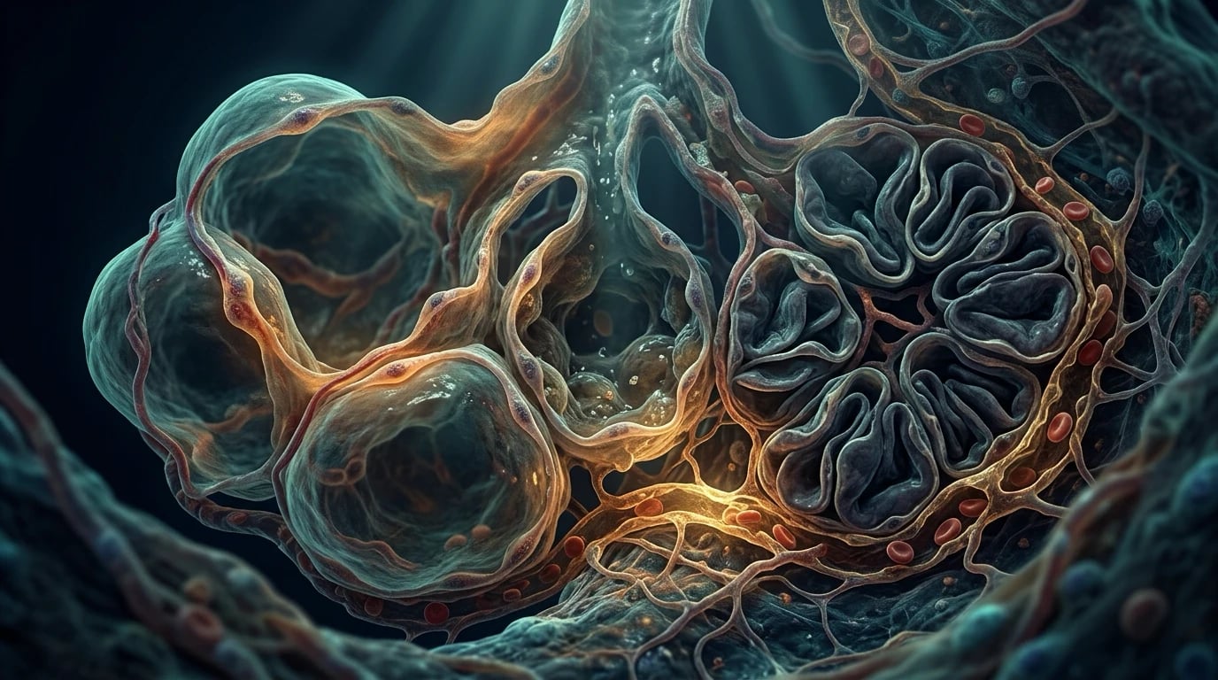
When a patient fails repeated SBTs (difficult weaning) or remains unweanable beyond a week or two (prolonged weaning), the priority shifts from performing the trial to diagnosing why the patient cannot sustain spontaneous breathing. The Boles framework reduces the problem to the load-versus-capacity balance, and the causes fall into four groups: [1]
Cardiac
Weaning-induced pulmonary oedema
- The commonest occult cause — the switch to negative-pressure breathing increases venous return and LV afterload, unmasking diastolic or systolic dysfunction
- Suspect in the elderly, known heart failure, ischaemic heart disease, rising filling pressures or BNP during the SBT
- Diagnose with bedside echo, BNP/NT-proBNP, and watching for a rising CVP or new pulmonary oedema on ultrasound/lung auscultation during the SBT
- Treat with diuresis, afterload reduction, and consideration of NIV as a bridge
Respiratory muscle weakness
ICU-acquired weakness
- Critical-illness polyneuromyopathy (CIP/CIM), disuse atrophy, electrolyte depletion
- Diagnosed clinically (MRC sum score <48), reduced MIP, and (definitively) electrophysiology and biopsy
- Risk factors: sepsis, prolonged immobility, hyperglycaemia, corticosteroids, neuromuscular blockers
- Prevention and recovery: glucose control, minimise steroids/paralytics, early mobilisation, nutrition, rehabilitation
Residual load
Unresolved lung disease
- Ongoing pneumonia, pulmonary oedema, COPD with dynamic hyperinflation and auto-PEEP, large pleural effusion, pneumothorax, pulmonary embolism
- Excessive secretions, bronchospasm, upper-airway obstruction
- Metabolic load: fever, sepsis, overfeeding (excess CO2 production), acidosis
- Re-assess the chest: imaging, gas exchange, auto-PEEP measurement
Neurological / metabolic
Drive and chemistry
- Over-sedation, delirium, encephalopathy (hepatic, uraemic, septic)
- Electrolyte derangement: hypokalaemia, hypomagnesaemia, hypophosphataemia (respiratory-muscle weakness)
- Hypothyroidism, neuromuscular disease (myasthenia, GBS)
- Correct each: lightest effective sedation, treat delirium, replete electrolytes, optimise thyroid
Investigating the difficult-to-wean patient
Characterise the failure pattern
Was it respiratory (rising RR, falling SpO2, shallow breathing, high RSBI), cardiac (rising HR, BP, CVP, new oedema, ischaemia on ECG), or neurological (agitation, somnolence, delirium)? The pattern points to the cause.
Bedside cardiac assessment
Perform a focused echocardiogram during the SBT: look for LV systolic/diastolic dysfunction, elevated filling pressures, new regional wall motion abnormality. Check BNP/NT-proBNP before and after the SBT. A rising value during the trial supports weaning-induced pulmonary oedema.
Respiratory mechanics and load
Measure auto-PEEP (expiratory hold) in COPD, assess compliance and resistance, review the chest X-ray and lung ultrasound for oedema, consolidation, effusion, or pneumothorax. Check the RSBI and consider P0.1.
Neuromuscular and metabolic
Examine for weakness (MRC score), measure MIP, check phosphate, magnesium, potassium, calcium, thyroid function, and review the cumulative sedation, steroid, and neuromuscular-blocker exposure. Screen for delirium (CAM-ICU).
Treat the cause and re-attempt
Diurese the cardiac patient, replete electrolytes, lighten sedation, treat delirium, mobilise early, drain effusions, relieve obstruction, treat infection. Re-attempt daily SBTs only as the reversible causes are corrected. Do not repeat failing trials mechanically.
Weaning-induced (cardiac) pulmonary oedema — the classic trap
The most exam-relevant and clinically under-recognised cause of difficult weaning is weaning-induced pulmonary oedema, also called weaning-induced cardiac dysfunction or, in COPD, COPD-weaning pulmonary oedema. The mechanism is a haemodynamic consequence of the transition from positive-pressure ventilation back to spontaneous (negative-pressure) breathing: [1]
[1]This is the answer to the classic fellowship MCQ: a COPD patient who repeatedly fails SBTs, developing tachypnoea and desaturation within minutes, whose cardiac biomarkers rise — the cause is weaning-induced pulmonary oedema from LV dysfunction unmasked by the removal of positive pressure, not bronchospasm or auto-PEEP. [1]
Respiratory muscle weakness — ICU-acquired weakness (ICU-AW)
ICU-acquired weakness (ICU-AW) is a major contributor to prolonged weaning, especially after severe sepsis, ARDS, or prolonged immobility. It encompasses critical-illness polyneuropathy (CIP), critical-illness myopathy (CIM), and their overlap, and produces a flaccid, symmetric, predominantly proximal weakness with preserved sensation (in pure myopathy) that disables the respiratory muscles as much as the limbs. [1]
ICU-AW — recognition and risk
Respiratory-muscle involvement manifests as a weak cough, low tidal volume, a high RSBI, and a low MIP — the patient simply cannot generate the pressures needed to breathe unaided. There is no specific drug treatment; management is prevention and rehabilitation: tight glucose control, minimise corticosteroids and neuromuscular blockers, early mobilisation, adequate nutrition, and a structured rehabilitation programme. Recovery is slow (weeks to months). [1]
Metabolic, electrolyte, and nutritional factors
Respiratory-muscle function is exquisitely sensitive to metabolic derangement. Three electrolytes deserve specific attention in the difficult-to-wean patient: [1]
- Hypophosphataemia (especially <0.5-0.8 mmol/L) impairs diaphragm contractility and ATP generation; repletion can measurably improve ventilatory capacity.
- Hypomagnesaemia and hypokalaemia cause generalized and respiratory-muscle weakness; replete both.
- Hypocalcaemia and acid-base disturbance contribute similarly. [1]
Nutrition cuts both ways: under-nutrition causes respiratory-muscle wasting and weakness, while overfeeding (particularly excess carbohydrate) increases CO2 production and the ventilatory load, worsening weaning. Aim for euglycaemia — both hyper- and hypoglycaemia are harmful. Thyroid disease (hypothyroidism) and adrenal insufficiency should be excluded as reversible causes of weakness and fatigue. [1]
[1]Tracheostomy for prolonged weaning
A tracheostomy is the airway of choice for the patient in prolonged weaning or anticipated prolonged ventilation. It offers real advantages over a translaryngeal tube: improved patient comfort (allowing less sedation), reduced laryngeal damage, easier and safer secretion clearance, oral nutrition and communication, secure airway, and the ability to wean by progressively longer T-piece or tracheostomy-mask off-periods. For many patients it is the gateway to liberation. [1]
The timing of tracheostomy, however, has been a long-standing controversy — early (within the first few days) versus late (after 10-14 days). The definitive answer came from the TracMan trial (Young et al., JAMA 2013), the largest randomised trial of tracheostomy timing: 909 patients predicted to need at least seven more days of ventilation were randomised to early tracheostomy (within four days) or late (after 10 days if still indicated).[6]
TracMan — the key numbers
TracMan showed no difference in 30-day mortality, ICU stay, hospital stay, or antibiotic use between early and late tracheostomy. Crucially, only 45 per cent of the late group ever received a tracheostomy — the rest had been extubated or had died — showing that clinicians cannot reliably predict who will need prolonged ventilation. The current practice, supported by TracMan, is to individualise the decision, typically performing a tracheostomy around 7-14 days if prolonged ventilation and weaning remain likely, rather than routinely at day 3-4. [1]
Evidence and landmark trials
ABC (Girard)
Lancet 2008
336 mechanically ventilated pts — paired daily SAT (sedation interruption) + SBT vs usual sedation + daily SBT
Key finding
More ventilator-free days (14.7 vs 11.6), shorter ICU (9.1 vs 12.9 d) and hospital stay, and LOWER 1-year mortality (HR 0.68); NNT 7 to save one life. Self-extubation more common but reintubation identical
Practice change
Established the "wake up and breathe" protocol — pair daily sedation interruption with daily SBT — as the standard of care
Brochard
AJRCCM 1994
109 pts who failed 2-h T-piece — pressure support vs SIMV vs T-piece for weaning
Key finding
PSV superior: fewer weaning failures (23% vs 42-43%) and shorter wean (5.7 vs 8.5-9.9 d); SIMV worst
Practice change
Established pressure support and the comparative framework for weaning methods; foundation for weaning-category definitions
Esteban
NEJM 1995
130 pts who failed initial SBT — once-daily SBT vs multiple SBT vs PSV vs IMV (4-way RCT)
Key finding
Once-daily SBT weaned ~3x faster than IMV (RR 2.83) and ~2x faster than PSV (RR 2.05); multiple daily SBTs equally good
Practice change
Established the once-daily spontaneous breathing trial as the most effective weaning method
Kress (Sedation)
NEJM 2000
128 ventilated pts on continuous sedation — daily sedation interruption vs clinician-directed
Key finding
Shorter ventilation (median 4.9 vs 7.3 d) and ICU stay (6.4 vs 9.9 d); fewer diagnostic tests; no excess self-extubation
Practice change
Daily sedation interruption became a cornerstone — later combined with the SBT in the ABC trial
Ferrer (NIV weaning)
AJRCCM 2003
43 pts (mostly COPD) with persistent weaning failure (3 failed days) — immediate extubation to NIV vs continued invasive weaning
Key finding
Shorter invasive ventilation (9.5 vs 20.1 d), shorter ICU stay, less nosocomial pneumonia (24% vs 59%), fewer tracheostomies, improved survival
Practice change
Extubation to NIV is effective for COPD/hypercapnic patients stuck in weaning failure
TracMan
JAMA 2013
909 pts expected to need >7 d more ventilation — early tracheostomy (≤4 d) vs late (≥10 d)
Key finding
No difference in 30-day mortality (30.8% vs 31.5%), ICU/hospital stay, or antibiotic use; only 45% of the late group ever received a tracheostomy
Practice change
Routine early tracheostomy abandoned; timing individualised (typically day 7-14)
Prognosis
The outlook is dictated by the weaning category. Simple weaning (about 70 per cent of patients) is usually successful within a day and carries the prognosis of the underlying illness. Difficult weaning roughly doubles the length of stay and carries a moderate mortality. Prolonged weaning (about 10 per cent) consumes the majority of ventilator-days and ICU resources and carries the highest mortality — often 30-50 per cent at one year — driven by the severity of the original illness, comorbidity, and the complications of prolonged ventilation (VAP, ICU-AW, delirium). [1]
Weaning outcomes — the numbers
Reintubation after a successful extubation occurs in 10-20 per cent of patients and is itself associated with a marked increase in mortality, which is why preventing extubation failure (cuff leak testing and prophylactic steroids for PES, prophylactic NIV for high-risk patients) matters as much as passing the SBT. Poor prognostic features include advanced age, severe comorbidity, prolonged ventilation, ICU-AW, persistent delirium, and cardiac failure. Long-term, many survivors of prolonged ventilation have persistent physical and cognitive impairment — the post-intensive-care syndrome (PICS) — and benefit from structured rehabilitation and follow-up. [1]
Clinical pearls
Red flags
[1]SAQ — Ventilator liberation and difficult weaning
10 minutes · 10 marks
A 68-year-old with COPD has been ventilated for five days after a severe exacerbation. Daily sedation holds are performed. Today FiO2 is 0.35, PEEP 5, he is awake and coughing, secretions are moderate. An SBT on PS 5/PEEP 5 is started; at 20 minutes RR is 38, SpO2 88% on FiO2 0.4, he is diaphoretic and distressed. After return to full support he improves. Echo shows moderate LV systolic impairment; he has a positive fluid balance of 4 L.
References
- [1]Girard TD, Kress JP, Fuchs BD, Thomason JW, Schweickert WD, Pun BT, Taichman DB, Dunn JG, Pohlman AS, Kinniry PA, Jackson JC, Canonico AE, Light RW, Shintani AK, Thompson JL, Gordon SM, Hall JB, Dittus RS, Bernard GR, Ely EW. Efficacy and safety of a paired sedation and ventilator weaning protocol for mechanically ventilated patients in intensive care (Awakening and Breathing Controlled trial): a randomised controlled trial Lancet, 2008.PMID 18191684
- [2]Brochard L, Rauss A, Benito S, Conti G, Mancebo J, Rekik N, Gasparetto A, Lemaire F. Comparison of three methods of gradual withdrawal from ventilatory support during weaning from mechanical ventilation Am J Respir Crit Care Med, 1994.PMID 7921460
- [3]Esteban A, Frutos F, Tobin MJ, Alía I, Solsona JF, Valverdú I, Fernández R, de la Cal MA, Benito S, Tomás R, et al. A comparison of four methods of weaning patients from mechanical ventilation. Spanish Lung Failure Collaborative Group N Engl J Med, 1995.PMID 7823995
- [4]Kress JP, Pohlman AS, O'Connor MF, Hall JB. Daily interruption of sedative infusions in critically ill patients undergoing mechanical ventilation N Engl J Med, 2000.PMID 10816184
- [5]Ferrer M, Esquinas A, Arancibia F, Bauer TT, Gonzalez G, Carrillo A, Rodriguez-Roisin R, Torres A. Noninvasive ventilation during persistent weaning failure: a randomized controlled trial Am J Respir Crit Care Med, 2003.PMID 12689847
- [6]Young D, Harrison DA, Cuthbertson BH, Rowan K; TracMan Collaborators. Effect of early vs late tracheostomy placement on survival in patients receiving mechanical ventilation: the TracMan randomized trial JAMA, 2013.PMID 23695482