ICU · respiratory
Ventilator Waveforms — Comprehensive (Scalars, Loops, Asynchrony)
Also known as Ventilator waveforms · Scalar waveforms · Pressure-volume loop · Flow-volume loop · Patient-ventilator asynchrony · Double triggering · Auto-PEEP waveform · Flow-time waveform · Pressure-time waveform
Ventilator waveforms — the graphical display of pressure, flow, and volume over time during mechanical ventilation. Three SCALAR waveforms (pressure-time, flow-time, volume-time) and two LOOPS (pressure-volume, flow-volume) provide real-time information about patient-ventilator interaction, lung mechanics, and asynchrony. VOLUME CONTROL: square flow waveform + decelerating pressure + linear volume rise. PRESSURE CONTROL: square pressure + decelerating flow + curvilinear volume rise. P-V LOOP: compliance (slope = ΔV/ΔP), inflection points (lower = recruitment opportunity, upper = overdistension). F-V LOOP: auto-PEEP (expiratory flow doesn't return to baseline before next inspiration), obstruction (concave expiratory limb). PATIENT-VENTILATOR ASYNCHRONY: trigger asynchrony (missed triggers, auto-triggering), flow asynchrony (double triggering — patient wants more flow than delivered), cycle asynchrony (premature/delayed cycling), expiratory asynchrony. Waveform analysis is a CRITICAL CICM exam skill — tested in vivas and SAQs.
On this page & tools
Your progress
Saved locally on this device.
Target exams
Red flags
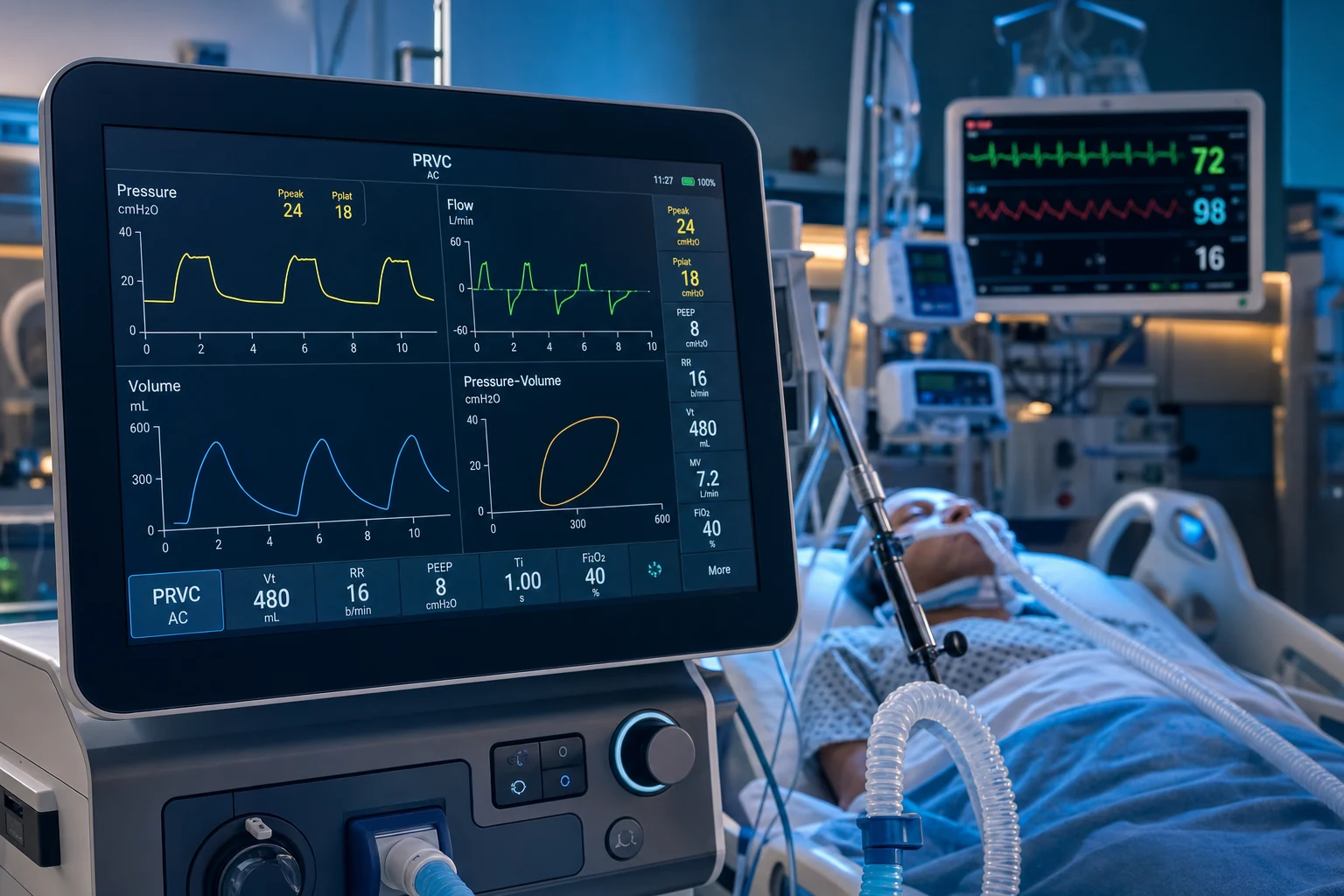
Overview
Scalar waveforms — pressure-time, flow-time, volume-time
Volume control vs pressure control waveform patterns
| Feature | Volume Control (VC) | Pressure Control (PC) |
|---|---|---|
| Flow waveform | SQUARE (constant flow — preset by ventilator) — flow is the INDEPENDENT variable | DECELERATING (starts high, falls exponentially as airway pressure rises to set target) |
| Pressure waveform | RISING (starts at PEEP, rises progressively as lung fills — depends on compliance + resistance — END pressure = plateau pressure) | SQUARE (instantly rises to preset pressure — held constant throughout inspiration) |
| Volume waveform | LINEAR RISE (constant volume delivery rate) | CURVILINEAR RISE (fast initially, slows as flow decelerates — most volume delivered early) |
| Tidal volume | GUARANTEED (preset — delivered regardless of lung mechanics) | VARIABLE (depends on compliance + resistance + inspiratory time — if compliance falls, Vt falls) |
| Peak pressure | VARIABLE (depends on compliance — risk of high pressure with low compliance) | GUARANTEED LIMITED (preset — cannot exceed set pressure — safer for ARDS) |
| Flow distribution | Prefers LOW-resistance regions (may overinflate healthy lung → VILI) | Prefers LOW-resistance regions initially but better distribution to slow-filling (stiff) regions |
| Best for | Guaranteed ventilation (neuromuscular disease, anaesthesia, guaranteed Vt) | ARDS (pressure-limited — safer), asynchrony (variable flow better matches patient demand) |
Detailed Volume Control (VC) scalar analysis
In VC the ventilator delivers a PRESET VOLUME at a PRESET FLOW. Flow is the independent variable (the ventilator controls it); pressure and the resulting volume are dependent on the patient's mechanics. The three scalars must always be read together — never interpret one in isolation. [1]
Pressure–time scalar in VC
The pressure–time waveform in VC rises from PEEP to a peak (PIP), then — during an inspiratory hold — drops to plateau (Pplat), before falling back to PEEP at expiration. The three-pressure "PIP-Pplat-PEEP" method splits the pressure waveform into its two physical components: [1]
The PIP-Pplat-PEEP method: splitting airway pressure into resistance and compliance
| Pressure segment | What it measures | Calculation | Normal range |
|---|---|---|---|
| PIP – Pplat | RESISTIVE pressure (airway + ETT + circuit resistance) | ΔP_resistance = PIP – Pplat | 4–6 cmH2O |
| Pplat – PEEP | ELASTIC pressure (lung + chest-wall compliance) = the DRIVING PRESSURE | ΔP = Pplat – PEEP | <15 cmH2O |
| PIP | Total inspiratory pressure (resistive + elastic) | — | <30 cmH2O |
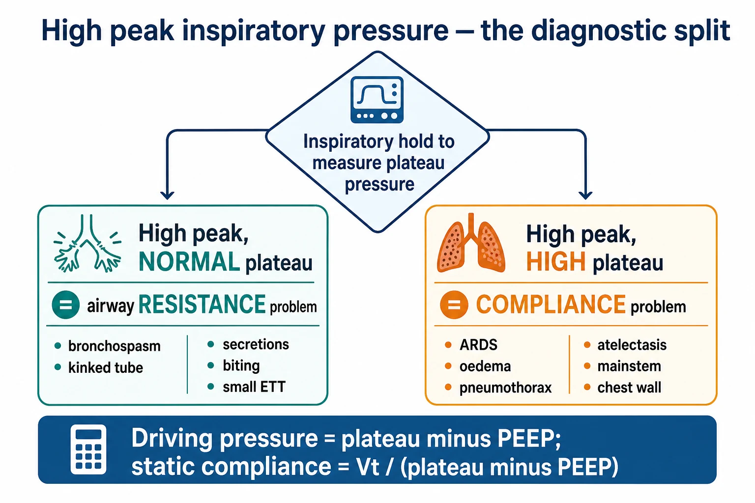
Reading the three numbers in sequence — the diagnostic algorithm:
- High PIP with NORMAL Pplat → the gradient (PIP – Pplat) is widened → RESISTANCE problem. Causes: bronchospasm, secretions/mucus plug, kinked or bitten ETT, ETT too small for patient size, circuit obstruction (water in tubing), patient coughing/bucking. Fix the airway, not the ventilator.
- High PIP with HIGH Pplat (gradient normal at 4–6) → COMPLIANCE problem. Causes: ARDS, pulmonary oedema, tension pneumothorax, atelectasis, main-stem intubation, chest-wall restriction (obesity, burns, ascites, prone positioning), or simply excessive Vt/PEEP. Reduce Vt or fix the cause (chest tube, tube repositioning, diuresis).
- Sudden rise in BOTH PIP and Pplat → acute derecruitment (mucus plug, endobronchial intubation, pneumothorax) or a change in chest-wall compliance (abdominal distension, patient fighting the ventilator).
- Low PIP and low Pplat → circuit leak or disconnect (the ventilator cannot build pressure) — check the circuit, cuff, and connections. [1]
Shape of the inspiratory pressure rise: in VC the pressure rises in a smooth concave-down curve as volume accumulates against compliance. A convex notch or downward deflection mid-inspiration = patient effort against the fixed flow = FLOW ASYNCHRONY (the patient wants more flow than the ventilator is delivering). A notch at the start of inspiration (before flow begins) = trigger asynchrony. [1]
Flow–time scalar in VC
The hallmark of VC is a SQUARE inspiratory flow waveform (constant flow set by the ventilator). The two available inspiratory flow patterns: [1]
Inspiratory flow patterns in volume control
| Flow pattern | Inspiratory flow shape | Expiratory flow shape | Best use |
|---|---|---|---|
| Constant (square) | Rectangular — flat plateau throughout inspiration | Decelerating exponential (returns to zero) | Standard; clearest for reading resistance and auto-PEEP |
| Decelerating | Starts high, falls toward end-inspiration | Decelerating exponential | Better gas distribution in heterogeneous lungs (ARDS); resembles PC |
The expiratory limb is where auto-PEEP is detected. In the normal breath, expiratory flow returns to ZERO (the zero-flow baseline) BEFORE the next inspiration begins. If expiratory flow does NOT reach zero → gas is still leaving the lung when the next breath starts → INCOMPLETE EXPIRATION → AUTO-PEEP (intrinsic PEEP). The magnitude of residual flow at end-expiration correlates with the severity of gas trapping. Confirm quantitatively with an expiratory hold. [1]
Inspiratory flow settings that matter:
- Peak flow (L/min): determines inspiratory time at a given Vt. Higher peak flow → shorter inspiratory time → longer expiratory time → better for obstructive disease (COPD, asthma — gives time to exhale, reduces auto-PEEP). Lower peak flow → longer inspiratory time → better gas distribution in ARDS/recruitable lung. Typical setting 40–60 L/min; in COPD may need 60–80 L/min to maximise expiratory time.
- Flow trigger: the inspiratory flow the patient must generate to trigger a breath. Too insensitive (high threshold) → missed triggers; too sensitive → auto-triggering from circuit noise. Typical 1–2 L/min flow trigger.
- Flow acceleration/rise time: how rapidly flow ramps to its peak. A fast rise time minimises trigger delay but can cause an initial pressure overshoot; a slow rise time is gentler but increases work of breathing if too sluggish. [1]
Volume–time scalar in VC
The volume–time scalar rises LINEARLY during inspiration (constant flow → constant rate of volume delivery) to the preset Vt, then falls during expiration. Two bedside checks:
- Inspiratory volume = the preset Vt (guaranteed in VC — if the displayed delivered Vt is LESS than set, there is a LEAK: cuff leak, circuit disconnect, bronchopleural fistula, cracked humidifier).
- Expiratory volume should return to the zero baseline at end-expiration. If it does NOT return to zero → auto-PEEP (the trapped volume is "stored" above functional residual capacity). If expiratory volume is less than inspiratory volume → leak. [1]
The 30-second VC scalar diagnostic sequence
- Look at PIP. Trend it over time. A rising PIP → something changed (resistance or compliance). Do an inspiratory hold.
- Inspiratory hold → read Pplat. Now you have all three pressures: PIP, Pplat, PEEP.
- PIP – Pplat > 5–6 cmH2O → RESISTANCE problem → suction, give bronchodilator, check ETT patency/position, exclude kink/bite.
- Pplat – PEEP (driving pressure) > 15 cmH2O → COMPLIANCE problem → reduce Vt, drain pneumothorax, reposition tube if main-stem, consider diuresis for oedema.
- Expiratory hold → read total PEEP. Total PEEP – set PEEP = auto-PEEP. If >5 cmH2O → gas trapping → lower RR, shorten inspiratory time, lengthen expiratory time (I:E 1:3 to 1:4).
- Check the flow–time expiratory limb — confirms auto-PEEP non-invasively at the bedside (residual flow at end-expiration).
- Check the volume–time scalar for delivered vs set Vt → screens for leak (inspired – expired Vt = leak volume).
- Check the pressure–time shape for notches/deflections → screens for asynchrony (trigger and flow).
Detailed Pressure Control (PC) scalar analysis
In PC the ventilator delivers a PRESET PRESSURE for a SET inspiratory time. Pressure is the independent variable (the ventilator controls it); flow and volume are dependent on the patient's mechanics. PC is preferred in ARDS (pressure-limited, safer) and in spontaneously breathing patients (variable flow better matches demand). [1]
Pressure–time scalar in PC
The hallmark of PC is a SQUARE pressure waveform: airway pressure rises rapidly to the preset target and is HELD CONSTANT throughout inspiration. This is the single most useful distinguishing feature — if the pressure is not constant (it sags or overshoots), there is a problem:
- Pressure overshoot at the start (a spike above the set pressure) → rise time too fast (pressure ramp set too aggressive) → reduce rise time. The overshoot is felt by the patient as discomfort and can cause barotrauma.
- Pressure sag during inspiration (the pressure drifts downward below target) → the patient is PULLING (high inspiratory demand) — the ventilator cannot sustain pressure against high patient flow demand → consider increasing the rise time or switching to a higher set pressure.
- Pressure NOT square (slow rise, never reaches plateau) → rise time too slow → increase rise time (faster pressure ramp). [1]
Because pressure is constant, PIP = Pplat = set pressure (there is no separate peak and plateau in PC — by design pressure is limited). This means you CANNOT separate resistance from compliance using the inspiratory hold in PC the way you can in VC. Instead, you assess resistance and compliance from the FLOW waveform. [1]
Flow–time scalar in PC — the diagnostic waveform in PC
In PC the inspiratory flow is DECELERATING: it starts HIGH (maximal gradient between set pressure and alveolar pressure at the start of inspiration) and falls EXPONENTIALLY as alveolar pressure rises toward the set pressure (the driving gradient narrows). The flow may reach zero before the end of the inspiratory time (no-flow plateau) or remain above zero throughout. [1]
The decay rate of the decelerating flow tells you about RESISTANCE and COMPLIANCE:
| Flow appearance | Interpretation | Mechanism |
|---|---|---|
| Flow decelerates RAPIDLY (reaches zero well before end of inspiration, with a flat no-flow tail) | LOW resistance OR HIGH compliance | Lung fills quickly — alveolar pressure equilibrates with set pressure early → gradient collapses → flow stops. Normal lung, or over-distended lung with little recruitable volume |
| Flow decelerates SLOWLY (remains well above zero at end of inspiration) | HIGH resistance OR LOW compliance | Lung fills slowly — alveolar pressure never catches up to set pressure → gradient persists → flow continues throughout. Bronchospasm, secretions (high resistance); ARDS, oedema, atelectasis (low compliance) |
| Inspiratory flow remains at peak (does not decelerate at all) | Severe obstruction or a circuit/valve problem | Fixed high resistance throughout |
Assessing bronchodilator response from the flow–time scalar: in a COPD/asthma patient on PC, observe the expiratory flow. Before bronchodilator: the expiratory limb is CONCAVE (scooped) — flow falls rapidly then plateaus at a low level (expiratory obstruction). After nebulised salbutamol: if the expiratory flow IMPROVES (the concavity reduces, peak expiratory flow increases, flow returns toward baseline faster) → bronchodilator response confirmed objectively at the bedside. [1]
Volume–time scalar in PC
In PC the volume–time scalar rises CURVILINEARLY: fast initially (when flow is high), slowing as flow decelerates (most volume is delivered in the first half of inspiration). The total delivered Vt is the integral of flow over the inspiratory time. Key principle: Vt is NOT guaranteed in PC — it is the dependent variable:
- If compliance falls (ARDS worsening, oedema, atelectasis) → Vt falls (the lung cannot accept the same volume at the same pressure) → hypoventilation, rising PaCO2.
- If resistance rises (bronchospasm) → Vt falls (more pressure dissipated in resistance, less reaches alveoli) → hypoventilation.
- If the patient actively expires against inspiration (cycle asynchrony) → effective Vt falls. [1]
The "set the pressure, read the volume" principle: in PC you SET the pressure and READ the resulting Vt. If Vt is too low, you increase the set pressure (recheck that plateau stays <30) or increase inspiratory time. NEVER assume Vt is being delivered in PC — always confirm with the displayed Vt and capnography. [1]
What each scalar tells you in VC vs PC — the dependent-variable principle
| Variable | Volume Control | Pressure Control |
|---|---|---|
| Independent (you SET) | Flow (and Vt, Ti) | Pressure (and Ti) |
| Dependent (you READ) | Pressure (PIP, Pplat) | Flow and Vt |
| Guaranteed | Vt | Airway pressure limit |
| Variable (potentially dangerous) | Peak pressure (risk of barotrauma) | Vt (risk of hypoventilation if mechanics worsen) |
| How to assess RESISTANCE | PIP – Pplat (inspiratory hold) | Rate of flow decay (faster decay = lower resistance) |
| How to assess COMPLIANCE | Vt / (Pplat – PEEP) | Vt delivered at a given pressure (low Vt = low compliance) |
P-V and F-V loops — the diagnostic loops
PRESSURE-VOLUME (P-V) LOOP:
- X-axis: pressure (airway). Y-axis: volume
- The loop is drawn CLOCKWISE (inspiration goes RIGHT, expiration goes LEFT)
- Slope = compliance (ΔV/ΔP — steeper slope = more compliant lung). In ARDS, the slope is FLAT (stiff lung)
- Lower inflection point (LIP): the pressure at which collapsed alveoli start to recruit (sudden increase in slope). SETTING PEEP ABOVE the LIP keeps alveoli open. Controversial — not always visible on dynamic loops (best on static/slow inflation)
- Upper inflection point (UIP): the pressure at which the lung is fully recruited and further pressure causes overdistension (flattening of slope). Vt/pressure should NOT exceed the UIP (overdistension → VILI). Amato 2015: driving pressure (Vt/compliance) = the distance between PEEP and end-inspiratory pressure on the P-V loop — the strongest predictor of survival in ARDS
- Hysteresis: the inspiratory limb is DIFFERENT from the expiratory limb (inspiratory curve is to the RIGHT of expiratory curve — the area between them = energy dissipated in tissue resistance + surfactant). Increased hysteresis = increased tissue resistance [1]
FLOW-VOLUME (F-V) LOOP:
- X-axis: volume. Y-axis: flow
- Inspiration is ABOVE the baseline (positive flow). Expiration is BELOW (negative flow)
- Auto-PEEP detection: if the expiratory flow does NOT return to the ZERO FLOW LINE before the next inspiration starts → gas is TRAPPED → auto-PEEP. The amount of trapped volume = the area between the expiratory flow curve and the zero line at end-expiration
- Obstruction: expiratory limb is CONCAVE (scooped) — flow falls rapidly initially then plateaus at a low level → expiratory obstruction (COPD, asthma) → gas trapping
- Leak: if the expiratory volume (area under expiratory flow) is LESS than inspiratory volume → LEAK (cuff leak, bronchopleural fistula, circuit disconnect) [1]
Detailed P-V loop interpretation — compliance, recruitment, overdistension
The pressure-volume (P-V) loop plots airway pressure (x-axis) against lung volume (y-axis). It is the most information-dense single graphic in mechanical ventilation — from one loop you can read compliance (slope), recruitment opportunity (lower inflection point), overdistension (upper inflection point), and energy dissipation (hysteresis). In CICM/FFICM exams you will be shown a P-V loop and asked to interpret it. [1]
The four features to extract from every P-V loop
The four P-V loop features — what each means and how to use it
| Feature | What it is | How to read it | Clinical action |
|---|---|---|---|
| Slope = compliance | ΔV/ΔP of the loop (steepness) | Draw a line along the inspiratory limb. Steeper = more compliant (healthier lung). Flatter = stiffer (ARDS, oedema, atelectasis, restriction) | Calculate static compliance = Vt / (Pplat – PEEP). Normal 50–100 mL/cmH2O |
| Lower inflection point (LIP) | The pressure at which collapsed alveoli begin to recruit (slope suddenly steepens at low pressure) | Find where the flat low-pressure segment transitions to a steeper mid-portion | Historically: SET PEEP just ABOVE the LIP to keep recruited alveoli open. Controversial — LIP not reliably seen on dynamic loops |
| Upper inflection point (UIP) | The pressure at which the lung is fully recruited and further pressure only overdistends (slope flattens at high pressure — "beaking") | Find where the steep mid-portion flattens toward the top of inspiration | Keep end-inspiratory (plateau) pressure BELOW the UIP — typically Pplat <30 cmH2O — to avoid overdistension and VILI |
| Hysteresis | The gap between the inspiratory and expiratory limbs (they don't overlap) — the area between them = energy dissipated in tissue resistance + surfactant | The inspiratory limb lies to the RIGHT of (below) the expiratory limb. Greater separation = more hysteresis | Increased hysteresis = increased tissue resistance, surfactant dysfunction, or recruitment/derecruitment with each breath |
How to perform a P-V curve (low-flow inflation / super-syringe method)
Dynamic P-V loops displayed by the ventilator in real time are useful but are affected by resistive pressure (they include the flow×resistance component). To get a TRUE static compliance curve (the gold standard for identifying inflection points), perform a LOW-FLOW INFLATION (quasi-static) P-V curve: [1]
Performing a low-flow inflation P-V curve
- Indication: suspected recruitable lung (ARDS with severe hypoxaemia), PEEP titration dilemma, suspected overdistension (high plateau pressure with low compliance).
- Pre-requisites: patient deeply sedated and PARALYSED (any patient effort distorts the curve). Haemodynamically stable. Adequate oxygenation during the manoeuvre.
- Method (ventilator-based low-flow inflation): Use the ventilator's built-in P-V tool. Set a constant low inspiratory flow (e.g., 5–8 L/min — slow enough to be quasi-static, minimising resistive pressure). Inflate from 0 cmH2O to a target pressure (e.g., 40–45 cmH2O) or a target volume.
- Read the inspiratory limb: identify the LOWER inflection point (LIP — pressure at which slope steepens) and UPPER inflection point (UIP — pressure at which slope flattens, "beaking").
- Interpretation: SET PEEP just above the LIP (recruitment) and ensure end-inspiratory pressure stays below the UIP (avoid overdistension). The ideal tidal excursion lives entirely on the STEEPEST, most compliant part of the curve between LIP and UIP.
- Limitations: the LIP is often NOT a sharp point (it's a gradual transition). Modern evidence favours PEEP titration by BEST COMPLIANCE (the PEEP that gives the highest compliance / lowest driving pressure) over inflection-point methods. The P-V curve does NOT predict recruitability reliably in all patients.
- Alternative — the decremental PEEP trial: after a recruitment manoeuvre, step PEEP DOWN from high to low and find the PEEP giving the BEST compliance (lowest driving pressure); set PEEP 2 cmH2O above that point. More practical at the bedside than inflection points.
Calculating compliance from the P-V loop
Static compliance (Cstat) = Vt / (Pplat – PEEP). Measured during an inspiratory hold (no flow → no resistive component). Normal 50–100 mL/cmH2O. This is the slope of the line connecting PEEP-volume to Pplat-volume on the P-V loop. [1]
Dynamic compliance (Cdyn) = Vt / (PIP – PEEP). Measured during dynamic flow (includes resistive component). Always LESS than static compliance in the presence of resistance. [1]
The ratio Cdyn/Cstat reflects resistance: if Cdyn ≪ Cstat → high resistance (the resistive pressure dominates). If Cdyn ≈ Cstat → low resistance. This is a quick screen for resistance vs compliance problems without an inspiratory hold. [1]
Driving pressure (ΔP) = Pplat – PEEP = Vt / Cstat. This is the elastic pressure applied to deliver the tidal volume. Amato 2015 (NEJM): ΔP is the STRONGEST ventilatory predictor of survival in ARDS — target ΔP <15 cmH2O. On the P-V loop, ΔP is the WIDTH of the loop along the pressure axis between PEEP and end-inspiration.[4]
What "beaking" means and why it matters
Beaking = flattening of the inspiratory limb at high pressure (the upper inflection point reached). At the beak, additional pressure produces almost NO additional volume — the lung is fully recruited and further pressure only OVERSTRETCHES the open alveoli → volutrauma/barotrauma → VILI. If you see beaking:
- The tidal volume is too large for the baby lung (reduce Vt).
- PEEP may be too high (the lung is already over-recruited at end-expiration — reduce PEEP and recheck).
- The plateau pressure is almost certainly >30 cmH2O (overdistension threshold). [1]
Hysteresis — the forgotten feature
The inspiratory and expiratory limbs do NOT overlap — the inspiratory limb lies to the right (at any given volume, the inspiratory pressure is higher than the expiratory pressure). The AREA between the limbs = HYSTERESIS = energy dissipated per breath (tissue viscoelastic resistance + surfactant action + recruitment/derecruitment cycling). Increased hysteresis is seen in ARDS (stiff, heterogeneous lung with ongoing recruitment-derecruitment). A loop with little hysteresis and a normal slope = healthy lung. [1]
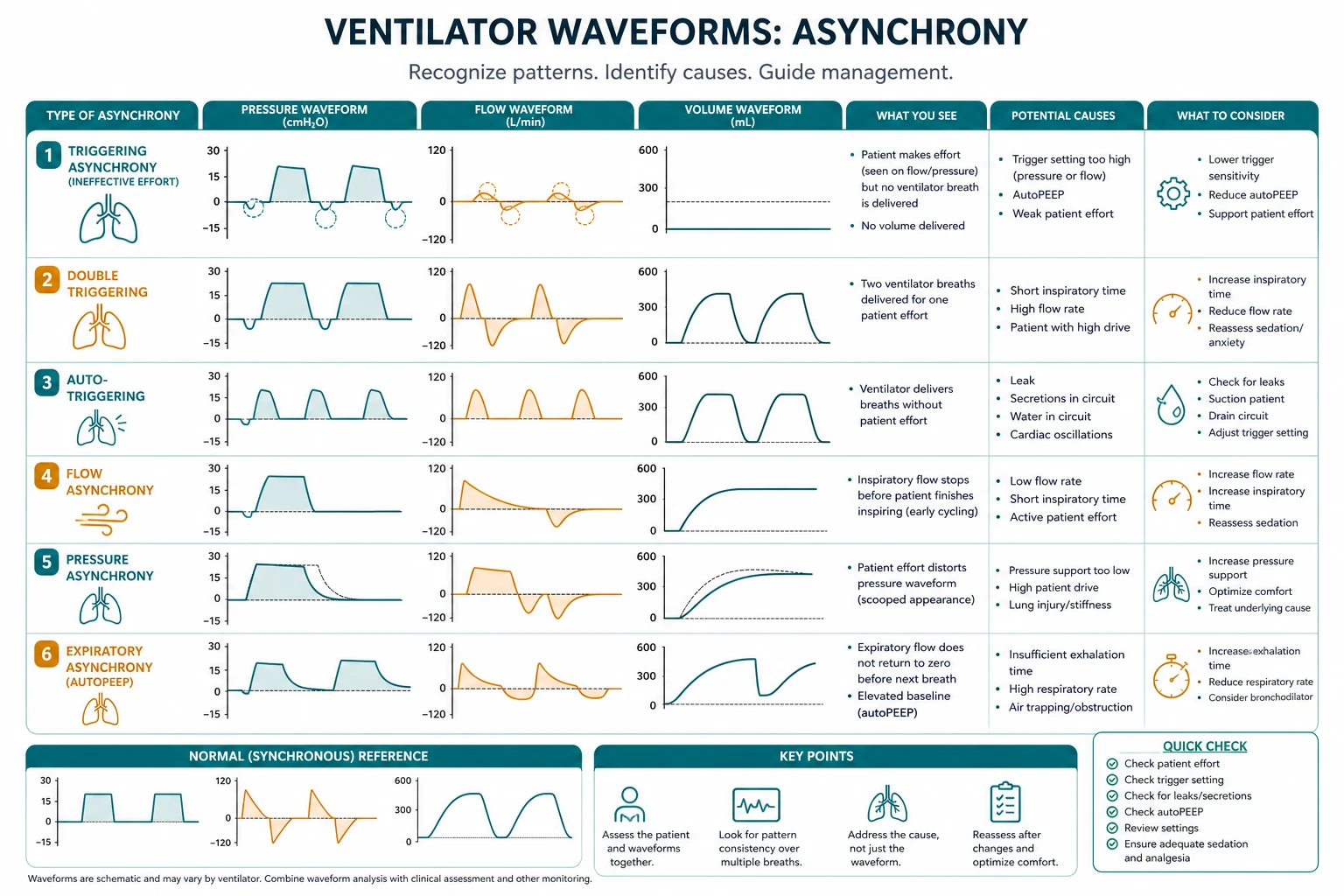
Patient-ventilator asynchrony — the 4 types
Patient-ventilator asynchrony — recognition and correction
| Type | Waveform appearance | Cause | Correction |
|---|---|---|---|
| TRIGGER ASYNCHRONY | |||
| - Missed triggers | Patient effort visible (pressure dips below baseline or flow changes) but NO ventilator breath delivered | Trigger sensitivity set too INSENSITIVE (high trigger threshold) OR auto-PEEP preventing the patient from generating enough negative pressure/flow to trigger | Make trigger MORE sensitive (flow trigger 1-2 L/min or pressure trigger -0.5 to -1 cmH2O). Reduce auto-PEEP (lower RR, shorter I time) |
| - Auto-triggering | Ventilator delivers breaths WITHOUT patient effort (rapid rate, irregular timing) | Trigger TOO sensitive (circuit movement, condensation in circuit, cardiac oscillations, water in tubing) | Make trigger LESS sensitive. Check for circuit leak or condensation |
| FLOW ASYNCHRONY | |||
| - Double triggering | TWO ventilator breaths within one patient inspiratory effort (the patient is STILL inhaling when the ventilator cycles to expiration — triggers a SECOND breath within 0.5s) | Delivered FLOW is INSUFFICIENT for patient demand — patient wants more flow → continues inhaling after ventilator has delivered the preset volume → triggers another breath | Increase FLOW RATE (VC) OR switch to PRESSURE SUPPORT/CONTROL (variable flow matches demand). Increase Vt. Increase inspiratory time |
| - Flow starvation | Pressure-time scalar shows a DIP during inspiration (patient is pulling against the ventilator — trying to get more flow than delivered) | Delivered flow < patient demand | Switch from VC to PC or PS (variable flow). Increase flow rate in VC |
| CYCLE ASYNCHRONY | |||
| - Premature cycling | Expiratory flow-time shows an abrupt cessation of flow + pressure spike → the ventilator cycled to expiration BEFORE the patient finished inhaling → patient continues to inhale against a closed inspiratory valve → active exhalation → flow spike | Inspiratory time TOO SHORT (volume control with fast flow) OR cycling criteria too sensitive (pressure support — cycling too early) | Increase inspiratory time (VC). Adjust cycling criteria (PS — increase cycling-off % from e.g., 25% to 40% of peak flow) |
| - Delayed cycling | The ventilator continues INSUFFLATING after the patient has started EXHALING → the patient actively EXHALES against positive pressure → pressure spike at end-inspiration | Inspiratory time TOO LONG (set too long in VC or PC) OR cycling criteria too insensitive (PS — doesn't cycle until very low flow) | Decrease inspiratory time. Adjust cycling criteria (PS — decrease cycling-off % to cycle earlier) |
| EXPIRATORY ASYNCHRONY | |||
| - Inadequate expiratory time | Expiratory flow does NOT return to zero → auto-PEEP (gas trapping). Patient cannot exhale completely before next breath | Inspiratory time too long → expiratory time too short. High RR. Obstructive disease (COPD, asthma — slow expiration) | Reduce RR. Shorten inspiratory time (increase I:E ratio to 1:3 or 1:4). Reduce Vt. Consider permissive hypercapnia |
Asynchrony quantification — measuring the burden at the bedside
Asynchrony is common (up to 25% of ICU patients have significant asynchrony) and is associated with prolonged ventilation, delayed weaning, discomfort, and worse outcomes. The key exam concept is that asynchrony can be QUANTIFIED from a short waveform recording — it is not just "eyeballed". [1]
The asynchrony index (AI)
The asynchrony Index (AI) = (number of asynchrony events / total number of breaths [triggered + machine]) × 100, expressed as a percentage. It is calculated over a representative recording (typically 5–30 minutes of waveform data, or via dedicated monitoring software). [1]
Severe asynchrony is defined as AI >10% — this threshold (from the Thille 2006 and de Wit 2009 studies) identifies patients in whom asynchrony is clinically meaningful and associated with prolonged mechanical ventilation and worse outcomes.[3]
The major asynchrony subtypes and how to count each
Counting each asynchrony type from the waveform
| Asynchrony type | Waveform signature | How to count | Calculation |
|---|---|---|---|
| Ineffective (missed) trigger | A patient effort (downward pressure deflection or expiratory flow distortion) with NO ventilator breath delivered | Count each aborted effort per minute | Ineffective trigger rate = events/min; or as % of total breathing efforts |
| Double trigger | Two ventilator cycles with the second starting <50% of the inspiratory time of the first (i.e., within one patient effort) | Count each doublet | Double-trigger rate = doublets / total breaths × 100 |
| Auto-trigger | A ventilator breath with NO preceding patient effort (irregular, often fast rate) | Compare breath timing to the patient's intrinsic pattern; breaths with no diaphragmatic/flow trigger signal | Auto-trigger rate = non-triggered breaths / total breaths × 100 |
| Premature / delayed cycling | Active expiration against inspiration (pressure spike at end-inspiration) OR continued inspiratory flow demand after cycling (flow spike at expiration start) | Count each breath with an end-inspiratory pressure spike or expiratory-flow spike | Cycling asynchrony rate = affected breaths / total breaths × 100 |
| Short cycling | A breath that cycles to expiration before the patient's neural inspiration ends → triggers the next breath (a double trigger) | Subset of double trigger | Counted with double triggers |
The effective triggering rate
A useful single metric: the effective triggering rate = number of breaths successfully triggered by the patient per minute. Compare the SET (backup) respiratory rate to the TOTAL rate: if the total rate barely exceeds the set rate, the patient is doing little work (or is oversedated); if there are many ineffective efforts BETWEEN triggered breaths, the patient is working hard but failing to trigger (high ineffective trigger rate). A high ineffective trigger rate in a COPD patient usually points to auto-PEEP — the patient must first overcome intrinsic PEEP before generating the trigger flow/pressure. [1]
Clinical implications of severe asynchrony (AI >10%)
- Prolonged mechanical ventilation and delayed weaning (the patient-ventilator mismatch prevents successful liberation).
- Increased work of breathing and discomfort.
- Higher risk of VAP (prolonged intubation).
- Worse outcomes: observational data link high AI to increased ICU mortality.
- Action: if AI >10%, systematically address each modifiable cause — trigger sensitivity, auto-PEEP (the commonest reversible cause of ineffective triggering in COPD), flow/demand matching (switch VC → PS for better demand matching), and cycling criteria.
Pressure Support (PS) and CPAP waveform analysis
Pressure Support (PS) and CPAP are the spontaneous breathing modes — the patient drives every breath and the ventilator ASSISTS. These are the weaning modes, and their waveforms are distinctive because they reflect patient-ventilator INTERACTION rather than ventilator control. [1]
Pressure Support (PS) waveform — the four phases
In PS the patient TRIGGERS every breath, the ventilator delivers a SET PRESSURE (above PEEP), flow is decelerating (variable, matches patient demand), and the breath CYCLES to expiration when inspiratory flow falls to a SET percentage of peak flow. Four phases are analysable on the waveform: [1]
The four phases of a PS breath — what to assess
| Phase | What happens | Waveform appearance | What to assess | Normal / abnormal |
|---|---|---|---|---|
| 1. Trigger | Patient generates inspiratory effort → ventilator detects it (flow or pressure trigger) | A downward deflection in airway pressure (or a flow change) BEFORE the inspiratory pressure rise | Trigger delay = time from patient effort onset to ventilator pressure delivery | Trigger delay <100 ms normal; >200 ms = prolonged → increase trigger sensitivity, check for auto-PEEP |
| 2. Pressure ramp (rise time) | Ventilator raises pressure from PEEP to set PS target | The steepness of the initial pressure rise | Rise time setting: too fast → pressure overshoot spike (patient discomfort); too slow → pressure does not reach target before flow falls (patient works against lag) | Match rise time to patient demand. Aggressive patients need fast rise |
| 3. Pressurisation / flow delivery | Constant pressure maintained; flow decelerates as alveolar pressure rises | Square pressure plateau + decelerating flow (like PC) | Whether pressure stays ON target (square) or sags (patient outpacing flow) | Square plateau = good match; sag = increase PS or rise time |
| 4. Cycling (E-to-E / cycle-off) | Ventilator cycles to expiration when flow falls to the cycle-off % of peak flow | The point where inspiratory flow stops and expiratory flow begins | Cycle-off % (typically 25%): too low (e.g., 5%) → delayed cycling → pressure spike at end-inspiration (patient exhaling against flow); too high (e.g., 50%) → premature cycling → double triggering | Observe for end-inspiratory pressure spike (delayed cycling) or double triggers (premature cycling) and adjust cycle-off % |
Trigger delay
Trigger delay = the time between the onset of the patient's neural inspiration (detectable as the start of the pressure/flow deflection) and the ventilator's response (pressure begins to rise). Prolonged trigger delay (>200 ms) increases the patient's work of breathing and is felt as dyspnoea. Causes:
- Trigger too insensitive (high threshold) → make more sensitive (flow trigger 1–2 L/min).
- Auto-PEEP (the patient must first decompress intrinsic PEEP before generating trigger flow) → the commonest cause in COPD → reduce auto-PEEP (lower RR, shorten I time, treat bronchospasm).
- Rise time too slow → the pressure ramps up too gradually → adjust rise time. [1]
Pressure ramp (rise time)
The rise time (or pressure ramp / pressurisation rate) controls how quickly airway pressure rises from PEEP to the set PS target at the start of inspiration.
- Rise time too fast → a pressure overshoot (spike above set PS at the start of inspiration) → patient discomfort, sometimes triggering premature cycling. Reduce the rise time (slower ramp).
- Rise time too slow → pressure fails to reach the PS target before flow has already begun to fall → the patient perceives inadequate support and works harder → increase rise time (faster ramp). A concave initial pressure upstroke (never reaching the target) signals a slow rise time or a leak. [1]
Cycling criteria (flow-based cycle-off)
In PS the breath cycles to expiration when inspiratory flow falls to a SET percentage of peak inspiratory flow (the cycle-off %, typically 25%, adjustable 5–80%). This is the LEVER for cycle asynchrony in PS:
- Premature cycling (cycle-off % too high, e.g., 50%) → the breath cycles before the patient's neural inspiration ends → the patient continues to inhale against a closed inspiratory valve → triggers a second breath → double triggering. Fix: DECREASE the cycle-off % (e.g., from 50% to 25%) to lengthen inspiration.
- Delayed cycling (cycle-off % too low, e.g., 5%) → the ventilator keeps insufflating after the patient has started to exhale → the patient actively expires against inspiratory flow → pressure spike at end-inspiration. Fix: INCREASE the cycle-off % (e.g., from 5% to 25%) to shorten inspiration. Delayed cycling is common in obstructive disease (long time constants) — the flow falls slowly and never reaches a low cycle-off threshold promptly.
- Leak and cycling: a circuit leak prevents inspiratory flow from falling to the cycle-off threshold → the breath never cycles → prolonged inspiration → pressure support effectively becomes pressure control. Modern ventilators use time-based backup cycling to terminate PS in the presence of a large leak. [1]
CPAP waveform — spontaneous breathing on PEEP
In CPAP the ventilator provides a CONTINUOUS POSITIVE AIRWAY PRESSURE with the patient doing ALL the work (no pressure support above PEEP). The waveform shows the patient's spontaneous breathing against a constant baseline pressure:
- Pressure–time: a flat baseline at the set CPAP (e.g., 5–10 cmH2O). Each spontaneous breath appears as a small downward deflection (the patient's inspiratory effort briefly drops airway pressure) followed by a return to baseline. The depth of the deflection reflects the patient's work of breathing — large deflections = high WOB, may indicate fatigue (consider adding PS).
- Flow–time: spontaneous biphasic flow — inspiratory flow above baseline, expiratory flow below, with a sinusoidal-ish pattern at the patient's own rate and depth. Normal spontaneous breaths have a smooth, rounded inspiratory flow curve (NOT square) decelerating to zero, then a passive decelerating expiratory limb.
- Volume–time: rises and falls with each spontaneous breath at variable Vt (the patient determines the size of each breath). [1]
What normal spontaneous breaths look like on CPAP: a regular rate (10–20/min in adults), smooth rounded inspiratory flow peaks of consistent amplitude, full expiration to baseline (no auto-PEEP), and small pressure deflections (minimal WOB). Irregular rate, large pressure deflections, or failure of expiratory flow to return to baseline = abnormal — assess for fatigue, obstruction, or asynchrony. [1]
Setting up and reading a PS breath — the four-step optimisation
- Trigger: set flow trigger 1–2 L/min (or pressure trigger -0.5 to -1 cmH2O). Check trigger delay on the pressure scalar — target <100 ms. If long → make more sensitive, exclude auto-PEEP.
- Rise time: set so the pressure reaches the PS target without overshoot. Look for a pressure spike at inspiration start (overshoot → slow the rise) or a concave upstroke that never reaches target (too slow → speed the rise).
- Pressurisation: confirm the pressure plateaus square (on target). A sag means the patient is outpacing flow → consider more PS. The resulting Vt should be 4–8 mL/kg predicted body weight.
- Cycle-off: set 25% (adjust 5–80%). Look at end-inspiration: a pressure spike = delayed cycling → increase cycle-off %. A double trigger = premature cycling → decrease cycle-off %. In obstructive disease, a higher cycle-off (30–40%) prevents delayed cycling; in restrictive disease, a lower cycle-off (10–20%) prevents premature cycling.
Clinical pearls
Red flags
Prognosis
Waveform analysis outcomes
| Finding | Clinical significance | Management |
|---|---|---|
| High driving pressure (ΔP >15) | Increased mortality (Amato 2015) | Reduce Vt or optimise PEEP |
| Auto-PEEP >5 | Gas trapping, hypotension, barotrauma | Reduce RR, shorten I time |
| Double triggering >10% of breaths | Flow asynchrony, delayed weaning | Increase flow or switch to PS |
| Missed triggers >10% | Ineffective triggering, wasted work | Adjust trigger, reduce auto-PEEP |
Key trials and evidence
Amato 2015 — Driving pressure and survival (PMID 25844785)
Source
NEJM — meta-analysis of 3,562 patients from 9 ARDS RCTs
Key finding
Driving pressure (ΔP = Pplat - PEEP) is the STRONGEST ventilatory predictor of survival
Threshold
ΔP >15 cmH2O = increased mortality
Clinical bottom line
Monitor ΔP on the P-V loop. If ΔP >15: reduce Vt or optimise PEEP to minimise ΔP
Thille 2006 — Patient-ventilator asynchrony during mechanical ventilation (PMID 17032638)
Source
American Journal of Respiratory and Critical Care Medicine — prospective observational study, 62 intubated patients
Study design
30-minute waveform recordings at least once daily; asynchrony index (AI) = asynchrony events / total breaths × 100
Key finding
Severe asynchrony (AI >10%) occurred in 24% of patients and was independently associated with prolonged mechanical ventilation
Asynchrony types
Ineffective triggering was the commonest (missed triggers), followed by double triggering
Clinical bottom line
Asynchrony is common, quantifiable from the waveform, and clinically meaningful when AI >10%. Daily waveform review and prompt correction (trigger sensitivity, auto-PEEP, demand matching) is part of standard ventilator care.
Why it matters for waveforms
This study established the 10% threshold used worldwide to define 'severe' asynchrony and validated waveform-based asynchrony counting
ART 2017 — Driving pressure and survival in ARDS (PMID 28615069)
Source
New England Journal of Medicine — multicentre RCT, 1,010 patients with moderate-to-severe ARDS
Study design
Strategy of PEEP titration to BEST COMPLIANCE (highest static compliance / lowest driving pressure) on the P-V loop vs standard ARDSNet PEEP/FiO2 table
Key finding
Higher PEEP guided by compliance (driving-pressure minimisation) did NOT reduce 28-day mortality; trend to HARM (28-day mortality 34.8% vs 27.8%)
Hypothesis
The compliance-guided approach recruited lung in some patients but OVERDISTENDED others — harm in patients with poorly recruitable lung
Clinical bottom line
Driving pressure and P-V-loop compliance are useful MONITORING tools, but aggressively maximising PEEP for all ARDS patients is not beneficial. Apply compliance-guided PEEP selectively to recruitable lung; the driving pressure should be kept <15 cmH2O where achievable, individualised to the patient.
Why it matters for waveforms
Cautionary tale — the P-V loop and driving pressure are powerful diagnostic tools, but using them to drive a one-size-fits-all protocol can cause harm. Waveform-guided ventilation must be individualised.
Additional red flags
Common exam scenarios — applying waveform analysis
Scenario A: Rising peak pressure in a ventilated patient
You are called: "The ventilator is alarming — high pressure." Approach with the PIP-Pplat-PEEP method. Do an inspiratory hold. If PIP is high but Pplat is normal (wide gradient) → RESISTANCE (suction, bronchodilate, check the tube is not kinked/bitten, check for circuit obstruction). If both PIP and Pplat are high → COMPLIANCE (examine for pneumothorax — asymmetric chest, tracheal deviation, hypotension; check for main-stem intubation; consider derecruitment/atelectasis; reduce Vt if overdistended). The waveform tells you WHICH problem before you order the chest X-ray.[3]
Scenario B: COPD patient cannot be weaned — high PaCO2, tachypnoeic
Look at the flow–time scalar and F-V loop. If expiratory flow does NOT return to baseline before the next inspiration → AUTO-PEEP (the commonest weaning failure mechanism in COPD). Confirm with an expiratory hold (total PEEP − set PEEP = auto-PEEP). The auto-PEEP also causes INEFFECTIVE TRIGGERING — the patient must decompress intrinsic PEEP before generating trigger flow, so many efforts are wasted (visible as downward pressure deflections without a delivered breath). Fix: reduce RR, shorten inspiratory time (lengthen expiratory time, I:E 1:3 to 1:4), treat bronchospasm, consider a larger ETT or extubation to NIV.[3]
Scenario C: Double triggering in ARDS on VC
The patient is still inhaling when the ventilator cycles → triggers a second breath → effectively double the Vt → risk of VILI and a high apparent RR. The patient wants more flow than the fixed VC flow provides. Fix: increase the inspiratory flow rate, increase inspiratory time, or switch to PC/PS where flow is variable and matches demand. Double triggering is the commonest asynchrony and, if >10% of breaths, warrants intervention.[3]
Scenario D: Titrating PEEP in ARDS using the P-V loop
Goal: maximise recruitable lung without overdistending. After a recruitment manoeuvre, perform a DECREMENTAL PEEP trial — step PEEP down from high to low and find the PEEP giving the BEST compliance (lowest driving pressure, ΔP). Set PEEP 2 cmH2O above that best-compliance point. Confirm no beaking on the P-V loop (UIP not exceeded, Pplat <30) and ΔP <15. This is more practical and better validated than chasing inflection points.[4]
Waveform-guided ventilator optimisation — a structured daily review
- Confirm mode and settings: VC, PC, or PS? Set Vt, RR, PEEP, FiO2, flow/trigger/cycle settings documented.
- Pressure scalar + inspiratory hold: read PIP, Pplat. Compute ΔP = Pplat − PEEP (target <15) and resistive gradient PIP − Pplat (target <6). Identify resistance vs compliance problem.
- Flow scalar + expiratory hold: confirm presence/absence of auto-PEEP (target <5). Check expiratory limb returns to baseline. If not → reduce RR, shorten I time.
- Volume scalar: confirm delivered Vt = set Vt (no leak). Check expired Vt returns to baseline (no trapping).
- P-V loop: confirm slope (compliance), no beaking (UIP), no excessive flatness. Use for PEEP/Vt titration.
- F-V loop: check expiratory limb (auto-PEEP, obstruction, leak).
- Asynchrony sweep: scan 5–10 minutes for ineffective triggers, double triggers, auto-triggers, cycling spikes. Compute AI; if >10%, intervene.
- PS-specific (if applicable): check trigger delay, rise time (overshoot/sag), and cycle-off (end-inspiratory spike / double trigger). Adjust each phase.
- Document and reassess: record the optimised settings and the objective metrics (ΔP, auto-PEEP, AI). Reassess after any change.
High-yield summary for exams
The high-yay exam one-liners for ventilator waveforms
| Waveform finding | One-line interpretation | Action |
|---|---|---|
| Square flow + rising pressure | Volume control | Read PIP/Pplat; assess resistance & compliance |
| Square pressure + decelerating flow | Pressure control | Read flow decay for resistance; Vt for compliance |
| PIP − Pplat > 6 cmH2O | High resistance (bronchospasm, secretions, kinked tube) | Suction, bronchodilate, check ETT |
| Pplat − PEEP > 15 (high ΔP) | Low compliance / overdistension | Reduce Vt, optimise PEEP |
| Expiratory flow not to baseline | Auto-PEEP (gas trapping) | Reduce RR, shorten I time, I:E 1:3–1:4 |
| P-V loop beaking | Overdistension (UIP reached) | Reduce Vt/PEEP, keep Pplat <30 |
| P-V loop flat slope | Low compliance (stiff lung) | Treat cause (ARDS, oedema); reduce Vt |
| Double triggering | Flow asynchrony (demand > supply) | Increase flow or switch to PS/PC |
| End-inspiratory pressure spike (PS) | Delayed cycling | Increase cycle-off % |
| Pressure overshoot at inspiration start (PS/PC) | Rise time too fast | Slow rise time |
| Sawtooth on flow/pressure | Secretions | Suction |
| AI > 10% | Severe asynchrony | Systematic correction; associated with worse outcome |
Written practice
SAQ — Waveform diagnosis of high airway pressure
10 minutes · 10 marks
A patient on volume-control ventilation alarms for high airway pressure. PIP 48 cmH2O, Pplat after inspiratory hold 24 cmH2O, set PEEP 8 cmH2O. Expiratory flow on the flow-time scalar does not return to baseline before the next breath.
References
- [1]Labeau SO, et al. [Natural killer cells: adaptation and memory in innate immunity] Med Sci (Paris), 2013.PMID 23621934
- [2]Esteban A, et al. Drowning N Engl J Med, 2012.PMID 22646632
- [3]Acute Respiratory Distress Syndrome Network Handling of hazardous materials Ann Emerg Med, 2000.PMID 10613956
- [4]Amato MB, et al. Novel psoriasis therapies and patient outcomes, part 1: topical medications Cutis, 2015.PMID 25844785
- [5]Stewart RM, et al. Causes and characteristics of injury in paediatric major trauma and trends over time Arch Dis Child, 2019.PMID 30279158
- [6]Pittman RN, et al. Commentary on providing guidance to patients: physicians' views about the relative responsibilities of doctors and religious communities South Med J, 2013.PMID 23820320