ICU · Respiratory
Weaning from mechanical ventilation
Also known as Ventilator weaning · Spontaneous breathing trial (SBT) · Extubation criteria · Rapid shallow breathing index (RSBI) · Daily sedation interruption
Weaning from mechanical ventilation is the process of transitioning from full ventilatory support to spontaneous breathing and extubation. The key principle: daily assessment of readiness + spontaneous breathing trial (SBT). Daily SAT (sedation awakening trial) + SBT pairing reduces ventilation days and mortality. SBT methods: T-piece, pressure support (5-8 cmH2O), or CPAP (5 cmH2O) for 30-120 minutes. Extubation success predictors: RSBI (f/VT) <105, strong cough, good secretion clearance, cuff leak present (if prolonged intubation). Failure indicators: RR 35, SpO2 <90%, HR 140, agitation, diaphoresis. Failed SBT → return to ventilator, investigate cause, retry in 24h.
On this page & tools
Your progress
Saved locally on this device.
Target exams
Red flags

Weaning process
Daily weaning assessment and protocol
Daily sedation awakening trial (SAT)
Stop or reduce sedation daily until patient awake enough to follow commands (RASS -1 to +1). Safety check: no active seizures, no agitation, no high ventilator settings. SAT reduces over-sedation, allows neurological assessment, prepares for SBT.
Assess readiness for SBT
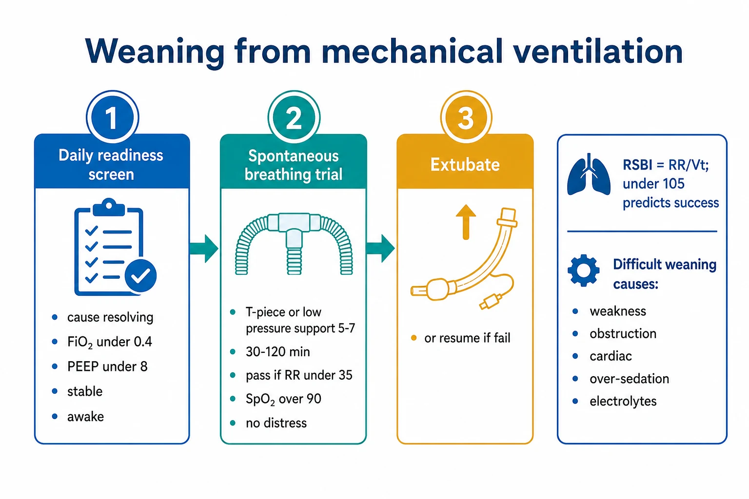
Criteria: (1) original illness resolving/reversed, (2) FiO2 <0.4, (3) PEEP <8 cmH2O, (4) haemodynamically stable (no or low-dose vasopressors), (5) able to follow commands, (6) adequate cough, (7) PaO2/FiO2 >150-200, (8) no significant respiratory acidosis. If criteria met → proceed to SBT.
Conduct SBT (30-120 minutes)
Method 1: T-piece (no ventilatory support — most challenging, most predictive). Method 2: Pressure support 5-8 cmH2O + PEEP 5 (most common). Method 3: CPAP 5 cmH2O. Duration: 30 min (first attempt) or 120 min (to confirm). Monitor: RR, SpO2, HR, BP, patient comfort.<Cite id="3" />}
Assess SBT result
PASS: RR <35, SpO2 >90%, HR <140 (or change <20%), no agitation, no diaphoresis, no distress. → Proceed to extubation assessment. FAIL: RR >35, SpO2 <90%, HR >140 (or >20% change), agitation, diaphoresis, distress, hypotension/hypertension. → Return to ventilator, investigate cause, retry in 24h.<Cite id="2" />}
Pre-extubation assessment
Assess: (1) cough strength (can patient clear secretions?), (2) secretion volume/viscosity (copious/thick = harder to manage post-extubation), (3) cuff leak test (if intubated >6 days — absent leak = post-extubation stridor risk), (4) upper airway patency (no laryngeal oedema suspected), (5) neurological status (can protect airway?).<Cite id="1" />}
Extubate if all criteria met
Suction airway. Deflate cuff. Remove ETT in one smooth motion. Apply face mask or nasal cannula. Monitor closely for 1-2h (RR, SpO2, HR, stridor). Have reintubation equipment ready. Consider NIV prophylactically for high-risk patients (COPD, cardiac failure, elderly).
Weaning classification: simple, difficult, prolonged
Not every patient weans at the same rate. Two frameworks classify weaning difficulty — the 1998/2007 International Consensus Conference (ICC) categories and the newer 2017 WIND (Weaning according to a New Definition) classification. Assigning the category on the day of the first separation trial predicts resource use, ICU length of stay and mortality, and triggers early escalation for the difficult/prolonged weaner.[4][8]
Simple weaning
ICC group 1 — ~60-70% of patients
- Passes the FIRST spontaneous breathing trial and is successfully extubated on that attempt
- Typically intubated for a short, fully reversible illness (e.g. drug overdose, post-operative, sedation)
- Lowest ICU and hospital mortality; shortest ventilation duration (median ~3 days)
- Needs nothing more than a daily SAT + SBT protocol
Difficult weaning
ICC group 2 — ~20-25%
- Up to THREE SBTs OR up to 7 days from the first SBT to successful extubation
- Usually a single dominant, correctable burden (fluid overload, ongoing infection, bronchospasm)
- Higher reintubation risk than the simple group
- Targeted workup: identify and treat the limiting factor, then re-trial
Prolonged weaning
ICC group 3 — ~10-15%
- FAILS 3 or more SBTs OR needs more than 7 days of weaning after the first SBT
- Often multifactorial: ICU-acquired weakness plus cardiac dysfunction plus retained secretions plus malnutrition
- Highest mortality (around 30%), longest ICU stay, highest tracheostomy rate
- Demands a structured multidisciplinary weaning programme and early tracheostomy consideration
Spontaneous breathing trial (SBT) methods in detail
The SBT is the gatekeeper of extubation. Three methods are in routine use, differing in the load they impose on the respiratory muscles. The T-piece is the most demanding and most predictive of true post-extubation work; pressure support with low PEEP is the most common because it is easy to set up and best tolerated.[3][6]
T-piece
No ventilatory support
- Patient disconnected from the ventilator, breathing through a low-resistance T-piece circuit with humidified supplemental oxygen
- Highest work of breathing — recruits accessory muscles and imposes the full resistive and elastic load
- Most closely mimics the load the patient will face after extubation (no tube compensation, no PEEP)
- Subira 2019: a T-piece SBT gave HIGHER successful-extubation rates than pressure support, but a higher in-trial failure rate
- Exhaled tidal volume must be measured separately to calculate RSBI
Pressure support 5-8 + PEEP 5
Most commonly used
- Low pressure support overcomes endotracheal-tube resistance; 5 cmH2O PEEP maintains functional residual capacity
- Best tolerated — lowest failure rate of the three methods during the trial itself
- Ventilator still measures exhaled VT and RR, so RSBI is easy to calculate
- May OVERESTIMATE success — slightly higher reintubation than T-piece in Subira 2019
- Current first-line method in most protocols; switch to T-piece for a borderline pass
CPAP 5 cmH2O
Continuous positive airway pressure
- No pressure support (PS = 0) but 5 cmH2O continuous pressure throughout the respiratory cycle
- Maintains alveolar recruitment — useful in obesity, basal atelectasis, cardiogenic pulmonary oedema
- Intermediate difficulty between T-piece and pressure support
- Now used less often as a sole SBT method
Daily SAT + SBT protocol: the evidence base
Pairing a Sedation Awakening Trial (SAT) with a Spontaneous Breathing Trial (SBT) every day is the single best-studied weaning intervention. It works because oversedation is the commonest reversible cause of delayed extubation, and a calm, awake patient can be screened for breathing readiness. [1]
SAT safety screen to SAT to SBT safety screen to SBT
SAT safety screen
Exclude: active seizures, agitation (RASS +2 or higher), alcohol or DT withdrawal, neuromuscular blockade in the last 24h, high ventilator demand (FiO2 0.6 or more, PEEP 10 or more), untreated myocardial ischaemia, untreated raised intracranial pressure. If any present, SKIP todays SAT and retry tomorrow.
Perform SAT
Stop ALL sedation (propofol, midazolam, dexmedetomidine) and opioids (unless treating active pain) until the patient is awake (RASS -1 to +1) or a safety failure occurs. Safety failure = agitation, distress, ventilator dyssynchrony, SpO2 under 88%, HR over 140 or under 50. Resume at half the prior infusion rate.
SBT safety screen
Original illness resolving, FiO2 0.4 or less, PEEP 8 or less, adequate cough, PaO2/FiO2 over 150-200, no significant acidosis, no new myocardial ischaemia, no high-dose vasopressors (norepinephrine about 5 mcg/min or less), no need for airway protection for secretions.
Conduct paired SBT (30-120 min)
T-piece OR pressure support 5-7 + PEEP 5. Monitor pass/fail criteria continuously. If pass, proceed to extubation assessment. If fail, resume prior settings, identify the limiting factor (load, capacity, drive or cardiac), treat it, and retry the next day.
Rapid Shallow Breathing Index (RSBI)
[1]Causes of weaning failure
Respiratory causes
Most common
- Respiratory muscle weakness (ICU-acquired weakness, fatigue)
- Excessive respiratory load (bronchospasm, secretions, pneumonia)
- Reduced respiratory drive (opioids, brainstem injury, sleep apnoea)
- Cardiac failure (weaning-induced pulmonary oedema — increased preload/afterload)
- Critical illness polyneuropathy/myopathy (CIP/CIM)
Non-respiratory causes
Systemic
- Sepsis/infection (increases metabolic demand, impairs muscle function)
- Electrolyte disturbance (hypophosphataemia, hypokalaemia, hypomagnesaemia)
- Malnutrition (insufficient respiratory muscle strength)
- Psychological: anxiety, panic, agitation
- Obesity (reduced chest wall compliance, high work of breathing)
Extubation criteria: the final checklist
Passing the SBT is necessary but not sufficient. Roughly 10-20% of patients who pass an SBT still fail extubation. The pre-extubation assessment checks whether the patient can protect and clear the airway once the tube is removed.[4]
[1]Predictors of SUCCESS
Safe to extubate
- Strong cough on command
- Minimal thin secretions; suction at most every 4 hours
- Awake and cooperative, RASS -1 to 0, follows commands
- Cuff leak present (over 110 mL or over 25% of delivered VT) if intubated more than 6 days
- Passed a 120-minute SBT with RSBI under 105
- PaO2/FiO2 over 200 on FiO2 0.4 / PEEP 5
Predictors of REINTUBATION
Delay or use NIV
- Weak or absent cough, especially with copious secretions
- Copious thick secretions, frequent suctioning
- Altered consciousness (GCS under 8, agitation, encephalopathy)
- Absent cuff leak after more than 6 days intubation (stridor risk)
- Older age, cardiac failure, COPD, obesity, hypercapnia during the SBT
- A failed prior extubation attempt
Weaning-induced cardiac failure
Removing positive-pressure ventilation suddenly increases venous return (preload) and left-ventricular afterload, and can unmask occult left-heart disease — producing weaning-induced pulmonary oedema. It is one of the most commonly missed causes of a difficult or prolonged wean.[4]
[1]Managing weaning-induced cardiac failure
Recognise the pattern
Recurrent SBT failure with pulmonary oedema signs — crackles, rising jugular venous pressure, falling SpO2. Look for raised BNP, new mitral regurgitation or an E-over-e-prime ratio above 14 on echocardiography during the trial.
Reduce preload
Diurese to euvolaemia with furosemide BEFORE the next SBT. Aim for a negative fluid balance and use daily weights to guide therapy.
Optimise cardiac medications
Continue or restart ACE-inhibitor, beta-blocker and diuretic. Avoid over-diuresis causing acute kidney injury. Treat any myocardial ischaemia. Add rate control for atrial fibrillation.
Modify the SBT
Use low-level pressure support plus PEEP (which maintains alveolar recruitment and reduces afterload) rather than a T-piece. Use shorter, more frequent trials. Arrange prophylactic NIV immediately after extubation.
ICU-acquired weakness and the difficult-to-wean patient
ICU-acquired weakness (ICU-AW) — critical illness polyneuropathy (CIP), critical illness myopathy (CIM), or both — is the dominant cause of respiratory-muscle failure in prolonged weaners. It affects up to half of patients ventilated for more than a week, and predicts delayed weaning, prolonged ICU stay and long-term disability. [1]
[1]Risk factors for ICU-AW
Preventable
- Sepsis and multi-organ failure
- Hyperglycaemia (aim for glucose 6-10 mmol/L)
- Immobilisation — avoid; start early mobilisation
- High-dose prolonged corticosteroids, especially with neuromuscular blockers
- Prolonged deep sedation and benzodiazepine use
Workup of the weak weaner
Diagnose
- MRC sum-score under 48 out of 80 (or weak hand-grip dynamometry) defines ICU-AW
- Bedside nerve-conduction studies and EMG separate CIP from CIM
- Exclude electrolyte causes (low K, Mg, PO4, Ca) and myasthenia or Guillain-Barre syndrome
- Engage physiotherapy for respiratory-muscle training and limb mobilisation
Post-extubation stridor
[1]Post-extubation respiratory support: NIV and HFNC
If the patient is at HIGH risk of reintubation, do not leave them on a simple face mask — apply prophylactic non-invasive ventilation (NIV) or high-flow nasal cannula (HFNC) immediately after extubation. The choice depends on the underlying problem: hypercapnia favours NIV; hypoxaemia or a low-risk patient favours HFNC.[11]
Prophylactic NIV
Hypercapnia / COPD / cardiac
- Indicated in COPD with hypercapnia during the SBT, obesity-hypoventilation, neuromuscular disease, cardiogenic pulmonary oedema, or PaCO2 over 45 mmHg at end of SBT
- Nava 2005: prophylactic NIV lowered reintubation and mortality in hypercapnic high-risk patients
- Set IPAP 10-15 and EPAP 4-5; titrate to comfort and PaCO2
- NOT effective, and possibly harmful, when used as RESCUE after overt post-extubation failure in non-COPD patients — use prophylactically, not as a bail-out
High-flow nasal cannula
Hypoxaemia / low-risk
- Hernandez 2016 (JAMA): in LOW-risk patients HFNC reduced reintubation versus standard oxygen (4.9% vs 12.2%)
- HIGH-WEAN (Thille 2018) and meta-analyses: HFNC is a reasonable alternative to NIV in non-hypercapnic patients
- Set flow 30-50 L/min and FiO2 to keep SpO2 at least 92%; it delivers low-level PEEP, washes out dead space and humidifies secretions
- Preferred over NIV when the patient cannot tolerate a mask, has facial trauma, or needs to communicate and eat
Standard oxygen
Conventional
- Acceptable only in genuinely low-risk patients with a normal PaCO2 and a strong cough
- Lower cost and simplest, but inferior to HFNC even in low-risk patients (Hernandez 2016)
- Always have NIV or HFNC ready at the bedside for any high-risk extubation
Tracheostomy for prolonged weaning
About 10-15% of ventilated patients need a tracheostomy — most often for prolonged weaning, airway protection, or inability to clear secretions. A tracheostomy reduces laryngeal injury, improves comfort, allows weaning to a speaking valve, and facilitates transfer to a weaning or rehabilitation ward — but the TIMING is contested.[16]
Potential benefits
Tracheostomy
- Lower work of breathing than an endotracheal tube (less resistance, less dead space)
- Improved comfort — the patient can talk (speaking valve), eat and mobilise
- Easier secretion clearance and airway toilet
- Avoids glottic and laryngeal injury from prolonged translaryngeal intubation
- Enables step-down from ICU to a dedicated weaning or rehabilitation ward
Indications
When to place
- Anticipated ventilation for more than 10-14 days
- Prolonged or difficult weaning (ICC group 3) failing 3 or more SBTs
- Inability to protect the airway (severe brain injury, neuromuscular disease)
- Upper-airway obstruction, copious secretions, or a failed extubation
- To facilitate transfer for long-term rehabilitation
Timing — early vs late
Evidence
- TracMan (Young 2013, JAMA, 909 patients): early (day 1-4) vs late (day 10 or later) tracheostomy showed NO mortality difference and no reduction in ventilation days, but less sedation in the early group
- Broad consensus: consider tracheostomy at around 7-10 days of ventilation, or earlier if a prolonged course is anticipated
- Percutaneous dilatational tracheostomy (bedside, bronchoscopy-guided) is preferred in most ICUs
Landmark trials in ventilator weaning
ABC (Girard 2008)
Lancet 2008
336 ventilated patients — paired SAT + SBT protocol vs standard sedation plus daily SBT
Key finding
More ventilator-free days (14.7 vs 11.6); on long-term follow-up lower one-year mortality (44% vs 58%, NNT about 7)
Practice change
Established daily SAT + SBT pairing as the cornerstone of weaning
Kress 2000
NEJM 2000
128 ventilated patients — daily interruption of sedatives vs sedation at physician discretion
Key finding
Median ventilation 4.9 vs 7.3 days; half the complications of prolonged ICU stay
Practice change
Daily sedation interruption (the SAT) became standard
Ely 1996
NEJM 1996
300 patients — nurse-implemented daily SBT screening vs physician discretion
Key finding
Median ventilation 4.5 vs 6 days; 50% fewer complications; earlier ICU discharge
Practice change
Nurse-driven daily SBT screening protocols widely adopted
Penuelas (Subira 2019)
JAMA 2019
1153 patients — pressure support 8 + PEEP 5 vs T-piece SBT for up to 2 hours
Key finding
Higher successful extubation at 72 hours with pressure support plus PEEP (82.3% vs 74.0%); less reintubation
Practice change
Pressure-support SBT validated as first-line; T-piece reserved for borderline cases
Esteban 1995
NEJM 1995
546 patients — four weaning methods: 2-hour T-piece vs pressure support vs SIMV vs once-daily T-piece
Key finding
Once-daily T-piece and pressure support weaned faster than SIMV (slowest); established T-piece and pressure support dominance
Practice change
SIMV de-emphasised for weaning; T-piece and pressure support became preferred
Brochard 1994
AJRCCM 1994
456 difficult-to-wean patients — pressure support vs T-piece vs SIMV
Key finding
Pressure support had fewer failures (shorter weaning, fewer reintubations) than SIMV or T-piece
Practice change
Pressure support established as a primary weaning mode
WIND (Beduneau 2017)
AJRCCM 2017
5721 patients across 36 French ICUs — prospective observational weaning classification
Key finding
About 63% short, 18% difficult, 19% prolonged; difficult and prolonged weaning independently predicted higher 90-day mortality
Practice change
Reframed weaning categories around the first separation trial; validated the WIND classification
TracMan (Young 2013)
JAMA 2013
909 patients — early (day 1-4) vs late (day 10 or later) percutaneous tracheostomy
Key finding
No difference in 30-day mortality (30% vs 31%) or ventilation duration; less sedation with early tracheostomy
Practice change
No mandate for early tracheostomy; decide at around 7-10 days on an individual basis
Nava 2005
Crit Care Med 2005
97 hypercapnic high-risk patients — prophylactic post-extubation NIV vs standard oxygen
Key finding
Lower reintubation rate and lower 90-day mortality in the NIV group
Practice change
Prophylactic NIV after extubation in hypercapnic and COPD high-risk patients
Ferrer 2002
ERJ 2002
43 intubated COPD patients who failed an SBT — NIV-facilitated weaning vs invasive pressure-support weaning
Key finding
Shorter ventilation, fewer complications, lower mortality with NIV-facilitated weaning
Practice change
NIV used to facilitate weaning in COPD after a partial SBT
Hernandez 2016
JAMA 2016
527 low-risk patients — post-extubation HFNC vs conventional oxygen
Key finding
Lower reintubation at 72 hours (4.9% vs 12.2%) with HFNC
Practice change
HFNC preferred over a simple mask after extubation, even in low-risk patients
HIGH-WEAN (Thille 2018)
BMJ Open 2018
Protocol for an RCT of HFNC alone vs HFNC plus NIV after extubation in high-risk patients
Key finding
Established the protocol and feasibility of combined HFNC and NIV strategies after extubation
Practice change
Informed post-extubation oxygen strategy in high-risk weaners
SAQ — Daily SAT + SBT protocol and the post-extubation plan
10 minutes · 10 marks
A 65-year-old man with severe COPD is on day 5 of mechanical ventilation for a hypercapnic exacerbation. He is on FiO2 0.35, PEEP 5, pressure-support 8, on a propofol infusion at 30 mL/h, RASS −3. He is haemodynamically stable on no vasopressors. Outline the structured weaning protocol you would apply today and the immediate post-extubation plan.
SAQ — Recurrent SBT failure from weaning-induced cardiac dysfunction
10 minutes · 10 marks
A 72-year-old woman with a background of hypertension and obesity has failed three consecutive SBTs on the respiratory ward. Each time she develops tachycardia (HR 130s), new bibasal crackles, and a falling SpO2 at around 30 minutes into the trial, with no obvious respiratory cause. Her BNP rises from 200 to 900 pg/mL during the trial. Diagnose the problem and outline management.
Clinical pearls
Red flags
Key takeaways
References
- [1]Girard TD, Kress JP, Fuchs BD, Thomason JW, Schweickert WD, Pun BT, Taichman DB, Dunn JG, Pohlman AS, Kress JP, Hall JB, Dittus RS, Bernard GR; Ely EW. Efficacy and safety of a paired sedation and ventilator weaning protocol for mechanically ventilated patients in intensive care (Awakening and Breathing Controlled trial): a randomised controlled trial Lancet, 2008.PMID 18191684
- [2]Ely EW, Baker AM, Dunagan DP, Burke HL, Smith AC, Kelly PT, Johnson MM, Browder RW, Bowton DL, Haponik EF. Effect on the duration of mechanical ventilation of identifying patients capable of breathing spontaneously N Engl J Med, 1996.PMID 8948561
- [3]Subirà C, Hernández G, Vázquez A, Rodríguez-García R, González-Castro A, García C, Rubio O, Ventura M, López MJ, Estrada L, Carrillo A, Caballero J, Figueira JC, Bordejé ML, Tomicic V, de Haro C, González-Hernández S, Maggiore SM, Esteban A, Ferguson ND, PEÑUELAS Group; AñaVEmIdo Group. Effect of Pressure Support vs T-Piece Ventilation Strategies During Spontaneous Breathing Trials on Successful Extubation Among Patients Receiving Mechanical Ventilation: A Randomized Clinical Trial JAMA, 2019.PMID 31184740
- [4]Boles JM, Bion J, Connors A, Herridge M, Marsh B, Melot C, Pearl R, Silverman H, Stanchina M, Vieillard-Baron A, Welte T. Weaning from mechanical ventilation Eur Respir J, 2007.PMID 17470624
- [5]Kress JP, Pohlman AS, O'Connor MF, Hall JB. Daily interruption of sedative infusions in critically ill patients undergoing mechanical ventilation N Engl J Med, 2000.PMID 10816184
- [6]Esteban A, Frutos F, Tobin MJ, Alía I, Solsona JF, Valverdú I, Fernández R, de la Cal MA, Benito S, Tomás R, et al. A comparison of four methods of weaning patients from mechanical ventilation. Spanish Lung Failure Collaborative Group N Engl J Med, 1995.PMID 7823995
- [7]Brochard L, Rauss A, Benito S, Conti G, Mancebo J, Rekik N, Gasparetto A, Lemaire F. Comparison of three methods of gradual withdrawal from ventilatory support during weaning from mechanical ventilation Am J Respir Crit Care Med, 1994.PMID 7921460
- [8]Béduneau G, Pham T, Schortgen F, Piquilloud L, Zogheib E, Jonas M, Grelon F, Runge I, Terzi N, Grangé S, Barberet G, Guitard PG, Frat JP, Constans J, Ayac F, Girault C, CADRE/REVA research network; WIND (Weaning according to a New Definition) study investigators. Epidemiology of Weaning Outcome according to a New Definition. The WIND Study Am J Respir Crit Care Med, 2017.PMID 27626706
- [9]Ferrer M, Esquinas A, Arancibia F, Bauer TT, González G, Carrillo A, Rodriguez-Roisin R, Torres A. Noninvasive ventilation after intubation and mechanical ventilation Eur Respir J, 2002.PMID 12030739
- [10]Nava S, Gregoretti C, Fanfulla F, Squadrone E, Grassi M, Carlucci A, Beltrame F, Navalesi P. Noninvasive ventilation to prevent respiratory failure after extubation in high-risk patients Crit Care Med, 2005.PMID 16276167
- [11]Rochwerg B, Brochard L, Elliott MW, Hess D, Hill NS, Nava S, Navalesi P, Antonelli M, Ferrer M, Grunauer AM, Mancebo J, Mehta S, Raoof S, Tobin MJ, Singh JM, Pang L, Esbrook A, Fraser GL, McConnell K, Ruhl L, Laghi F, Amin AH, Karhu J, Kragholm K, Smit AJ, Shashank P, Zegenhagen S, Ramsay MAE, Kondili E, Bollini CA, Bolliger CT, Bosma K, Booth S, Burgos C, Ciccalini P, Cinesi-Gómez C, Coleman JM, Cox CE, Currow DC, Douglas IS, Ehrmann C, Farias FA, Galinski M, Gay P, Guarracino F, Heunks L, Holanda-Peña M, Hug MI, Hurtado M, Jarava PM, Joynt GM, Kawasaki T, Kelly CR, Khanna AK, Kuo YW, Laubscher T, Linde-Zwirble W, Luyt CE, Manthous CA, McGee WT, Mélot C, Menger MN, Mokhlesi B, Murthy S, Newman RW, Nishimura K, Northwood D, Olaposi-Ogundipe A, Parthasarathi S, Pellegrini M, Pravikoff DS, Preseaux AL, Schönhofer B, Scröder M, Serrano-Mollar A, Shrestha-Munar H, Sivaloganathan S, Srinivasan V, Stämpfle DF, Subirà C, Tänzler K, Tinoco-Velasco-Rivera P, Veiga M, Vitacca M, Williams JW, Younes M, Yusuff HO, Zhou L, Djärv T, Alonso-Coello P. Official ERS/ATS clinical practice guidelines: noninvasive ventilation for acute respiratory failure Eur Respir J, 2017.PMID 28860265
- [12]Lee CH, Peng MJ, Wu CL. Dexamethasone to prevent postextubation airway obstruction in adults: a prospective, randomized, double-blind, placebo-controlled study Crit Care, 2007.PMID 17605780
- [13]Thille AW, Müller G, Gacouin A, Coudroy R, Decavèle M, Bourdin G, lzq Tessier W, Ehrmann S, Kimmoun A, Tonnelier JM, Prat G, Rol A, Bréchot N, Sirodot M, Béduneau G, Gouëllo JP, Rolland I, Guitard PG, Frat JP, Robert R, Vivier E, Roze H, Liet JM, Barberet G, Salhi A, Chouihed T, Dubreuil J, Amectier M, Mercat A, Reignier J, Darget R, Boulain T; HIGH-WEAN Investigators; Réseau REVA. High-flow nasal cannula oxygen therapy alone or with non-invasive ventilation during the weaning period after extubation in ICU: the prospective randomised controlled HIGH-WEAN protocol BMJ Open, 2018.PMID 30185583
- [14]Hernández G, Vaquero C, González P, Subira C, Frutos-Vivar F, Rialp G, Laborda C, Colinas L, Cuena R, Fernández R. Effect of Postextubation High-Flow Nasal Cannula vs Conventional Oxygen Therapy on Reintubation in Low-Risk Patients: A Randomized Clinical Trial JAMA, 2016.PMID 26975498
- [15]Granton DN, Noble M, Zeraatkar D, Hann S, Slobogean GP, Mbuagbaw L. High-Flow Nasal Cannula Compared With Conventional Oxygen Therapy or Noninvasive Ventilation Immediately Postextubation: A Systematic Review and Meta-Analysis Crit Care Med, 2020.PMID 32947472
- [16]Young D, Harrison DA, Cuthbertson BH, Rowan K; TracMan Collaborators. Effect of early vs late tracheostomy placement on survival in patients receiving mechanical ventilation: the TracMan randomized trial JAMA, 2013.PMID 23695482