ICU · Respiratory / ventilation
Weaning from Mechanical Ventilation — SBT, Protocols & Prolonged Weaning
Also known as Weaning · Liberation · Spontaneous breathing trial · SBT · T-piece trial · Rapid shallow breathing index · RSBI · ABC trial · Daily sedation interruption · Weaning protocol · Prolonged weaning
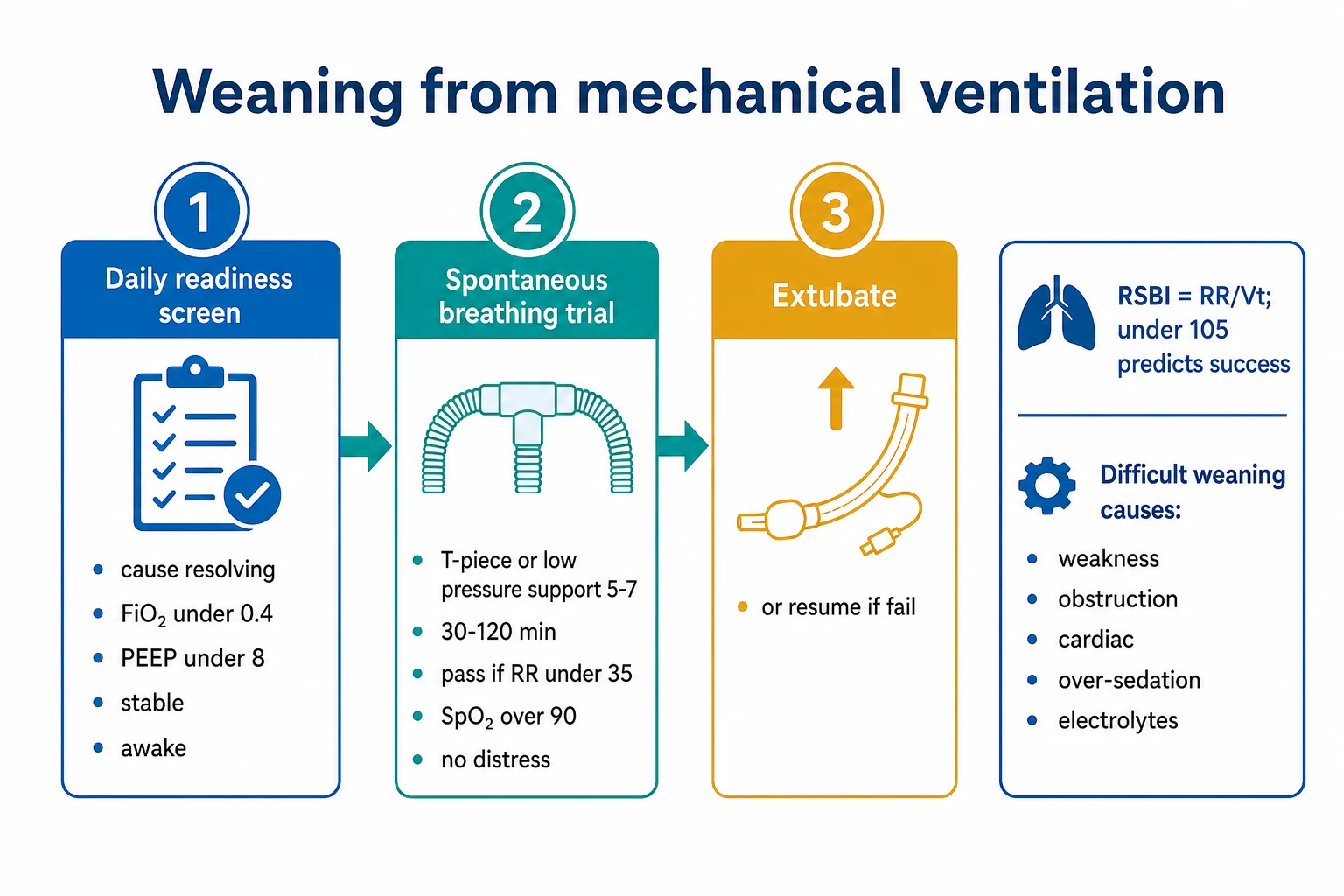
Weaning is the process of discontinuing mechanical ventilation: a daily readiness screen, a spontaneous breathing trial (SBT), and extubation if the trial is tolerated. Around 70 per cent of patients wean simply after one SBT, 20 per cent wean with difficulty (up to three failed SBTs or up to seven days), and 10 per cent have prolonged weaning (over three failed SBTs or over seven days). The SBT uses minimal support — a T-piece, low pressure support 5-7 cmH2O, or CPAP 5 cmH2O — for 30-120 minutes; tolerance means a respiratory rate under 35, an SpO2 over 90 per cent, a heart rate under 140, no distress, and stable haemodynamics. The ABC trial (Girard, Lancet 2008) paired a daily sedation interruption (SAT) with the daily SBT and improved outcomes. The rapid shallow breathing index (RSBI = RR/Vt in litres), under 105, predicts success (Yang & Tobin, NEJM 1991). Before extubating, assess airway patency with a cuff leak test and the patient's ability to protect the airway (cough on suction, gag); anticipate post-extubation stridor (especially in females, prolonged intubation, and traumatic intubation) and have a low threshold for prophylactic steroids and post-extubation high-flow nasal cannula or NIV. Reintubation rates run 10-20 per cent; a failed extubation carries a markedly higher mortality. Protocolised, nurse- or therapist-driven weaning reduces the duration of mechanical ventilation versus physician-directed weaning (Ely 1996).
On this page & tools
Your progress
Saved locally on this device.
Target exams
Red flags
Overview & definition
Weaning (liberation) from mechanical ventilation is the process of discontinuing ventilatory support. It has three steps: a daily readiness screen, a spontaneous breathing trial (SBT), and extubation if the trial is tolerated. Around 70 per cent of patients wean easily after a single SBT; a minority wean with difficulty and need the underlying cause addressed.[1][1]
Weaning classification — simple, difficult, prolonged
The WIND (Weaning according to a New Definition) classification is the modern framework used to stratify weaning. Patients fall into one of three groups according to the number of failed SBTs and the time from the first SBT to successful extubation:[1]
Simple weaning
~70% of patients
- Passes the first SBT and is successfully extubated
- Median duration from first SBT to extubation: under 24 hours
- Typical course: short medical/surgical ICU admission, postoperative ventilation, drug overdose
- Outcomes are excellent — focus on protocolising the screen to avoid delay
Difficult weaning
~20% of patients
- Up to three failed SBTs, or up to seven days from the first SBT to successful extubation
- Usually has a single dominant and reversible barrier — over-sedation, secretions, mild weakness
- Targets: address the cause, repeat SBTs daily, early mobility, sleep hygiene
- Most progress to successful weaning within a week once the cause is treated
Prolonged weaning
~10% of patients
- More than three failed SBTs, or more than seven days from the first SBT to successful extubation
- Often multiple interacting causes — critical-illness neuromyopathy, chronic lung disease, cardiac failure
- Consider a tracheostomy to reduce dead space, ease secretion clearance, and enable rehabilitation (TracMan: timing does not change mortality)
- Mortality is high — driven by the underlying disease, not the weaning itself
- Multidisciplinary approach: nutrition, mobilisation, speech and language therapy, psychological support
Why the 70/20/10 split matters for the exam
Simple (70 per cent), difficult (20 per cent), prolonged (10 per cent) — this is the single most testable number in weaning. The exam wants the candidate to recognise that the vast majority of patients wean easily with a protocolised daily screen and SBT, and that the difficult and prolonged groups are where the mark is won or lost. The candidate is expected to recite the causes of difficult weaning (load-versus-capacity imbalance) and to discuss a structured approach: address the cause, repeat SBTs, avoid SIMV weaning, consider tracheostomy for prolonged weaning, and recognise weaning-induced cardiac failure.[1][1]
Step 1 — the daily readiness screen
Before each SBT, screen for readiness:[1]
- The underlying cause of respiratory failure is resolving.
- Adequate oxygenation — a PaO2/FiO2 over about 150-200, PEEP under about 5-8 cmH2O, FiO2 under about 0.4-0.5.
- Haemodynamically stable — no or low-dose vasopressors, no active myocardial ischaemia.
- Awake and cooperative — able to follow commands, initiate inspiration, and cough.
- No new serious problem (no ongoing deep sedation, no severe acidosis).
A daily screen, combined with a daily sedation interruption (a spontaneous awakening trial), is the foundation of efficient weaning.[1]
The daily readiness screen is a filter — its job is to safely and quickly identify patients who can attempt an SBT without missing anyone capable of breathing. The screen has positive criteria (must be present) and negative criteria (must be absent). A protocolised screen, run by nurses or respiratory therapists, is faster than waiting for a doctor and reduces ventilator days.[5]
Positive criteria (must be present)
Resolution + capacity
- Underlying cause of respiratory failure resolving or resolved
- PaO2/FiO2 > 150-200 on PEEP ≤ 5-8 cmH2O and FiO2 ≤ 0.4-0.5
- Patient initiates inspiration (triggering the ventilator), cough on suction
- Neurology: awake or rousable (RASS -1 to 0), able to follow commands — gag and cough intact
- Haemodynamics: no or low-dose vasopressors (e.g. noradrenaline < 0.1 mcg/kg/min), no active myocardial ischaemia
Negative criteria (must be absent)
Barriers to weaning
- No significant respiratory acidosis (pH > 7.32; rising PaCO2 not acceptable)
- No active agitation or uncontrolled delirium; no ongoing deep sedation requirement
- No new serious problem in the last 24 hours (e.g. cardiac arrest, re-intubation, dialysis initiation)
- No copious secretions exceeding the ability to clear
- No fever or active significant bleeding
The cuff leak, cough, and gag — airway protection before the trial
A readiness screen that ignores the airway is incomplete. Before extubating you must confirm three things: (i) cuff leak is adequate (no laryngeal oedema — see the extubation section below); (ii) cough peak flow is at least 60 L/min (the patient can clear secretions — a strong cough on suctioning is the bedside surrogate); and (iii) gag and swallow are intact so the patient can protect the airway from aspiration. A weak cough (peak flow under 60 L/min, Smina Chest 2003) and copious secretions predict extubation failure independent of the SBT result.[11]
Running the daily readiness screen and SBT
Step A — screen at the morning round
Run the readiness screen on every ventilated patient every day. Use a written checklist. If any negative criterion is present, fix it and reassess the next day; do not start the SBT.
Step B — pair with a spontaneous awakening trial (SAT)
Reduce or stop sedation (RASS target -1 to 0). If the patient tolerates the SAT (no agitation, distress, or desaturation), proceed to the SBT. The ABC trial proved this pairing reduces ventilator days and improves outcomes.
Step C — start the SBT on minimal support
T-piece (no support), or PSV 5-7 cmH2O + PEEP 5, or CPAP 5. Duration 30-120 minutes (30-60 minutes for simple weaning; up to 120 minutes for the difficult/chronic patient). Subirà (JAMA 2019): a 30-minute trial is non-inferior to 120 minutes.
Step D — observe and reassess
Watch continuously. Pass criteria: RR < 35, SpO2 > 90%, HR < 140 (change < 20%), SBP change < 20%, no distress, no arrhythmia. Fail criteria: RR > 35, SpO2 < 90%, HR > 140, agitation, diaphoresis, accessory-muscle use, rising PaCO2, arrhythmia. Resume support the moment any fail criterion appears.
Step E — extubate or restart
If the SBT passes and the airway is safe (cuff leak adequate, cough strong, gag intact), extubate. If the SBT fails, restart support, document the failure mode (see the failed-SBT section), and address the cause. Reattempt the next day.
The screen + SBT takes a few minutes to set up and an hour to run; it should be a daily routine, not a special event. A patient who passes is extubated; a patient who fails goes back on support, the cause is addressed, and the trial is repeated the next day. [1]
Step 2 — the spontaneous breathing trial (SBT)
The SBT tests whether the patient can breathe with minimal support:[1]
- Method — a T-piece (no ventilatory support), or low pressure support (5-7 cmH2O) with PEEP 5, for 30-120 minutes.
- Tolerance (pass) — respiratory rate under 35, SpO2 over 90 per cent, heart rate under 140 (and not changing by more than 20 per cent), no respiratory distress (no accessory-muscle use, no diaphoresis, no agitation), stable blood pressure, and a stable gas exchange.
- Failure — a rising respiratory rate, falling SpO2, distress (accessory muscles, diaphoresis, agitation), haemodynamic instability, or arrhythmia — resume ventilatory support and address the cause.
A patient who tolerates the SBT proceeds to extubation (after confirming airway protection — see the extubation topic).[1]
The ideal SBT loads the respiratory muscles with the work they will face after extubation (an endotracheal tube adds resistive work, so the post-extubation work is similar). Three techniques are accepted and roughly equivalent in trial populations; the T-piece most closely mimics unassisted breathing, while PSV 5-7 cmH2O + PEEP 5 partly compensates for the resistance of the endotracheal tube.[3][4]
T-piece
No ventilatory support
- Most physiological — patient does all the work; closest to post-extubation breathing
- Disconnect the endotracheal tube from the ventilator; attach a T-piece with humidified oxygen (FiO2 same as the ventilator)
- No positive pressure to overcome endotracheal-tube resistance (which can over-estimate work)
- Use 30-120 minutes; preferred in trials and for the harder/chronic patient
- Risk: rapidly tiring a borderline patient; monitor continuously
PSV 5-7 + PEEP 5
Low pressure support
- Pressure support 5-7 cmH2O + PEEP 5 cmH2O; compensates for endotracheal-tube resistance
- Most popular in practice — easy to set on the ventilator, no circuit change
- Brochard (1994) found pressure-support weaning faster than SIMV or T-piece weaning in difficult weaners
- May slightly over-estimate tolerance (the patient gets some help)
- Most useful as a daily screen — easy to repeat
CPAP 5
Continuous positive airway pressure
- CPAP 5 cmH2O alone — no pressure support, just a constant pressure
- Useful in COPD (offsets intrinsic PEEP and the work of triggering) and cardiogenic pulmonary oedema
- Maintains alveolar recruitment; less fatiguing in stiff lungs
- Esteban (1995) found a CPAP wean comparable to T-piece weaning
- Bridges to extubation in the difficult patient
SBT duration — 30-120 minutes
The standard duration is 30 to 120 minutes. Subirà (JAMA 2019) randomised patients to a 30-minute vs a 120-minute SBT and found the shorter trial was non-inferior for successful extubation — there is no benefit in prolonging the trial in patients who are passing. The corollary: a patient who is going to fail usually does so within the first 20-30 minutes (rising rate, falling saturation, distress). A trial of 120 minutes is reserved for the difficult or chronically ventilated patient where longer observation is informative.[12]
Step 3 — weaning protocols and the ABC trial
Protocolised weaning (a nurse- or respiratory-therapist-driven daily screen and SBT) reduces the duration of mechanical ventilation compared with physician-directed weaning, because it removes the delay of waiting for a doctor to assess the patient.[1]
The ABC trial (Awakening and Breathing Controlled, Girard, Lancet 2008) paired a daily sedation interruption (a spontaneous awakening trial) with a daily SBT. The combined protocol reduced ventilator-free days and improved outcomes (with no excess self-extubation), establishing the paired SAT plus SBT as standard.[1]
The evidence for protocolised weaning is strong. Ely (NEJM 1996) randomised patients to a nurse/therapist-driven protocol versus physician-directed weaning and found the protocol group was extubated a median of 1.5-2 days earlier, with no increase in reintubation. The mechanism is simple: doctors round once or twice a day; nurses are at the bedside continuously, so a protocolised screen + SBT removes the decision-to-extubate delay.[5]
Protocolised weaning
Nurse/RT-driven
- A written daily screen + SBT protocol executed by nurses or respiratory therapists
- Ely (NEJM 1996): reduced duration of mechanical ventilation by ~1.5-2 days vs physician-directed
- Removes decision delay — runs every day, every shift, at the bedside
- No increase in self-extubation, reintubation, or reintubation-related morbidity
- Pairs naturally with a daily sedation interruption (SAT) — the ABC protocol
Physician-directed weaning
Clinician-led
- Doctor reviews the patient, decides readiness, orders the SBT, reviews the result, orders extubation
- Slow — limited by rounding frequency and competing clinical priorities
- Inherently variable — depends on individual clinician enthusiasm and experience
- Acceptable where a robust protocol does not exist; inferior to a protocol in trial settings
- Still needed for the difficult/prolonged weaner who needs cause-directed treatment
The ABC trial — pairing sedation interruption with the SBT
The Awakening and Breathing Controlled trial (Girard, Kress, Fuchs; Lancet 2008) was a multicentre randomised trial pairing a spontaneous awakening trial (SAT, a daily sedation interruption) with a spontaneous breathing trial (SBT) versus usual care with sedation directed by the clinician. The combined protocol increased ventilator-free days, ICU days, and 28-day survival, with no excess self-extubation or reintubation. The principle: the patient must be awake enough to breathe before you can test whether they can breathe. This pairing is now the global standard.[1]
The paired SAT + SBT (the ABC protocol)
Step 1 — Safety screen before SAT
Exclude if: active seizures, alcohol withdrawal, agitation, neuromuscular blockade, myocardial ischaemia, raised ICP. If safe, proceed to stop or reduce sedation.
Step 2 — Spontaneous Awakening Trial (SAT)
Stop all sedatives and opioids (or reduce to a light RASS -1 to 0). Wait 30-60 minutes. Fail if: agitation, distress, desaturation, arrhythmia, or ventilator asynchrony requiring restart — resume sedation at half the previous dose.
Step 3 — Daily readiness screen
If the patient passes the SAT, run the readiness screen (see Step 1 above). Resolution of cause, PaO2/FiO2 > 150, PEEP ≤ 5-8, FiO2 ≤ 0.4, haemodynamically stable, no new serious problem.
Step 4 — Spontaneous Breathing Trial (SBT)
30-120 min on T-piece or PSV 5-7 + PEEP 5. Pass criteria: RR < 35, SpO2 > 90%, HR < 140, no distress, stable BP. Fail: resume support and address the cause.
Step 5 — Extubate
Confirm airway is safe (cuff leak, cough on suction, gag). Extubate. Plan post-extubation support (HFNC or prophylactic NIV for the high-risk patient — see below).
Daily sedation interruption — the Kress evidence
Kress (NEJM 2000) showed that a daily interruption of sedative infusions (until the patient was awake) halved the duration of mechanical ventilation and the length of ICU stay, with no excess psychological harm or agitated delirium. It is the single most effective, cheapest weaning intervention. Pair it with the SBT (the ABC trial) for the maximum effect.[6]
The rapid shallow breathing index (RSBI)
The RSBI = respiratory rate divided by tidal volume in litres, measured during a brief unsupported breath. An RSBI under 105 predicts SBT success; over 105 predicts failure (shallow, rapid breathing — the pattern of fatigue). It is a guide, not absolute, and is used alongside the clinical SBT.[1]
How the RSBI was derived and how to measure it
Yang & Tobin (NEJM 1991) prospectively measured a battery of physiological indices in ventilated patients being tested for weaning, and asked which one best predicted success or failure. The f/VT ratio (respiratory frequency divided by tidal volume in litres) outperformed every other index — a threshold of 105 breaths/min/L gave a sensitivity and specificity of ~0.80 for SBT success. The physiology: as the diaphragm fatigues, the patient adopts a rapid, shallow breathing pattern (high rate, small tidal volume) — a textbook fatigue pattern. To measure it, place the patient on a T-piece or CPAP 5 (no pressure support) for one minute, then read the rate and the exhaled Vt (in litres) from the ventilator.[2]
Pitfalls of the RSBI
The RSBI is a guide, not absolute. It over-estimates failure (a falsely high value) when the endotracheal tube is narrow or partly blocked (high resistance inflates the rate), and during the first minute of a trial before the patient settles. It under-estimates failure when the cuff is leaking (Vt measured by the ventilator is spuriously low, lowering the RSBI). Use it alongside the clinical SBT — a patient with an RSBI of 100 who is in distress has failed; a patient with an RSBI of 120 who is comfortable and passing the trial can still be extubated.[2]
Other predictors of weaning outcome
The RSBI is the headline index, but a modern difficult-weaning assessment adds:
- Maximal inspiratory pressure (MIP or NIF) — under -20 to -30 cmH2O suggests adequate reserve; over -20 (i.e. weaker) predicts failure.
- Cough peak flow — under 60 L/min predicts extubation failure (Smina, Chest 2003).[11]
- Diaphragmatic thickening fraction on ultrasound — under 30-36 per cent on M-mode predicts failure; ultrasound is increasingly used at the bedside.
- Lung ultrasound score — a high B-line count predicts weaning-induced pulmonary oedema.
- Cardiac biomarkers (BNP/NT-proBNP) — a rise during the SBT predicts weaning-induced cardiac failure.
Failed SBT — explicit criteria and what to do
The failed SBT is defined by any one of the following during the trial:[1]
Respiratory
Breathing failure
- Respiratory rate > 35/min, or a rise of > 50% from baseline
- SpO2 < 90% (or PaO2 < 60 mmHg) on the trial FiO2
- PaCO2 rise > 8 mmHg with pH fall
- Marked accessory-muscle use, paradoxical abdominal breathing, diaphoresis
Cardiovascular
Haemodynamic instability
- Heart rate > 140/min, or sustained rise/fall > 20%
- SBP > 180 or < 90 mmHg
- New arrhythmia, chest pain, or ST change suggesting ischaemia
Neurological
Loss of drive or distress
- Agitation, anxiety, diaphoresis, or new confusion
- Somnolence or loss of respiratory drive (over-sedation)
- Clinician judgement: the patient looks like they are failing
When the SBT fails, resume the previous ventilatory support immediately and address the cause. Document the mode of failure — respiratory (rate, saturation), cardiac (rate, BP, echo), or neurological (agitation, sedation). Each failure should teach you something about the dominant barrier, and the next day's plan should target it. Repeat the SBT daily (or every other day in the prolonged weaner); do not abandon daily trials because the patient has failed a few times.[1]
Difficult and prolonged weaning
Weaning difficulty is categorised:[1]
- Simple — passes the first SBT and is extubated.
- Difficult — up to three failed SBTs, or up to seven days from the first SBT.
- Prolonged — more than three failed SBTs, or more than seven days.
The fundamental problem in difficult weaning is a respiratory load that exceeds the capacity of the respiratory muscles. The causes:[1]
- Reduced capacity (respiratory-muscle weakness) — critical-illness polyneuromyopathy, prolonged neuromuscular blockade, malnutrition, and electrolyte disturbance (hypophosphataemia, hypokalaemia, hypomagnesaemia).
- Increased load — obstructive disease (COPD with high resistive load and dynamic hyperinflation), pneumonia and copious secretions, pulmonary oedema, obesity and chest-wall restriction.
- Reduced drive — over-sedation, central nervous system depression.
- Cardiac — weaning-induced cardiac failure: resuming spontaneous breathing increases venous return (preload) and left-ventricular afterload (the loss of positive intrathoracic pressure), unmasking occult LV dysfunction and causing pulmonary oedema (suspect a patient who fails the SBT with a rising respiratory rate and crackles; diagnose with an echo during a trial).
- Metabolic and psychological — acidosis, delirium, anxiety.
The framework to keep in your head is the load-versus-capacity balance: the respiratory muscles pump air against a load (resistance, compliance, secretions). When capacity falls (weakness) or load rises (obstruction, oedema), the pump fails — and the patient weans slowly or not at all. A separate axis is drive: even with adequate capacity and load, over-sedation abolishes the central drive to breathe. Diagnose the dominant axis at each failed SBT.[1]
Reduced capacity
Muscle weakness
- Critical-illness polyneuromyopathy (CIPNM) — the leading cause in prolonged ICU stays
- Prolonged neuromuscular blockade (aminosteroids, especially in ARDS, sepsis, steroids)
- Malnutrition, hypoalbuminaemia, ICU-acquired weakness
- Electrolytes: hypophosphataemia, hypokalaemia, hypomagnesaemia, hypocalcaemia
- Diaphragmatic dysfunction from prolonged ventilation (Dres, 2014; ultrasound diagnosis)
Increased load
High work of breathing
- COPD — high resistive load, dynamic hyperinflation (auto-PEEP), increased work of triggering
- Pneumonia, copious secretions, atelectasis, pleural effusion
- Pulmonary oedema — reduced compliance, raised work
- Obesity hypoventilation, chest-wall restriction (flail chest, kyphoscoliosis)
- Bronchospasm, endotracheal tube too narrow or partly blocked
Reduced drive
Loss of respiratory drive
- Over-sedation — the commonest reversible cause (interrupt daily, ABC trial)
- CNS depression — stroke, encephalopathy, opiates
- Metabolic alkalosis (chronic CO2 retainer, diuretic-induced) blunts drive
- Sleep deprivation, delirium — disrupts respiratory pattern
Cardiac
Weaning-induced cardiac failure
- Switch to negative-pressure breathing raises venous return (preload) and LV afterload
- Unmasks occult LV dysfunction, diastolic dysfunction, ischaemia
- Presents as SBT failure with rising rate, falling SpO2, new crackles, rising BNP
- Diagnose with an echo during a failed trial; treat with diuresis and afterload reduction
Approach to a failed SBT — find the dominant barrier
1. Was the failure respiratory, cardiac, or neurological?
Read the trial chart: rising RR with falling SpO2 = respiratory. Rising HR/BP with new crackles or ST change = cardiac. Agitation or somnolence = neurological/drive. This directs the work-up.
2. Respiratory failure — check load and capacity
Listen for wheeze (bronchodilate), check secretions (suction, treat infection), CXR for new infiltrate or effusion. Ultrasound the diaphragm (thickening fraction). Check ABG — is PaCO2 rising (fatigue) or PaO2 falling (load)?
3. Cardiac failure — echo during the next trial
Bedside echo during a failed SBT: new wall-motion abnormality, rising E/e, dilated IVC, or new B-lines indicate weaning-induced cardiac failure. Treat with diuresis, afterload reduction, rate control; consider an echo-guided heart-failure regimen.
4. Drive failure — review sedation
List every sedative and opiate. Reduce to RASS -1 to 0 (daily SAT). Stop benzodiazepines (delirium). Check the delirium screen (CAM-ICU/ICDSC); treat with a low-dose antipsychotic if needed.
5. Capacity failure — feed, mobilise, correct electrolytes
Check phosphate, potassium, magnesium, calcium. Start or advance enteral nutrition. Begin early mobilisation (Schweickert, Lancet 2009 — early PT/OT improves functional outcomes). Reassess daily.
6. Repeat the SBT the next day
Once the dominant barrier is treated, repeat the SBT. Do not abandon daily trials because the patient has failed a few times. For the prolonged weaner, consider a tracheostomy (TracMan — timing does not change mortality but improves comfort and secretion clearance).
Management of difficult weaning

- Muscle weakness — nutrition, correct electrolytes (phosphate, potassium, magnesium), avoid prolonged paralysis, early mobility.
- Increased load — bronchodilate obstruction, treat infection and clear secretions, diurese pulmonary oedema.
- Reduced drive — reduce sedation (daily SAT).
- Cardiac failure — diurese, afterload reduction, treat ischaemia; an echo during a failed SBT confirms weaning-induced pulmonary oedema.
- Weaning strategy — repeat daily SBTs (the preferred approach); gradual pressure-support weaning as an alternative. Avoid SIMV weaning, which prolongs weaning. For prolonged weaning, a tracheostomy reduces dead space and eases secretion clearance and weaning (timing does not change mortality, per TracMan).
- Treat delirium, anxiety, and optimise sleep.[1]
Extubation — cuff leak, post-extubation stridor, reintubation
Passing the SBT is necessary but not sufficient for extubation. The airway must be safe: there must be no upper-airway obstruction (laryngeal oedema), and the patient must protect the airway (cough, gag, swallow). Two bedside tests answer these questions: the cuff leak test and the cough on suction. [1]
The cuff leak test — predict laryngeal oedema
The cuff leak is the difference between the inspired and the expired tidal volume when the cuff is deflated. A small cuff leak (under 110 mL, or under 25 per cent of the delivered tidal volume) suggests that the tube fills the trachea snugly because the surrounding laryngeal and tracheal tissues are oedematous — predicting post-extubation stridor. Risk factors for a low cuff leak include a traumatic or difficult intubation, prolonged intubation (over 7 days), large-bore tube, female sex, and active cuff infection. A patient at high risk of post-extubation stridor should receive prophylactic steroids (methylprednisolone 20-40 mg every 4-6 hours for 4 doses, starting 12-24 hours before extubation), which reduce the rate of reintubation for stridor.[15]
Cuff leak > 110 mL
Low risk of stridor
- Cuff leak exceeds 25% of the delivered tidal volume
- No prophylactic intervention required; routine extubation
- Negative predictive value for stridor is high (> 95%)
- Confirm adequate cough and gag, then extubate
Cuff leak < 110 mL
High risk of stridor
- Suggests laryngeal oedema — risk of post-extubation stridor
- Give prophylactic steroids (methylprednisolone 20-40 mg q4-6h x 4 doses, start 12-24h pre-extubation)
- Recheck the cuff leak after steroid course; the leak often improves
- Plan post-extubation HFNC or NIV to support the upper airway
- Have a low threshold to reintubate early for stridor (it worsens rapidly)
Post-extubation stridor
Post-extubation stridor is inspiratory stridor within 24-48 hours of extubation caused by laryngeal oedema (or, less commonly, bilateral vocal-cord paralysis). It occurs in 2-15 per cent of extubations and carries a higher reintubation and mortality rate. Risk factors (Jaber, CCM 2003): female sex, prolonged intubation (over 36-48 hours), traumatic or repeated intubation, large tube, high cuff pressure, and aggressive suctioning.[15]
Management:
- Racemic adrenaline nebulised (1:1000 adrenaline 5 mL) — topical vasoconstrictor, fast onset.
- Dexamethasone IV (systemic anti-inflammatory; 4-8 mg) — onset over hours, treats laryngeal oedema.
- Heliox (helium-oxygen mixture) — lower density reduces turbulent-flow work of breathing across the obstruction; a temporising bridge.
- Non-invasive ventilation — CPAP/PSV stents the upper airway open; useful if the patient is tiring but not in failure.
- Reintubate early — a worsening stridor with rising respiratory rate is a reintubation indication; do not wait for a respiratory arrest. Use a smaller tube this time. [1]
Reintubation — 10-20 per cent of extubations
Reintubation rates run 10-20 per cent overall, and higher in medical and neurology ICUs. Reintubation within 48-72 hours is an extubation failure; the cause is usually one of: upper-airway obstruction (stridor), inadequate cough or secretion clearance, encephalopathy with loss of airway protection, or residual cardiac/respiratory failure. A failed extubation carries a markedly higher mortality (some studies show mortality 2-5 times higher than successful extubation), partly because reintubation is itself hazardous (aspiration, haemodynamic instability, desaturation) and partly because failure marks a sicker patient.[8][9]
Prophylactic post-extubation NIV
High-risk hypercapnic
- Ferrer (Am J Respir Crit Care Med 2003; BMJ 2009): prophylactic NIV after extubation in COPD/hypercapnic patients reduces reintubation and mortality
- Use for: chronic respiratory disease, hypercapnia during the SBT, older age, cardiac failure
- Start immediately after extubation; bilevel for hypercapnia, CPAP for cardiac failure
Rescue NIV for extubation failure
Treat with caution
- Esteban (NEJM 2004): rescue NIV after extubation failure did NOT reduce mortality and may delay needed reintubation
- A rescue NIV trial is acceptable if the failure is hypercapnic/COPD and the patient is stable, with a strict reassessment at 1 hour
- Do NOT use rescue NIV as a routine blanket strategy in non-selected patients — it delays reintubation
Post-extubation HFNC
Low-risk support
- High-flow nasal cannula (50 L/min, FiO2 0.3-0.5) reduces work of breathing and provides mild positive pressure
- Useful for the moderate-risk patient; lower reintubation rates than standard oxygen in low-risk cardiac and respiratory patients (OPTINC trial)
- Easy to apply, well-tolerated, does not impede speech or feeding
The cuff leak, the cough, and the swallow — the airway-protection bundle
Before pulling the tube, complete the airway bundle: (1) cuff leak over 110 mL (or a positive leak after steroids); (2) strong cough on suctioning (peak cough flow over 60 L/min); (3) gag reflex intact and the patient able to swallow secretions. A patient who passes the SBT but fails the airway bundle (weak cough, absent gag, copious secretions) will likely fail extubation — leave the tube in, address the cause (suction, treat delirium, mobilise), and reassess daily.[11]
The extubation procedure
1. Pre-extubation checklist
SBT passed. Cuff leak > 110 mL (or steroids given for high-risk). Strong cough on suction. Gag intact. FiO2 ≤ 0.4, PEEP ≤ 5-8. Haemodynamically stable. Vasopressors minimal. Plan for post-extubation support (HFNC or NIV) ready.
2. Position and prepare
Sit the patient upright. Pre-oxygenase with 100% FiO2 for 1-2 minutes. Suction the oral cavity, the back of the throat, and above the cuff (to clear pooled secretions that would otherwise be aspirated on cuff deflation).
3. Deflate the cuff and remove the tube
Deflate the cuff fully. Insert a suction catheter through the tube. On inspiration, withdraw the tube and the suction catheter together (clearing secretions as you withdraw). Apply a face mask or HFNC at once.
4. Immediate post-extubation
Encourage cough and deep breathing. Start HFNC or NIV if high-risk. Listen for stridor. Monitor SpO2, RR, HR, and work of breathing for the next 4 hours — the highest-risk period for failure.
5. Watch for failure
A rising RR, falling SpO2, new stridor, agitation, or somnolence in the first 24-72 hours is extubation failure. Treat the cause (racemic adrenaline for stridor, NIV for hypercapnia, diuresis for oedema); reintubate early if deteriorating.
Tracheostomy for prolonged weaning
For the prolonged weaner (over three failed SBTs, over seven days), a tracheostomy reduces dead space, lowers the work of breathing, eases secretion clearance, and is more comfortable than an endotracheal tube (the patient can eat, speak, mobilise). The TracMan trial (NEJM 2013) compared early (day 1-4) versus late (day 10+) tracheostomy and found no difference in mortality or ventilator-free days — so the timing is driven by patient comfort and expected duration of ventilation rather than a hard deadline. A percutaneous dilatational tracheostomy at the bedside (Ciaglia technique) is the standard approach in ICU. Decannulation follows once the patient tolerates a speaking valve and a capped tracheostomy on room air for 24-48 hours.[7]
The evidence base — landmark trials
ABC (Girard)
Lancet 2008
336 mechanically ventilated pts — paired SAT + SBT protocol vs usual care
Key finding
More ventilator-free days (14.7 vs 11.6, p=0.02), shorter ICU stay, improved 28-day survival (HR 0.68); no excess self-extubation
Practice change
Pair daily sedation interruption with daily SBT — the standard of care
Kress (Daily SAT)
NEJM 2000
128 ventilated pts — daily interruption of sedatives vs usual (clinician-directed) sedation
Key finding
Halved duration of mechanical ventilation (median 4.9 vs 7.3 days, p=0.004) and ICU stay; no excess psychological harm
Practice change
Daily sedation interruption became routine for ventilated patients
Yang & Tobin (RSBI)
NEJM 1991
36 pts — derivation cohort of physiological indices predicting weaning success
Key finding
f/Vt (RSBI) under 105 predicted SBT success with sensitivity/specificity ~0.80; outperformed all other indices
Practice change
RSBI <105 became the standard bedside predictor of SBT success
Ely (Protocol weaning)
Ann Intern Med 1996
300 pts — nurse/RT-driven weaning protocol vs physician-directed weaning
Key finding
Reduced duration of mechanical ventilation (median 1.5-2 days shorter); no increase in reintubation
Practice change
Protocolised, nurse/RT-driven weaning adopted in modern ICUs
Esteban (4 methods)
NEJM 1995
546 pts — daily T-piece trials vs PSV weaning vs SIMV weaning vs a combination
Key finding
PSV weaning shorter than SIMV or once-daily T-piece; SIMV slowest of all
Practice change
SIMV weaning discouraged; daily SBT or PSV preferred
Brochard (3 methods)
AJRCCM 1994
456 difficult-to-wean pts — PSV vs SIMV vs T-piece weaning
Key finding
PSV weaning had fewer failures and shorter weaning than SIMV or T-piece in difficult weaners
Practice change
PSV weaning validated for difficult weaning
Subirà (SBT duration)
JAMA 2019
970 pts — 30-min vs 120-min SBT
Key finding
30-min SBT non-inferior to 120-min for successful extubation (82.3% vs 83.5%)
Practice change
A 30-min SBT is sufficient for most patients
TracMan
NEJM 2013
909 ventilated pts — early (day 1-4) vs late (day 10+) tracheostomy
Key finding
No difference in 30-day mortality (30% vs 31%) or 2-year survival
Practice change
Tracheostomy timing driven by patient comfort, not a hard deadline
Ferrer (NIV post-ext)
AJRCCM 2003
43 COPD pts hypercapnic at SBT — prophylactic NIV after extubation vs standard oxygen
Key finding
Reduced reintubation (20% vs 50%) and 90-day mortality (0% vs 19%)
Practice change
Prophylactic NIV after extubation in hypercapnic COPD patients
Esteban (rescue NIV)
NEJM 2004
221 pts with extubation failure — NIV vs standard oxygen
Key finding
No difference in reintubation or mortality; NIV may delay needed reintubation in non-selected patients
Practice change
Rescue NIV is not routine for extubation failure — only selected hypercapnic/COPD patients
Schweickert (early mob)
Lancet 2009
104 ventilated pts — early PT/OT vs usual care
Key finding
More patients returned to independent function (59% vs 35%); shorter delirium and more ventilator-free days
Practice change
Early mobilisation added to the ABC bundle (now SAT + SBT + mobility)
Jaber (stridor)
Crit Care Med 2003
Cohort — risk factors and outcomes of post-extubation stridor
Key finding
Stridor in ~2-15% of extubations; female sex, prolonged intubation, traumatic intubation predict it; stridor raises reintubation and mortality
Practice change
Steroid prophylaxis and HFNC/NIV for high-risk extubations
High-yield points for the CICM/FFICM/EDIC exam
[1]SAQ — SBT method, duration, and protocolised weaning
10 minutes · 10 marks
A 60-year-old man on day 4 of ventilation for a severe CAP is ready for an SBT. He is on FiO2 0.35, PEEP 5, fully rousable to command (RASS −1), haemodynamically stable. Outline the spontaneous breathing trial you would perform, the criteria for pass/fail, and the rationale for protocolising the process.
SAQ — Difficult weaning: load-versus-capacity and ICU-acquired weakness
10 minutes · 10 marks
A 55-year-old man with severe ARDS from influenza has been ventilated for 18 days and has failed four consecutive SBTs, each time with a rising respiratory rate, shallow breathing, and accessory-muscle recruitment. He has MRC sum-score 38/80, phosphate 0.45 mmol/L, and albumin 22 g/L. Classify his weaning and outline a structured assessment and management plan.
Red flags
References
- [1]Girard TD, Kress JP, Fuchs BD, et al. Efficacy and safety of a paired sedation and ventilator weaning protocol for mechanically ventilated patients in intensive care (Awakening and Breathing Controlled trial): a randomised controlled trial Lancet, 2008.PMID 18191684
- [2]Yang KL, Tobin MJ. In vitro release of growth hormone-releasing factor (GRF) from the hypothalamus: somatostatin inhibits GRF release Regul Pept, 1991.PMID 1675472
- [3]Esteban A, Frutos F, Tobin MJ, et al. Marijuana, the AIDS wasting syndrome, and the U.S. government N Engl J Med, 1995.PMID 7637743
- [4]Brochard L, Rauss A, Benito S, et al. Ozone dose and effect in humans and rats. A comparison using oxygen-18 labeling and bronchoalveolar lavage Am J Respir Crit Care Med, 1994.PMID 8087337
- [5]Ely EW, Baker AM, Dunagan DP, et al. Persistent hepatitis C viremia predicts late relapse after sustained response to interferon-alpha in chronic hepatitis C. TriVeneto Viral Hepatitis Group Ann Intern Med, 1996.PMID 8633819
- [6]Kress JP, Pohlman AS, O'Connor MF, Hall JB. Daily interruption of sedative infusions in critically ill patients undergoing mechanical ventilation N Engl J Med, 2000.PMID 10816184
- [7]Young D, Harrison DA, Cuthbertson BH, Rowan K; TracMan Collaborators. Start codon targeted (SCoT) polymorphism in toxic and non-toxic accessions of Jatropha curcas L. and development of a codominant SCAR marker Plant Sci, 2013.PMID 23602106
- [8]Ferrer M, Esquinas A, Arancibia F, et al. Complement receptor 1 gene polymorphisms are associated with idiopathic pulmonary fibrosis Am J Respir Crit Care Med, 2003.PMID 12773320
- [9]Esteban A, Frutos-Vivar F, Ferguson ND, et al. Racial variations in causes of vision loss in nursing homes: The Salisbury Eye Evaluation in Nursing Home Groups (SEEING) Study Arch Ophthalmol, 2004.PMID 15249367
- [10]Schweickert WD, Pohlman MC, Pohlman AS, et al. Early physical and occupational therapy in mechanically ventilated, critically ill patients: a randomised controlled trial Lancet, 2009.PMID 19446324
- [11]Smina M, Salam A, Khamiees M, Gada P, Amoateng-Adjepong Y, Manthous CA. Left ventricular hypertrophy is a common echocardiographic abnormality in severe obstructive sleep apnea and reverses with nasal continuous positive airway pressure Chest, 2003.PMID 12907548
- [12]Subirà C, Hernández-Tejedor A, Delgado M, et al. Association of Changes in Air Quality With Incident Asthma in Children in California, 1993-2014 JAMA, 2019.PMID 31112259
- [13]Dres M, Schmidt M, Ferre A, Demoule A. [Ethical principles in psychiatric action] Psychiatr Prax, 2014.PMID 24983582
- [14]Ferrer M, Sellares J, Valencia M, et al. Estimation of Potential Availability of Essential Oil in Some Brands of Herbal Teas and Herbal Dietary Supplements PLoS One, 2015.PMID 26110869
- [15]Jaber S, Chanques G, Matecki S, et al. A germin-like protein of wheat leaf apoplast inhibits serine proteases J Exp Bot, 2003.PMID 12709479