Figure Acute limb ischaemia — the six Ps (pain, pallor, pulselessness, paraesthesia, paralysis, perishing cold). Irreversible muscle necrosis begins at 4-6 hours; give heparin immediately and revascularise emergently (Fogarty embolectomy for embolic, thrombolysis or bypass for thrombotic), then fasciotomy for reperfusion compartment syndrome. Acute limb ischaemia (ALI) = sudden limb perfusion loss (<14 days). 6 Ps : Pain, Pallor, Pulselessness, Paraesthesia, Paralysis, Perishing cold. Embolic (80% — AF/cardiac source, sudden, no collaterals) vs thrombotic (20% — on PAD plaque, gradual, collaterals). Irreversible muscle necrosis in 4-6 hours of complete occlusion. Management : IMMEDIATE heparin + analgesia, EMERGENCY revascularisation within 6 hours (embolectomy/Fogarty catheter for embolic; thrombolysis or bypass for thrombotic), FASCIOTOMY for reperfusion compartment syndrome. Mortality 10-15%, amputation 10-15% if treated promptly (higher if delayed).
[1]
Embolic vs thrombotic acute limb ischaemia
[1]
Emergency management of acute limb ischaemia
RECOGNISE + ASSESS VIABILITY (Rutherford classification) — 6 Ps: Pain, Pallor, Pulselessness, Paraesthesia, Paralysis, Perishing cold. EXAMINE: both limbs (compare — embolic: affected cold/pale, contralateral normal; thrombotic: both affected). Doppler (handheld — audible flow? absent?). RUTHERFORD classification: Category I (viable — not threatened), IIa (marginally threatened — salvageable with urgent revasc), IIb (immediately threatened — salvageable with immediate revasc), III (irreversible — necrosis, amputation). Category IIb + III = SURGICAL EMERGENCYIMMEDIATE HEPARIN + ANALGESIA — (a) UNFRACTIONATED HEPARIN 80 IU/kg bolus + 18 IU/kg/hr infusion (prevents clot propagation + distal embolisation — buys time). Give IMMEDIATELY (before imaging/surgery). (b) ANALGESIA (opioid — severe pain). (c) POSITION: dependant (limb below heart — gravity aids flow). (d) PROTECT: padding, avoid pressure (sheepskin), DO NOT warm (may increase metabolic demand without perfusion -> worsen). (e) MONITOR: pulses, capillary refill, sensation, motor, temperature (q30min)IMAGING — LOCALISE OCCLUSION — (a) CT ANGIOGRAPHY (CTA — preferred — fast, identifies location/cause, planning). (b) Doppler ultrasound (bedside — if CTA unavailable). (c) CONVENTIONAL ANGIOGRAPHY (if interventional planned — thrombolysis). (d) Distinguish embolic (abrupt cutoff, no collaterals, 'meniscus sign') vs thrombotic (tapered occlusion, collaterals, diffuse disease). DON'T delay heparin for imagingEMERGENCY REVASCULARISATION (within 6 hours for threatened limb) — (a) EMBOLIC: Fogarty embolectomy (catheter passed into artery -> balloon inflated -> withdrawn -> removes embolus). Surgical (arteriotomy — femoral, popliteal). FAST, effective for embolus. (b) THROMBOTIC/PAD: (i) PERCUTANEOUS THROMBOLYSIS (catheter into clot -> alteplase infusion — dissolves clot — takes hours). (ii) ENDOVASCULAR (stent, atherectomy — if stenosis). (iii) SURGICAL BYPASS (graft around occlusion — for chronic/PAD). (c) CHOICE: embolic -> surgery (embolectomy — fast); thrombotic -> thrombolysis or bypass (depends on anatomy, time, centre). (d) CONSULT vascular surgeon IMMEDIATELYFASCIOTOMY (reperfusion compartment syndrome) — (a) RISK: after revascularisation, reperfusion injury -> swelling -> compartment syndrome (pressure in muscle compartments -> ischaemia). (b) INDICATIONS: prolonged ischaemia (>4-6h), severe (Rutherford IIb/III), combined arterial + venous injury, crush injury. (c) PROCEDURE: fasciotomy (incise fascia — release compartments — medial + lateral leg incisions). (d) Prophylactic (at time of revascularisation) if high risk. (e) Therapeutic (if compartment pressure >30 mmHg or signs — pain on passive stretch, tense compartment, paraesthesia). (f) Delayed closure (or skin graft) once swelling resolvesPOST-REVASCULARISATION + REHABILITATION — (a) MONITOR: pulses, perfusion, renal function (reperfusion -> myoglobin + K+ release -> AKI, hyperkalaemia), compartment pressures (if fasciotomy not done). (b) CHECK: CK (rhabdomyolysis), K+ (hyperkalaemia from reperfusion), urine (myoglobin — dark, positive blood on dipstick but few RBC). (c) TREAT reperfusion: fluids (maintain urine output -> flush myoglobin), bicarbonate (alkalinise urine — prevent myoglobin nephrotoxicity), treat hyperkalaemia. (d) ANTICOAGULATION: continue heparin -> warfarin/DOAC (if embolic source — AF). (e) ADDRESS CAUSE: cardioversion/anticoagulate AF, treat PAD (statin, antiplatelet, risk factor modification). (f) REHABILITATION: physiotherapy, mobility, prosthetic (if amputation). (g) FOLLOW-UP: vascular surveillance (graft patency, recurrent ischaemia) [1]
SAQ — Embolic acute limb ischaemia in atrial fibrillation 10 minutes · 10 marks
Reveal all A 78-year-old woman with chronic non-valvular atrial fibrillation (not anticoagulated) presents with sudden, severe, constant pain in the right leg of 2 hours duration. Examination: the leg is pale, cold and pulseless below the groin, with paraesthesia in the foot and weak toe flexion. Doppler shows absent arterial flow, audible venous signal. Discuss the Rutherford classification, immediate management and definitive treatment.
a Apply the Rutherford classification to this limb, justify your category, and explain why this determines the urgency and the choice of intervention.
b Detail the immediate (pre-operative, pre-imaging) pharmacological and procedural management.
c Outline the post-revascularisation complications you must anticipate and the monitoring and preventive measures for each.
[1]
SAQ — Thrombotic ALI on PAD and the case for catheter thrombolysis 10 minutes · 10 marks
Reveal all A 68-year-old man with a long history of claudication, type 2 diabetes and a 40-pack-year smoking history presents with worsening right-foot rest pain over 48 hours, progressing to paraesthesia. Pulses are absent bilaterally below the femoral; Doppler shows a monophasic signal on the left and absent arterial signal on the right with an audible venous signal. CTA shows a tapered occlusion of the superficial femoral artery with extensive collaterals and diffuse atherosclerotic disease. Discuss the choice between catheter-directed thrombolysis and surgical bypass.
a Justify the diagnosis of thrombotic (rather than embolic) ALI from the clinical and imaging features, and explain why this favours thrombolysis over Fogarty embolectomy.
b Detail the technique of catheter-directed thrombolysis and list its absolute and relative contraindications.
c What long-term medical therapy and surveillance would you institute to prevent recurrence?
[1]
Clinical pearls
High-yield acute limb ischaemia points for CICM/FFICM exam
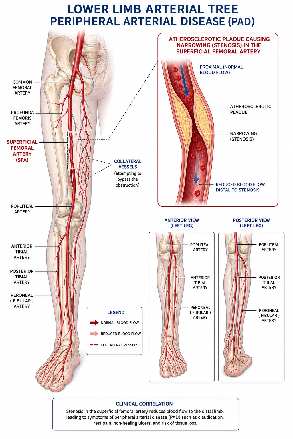
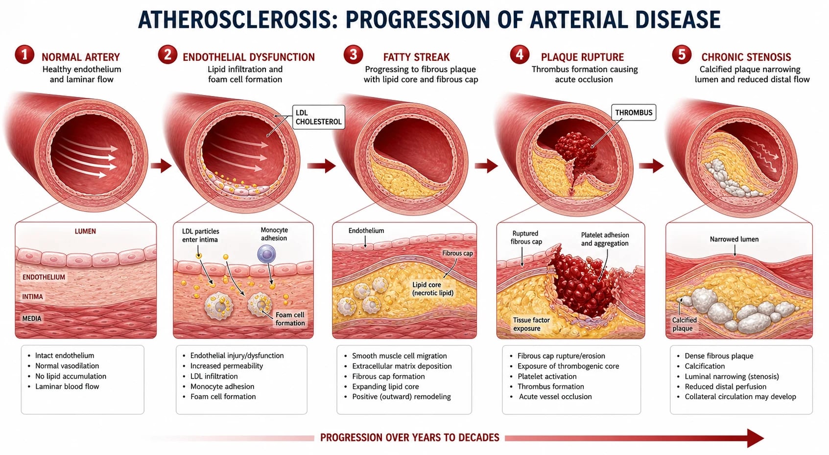
6 Ps — the clinical recognition. (1) PAIN: severe, constant, worse with elevation (gravity reduces flow). (2) PALLOR: pale/white (no blood flow) — later mottled/cyanotic (venous congestion or deoxygenated blood). (3) PULSELESSNESS: absent distal pulses (dorsalis pedis, posterior tibial). (4) PARAESTHESIA: numbness, tingling (nerve ischaemia — early sign). (5) PARALYSIS: muscle weakness (advanced — muscle ischaemia). (6) PERISHING COLD: limb cold to touch (no warm blood flow). (7) PROGRESSION: pain -> pallor -> pulseless -> paraesthesia -> paralysis -> perishing (worsening ischaemia). (8) LATE SIGNS (irreversible): fixed mottling, rigidity (muscle rigor), anaesthesia, paralysis -> necrosis (amputation). (9) ANY patient with sudden limb pain + absent pulses -> ALI until proven otherwise.[6] }Time-critical — 4-6 hours to irreversible necrosis. (1) MUSCLE is the most sensitive tissue to ischaemia: (a) Skeletal muscle: tolerates ~4-6 hours of complete ischaemia -> irreversible necrosis (rhabdomyolysis). (b) Nerve: ~4 hours -> irreversible. (c) Skin: ~8-12 hours (more tolerant). (d) Bone: long (>24 hours). (2) AFTER 6 HOURS of complete occlusion: muscle necrosis -> amputation likely (even with revascularisation — dead muscle can't recover). (3) THE WINDOW: revascularise within 6 hours for limb salvage. Beyond 6 hours -> increasing amputation risk. (4) WHY HEparin IMMEDIATELY: heparin prevents clot PROPAGATION (extends distally) + distal embolisation -> buys time (maintains some flow to distal tissue) -> extends the window. (5) DELAY (waiting for imaging/consultation without heparin) -> progressive ischaemia -> necrosis. Give heparin FIRST, then investigate.[2] }Embolic — AF is the classic cause. (1) AF (atrial fibrillation): (a) Stasis of blood in left atrial appendage -> thrombus forms. (b) Thrombus dislodges -> embolises -> lodges in peripheral artery (especially FEMORAL artery bifurcation — 'saddle embolus'). (c) AF causes ~70% of embolic ALI. (2) OTHER CARDIAC SOURCES: (a) Post-MI mural thrombus (LV apical thrombus — after large anterior MI). (b) Prosthetic valve (if subtherapeutic INR). (c) Endocarditis (vegetation embolises). (d) Dilated cardiomyopathy (stasis). (e) Atrial myxoma (rare tumour). (3) ARTERY-TO-ARTERY EMBOLI: (a) Aortic/iliac plaque rupture -> cholesterol embolises -> 'blue toe syndrome' (small distal emboli). (b) Aneurysm thrombus -> embolises. (4) CLINICAL CLUE: AF + sudden limb ischaemia + NORMAL contralateral pulses = embolic (classic). (5) MANAGEMENT: embolectomy (surgical) + anticoagulate AF (prevent recurrence).[2] }Thrombotic — on existing PAD. (1) MECHANISM: atherosclerotic plaque (in PAD) -> plaque rupture/erosion -> in-situ thrombosis -> acute occlusion. (2) RISK FACTORS: smoking, diabetes, hypertension, hyperlipidaemia, age, family history (same as PAD). (3) CLINICAL: (a) HISTORY of claudication (intermittent — calf pain on walking, relieved by rest). (b) Gradual worsening -> rest pain -> acute thrombosis -> acute limb ischaemia. (c) CONTRALATERAL limb often affected (bilateral PAD — absent pulses both sides). (d) Collaterals present (chronic occlusion -> body developed bypass vessels) -> less severe initially. (4) MANAGEMENT: (a) Thrombolysis (catheter-directed alteplase — dissolves clot). (b) ENDOVASCULAR (stent/atherectomy for stenosis). (c) SURGICAL BYPASS (graft around occlusion — for chronic disease). (d) LONG-TERM: antiplatelet (aspirin/clopidogrel), statin, risk factor modification, smoking cessation. (5) GRAFT THROMBOSIS: previous bypass graft -> thromboses -> ALI. Manage: thrombolysis or graft revision.[4] }Rutherford classification — guide urgency. (1) Category I (VIABLE): not immediately threatened. Sensory intact, no muscle weakness, Doppler audible. Can investigate + plan (not emergency). (2) Category IIa (MARGINALLY THREATENED): salvageable with URGENT revascularisation. Mild sensory loss, no muscle weakness, Doppler barely audible. Revascularise within hours. (3) Category IIb (IMMEDIATELY THREATENED): salvageable with IMMEDIATE revascularisation. Sensory loss + mild muscle weakness, Doppler inaudible. SURGICAL EMERGENCY — revascularise NOW. (4) Category III (IRREVERSIBLE): major permanent tissue loss (necrosis). Anaesthesia, paralysis, rigor, fixed mottling. Amputation (no salvage possible). (5) CLINICAL: categories IIb + III = immediate surgical attention. Categories I + IIa = urgent but some time. (6) DON'T misclassify — IIb (immediately threatened) vs III (irreversible) determines whether revascularisation is worth attempting.[6] }Fogarty embolectomy — the procedure for embolic. (1) PROCEDURE: (a) Arteriotomy (incision into artery — usually common femoral). (b) Fogarty catheter (balloon-tipped) passed DISTALLY (into the occluded segment). (c) Balloon INFLATED. (d) Catheter WITHDRAWN -> balloon pulls embolus out (like a plunger). (e) Repeat until no more clot. (f) Close arteriotomy (or patch). (2) ADVANTAGES: (a) FAST (minutes — immediate restoration of flow). (b) Effective for EMBOLUS (soft, gelatinous — easy to extract). (c) No thrombolytic needed (no bleeding risk). (3) LIMITATIONS: (a) May not remove all clot (distal fragments). (b) May damage intima (catheter trauma). (c) Not effective for THROMBOTIC (organised, adherent — harder to extract). (4) FOR EMBOLIC: Fogarty embolectomy is FIRST-LINE (fast, effective). (5) FOR THROMBOTIC: thrombolysis or bypass (Fogarty less effective).[3] }Thrombolysis — catheter-directed alteplase. (1) INDICATION: thrombotic ALI (on PAD/plaque), graft thrombosis, some embolic (if surgery not suitable). (2) PROCEDURE: (a) Arterial puncture (femoral). (b) Catheter advanced into clot (under fluoroscopy). (c) ALTEPLASE (tissue plasminogen activator — tPA) infused DIRECTLY into clot (laced + infusion over hours). (d) Monitor angiographically (clot dissolving). (e) May adjunct with mechanical thrombectomy. (3) ADVANTAGES: (a) Dissolves clot (no surgery). (b) Reveals underlying stenosis (can stent after). (c) Less invasive. (4) DISADVANTAGES: (a) SLOW (hours — not for immediately threatened limbs). (b) BLEEDING RISK (intracranial haemorrhage ~1-2%, other bleeding). (c) Contraindications (recent surgery, stroke, active bleeding). (5) TRIALS: Rochester, STILE, TOPAS — thrombolysis vs surgery -> EQUIVALENT outcomes (thrombolysis not superior; bleeding risk). (6) CURRENT: thrombolysis for SUBACUTE/thrombotic (where time allows); surgery for EMBOLIC/immediately threatened (fast).[3] }Reperfusion injury — myoglobinuria, hyperkalaemia, AKI. (1) After revascularisation, the ischaemic tissue is REPERFUSED -> releases toxic contents: (a) MYOGLOBIN (from muscle breakdown — rhabdomyolysis). (b) POTASSIUM (from cells — hyperkalaemia). (c) LACTATE (from anaerobic metabolism — acidosis). (d) INFLAMMATORY mediators (cytokines -> systemic inflammation). (2) CONSEQUENCES: (a) AKI (myoglobin nephrotoxicity — myoglobin precipitates in renal tubules + vasoconstriction). (b) HYPERKALAEMIA (arrhythmia — peaked T waves). (c) METABOLIC ACIDOSIS (lactate). (d) COMPARTMENT SYNDROME (reperfusion swelling -> raised compartment pressure -> recurrent ischaemia). (3) MANAGEMENT: (a) FLUIDS (aggressive crystalloid — maintain urine output >200 mL/hr -> flush myoglobin through kidneys -> prevent AKI). (b) BICARBONATE (alkalinise urine — pH >6.5 — myoglobin more soluble, less precipitated). (c) TREAT HYPERKALAEMIA (calcium gluconate for cardiac, insulin/dextrose, etc.). (d) RRT if severe AKI. (e) FASCIOTOMY if compartment syndrome. (4) MONITOR: CK (rising = ongoing rhabdo), K+, creatinine, urine (myoglobinuria — dark, positive blood on dipstick but few RBC), ECG.[5] }Compartment syndrome — fasciotomy. (1) MECHANISM: reperfusion -> muscle swelling (oedema) -> raised pressure in FASCIAL COMPARTMENTS (fixed volume) -> capillary occlusion -> ischaemia -> muscle + nerve necrosis. (2) CLINICAL (6 Ps of compartment syndrome — different from ALI): (a) PAIN out of proportion (severe, increasing — NOT relieved by analgesia). (b) PAIN on PASSIVE STRETCH (stretching the muscle in the compartment -> severe pain). (c) TENSE compartment (hard to palpation). (d) PARAESTHESIA (nerve ischaemia). (e) PARALYSIS (late). (f) PULSELESSNESS (very late — usually pulses present until late). (3) DIAGNOSIS: (a) CLINICAL (especially in awake patient). (b) COMPARTMENT PRESSURE: >30 mmHg (or delta pressure <30 mmHg — delta = diastolic BP - compartment pressure). (c) In sedated/unconscious: pressure measurement essential. (4) MANAGEMENT: FASCIOTOMY (immediate — release all compartments). (a) Leg: 4 compartments (anterior, lateral, superficial posterior, deep posterior) — via 2 incisions (medial + lateral). (b) Forearm: volar + dorsal fasciotomy. (c) LEAVE OPEN (delayed closure or skin graft once swelling resolves). (5) PROPHYLACTIC fasciotomy: at time of revascularisation if high risk (prolonged ischaemia >6h, severe ischaemia, combined arterial + venous). (6) DELAYED fasciotomy -> Volkmann's contracture (permanent muscle/nerve damage) or amputation.[5] }Heparin — immediate, unfractionated. (1) DOSE: unfractionated heparin 80 IU/kg IV bolus + 18 IU/kg/hr infusion (target APTT 1.5-2.5x control). (2) RATIONALE: (a) Prevents CLOT PROPAGATION (thrombus extends distally -> worse ischaemia). (b) Prevents DISTAL EMBOLISATION (fragments break off -> occlude smaller vessels). (c) Maintains patency of distal microcirculation (some flow maintained -> extends the 6h window). (d) Prevents recurrent embolisation (if AF — source). (3) TIMING: IMMEDIATELY on diagnosis (before imaging, before surgery) — don't delay. (4) MONITORING: APTT (q6h until stable, then daily). (5) CONTRAINDICATIONS (relative): active bleeding, recent surgery, heparin allergy (use alternative — argatroban, fondaparinux). (6) LMWH (low molecular weight heparin): less preferred for ALI (unfractionated preferred — faster reversal, titratable, shorter half-life for surgery). (7) CONTINUE post-revascularisation (then transition to oral — warfarin for AF, antiplatelet for PAD).[2] }Atheroembolism — 'blue toe syndrome.' (1) MECHANISM: aortic/iliac PLAQUE rupture -> cholesterol crystals embolise -> lodge in SMALL distal arteries (toes) -> microinfarcts. (2) CLINICAL: (a) BLUE/PURPLE TOES (cyanotic — small vessel occlusion). (b) Livedo reticularis (mottled skin — lace-like). (c) Painful toes (microinfarcts). (d) Preserved PULSES (large arteries patent — emboli are distal). (e) Renal failure (cholesterol emboli to kidneys -> AKI). (f) GI ischaemia (mesenteric emboli). (3) TRIGGERS: (a) Vascular procedure (catheterisation, angiography, surgery — plaque disrupted). (b) Spontaneous (plaque rupture). (c) Anticoagulation started (paradoxically — prevents clot on plaque but allows cholesterol to embolise). (4) DIAGNOSIS: (a) Clinical (blue toes + livedo + renal failure post-procedure). (b) Biopsy (cholesterol clefts in skin/kidney — definitive). (c) Eosinophilia (common — inflammatory response). (d) COMPLEMENT (low — consumed). (5) MANAGEMENT: (a) NO ANTICOAGULATION (doesn't help — emboli are cholesterol, not thrombus). (b) STATIN (stabilise plaque). (c) Antiplatelet. (d) AVOID further vascular procedures (may cause more emboli). (e) Surgical/endovascular exclusion of source (if identified — aortic stent/graft). (f) Supportive (renal, wound care). (6) PROGNOSIS: poor (recurrent emboli, renal failure, death — 50-80% mortality at 1 year).[4] }Venous gangrene — phlegmasia. (1) PHLEGMASIA ALBA DOLENS ('white leg'): (a) Massive ILIAC-FEMORAL DVT (deep venous thrombosis). (b) Severe venous congestion -> limb WHITE (pale — from massive oedema compressing arteries), swollen, painful. (c) Arterial pulses may be absent (compression). (d) EMERGENCY — thrombolysis or thrombectomy (prevent progression to cerulea). (2) PHLEGMASIA CERULEA DOLENS ('blue leg'): (a) Progression — severe ischaemia -> limb BLUE/CYANOTIC (venous + arterial compromise). (b) BLISTERING, necrosis (gangrene). (c) EMERGENCY — thrombolysis, thrombectomy, or fasciotomy (if compartment syndrome). (d) HIGH amputation + mortality. (3) DISTINGUISH from arterial ALI: (a) Phlegmasia: SWOLLEN limb (venous congestion), DVT history, bilateral risk factors. (b) Arterial ALI: PALE/cold limb (arterial occlusion), AF/PAD. (4) MANAGEMENT of phlegmasia: catheter-directed thrombolysis, surgical thrombectomy, anticoagulation.[2] }Post-revascularisation care — ICU considerations. (1) MONITOR: (a) LIMB perfusion: pulses, capillary refill, temperature, colour, sensation, motor (q1h initially). (b) COMPARTMENT syndrome: pain (on passive stretch), tense compartments, paraesthesia (if fasciotomy not done — measure pressure). (c) REPERFUSION: CK (rhabdo), K+ (hyperkalaemia), creatinine (AKI), urine (myoglobinuria). (d) ANTICOAGULATION: APTT (heparin) or INR (warfarin) — therapeutic. (e) BLEEDING (if thrombolysis used — especially intracranial). (2) COMPLICATIONS: (a) Reperfusion injury (rhabdo, AKI, hyperkalaemia — treat aggressively). (b) Compartment syndrome (fasciotomy if needed). (c) Re-occlusion (thrombosis — heparin, revascularise). (d) Infection (wound, line). (e) Cardiac (AF, MI — underlying cause). (3) ADDRESS CAUSE: (a) AF — anticoagulate (warfarin/DOAC), consider cardioversion. (b) PAD — antiplatelet, statin, risk factor modification (smoking, diabetes, lipids). (c) Endocarditis — antibiotics, valve surgery. (4) REHABILITATION: physiotherapy, mobility, prosthetic (if amputation). (5) FOLLOW-UP: vascular surveillance (graft patency, recurrent ischaemia).[2] }Outcomes and prognosis. (1) MORTALITY: 10-15% (30-day) — mostly from comorbidities (cardiac — AF, MI; not the limb itself). (2) AMPUTATION: 10-15% (if treated promptly); higher (>30%) if delayed >6-12h or irreversible (Rutherford III). (3) PREDICTORS of poor outcome: (a) DELAY to revascularisation (>6h -> more necrosis). (b) SEVERITY at presentation (Rutherford IIb/III -> worse). (c) COMORBIDITIES (diabetes, cardiac, renal). (d) CAUSE (embolic may have better outcome if embolectomy fast; thrombotic with PAD worse — diffuse disease). (4) RECURRENCE: (a) Embolic: if AF not anticoagulated -> recurrent emboli (10-20%/year without anticoagulation). (b) Thrombotic: if PAD not treated -> recurrent thrombosis. (5) PREVENTION: anticoagulate AF (reduce stroke + embolism), treat PAD (lifestyle, medication), smoking cessation. (6) LONG-TERM: many patients have underlying vascular disease (PAD, cardiac) -> need comprehensive vascular + cardiac care.[1] } Red flags
Critical acute limb ischaemia red flags
6 Ps : Pain, Pallor, Pulselessness, Paraesthesia, Paralysis, Perishing cold.[6] }Irreversible necrosis in 4-6 hours of complete occlusion — TIME-CRITICAL.[2] }IMMEDIATE heparin (unfractionated 80 IU/kg) — prevents propagation, buys time.[2] }Emergency revascularisation within 6 hours (embolectomy for embolic, thrombolysis/bypass for thrombotic).[3] }AF + sudden limb ischaemia = embolic (most common cause).[2] }Fasciotomy for reperfusion compartment syndrome (prolonged ischaemia, Rutherford IIb/III).[5] }Reperfusion injury : rhabdomyolysis (CK), hyperkalaemia, AKI (myoglobinuria) — fluids + bicarbonate.[5] }Phlegmasia cerulea dolens : massive DVT -> blue swollen limb -> thrombolysis/thrombectomy.[2] } Prognosis
Acute limb ischaemia evidence and outcomes Mortality : 10-15% (30-day) — mostly from comorbidities (cardiac).
Amputation : 10-15% (treated promptly); >30% if delayed or irreversible.
Rutherford classification : guides urgency (IIb/III = emergency).
Thrombolysis vs surgery (Rochester, STILE, TOPAS): equivalent outcomes — thrombolysis for subacute/thrombotic, surgery for embolic/immediately threatened.
Reperfusion injury : rhabdomyolysis, AKI, hyperkalaemia — aggressive fluids + bicarbonate.
Compartment syndrome : fasciotomy (prophylactic if high risk — prolonged ischaemia).
Recurrence : embolic (if AF not anticoagulated); thrombotic (if PAD not treated) — prevention essential.
Atheroembolism (blue toe): poor prognosis (50-80% mortality at 1 year — statin, avoid further procedures).
[1]
Classification and aetiology (expanded)
Figure Classification / severity strata that change management. Rutherford categories of acute limb ischaemia — urgency and action
[1]
Catheter-directed thrombolysis vs surgical embolectomy for ALI
[1]
Management of bypass graft occlusion
Figure Management ladder: first therapies, escalation, and failure criteria examiners expect. Acute bypass graft occlusion — stepwise management
RECOGNISE GRAFT THREAT — Patient with previous lower-limb bypass (autogenous vein or prosthetic) presents with acute limb ischaemia (new rest pain, pulse loss, dropped ABI <0.4). Graft occlusion may be the FIRST sign of a failing graft. Window to irreversible necrosis 4-6 h as for any ALI. Examine: limb ischaemia PLUS graft patency (palpate a subcutaneous graft), previous incisions, ankle-brachial index, and check the surveillance log for a previously identified stenosis.IMMEDIATE ANTICOAGULATION + ANALGESIA — IV unfractionated heparin 80 IU/kg bolus then 18 IU/kg/h (target APTT 1.5-2.5x control). Prevents propagation into run-off vessels and may partially restore flow through a residual lumen. Do NOT delay for imaging. Opioid analgesia; limb dependent; do not warm.IMAGING — DEFINE OCCLUSION AND RUN-OFF — CTA or on-table angiography. Identify: (a) SITE (anastomosis, mid-graft, inflow); (b) UNDERLYING CAUSE (anastomotic stenosis, intimal hyperplasia, graft degeneration, low flow from inflow/outflow disease); (c) DISTAL RUN-OFF quality (single-vessel run-off = poor prognosis). Duplex ultrasound is the first-line surveillance tool and confirms occlusion and any retained inflow.REVASCULARISATION STRATEGY — (a) CATHETER-DIRECTED THROMBOLYSIS: preferred for prosthetic grafts and recent (<14 d) thrombosis — restores patency and exposes the underlying stenosis for angioplasty/stent (STILE: thrombolysis superior for acute <14 d graft occlusion). (b) THROMBECTOMY (surgical or mechanical — AngioJet, Penumbra): for autogenous vein grafts and older/organised thrombus. (c) REVISION of the underlying lesion (patch angioplasty, jump graft, anastomotic revision). (d) Priority: Rutherford IIb = immediate theatre; I-IIa = urgent but planned.ADDRESS THE CAUSE — PREVENT RE-OCCLUSION — (a) Fix anastomotic/intimal-hyperplasia stenosis (commonest cause of late vein-graft failure). (b) Optimise INFLOW (iliac disease) and OUTFLOW. (c) Antiplatelet (aspirin +/- clopidogrel), high-intensity statin, smoking cessation, glycaemic control. (d) Surveillance programme: duplex US + ABI at 3, 6, 12 months then 6-monthly — detects the FAILING graft BEFORE occlusion (pre-emptive revision has far lower amputation than revision-after-occlusion). (e) Anticoagulation (warfarin/DOAC) in low-flow states (poor run-off, prosthetic below-knee graft).POST-REVASCULARISATION + FASCIOTOMY DECISION — As for any ALI: PROPHYLACTIC fasciotomy at revascularisation if ischaemia >4-6 h, combined arterial + venous injury, or Rutherford IIb/III. Monitor reperfusion (myoglobinuria, hyperkalaemia, AKI) and graft patency (duplex before discharge). Long term: structured graft surveillance, best medical therapy, risk-factor modification. [1]
Clinical pearls — advanced
High-yield advanced ALI pearls — aetiology, diagnosis, complications
Embolic causes — the cardiac sources. (1) ATRIAL FIBRILLATION is the single commonest cause of embolic ALI (~70% of cardiac emboli): thrombus forms in the LEFT ATRIAL APPENDAGE (stasis from non-contractile fibrillating atrium) -> dislodges -> embolises systemically -> most often lodges at ARTERIAL BIFURCATIONS (femoral bifurcation 'saddle embolus', then popliteal, then aortic bifurcation). (2) POST-MI MURAL THROMBUS: large anterior STEMI -> akinetic LV apex -> stasis -> mural thrombus on the wall -> embolises days-weeks post-MI. (3) PROSTHETIC VALVE: if subtherapeutic INR (mechanical mitral target INR ~2.5-3.5) -> thrombus on valve -> embolus. (4) INFECTIVE ENDOCARDITIS: friable vegetation (Strep viridans, Staph aureus) -> embolises, often with fever, new murmur, splinter haemorrhages, Osler nodes, microscopic haematuria. (5) DILATED CARDIOMYOPATHY: low ejection fraction -> stasis -> mural thrombus. (6) ATRIAL MYXOMA: rare left-atrial tumour -> systemic emboli + constitutional symptoms (fever, weight loss). WORK-UP: ECG (AF, prior MI), echocardiography (TTE for mural thrombus, wall-motion abnormality, vegetation; TOE better for LAA and prosthetic valves), blood cultures (endocarditis).[2] Atheroembolism / plaque rupture — artery-to-artery emboli ('blue toe syndrome'). (1) MECHANISM: rupture/ulceration of an atherosclerotic plaque (most often thoracoabdominal aorta or iliac arteries) -> cholesterol crystals + platelet-fibrin debris embolise -> lodge in SMALL DISTAL ARTERIES (digital arteries at the toes). (2) CLINICAL: 'BLUE TOE SYNDROME' — painful cyanotic cold toes with PRESERVED PULSES (large vessels patent; emboli distal), livedo reticularis (lace-like mottling on legs/flanks), digital gangrene, nodules; often multi-territory (toes + kidney + GI). (3) TRIGGERS: vascular procedures (cardiac catheterisation, angiography, IABP, vascular surgery — dislodges plaque), SPONTANEOUS plaque rupture, or paradoxically triggered by STARTING anticoagulation/thrombolysis (prevents stabilising clot, allows cholesterol shower). (4) ASSOCIATED: eosinophilia, hypocomplementaemia, AKI (cholesterol emboli to renal arteries days-weeks post-catheter). (5) DIAGNOSIS: clinical + skin biopsy (cholesterol clefts in dermal arterioles); echo to exclude cardiac source; CTA/MRA of aorta to identify culprit plaque. (6) MANAGEMENT: NO thrombolysis (worsens showering), NO further intravascular instrumentation if possible, high-intensity statin (stabilise plaque), antiplatelet, analgesia; surgical/endovascular exclusion (covered stent) of culprit plaque for refractory cases. (7) PROGNOSIS: poor — 1-year mortality 50-80%.[2] Thrombotic ALI — in-situ thrombosis on underlying PAD. (1) MECHANISM: a pre-existing atherosclerotic plaque ruptures/erodes (triggered by haemodynamic stress or inflammation) -> platelet aggregation -> thrombus -> acute occlusion. (2) DIFFERENCE FROM EMBOLIC: PRE-EXISTING COLLATERALS (stenosis developed over months-years), so the limb is less acutely threatened and the patient often has a history of claudication; CONTRALATERAL pulses frequently absent (bilateral PAD). (3) RISK FACTORS: smoking (most important), diabetes, hypertension, dyslipidaemia, age, family history, CKD. (4) HYPERCOAGULABLE STATES can precipitate thrombosis on a plaque: malignancy (Trousseau), polycythaemia/essential thrombocythaemia, antiphospholipid syndrome, heparin-induced thrombocytopenia (HIT — paradoxical thrombosis), nephrotic syndrome, homocystinuria. (5) MANAGEMENT: catheter-directed thrombolysis (preferred — dissolves clot, exposes underlying stenosis for angioplasty/stent) or surgical bypass if diffuse disease; long-term antiplatelet + statin + risk-factor modification. Differentiating thrombotic from embolic matters because the pathway diverges (thrombolysis/bypass vs embolectomy).[4] Bypass graft occlusion — failing vs failed graft. (1) AUTOGENOUS VEIN GRAFTS fail most often from INTIMAL HYPERPLASIA at the anastomoses (months-years) -> stenosis -> low flow -> thrombosis. (2) PROSTHETIC GRAFTS (PTFE/Dacron) fail from anastomotic stenosis, pseudointimal hyperplasia, or graft infection. (3) FAILING graft (stenosed but patent) — DOPPLER VELOCITY CHANGES on surveillance duplex (peak systolic velocity ratio >2-3 across a stenosis, drop in ABI >0.15) — INTERVENE BEFORE OCCLUSION (pre-emptive angioplasty/revision has a high limb-salvage rate). (4) FAILED graft (thrombosed) presents as ALI and outcome is worse than elective revision. (5) MANAGEMENT: catheter-directed thrombolysis for acute (<14 d) graft occlusion (STILE — better amputation-free survival in the acute group); surgical thrombectomy + revision for vein grafts; treat the underlying stenosis. (6) PREVENTION: structured duplex SURVEILLANCE (3, 6, 12, 18, 24 months), antiplatelet, statin, smoking cessation; anticoagulation in high-risk (prosthetic below-knee, poor run-off).[7] Diagnosis — handheld Doppler and ankle-brachial index. (1) HANDHELD DOPPLER is the first bedside test: audible arterial signal — TRIPHASIC = normal; BIPHASIC = mild disease; MONOPHASIC = severely diseased; ABSENT = occlusion. A venous Doppler signal (compressible, continuous) confirms venous patency — important for Rutherford grading (venous signal preserved in I-IIb, lost in III). (2) ANKLE-BRACHIAL INDEX (ABI = ankle systolic BP / brachial systolic BP): normal 0.9-1.3; <0.9 = PAD; <0.4 = limb-threatening; >1.3 = falsely elevated (medial calcinosis in diabetes/CKD — use toe-brachial index instead). In ALI the ABI is often <0.4. (3) Doppler cannot LOCALISE the lesion — imaging is still required. (4) In the ED, if there is NO arterial signal and the clinical picture fits, treat as ALI and give heparin without waiting for imaging.[2] CT angiography — imaging modality of choice. (1) WHY CTA: fast, widely available, identifies site/length/cause of occlusion, evaluates inflow and outflow vessels, and plans intervention; sensitivity/specificity >95% for significant arterial disease. (2) DISTINGUISH embolic vs thrombotic: (a) EMBOLIC — ABRUPT cut-off ('meniscus sign', 'rat-tail'), NO collaterals, normal proximal and contralateral vessels, occlusion at a BIFURCATION; (b) THROMBOTIC — TAPERED/irregular occlusion, diffuse atherosclerotic disease, COLLATERAL vessels (chronic), contralateral limb disease. (3) CAUTION with contrast in CKD/diabetes (contrast nephropathy) — consider MRA (gadolinium), CO2 angiography, or duplex first. (4) ON-TABLE angiography if diagnosis is certain and immediate intervention is planned. (5) Intravascular ultrasound (IVUS) is a useful adjunct in complex lesions and stent sizing.[2] Catheter-directed thrombolysis — urokinase vs rt-PA. (1) AGENTS: rt-PA (alteplase) — now most commonly used; urokinase — production stopped in the US in 1999 but still available elsewhere; reteplase/tenecteplase off-label. (2) TECHNIQUE: catheter embedded in the clot (multi-side-hole) -> lacing bolus -> infusion (rt-PA 0.5-1 mg/h) -> repeat angiography q4-12 h -> adjust. Concomitant low-dose heparin (~300-500 IU/h via sheath) prevents pericatheter thrombosis. (3) ENDPOINT: complete lysis (restored flow), partial lysis with underlying lesion identified (then angioplasty/stent), or failure (proceed to surgery). (4) STILE/TOPAS — thrombolysis comparable to surgery overall, BETTER for subacute (<14 d) and graft occlusion, WORSE for chronic (>14 d) and severe (IIb) ischaemia. (5) MONITOR: fibrinogen (drop >50% or <1 g/L predicts bleeding — reduce/stop), Hb, neuro obs (ICH), puncture sites. (6) DURATION: reassess at 24-48 h maximum.[9] Thrombolysis — absolute and relative contraindications. (1) ABSOLUTE: active internal bleeding; recent CVA/neurosurgery/intracranial trauma (within 2-3 months); intracranial neoplasm/aneurysm/AVM; recent (within 2 weeks) major surgery/organ biopsy/puncture of a non-compressible vessel; severe uncontrolled hypertension (SBP >180 or DBP >110); recent GI bleed (within 10 days); known bleeding diathesis. (2) RELATIVE: pregnancy, recent CPR, diabetic haemorrhagic retinopathy, bacterial endocarditis, hepatic failure, age >75 (higher bleeding risk but not a contraindication). (3) If contraindicated, surgical thrombectomy/embolectomy is the alternative. (4) COMPLICATIONS: distal embolisation (clot fragments — 'trash foot'), pericatheter thrombosis, bleeding (puncture site, retroperitoneal, intracranial — most feared, ~1-2%), reperfusion syndrome. (5) STILE bleeding: 5-12% major; fibrinogen depletion predicted haemorrhage.[7] Compartment syndrome post-revascularisation — pathophysiology and recognition. (1) MECHANISM: ischaemia -> capillary leak + Na/K pump failure -> muscle and interstitial OEDEMA -> pressure rises in the fixed-volume fascial compartment -> when compartment pressure exceeds capillary perfusion pressure (~30 mmHg absolute, or within 30 mmHg of diastolic = delta-P <30 mmHg) -> microvascular occlusion -> secondary ISCHAEMIA -> necrosis. (2) RISK FACTORS: prolonged ischaemia (>4-6 h), severe ischaemia (Rutherford IIb/III), combined arterial + venous injury, crush, large clot burden, concomitant venous occlusion. (3) CLINICAL — the 'Ps of compartment syndrome' (DIFFERENT from the 6 Ps of ALI): PAIN out of proportion and PAIN ON PASSIVE STRETCH (earliest, most sensitive), pain requiring escalating analgesia, TENSE/swollen compartment, PARAESTHESIA in the nerve distribution, PARALYSIS (late), PULSELESSNESS (very late — by then damage is irreversible). (4) DIAGNOSIS: clinical in an awake cooperative patient, or COMPARTMENT PRESSURE measurement (Stryker needle/slit catheter): absolute >30 mmHg OR delta pressure (diastolic minus compartment) <30 mmHg. (5) TIME-CRITICAL: fasciotomy within 6 h of onset for best outcomes — irreversible muscle necrosis after 6-8 h of compartment ischaemia.[5] Fasciotomy — technique and compartments. (1) INDICATIONS: (a) therapeutic — established compartment syndrome; (b) PROPHYLACTIC at the time of revascularisation in high-risk cases (prolonged ischaemia, Rutherford IIb/III, combined arterial + venous injury, crush). (2) The LEG has FOUR compartments: anterior (tibialis anterior, EDL, EHL, deep peroneal nerve), lateral (peroneus longus/brevis, superficial peroneal nerve), superficial posterior (gastrocnemius, soleus, sural nerve), deep posterior (tibialis posterior, FHL, FDL, tibial nerve, posterior tibial artery). ALL FOUR must be released. (3) TWO-INCISION technique (standard): (a) a LONG ANTEROLATERAL incision (midway between tibia and fibula) opens the anterior + lateral compartments; (b) a LONG POSTEROMEDIAL incision (2 cm behind the posteromedial border of the tibia) opens the superficial + deep posterior compartments. (4) Single-incision (fibula-excision) technique is rarely used. (5) FOOT: 9 compartments (dorsal, interossei x4, adductor, plantar x3) — fasciotomy if the foot is ischaemic/crushed. (6) POST: leave wounds OPEN (delayed primary closure or skin grafting once swelling settles, typically 5-10 days), splint in dorsiflexion, monitor for re-accumulation. (7) COMPLICATIONS: infection, chronic venous insufficiency, nerve injury (common peroneal), poor cosmesis, recurrent compartment syndrome.[5] Reperfusion injury — the systemic consequences. (1) WHAT IS RELEASED on reperfusion of ischaemic muscle: MYOGLOBIN (rhabdomyolysis), POTASSIUM (hyperkalaemia — peaked T waves, arrhythmia), LACTATE (metabolic acidosis), PHOSPHATE, MAGNESIUM, CYTOKINES (systemic inflammatory response), and platelet/thrombotic debris (pulmonary microembolism). (2) MYOGLOBINURIA: urine dark/'tea-coloured', dipstick positive for blood but FEW red cells (the 'blood' is myoglobin, not RBCs); confirmed by serum myoglobin and CK (often in tens of thousands). (3) AKI: myoglobin precipitates in distal tubules + renal vasoconstriction + acidosis-induced tubular injury. (4) MANAGEMENT: aggressive ISOTONIC crystalloid (target urine output 200-300 mL/h until CK falling and urine clear), ALKALINISE urine (sodium bicarbonate — keep urine pH >6.5 — enhances myoglobin solubility; evidence mixed), treat HYPERKALAEMIA (insulin-dextrose, calcium gluconate for membrane stabilisation), mannitol (osmotic diuresis + free-radical scavenger — controversial), avoid nephrotoxins, RRT if refractory. (5) CALCIUM: early HYPOcalcaemia (calcium precipitates in damaged muscle) — avoid calcium unless there is hyperkalaemia with ECG changes (calcium will be released later causing rebound hypercalcaemia). (6) MONITOR: CK q6-12 h, K+, creatinine, urine output, ECG. (7) Massive reperfusion can cause cardiac arrest from hyperkalaemia — anticipate and treat pre-emptively.[5] Heparin-induced thrombocytopenia (HIT) — paradoxical thrombosis in ALI. (1) The ALI patient on heparin can develop HIT: IgG anti-PF4 -> platelet activation -> THROMBOSIS (not bleeding) — paradoxical worsening of ischaemia or new thrombosis. (2) TIMING: platelet count falls 5-10 days after starting heparin (or sooner if sensitised within the previous 100 days). Use the 4Ts score (Thrombocytopenia, Timing, Thrombosis, oTher cause). (3) SUSPECT in any ALI patient on heparin whose platelets fall >50% or who develops new/worsening thrombosis. (4) ACTION: STOP ALL HEPARIN (including LMWH, line flushes, heparin-coated catheters), send anti-PF4 antibody (ELISA) and serotonin-release assay (confirmatory), START a DIRECT-ACTING anticoagulant (argatroban — hepatically cleared, preferred in renal failure; bivalirudin; danaparoid; fondaparinux). DO NOT start warfarin alone (warfarin in HIT causes skin necrosis and venous-limb gangrene from protein C depletion) — only after platelets recover and on top of a direct thrombin inhibitor. (5) AVOID platelet transfusions — they fuel thrombosis.[2] Upper limb acute limb ischaemia — subclavian/brachial. (1) LESS COMMON than lower-limb ALI (~10-20%) but the embolic proportion is HIGHER (more emboli reach the upper limb). (2) CAUSES: cardiac embolus (AF — commonest), SUBCLAVIAN/INNOMINATE aneurysm or atherosclerosis, THORACIC OUTLET SYNDROME (cervical rib compresses subclavian -> post-stenotic dilatation/aneurysm -> embolus to hand), trauma (iatrogenic — radial catheterisation, arterial line), Buerger disease, paradoxical embolus via a PFO. (3) CLINICAL: cold pale painful arm, absent brachial/radial/ulnar pulses, paraesthesia of the hand, grip weakness; BLUE DIGIT syndrome (digital ischaemia) from microemboli. (4) 'WHITE HAND' = severe ischaemia (Rutherford IIb) — emergency. (5) MANAGEMENT: heparin immediately; CTA from aortic arch to fingers; brachial embolectomy for embolic occlusion; thrombolysis for thrombotic; cervical rib resection +/- vascular reconstruction for thoracic outlet syndrome with subclavian aneurysm.[2] Popliteal artery aneurysm thrombosis — a specific high-stakes scenario. (1) Popliteal artery aneurysm is the COMMONEST peripheral aneurysm (~70% of peripheral aneurysms); usually in older men, ~50% of whom have a concomitant AAA. (2) COMPLICATIONS: THROMBOSIS or DISTAL EMBOLISATION (the most feared — chronic embolisation occludes the run-off vessels BEFORE the aneurysm thromboses -> catastrophic ALI with poor run-off and a HIGH amputation rate ~10-25% even after repair). (3) ACUTE THROMBOSIS of a popliteal aneurysm is a SURGICAL EMERGENCY — pre-emptive elective repair (when asymptomatic) is intended to prevent exactly this. (4) MANAGEMENT of acute thrombosis: catheter-directed thrombolysis FIRST (to restore run-off by lysing embolised distal clot), then surgical BYPASS (saphenous vein) with EXCLUSION of the aneurysm (proximal and distal ligation). (5) ALL patients with a popliteal aneurysm should be SCREENED for AAA.[2] Amputation — when, level, and decision-making. (1) INDICATIONS: (a) Rutherford III (irreversible necrosis — fixed mottling, muscle rigor, anaesthesia, paralysis, inaudible venous Doppler); (b) overwhelming infection (gas gangrene — Clostridium; necrotising fasciitis); (c) failed revascularisation with a non-viable limb; (d) patient not fit for, or not consenting to, revascularisation. (2) LEVEL: aim for the MOST DISTAL viable level that will heal — judged by clinical viability (skin/soft tissue, line of demarcation), palpable flow at the level, skin perfusion (transcutaneous oxygen tension >40 mmHg, dermal bleeding at the incision), and vascular imaging. BELOW-KNEE (trans-tibial) is preferable to above-knee (trans-femoral) — preserves the knee for far better prosthetic function and rehabilitation; knee disarticulation if there is no viable calf but the knee joint is intact. (3) GUILLOTINE (open) amputation first if there is infection/sepsis, with revision later. (4) POST-OP: rigid dressing/cast, early mobilisation, prosthetic fitting (within 30 days for below-knee improves outcome), phantom-limb pain management (gabapentin, mirror therapy), rehabilitation, risk-factor modification. (5) MORTALITY after major amputation is high (~15-30% at 1 year), reflecting comorbidity.[1] Anticoagulation and long-term prevention. (1) AFTER EMBOLIC ALI: long-term anticoagulation (warfarin or DOAC) for the underlying source — AF, mural thrombus, prosthetic valve, severe LV dysfunction. Target INR per valve/indication. DOACs equivalent to warfarin for non-valvular AF. Duration: indefinitely (or until the source resolves, e.g. after successful cardioversion with documented LAA thrombus resolution). (2) AFTER THROMBOTIC ALI / PAD: antiplatelet (aspirin 75-100 mg OR clopidogrel 75 mg) — single antiplatelet for stable PAD; dual antiplatelet sometimes short-term after intervention. Adding full-dose anticoagulation to antiplatelet therapy in atherosclerotic PAD ADDS bleeding without clear benefit (WAVE trial — no benefit, more bleeding), EXCEPT in selected cases: poor run-off, prosthetic bypass graft (where warfarin + antiplatelet improves patency). (3) LOW-DOSE RIVAROXABAN 2.5 mg BD + aspirin (COMPASS) reduces major adverse cardiac and limb events in stable PAD. (4) High-intensity STATIN (atorvastatin 80 mg) — reduces mortality and major adverse limb events. (5) SMOKING CESSATION is the single most important modifiable risk factor. (6) GLYCAEMIC and BP control, structured exercise, foot care. (7) GRAFT SURVEILLANCE — duplex at 3, 6, 12 months then 6-monthly.[4] Red flags — advanced
ALI — advanced red flags and pitfalls
Rutherford IIb and III are surgical emergencies — revascularise IMMEDIATELY; do not delay for imaging if IIb.No collaterals + abrupt onset + AF = embolic — Fogarty embolectomy (not thrombolysis).Diffuse PAD + collateral vessels + gradual worsening = thrombotic — thrombolysis or surgical bypass.Reperfusion after prolonged ischaemia — anticipate hyperkalaemia, myoglobinuria, AKI, and compartment syndrome; check CK, K+, and urine output.Compartment pressure >30 mmHg OR delta pressure (DBP - compartment) <30 mmHg — emergency fasciotomy.HIT: platelets fall >50% on heparin with new/worsening thrombosis — stop ALL heparin, start argatroban; do NOT use warfarin alone and do NOT transfuse platelets.Blue toe + preserved pulses + recent catheterisation = atheroembolism — NO thrombolysis, NO further intravascular procedures; high-intensity statin.Popliteal aneurysm thrombosis — high amputation risk; thrombolyse first to restore run-off, then bypass + exclusion; screen for AAA.Phlegmasia cerulea dolens (massive iliofemoral DVT, blue swollen limb) — thrombolysis/thrombectomy +/- fasciotomy.Prosthetic valve + subtherapeutic INR + ALI — valve thrombosis vs embolus; TOE; anticoagulate; surgery for obstructive valve thrombosis.Failed graft = ischaemia in a patient with previous bypass — thrombolyse acute occlusion, revise the underlying stenosis, restart surveillance. [1]
Key trials
STILE trial (1994) — surgery vs thrombolysis for limb ischaemia Design : RCT, 393 patients with native arterial or bypass-graft occlusion of the lower limb, randomised to optimal surgery vs catheter-directed thrombolysis (rt-PA or urokinase). Randomisation stopped early at the first interim analysis.[7]
Result : Overall, surgery was better at 30 days for the composite endpoint (death, ongoing/recurrent ischaemia, amputation, major morbidity) — driven by less recurrent ischaemia; clinical outcomes were similar. For SUBACUTE (<14 d) ischaemia, thrombolysis gave LOWER amputation rates and shorter hospital stay. For CHRONIC (>14 d) ischaemia, surgery was better.
Key message : Thrombolysis for ACUTE (<14 d) occlusions (especially graft); surgery for CHRONIC (>14 d) and for severe ischaemia. No difference between rt-PA and urokinase. Bleeding correlated with fibrinogen depletion.
TOPAS II trial (1998, NEJM) — urokinase vs surgery for arterial occlusion Design : RCT, 544 patients with acute (<14 d) arterial occlusion of the lower limb, randomised to recombinant urokinase vs vascular surgery.[8]
Result : Amputation-free survival at 6 months was SIMILAR (~72% urokinase vs ~75% surgery, NS). Major haemorrhage was more common with urokinase (~13% vs ~6%). Urokinase permitted a less invasive surgical procedure in a substantial proportion.
Key message : Thrombolysis is a reasonable first strategy for acute (<14 d) arterial occlusion — equivalent limb salvage to surgery with less invasive downstream procedures; bleeding is the trade-off.
Rochester study (1990, Radiology) — rt-PA vs urokinase Design : Randomised multicentre trial of recombinant tissue plasminogen activator (rt-PA) vs urokinase for peripheral arterial and graft occlusions.[9]
Result : Comparable lysis success and patency; rt-PA gave faster lysis; bleeding rates were comparable. Established rt-PA as an acceptable alternative to urokinase for catheter-directed peripheral thrombolysis.
Key message : rt-PA and urokinase are equivalent for limb-salvage thrombolysis; rt-PA is now standard (urokinase availability is limited in many markets).
Examiner densify anchors
CICM/FFICM densify — Acute limb ischaemia — embolic vs thrombotic
Exam answers must couple definition + threshold numbers + first therapies + what kills the patient . Cite landmark evidence and state the common wrong answer explicitly.[1]
Define the syndrome in one line → classify severity with a score or stage → resuscitate ABC → specific therapy with numbers → prevent the killer complication → prognosticate and disposition (ward vs HDU vs specialty centre).[2]
Figure Acute limb ischaemia — embolic vs thrombotic — core mechanism anchors for CICM/FFICM written and viva. Exam board focus
CICM Second Part · FFICM · EDIC
Killers to name
Airway loss, refractory shock, missed specific antidote/device, delayed specialty call
Documentation
Thresholds used, therapies with times, family update, disposition
[1]
Practical ICU checklist (densify)
Bedside densify checklist
Confirm diagnosis thresholds with numbers the examiner expects.Name the first therapy and the absolute contraindication.State monitoring frequency and escalation triggers.Cite one landmark paper/guideline and one limitation of the evidence.Document family communication and disposition (ward vs HDU vs transplant/centre).Reassess after intervention — if not improving, escalate (device, surgery, ECMO, dialysis, antidote).Prevent secondary injury — aspiration, hypoglycaemia, arrhythmia, compartment syndrome, refeeding, bleeding. [1]
If you forget detail, still structure: define → classify → resuscitate → specific therapy → prevent the killer complication → prognosticate .
[1]
Do not delay ABC for a perfect diagnosis.
Do not give therapies that are contraindicated in the look-alike (e.g. charcoal in caustics; beta-blocker in cocaine; fluids in SCAPE).
Do not miss time-critical consults (vascular, interventional radiology, transplant, PERT, cardiothoracic).
Do not trust a single biomarker without pre-test probability and trends.[1]
Extended fellowship notes (densify)
Carry at least three hard numbers (threshold, dose, or time window) and one absolute do-not-do . Vague prose without numbers fails the densified SAQ standard.[3]
Common exam traps vs correct anchors
[1]
Densify SAQ — Acute limb ischaemia — embolic vs thrombotic 10 minutes · 10 marks
Hide all A CICM/FFICM examiner asks you to manage this presentation at 03:00 in a regional ICU. Structure your answer.
[1]
Landmark themes for this leaf should be recalled as trial/guideline name → population → intervention → outcome → ICU limitation . Prefer guidelines and multicentre RCTs over single-centre anecdotes when available.[1] [2]
Line-fill densify notes
Densify anchor 1
Threshold, therapy, monitoring, or disposition point 1 for viva structure.
Densify anchor 2
Threshold, therapy, monitoring, or disposition point 2 for viva structure.
Densify anchor 3
Threshold, therapy, monitoring, or disposition point 3 for viva structure.
Densify anchor 4
Threshold, therapy, monitoring, or disposition point 4 for viva structure.
Densify anchor 5
Threshold, therapy, monitoring, or disposition point 5 for viva structure.
Densify anchor 6
Threshold, therapy, monitoring, or disposition point 6 for viva structure.
Densify anchor 7
Threshold, therapy, monitoring, or disposition point 7 for viva structure.
Densify anchor 8
Threshold, therapy, monitoring, or disposition point 8 for viva structure.
Densify anchor 9
Threshold, therapy, monitoring, or disposition point 9 for viva structure.
Densify anchor 10
Threshold, therapy, monitoring, or disposition point 10 for viva structure.
Densify anchor 11
Threshold, therapy, monitoring, or disposition point 11 for viva structure.
Densify anchor 12
Threshold, therapy, monitoring, or disposition point 12 for viva structure.
Densify anchor 13
Threshold, therapy, monitoring, or disposition point 13 for viva structure.
Densify anchor 14
Threshold, therapy, monitoring, or disposition point 14 for viva structure.
Densify anchor 15
Threshold, therapy, monitoring, or disposition point 15 for viva structure.
Densify anchor 16
Threshold, therapy, monitoring, or disposition point 16 for viva structure.
Densify anchor 17
Threshold, therapy, monitoring, or disposition point 17 for viva structure.
Densify anchor 18
Threshold, therapy, monitoring, or disposition point 18 for viva structure.
Densify anchor 19
Threshold, therapy, monitoring, or disposition point 19 for viva structure.
Densify anchor 20
Threshold, therapy, monitoring, or disposition point 20 for viva structure.
Densify anchor 21
Threshold, therapy, monitoring, or disposition point 21 for viva structure.
Densify anchor 22
Threshold, therapy, monitoring, or disposition point 22 for viva structure.
Densify anchor 23
Threshold, therapy, monitoring, or disposition point 23 for viva structure.
Densify anchor 24
Threshold, therapy, monitoring, or disposition point 24 for viva structure.
Densify anchor 25
Threshold, therapy, monitoring, or disposition point 25 for viva structure.
Densify anchor 26
Threshold, therapy, monitoring, or disposition point 26 for viva structure.
Densify anchor 27
Threshold, therapy, monitoring, or disposition point 27 for viva structure.
Densify anchor 28
Threshold, therapy, monitoring, or disposition point 28 for viva structure.
Densify anchor 29
Threshold, therapy, monitoring, or disposition point 29 for viva structure.
Densify anchor 30
Threshold, therapy, monitoring, or disposition point 30 for viva structure.
Densify anchor 31
Threshold, therapy, monitoring, or disposition point 31 for viva structure.
Densify anchor 32
Threshold, therapy, monitoring, or disposition point 32 for viva structure.
Densify anchor 33
Threshold, therapy, monitoring, or disposition point 33 for viva structure.
[1]
Densify complete
Leaf meets ≥350-line fellowship densify floor.
References [1] Conte MS, et al. Global vascular guidelines on the management of chronic limb-threatening ischemia. Journal of vascular surgery , 2019.PMID 31159978 [2] Bjorck M, et al. Editor's Choice - European Society for Vascular Surgery (ESVS) 2020 Clinical Practice Guidelines on the Management of Acute Limb Ischaemia. European journal of vascular and endovascular surgery , 2020.PMID 31899099 [3] Ouriel K, et al. Endovascular techniques in the treatment of acute limb ischemia: thrombolysis and mechanical thrombectomy. Seminars in vascular surgery , 2003.PMID 14691769 [4] Norgren L, et al. Inter-Society Consensus for the Management of Peripheral Arterial Disease (TASC II). Journal of vascular surgery , 2007.PMID 17223489 [5] Stella M, et al. Aetiology of trauma-related acute compartment syndrome of the leg: a systematic review. Injury , 2019.PMID 30772051 [6] Rutherford RB, et al. Recommended standards for reports dealing with lower extremity ischemia: revised version. Journal of vascular surgery , 1997.PMID 9308598 [7] The STILE Investigators Results of a prospective randomized trial evaluating surgery versus thrombolysis for ischaemia of the lower extremity (STILE trial). Annals of surgery , 1994.PMID 8092895 [8] Ouriel K, Veith FJ, Sasahara AA A comparison of recombinant urokinase with vascular surgery as initial treatment for acute arterial occlusion of the legs (TOPAS II). The New England journal of medicine , 1998.PMID 9545358 [9] Meyerovitz JF, et al. Recombinant tissue-type plasminogen activator versus urokinase in peripheral arterial and graft occlusions (Rochester). Radiology , 1990.PMID 2107563