ICU · Airway management
Awake Fibre-Optic Intubation
Also known as Awake fibre-optic intubation · AFOI · Awake intubation · Fibre-optic intubation · Topical airway anaesthesia · Spray-as-you-go · Cooperative sedation · Dexmedetomidine for intubation · Local anaesthetic airway · Superior laryngeal nerve block · Glossopharyngeal nerve block · Anticipated difficult airway
Awake fibre-optic intubation (AFOI) is the safest approach to the anticipated difficult airway when the patient can cooperate and maintain oxygenation: topicalise the upper airway with local anaesthetic, sedate lightly so the patient stays breathing with airway reflexes preserved, navigate a flexible scope through the cords on inspiration, and railroad an endotracheal tube. The principle is that the patient never loses their airway. Indications are a predicted difficult airway (prior difficulty, cervical instability, limited mouth opening, distorted anatomy, head-and-neck mass, obesity, risk of 'can't intubate, can't ventilate'); contraindications are inability to cooperate, inability to tolerate apnoea, complete upper-airway obstruction, an airway flooded with blood or secretions, and local-anaesthetic allergy. Technique: topicalisation with lidocaine 1–4% (spray, nebulised, gargle, paste, or spray-as-you-go), optional nerve blocks (superior laryngeal and glossopharyngeal), cooperative sedation with dexmedetomidine or remifentanil target-controlled infusion, scope handling, and railroading of the ETT over the scope. Complications include laryngospasm, local-anaesthetic systemic toxicity (LAST), and airway trauma.
On this page & tools
Your progress
Saved locally on this device.
Target exams
Overview & definition
Awake fibre-optic intubation (AFOI) is intubation of a conscious, spontaneously breathing patient whose upper airway has been topicalised with local anaesthetic, lightly sedated, using a flexible fibre-optic scope to guide the tube through the cords. Its great advantage is that the patient never loses their airway — they keep breathing and maintain their own airway tone throughout — which makes it the safest approach to the anticipated difficult airway.[1][1]
It is the opposite of rapid sequence intubation: instead of rendering the patient unconscious and apnoeic (and risking the loss of the airway in a known-difficult patient), AFOI keeps the patient cooperative and oxygenating while the airway is secured under vision.[1]
Key terminology for the viva. 'Awake' is a misnomer — the patient is cooperatively sedated, not alert. 'Fibre-optic' strictly refers to the flexible fibrescope, but the term now embraces flexible video-scopes (single-use and reusable) that are the modern standard. The unifying principle is a flexible endoscope, navigated under vision through a topicalised upper airway in a breathing patient, and an endotracheal tube railroaded over it.[1][6]
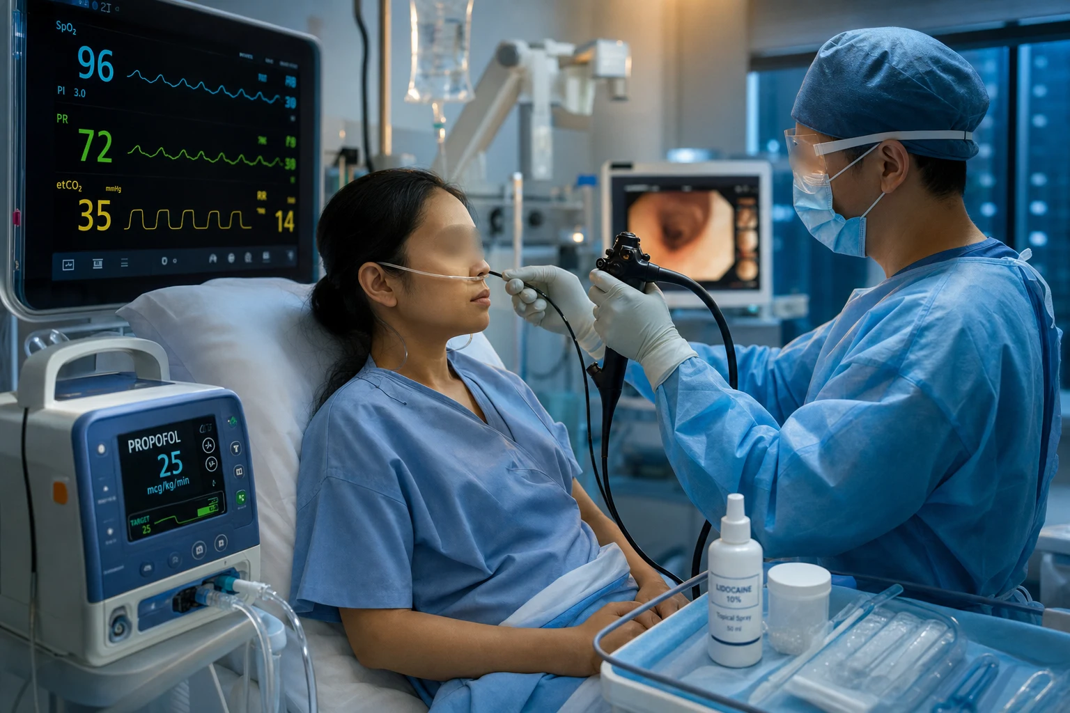
Indications
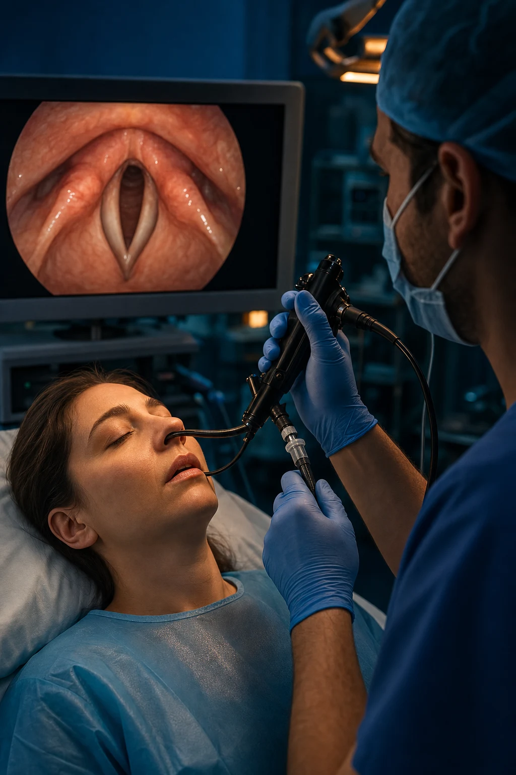
AFOI is for the anticipated difficult airway in a patient who can cooperate and oxygenate:[1][1][1]
- A history of difficult intubation or predicted difficulty (poor LEMON/MACOCHA).
- Cervical-spine instability (in-line stabilisation makes laryngoscopy difficult; AFOI via the nose avoids neck movement).
- Morbid obesity and the obstructed/blocked airway.
- Head-and-neck mass, distorted anatomy, or a compromised airway where inducing anaesthesia would precipitate "can't intubate, can't ventilate".
- Risk of regurgitation (the awake patient protects their own airway).[1]
The unifying decision rule is: predict a difficult airway AND the patient can cooperate and oxygenate → AFOI is the safest plan. The DAS 2018 ICU guideline places awake intubation as a first-line option for the predicted-difficult critically ill airway, and the ASA 2022 guideline recommends it whenever there is a known or predicted problem with facemask ventilation, supraglottic-airway ventilation, or intubation.[1][2]
| [{"header":"Indication"},{"header":"Why standard RSI is dangerous"},{"header":"Why AFOI is safer"}] |
|---|
Obstetric anticipated difficult airway deserves a special mention: physiological changes of pregnancy (oedema, friability, breast engorgement, rapid desaturation) plus an urgent surgical context make awake intubation a recommended default in many high-risk cases where the airway looks bad on assessment.[5]
Pediatric AFOI is feasible in selected cooperative older children but is rarely the ICU tool it is in adults; paediatric anticipated difficulty more often defaults to inhalational induction by a senior paediatric anaesthetist. [1]
Contraindications
- Inability to cooperate (agitated, confused, intoxicated, or paediatric patient).
- Inability to tolerate apnoea or maintain oxygenation (the critically hypoxaemic patient may not withstand the time taken).
- An airway flooded with blood or copious secretions (the scope cannot see).
- Allergy to the local anaesthetic.
- Patient refusal.[1]
For the deteriorating upper-airway obstruction with stridor at rest, AFOI is contentious: some centres attempt it in a prepared theatre with a surgical team scrubbed, but if the patient cannot lie flat or maintain oxygenation, an awake or primary surgical airway is safer.[1][1]
| [{"header":"Contraindication"},{"header":"Type"},{"header":"Reason"},{"header":"Alternative"}] |
|---|
A useful framing for the viva: AFOI is for the patient who is breathing, oxygenating, and cooperative; it is the wrong tool for the patient who is apnoeic, periarrest, or thrashing. [1]
The technique
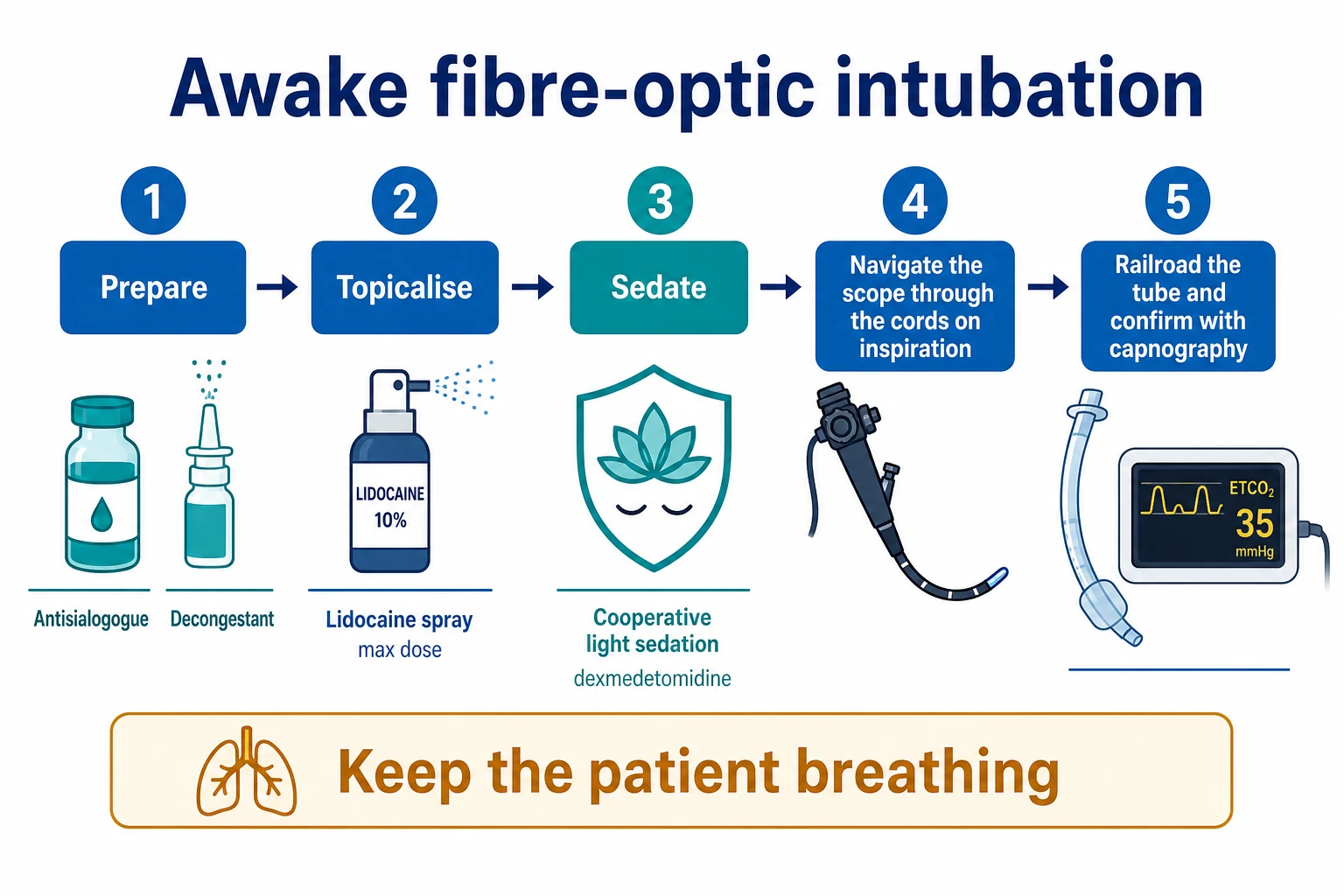
The AFOI sequence — seven steps from decision to secured tube
Assess and brief
Confirm a true anticipated difficult airway and that the patient can cooperate and oxygenate. Apply MACOCHA/LEMON. Brief the team on the plan (AFOI), the backup (optimised RSI), and the rescue (eFONA). Position a surgical-airway kit and a second skilled operator at the bedside.
Prepare the patient and the airway
Starve where possible. Full monitoring (ECG, SpO₂, NIBP, capnography). Antisialogogue — glycopyrrolate 200–400 μg IV (best given 15–30 min ahead, dries secretions so LA contacts mucosa and the view is clear). Nasal vasoconstrictor — xylometazoline 0.1% or phenylephrine, both nostrils, to shrink mucosa and reduce epistaxis. Pre-oxygenase. Sit the patient semi-upright.
Topicalise the upper airway
Lidocaine 1–4% by multiple routes — nebulised, gargle, paste/gel, and spray-as-you-go through the working channel at each anatomical level (tongue base, epiglottis, cords, trachea). Sum every dose; maximum ~3 mg/kg plain lidocaine (~4.5 mg/kg with adrenaline). Optional nerve blocks (superior laryngeal, glossopharyngeal) for dense anaesthesia in the difficult airway.
Sedate cooperatively
Goal: a calm, cooperative, breathing patient — NOT anaesthetised. Dexmedetomidine 0.2–1 μg/kg/h (± 0.5 μg/kg loading over 10 min) is close to ideal — anxiolysis, analgesia, sedation with minimal respiratory depression. Remifentanil target-controlled infusion (effect-site 1–3 ng/mL) is the analgesic alternative. Avoid propofol boluses and opioid-midazolam combinations that cause apnoea.
Navigate the scope
Load a lubricated ETT onto the scope. Pass the scope via the nose (preferred — better angle to glottis, less gag) or mouth (through a bite block). Advance under continuous vision through the nose/nasopharynx, around the uvula, to the tongue base, identify the epiglottis, then the cords. Pass through the cords DURING INSPIRATION (they open widest). Identify the carina and position the scope tip just above it.
Railroad the tube and confirm
Slide the lubricated ETT off the scope into the trachea. If resistance is felt at the glottis, withdraw 1–2 cm, rotate 90° counter-clockwise (so the Murphy eye faces the anterior commissure), and re-advance — this avoids the tube tip catching on the right arytenoid. Withdraw the scope. Inflate the cuff. Confirm with carinal vision and waveform capnography.
Induce and secure
Only AFTER capnographic confirmation, induce anaesthesia (propofol ± neuromuscular blocker), secure the tube, re-check ventilation and capnography, and document the grade of view, time taken, agents and doses, and any complications. Debrief and enter the airway alert in the chart.
1. Prepare
Starve the patient where possible; attach full monitoring (ECG, SpO2, blood pressure, capnography); give an antisialogogue (glycopyrrolate) to dry secretions so the local anaesthetic contacts mucosa and the view is clear; apply a topical vasoconstrictor (xylometazoline or phenylephrine) to the nose to reduce bleeding and mucosal oedema; pre-oxygenase; and have a rescue plan if AFOI fails.[1]
The single most important pre-procedural step is the team brief. The DAS 2018 ICU intubation bundle mandates a structured brief that names: (i) the intubator; (ii) the assistant; (iii) the drugs (induction, NMBA, vasopressor); (iv) the difficult-airway adjuncts ready (videolaryngoscope, bougie, supraglottic airway); (v) the eFONA kit and operator. For AFOI, add: (vi) who will topicalise; (vii) the total lidocaine dose budget for the patient; (viii) the sedation plan and the agent to reverse oversedation. NAP4 showed that airway disasters cluster where this brief is omitted.[1][3]
2. Topicalise the airway
Anaesthetise the upper airway with lidocaine by several routes: nebulised, gargle, paste/gel, and the spray-as-you-go technique (advancing the scope and instilling local anaesthetic through the working channel at each level — tongue base, epiglottis, vocal cords, trachea).[1]
- Sum every route and dose of lidocaine; the maximum is about 3 mg/kg of plain lidocaine (more with adrenaline), and toxicity is a real risk because absorption from the vascular airway mucosa is rapid.[1]
- Watch for local-anaesthetic systemic toxicity (LAST): early signs are perioral tingling, a metallic taste, tinnitus, and agitation; then seizures, and cardiovascular collapse (arrhythmia, arrest). Stop and treat with intralipid if toxicity develops.[1]
| [{"header":"Agent / route"},{"header":"Typical dose"},{"header":"Onset"},{"header":"Duration"},{"header":"Caveat"}] |
|---|
Maximum lidocaine dose rule of thumb. Plain lidocaine: ~3 mg/kg (≈ 200 mg in a 70 kg adult; ≈ seven 4% sprays of 2 mL). With adrenaline (which slows mucosal absorption): up to ~4.5–5 mg/kg. Plasma levels after airway topicalisation peak at 10–20 min and can approach toxic thresholds — the airway mucosa is a high-vascularity surface, second only to IV injection for rate of absorption. Always calculate the per-patient budget before the case and tally doses aloud as they are given.[10]
Nerve blocks — when topicalisation is not enough
Topicalisation alone anaesthetises most patients adequately. Nerve blocks are reserved for the airway that topicalisation cannot reach (densely reactive gag reflex, an anxious patient needing complete blocks, or a surgeon-neurointerventionalist who needs the patient still and unreactive). They add precision but also risk, and demand training.[1]
| [{"header":"Block"},{"header":"Nerve"},{"header":"Sensory territory"},{"header":"Technique"},{"header":"Pitfall"}] |
|---|
The cranial-nerve anatomy worth reciting for the viva: the glossopharyngeal (CN IX) carries sensation from the posterior third of the tongue and oropharynx; the vagus (CN X) via its superior laryngeal branch carries sensation above the cords and via the recurrent laryngeal branch below the cords. Topicalising these territories (or blocking them) is what makes AFOI tolerable.[1]
3. Sedate (cooperatively)
The goal is a calm, cooperative patient who is breathing and protecting their airway — not anaesthetised. Oversedation defeats the purpose (apnoea and loss of the airway).[1]
- Dexmedetomidine — close to ideal: anxiolysis, analgesia, and sedation with minimal respiratory depression (the patient remains rousable and breathing).
- Remifentanil — potent analgesic; careful titration, with risk of apnoea.
- Propofol in a low-dose target-controlled infusion.
- Older combinations (midazolam and fentanyl) cause more respiratory depression.[1]
| [{"header":"Agent"},{"header":"Mechanism"},{"header":"Dose / setup"},{"header":"Respiratory depression"},{"header":"Why use / avoid"}] |
|---|
The meta-analytic evidence is clear: dexmedetomidine-based regimens produce better intubating conditions, fewer episodes of apnoea, and fewer desaturations than remifentanil- or midazolam-based regimens for awake fiberoptic intubation.[7] The Cabrini systematic review of randomised trials of AFOI protocols similarly found dexmedetomidine to be the most consistently favourable sedative.[6]
Target-controlled infusion (TCI) is the modern way to administer remifentanil and propofol for AFOI. The pump models the effect-site concentration from a pharmacokinetic model (Minto for remifentanil, Marsh/Schnider for propofol) so you set a concentration rather than a rate. This allows smooth titration to the patient's response and rapid, predictable offset when the target is lowered — ideal for the fine balance of cooperative sedation. [1]
4. Navigate the scope
Pass the flexible scope via the nose (the preferred route — a better angle to the glottis and less gag reflex) or the mouth (through a bite block). Advance under vision, advance through the vocal cords during inspiration (the cords open widest), and identify the carina.[1]
Scope-handling pearls for the viva. Hold the insertion cord with the left hand near the patient, the right hand on the angulation lever and the body, the same way a clinician handles a bronchoscope. Stand at the head of the bed, monitor on the wall opposite. Keep the cord as straight as possible — every twist reduces tip control. Make small, deliberate movements; the lever is sensitive. Identify the uvula, then the epiglottis, then the cords, then the carina — losing anatomical landmarks is the commonest reason for failure. If the lens fogs, withdraw slightly and touch mucosa (wipes the lens) or use the working-channel flush.[1]
Nasal vs oral route. Nasal: better angle to the glottis (the scope drops naturally from the nasopharynx), less gag, requires a smaller tube (size 6.0–6.5), risks epistaxis, and is unsuitable if the nose is fractured or packed. Oral: requires a bite block (the patient can still occlude the scope), more gag reflex, but accepts a larger tube and has no nasal trauma. For cervical-spine trauma, the nasal route is preferred as the head stays neutral.[11]
5. Railroad the tube and confirm
Railroad a lubricated endotracheal tube over the scope into the trachea, withdraw the scope, inflate the cuff, and confirm with direct vision of the carina and waveform capnography.[1]
The railroad is the step that most often fails. The tube catches on the right arytenoid or the anterior glottic commissure as it is advanced off the scope. The fixes are: (i) liberally lubricate the tube and scope with water-soluble gel; (ii) use a wire-reinforced or Parker-tip tube if available (these have a bevel that rides over the glottis); (iii) if resistance is felt, withdraw the tube 1–2 cm and rotate 90° counter-clockwise so the bevel faces posteriorly and the Murphy eye faces the anterior commissure; (iv) re-advance slowly. Never force — the tube or the scope can be damaged, and the glottis traumatised.[1]
Only after capnographic confirmation, induce anaesthesia (the patient was awake and breathing throughout — now they may be induced with propofol and given a neuromuscular blocker as for any intubation). Securing the tube and re-checking capnography completes the procedure. [1]
AFOI vs videolaryngoscopy — when to choose which
Videolaryngoscopy (VL) has transformed predicted-difficult intubation in the unconscious patient, and the question of whether VL has replaced AFOI is a frequent viva topic. The honest answer: they are complementary. VL gives a view of the glottis without aligning the axes; AFOI secures the airway without losing the patient. The choice hinges on (i) whether the patient can be safely rendered apnoeic, (ii) whether the airway can be topicalised, and (iii) operator skill with each.[1][9]
| [{"header":"Feature"},{"header":"AFOI"},{"header":"Videolaryngoscopy"}] |
|---|
Clinical bottom line: AFOI remains the default for the predicted-difficult airway in a patient who can cooperate and oxygenate, especially where induction risks airway loss (obstructing mass, severely limited mouth opening, unstable c-spine). Videolaryngoscopy is the default for the unanticipated difficult intubation (after induction) and a strong backup for the predicted-difficult case when AFOI is contraindicated. Awake videolaryngoscopy is an emerging technique that may displace AFOI in centres with high VL expertise, but AFOI retains its advantage in the bloodless-but-distorted airway and the unstable c-spine.[1][9]
Advantages
- The patient keeps breathing and maintains their own airway — the airway is never lost.
- Direct visual confirmation of tube placement.
- Tolerated at lighter planes than an RSI, with less cardiovascular disturbance.
- Avoids neck movement (cervical-spine safety).[1]
- Preserves airway reflexes — safer in the high-aspiration-risk patient than an RSI that fails.
- Can be performed in any position (sitting, semi-upright, lateral) — vital for the patient who cannot lie flat (severe orthopnoea, raised ICP, late pregnancy).
- Can be used to intubate through a stoma, an existing tracheostomy tract, or a narrow laryngectomy stoma with the right scope.
- Provides diagnostic information en route — vocal cord mobility, mucosal lesions, the level of an obstruction — that a laryngoscope cannot.
Limitations and complications
- Time — slower than an RSI; unsuitable for the apnoeic or periarrest patient.
- Skill-dependent — requires practice and a functioning scope.
- Failure if the airway bleeds or floods with secretions, or if the patient cannot cooperate.
- Local-anaesthetic toxicity (LAST) from over-dosing or rapid mucosal absorption.
- Oversedation with apnoea and loss of the airway (the very thing AFOI avoids).
- Airway trauma, epistaxis (nasal route), and the rare risk of awareness if sedation is too light.[1]
Complications in detail
| [{"header":"Complication"},{"header":"Mechanism / cause"},{"header":"Recognition"},{"header":"Immediate management"}] |
|---|
Key trials and guidelines
DAS 2018 — intubation in critically ill adults (PMID 29406182)
Document type
Consensus guideline — Difficult Airway Society, Intensive Care Society, Faculty of Intensive Care Medicine, Royal College of Anaesthetists (UK)
Scope
Tracheal intubation in the critically ill adult (the ICU guideline; DAS 2015 covers the perioperative setting)
Key recommendations for awake intubation
Awake intubation is a first-line option for the predicted difficult ICU airway; standardise equipment (PEDI — Person, Equipment, Drugs, monitoring); use a difficult-airway bundle; allocate roles; have a declared eFONA plan
Clinical bottom line
The ICU intubation guideline. Every CICM/FFICM/EDIC candidate should know the intubation bundle, the role of awake intubation, and the explicit eFONA plan.
ASA 2022 — Practice Guidelines for Management of the Difficult Airway (PMID 34762729)
Document type
Practice guideline — American Society of Anesthesiologists (update of the 2013 guideline)
Scope
All settings (perioperative, ICU, ED); both anticipated and unanticipated difficult airway
Key recommendations
Recommend awake intubation when difficulty with facemask ventilation, SGA ventilation, or intubation is known or predicted; emphasise video laryngoscopy as initial attempt; explicit cognitive aids; multiple-device availability
Clinical bottom line
The North American counterpart to DAS. Cites AFOI as a primary strategy for the predicted difficult airway alongside VL.
NAP4 — Fourth National Audit Project of airway management (PMID 21447488)
Document type
National prospective audit of major airway complications — Royal College of Anaesthetists and Difficult Airway Society, UK
Population
All major complications of airway management reported over 1 year across UK NHS hospitals (anaesthesia, ICU, ED)
Key findings — awake intubation
Awake intubation was under-used in predicted-difficult ICU patients; several deaths occurred when awake intubation should have been the primary plan but was not attempted; poor planning, poor equipment availability, and failure to anticipate difficulty were recurring themes
Clinical bottom line
NAP4 is the foundational document for modern difficult-airway practice in ICU. It argues for earlier and more liberal use of awake intubation in the predicted difficult ICU airway.
Cabrini 2019 — systematic review of AFOI protocols (PMID 30896601)
Document type
Systematic review and meta-analysis of randomised controlled trials — Anesthesia & Analgesia
Population
Adults undergoing awake fibre-optic intubation for anticipated difficult airway in the operating room; 19 RCTs included
Key findings
Dexmedetomidine-based sedation provided better intubating conditions and patient comfort than remifentanil or propofol-based regimens, with fewer episodes of apnoea; antisialogogue pre-treatment improved conditions; remifentanil TCI was effective but apnoea-prone
Clinical bottom line
The definitive synthesis of sedation protocols for AFOI. Supports dexmedetomidine as the modern first-line sedative for awake intubation.
Tang 2021 — dexmedetomidine vs remifentanil for AFOI meta-analysis (PMID 33832107)
Document type
Systematic review and meta-analysis — Medicine (Baltimore)
Comparison
Dexmedetomidine vs remifentanil for awake fiberoptic intubation; multiple RCTs
Key findings
Dexmedetomidine associated with higher patient satisfaction and intubating-condition scores and fewer respiratory-depression events than remifentanil; remifentanil provided superior cough suppression but at the cost of more apnoea
Clinical bottom line
Reinforces dexmedetomidine as first-line for AFOI sedation; remifentanil is a reasonable adjunct or alternative in haemodynamically stable patients, with close respiratory monitoring.
Avitsian 2005 — dexmedetomidine for cervical spine AFOI (PMID 15840996)
Document type
Clinical series — Journal of Neurosurgical Anesthesiology
Population
Patients with possible cervical spine myelopathy undergoing awake fibre-optic intubation
Key findings
Dexmedetomidine provided cooperative sedation with preserved respiration and haemodynamic stability during AFOI for cervical spine disease; no cases of airway loss or new neurological deficit attributable to sedation
Clinical bottom line
An early and influential series establishing dexmedetomidine as a safe sedative for AFOI in the cervical-spine-at-risk patient — a high-yield historical citation for the viva.
Xu 2025 — awake videolaryngoscopy vs fibre-optic bronchoscope (PMID 40731382)
Document type
Randomised clinical trial — BMC Anesthesiology
Comparison
SEEK(flex) videolaryngoscope vs fibre-optic bronchoscope for awake tracheal intubation in patients with predicted difficult airway
Key findings
Awake videolaryngoscopy achieved comparable intubation success and time to intubation to fibre-optic bronchoscopy in expert hands; the SEEK(flex) stylet allowed direct visualised tube delivery
Clinical bottom line
Awake videolaryngoscopy is a credible alternative to AFOI in centres with the expertise; AFOI retains an advantage where the airway is distorted or blood is present. Supports the 'complementary, not competing' framing.
Mushambi 2020 — obstetric anticipated difficult airway (PMID 32144770)
Document type
Narrative literature review and management recommendations — Anaesthesia (Obstetric Anaesthetists' Association / Difficult Airway Society)
Scope
Anticipated difficult airway during obstetric general anaesthesia
Key recommendations
Awake fibre-optic intubation is recommended as a primary strategy in the predicted-difficult obstetric airway where time and cooperation allow; emphasises senior help, full monitoring, and a declared eFONA plan
Clinical bottom line
The obstetric-specific reference for AFOI; a high-yield citation for any question about pregnancy + difficult airway.
Cabrini 2020 — intubation in cervical spinal cord injury (PMID 31837227)
Document type
Systematic review — Acta Anaesthesiologica Scandinavica
Scope
Tracheal intubation in patients at risk of cervical spinal cord injury
Key findings
Awake fibre-optic intubation produces the least cervical spine movement of any intubation technique; manual in-line stabilisation reduces but does not eliminate movement with direct laryngoscopy; videolaryngoscopy is intermediate
Clinical bottom line
Supports nasal AFOI as the airway technique of choice when cervical spinal cord injury must be presumed and the patient can cooperate.
Neal 2012 — ASRA LAST checklist (PMID 22157743)
Document type
Simulation study — Regional Anesthesia and Pain Medicine
Intervention
ASRA-recommended checklist for the management of local anaesthetic systemic toxicity
Key findings
Use of the checklist improved trainee performance during simulated LAST — fewer omissions of lipid emulsion and correct dosing; checklists improve the management of this rare, high-stakes event
Clinical bottom line
Have the ASRA LAST checklist and lipid emulsion immediately available wherever AFOI is performed. The checklist saves lives in the rare event of toxicity.
High-yield clinical pearls
[1]SAQ — Anticipated difficult airway from an obstructing laryngeal mass
10 minutes · 10 marks
A 68-year-old man with a known supraglottic tumour and recent worsening stridor on lying flat is admitted for elective debulking surgery. He has limited mouth opening and an oropharyngeal mass displacing the epiglottis. He is cooperative, oxygenating well on room air, and fasted. Justify the choice of awake fibre-optic intubation (AFOI) and describe the technique, including how you would topicalise and sedate him.
SAQ — AFOI in the suspected cervical-spine-injured patient
10 minutes · 10 marks
A 30-year-old man is brought to the ED after a high-speed motor vehicle crash. He is in a hard collar, GCS 15, saturating at 97% on room air, with motor and sensory deficits suggesting an incomplete cervical cord injury. CT cannot exclude ligamentous instability. He needs intubation for an urgent MRI. Discuss the airway options and defend your chosen technique.
Red flags
Prognosis and follow-up
AFOI, performed well, has a high first-pass success rate (90%+ in elective series; lower in ICU where anatomy is more deranged) and a low rate of serious complications when doses are tallied and the team is briefed. The DAS 2018 and NAP4 literature converge on the message that most AFOI 'failures' are failures of selection, preparation, or sedation — not of the technique itself.[1][3]
After AFOI: document the airway findings (view, time taken, agents, total lidocaine dose, complications); place an airway alert in the chart; arrange difficult-airway identification (MedicAlert, patient-held letter); and debrief the team, especially if any step did not go to plan. Patients with structurally difficult airways (radiotherapy, mass, ankylosing spondylitis) should be flagged for future AFOI as the default technique.[1]
References
- [1]Higgs A, McGrath BA, Goddard C, et al.; Difficult Airway Society et al. Guidelines for the management of tracheal intubation in critically ill adults Br J Anaesth, 2018.PMID 29406182
- [2]Apfelbaum JL, Hagberg CA, Connis RT, et al. 2022 American Society of Anesthesiologists Practice Guidelines for Management of the Difficult Airway Anesthesiology, 2022.PMID 34762729
- [3]Cook TM, Woodall N, Frerk C; Fourth National Audit Project Major complications of airway management in the UK: results of the Fourth National Audit Project of the Royal College of Anaesthetists and the Difficult Airway Society. Part 1: anaesthesia Br J Anaesth, 2011.PMID 21447488
- [4]Woodall N, Frerk C, Cook TM Can we make airway management (even) safer?--lessons from national audit Anaesthesia, 2011.PMID 22074076
- [5]Mushambi MC, Athanassoglou V, Kinsella SM, et al. Anticipated difficult airway during obstetric general anaesthesia: narrative literature review and management recommendations Anaesthesia, 2020.PMID 32144770
- [6]Cabrini L, Baiardo Redaelli M, Ball L, et al. Awake Fiberoptic Intubation Protocols in the Operating Room for Anticipated Difficult Airway: A Systematic Review and Meta-analysis of Randomized Controlled Trials Anesth Analg, 2019.PMID 30896601
- [7]Tang ZH, Chen Q, Wang X, et al. A systematic review and meta-analysis of the safety and efficacy of remifentanil and dexmedetomidine for awake fiberoptic endoscope intubation Medicine (Baltimore), 2021.PMID 33832107
- [8]Avitsian R, Lin J, Lotto M, Ebrahim ZY Dexmedetomidine and awake fiberoptic intubation for possible cervical spine myelopathy: a clinical series J Neurosurg Anesthesiol, 2005.PMID 15840996
- [9]Xu W, Zhu C, Wu Q, et al. Comparison of SEEK(flex)/videolaryngoscopy and fibreoptic bronchoscope for awake tracheal intubation: a randomized clinical trial BMC Anesthesiol, 2025.PMID 40731382
- [10]Neal JM, Hsiung RL, Mulroy MF, et al. ASRA checklist improves trainee performance during a simulated episode of local anesthetic systemic toxicity Reg Anesth Pain Med, 2012.PMID 22157743
- [11]Cabrini L, Baiardo Redaelli M, Filippini M, et al. Tracheal intubation in patients at risk for cervical spinal cord injury: A systematic review Acta Anaesthesiol Scand, 2020.PMID 31837227