ICU · Airway management
Basic Airway Manoeuvres, Adjuncts & Bag-Valve-Mask
Also known as Basic airway skills · Bag-valve-mask · BVM · Guedel airway · Oropharyngeal airway · Nasopharyngeal airway · Jaw thrust · Head tilt chin lift · EC clamp · Apnoeic oxygenation
The 'A' of ABCDE. Basic airway skills are the non-invasive manoeuvres and adjuncts that establish and maintain a patent airway and oxygenate the patient before a definitive airway. Jaw thrust and head-tilt/chin-lift relieve obstruction by the tongue; the oropharyngeal (Guedel) and nasopharyngeal airways stent the pharynx; and the bag-valve-mask with a two-handed C-E grip, reservoir and PEEP valve ventilates the unprotected airway. Most 'can't intubate' situations are rescued by excellent basic airway skills and bag-valve-mask ventilation. Apnoeic oxygenation (THRIVE) extends safe apnoea time.
On this page & tools
Your progress
Saved locally on this device.
Target exams
Overview & definition
The airway is the first priority of resuscitation (the "A" of ABCDE). Basic airway skills are the non-invasive manoeuvres and adjuncts that open, hold open, and oxygenate the unprotected airway before a definitive airway (supraglottic device, endotracheal tube, or front-of-neck access) is placed. They include the jaw thrust and head-tilt/chin-lift, the oropharyngeal and nasopharyngeal airways, and the bag-valve-mask (BVM). Mastery of these is what rescues the patient when intubation fails — the commonest cause of airway-related death is hypoxia from poor basic airway management, not a failed tube.[1][1]
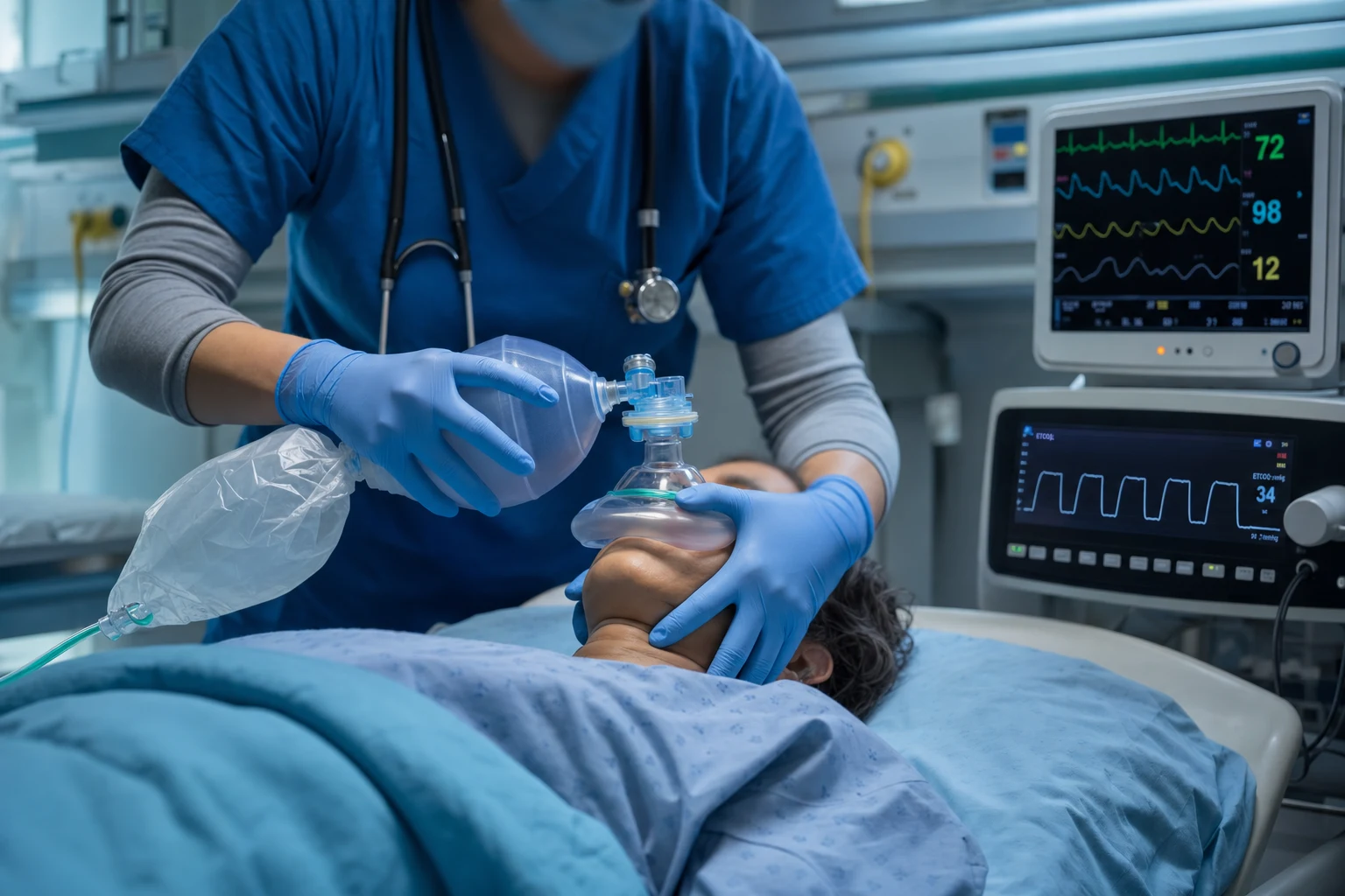
The obstructed airway
In the unconscious patient the commonest cause of airway obstruction is the tongue falling back against the posterior pharyngeal wall, followed by soft palate, epiglottis, and foreign material or secretions.[1]
Recognise obstruction with look, listen, feel:[1]
- Look — paradoxical "see-saw" chest and abdominal movement, tracheal tug, intercostal recession, cyanosis; little or no chest movement.
- Listen — snoring (pharyngeal, tongue/palate), gurgling (fluid in the airway), stridor (laryngeal, fixed upper-airway obstruction), or, ominously, silence (complete obstruction).
- Feel — little or no airflow at the mouth and nose.
Total obstruction is silent — silence in a patient with respiratory effort is an emergency. [1]
Basic manoeuvres
1. Head tilt + chin lift.[1][1]
- One hand on the forehead tilts the head back, the fingers of the other hand lift the chin forward. This lifts the tongue off the posterior pharyngeal wall.
- Contraindicated in suspected cervical-spine injury — use jaw thrust instead.
- The definitive basic manoeuvre: the index and middle fingers of both hands are placed behind the angles of the mandible and displaced upward and forward, lifting the jaw and tongue.
- It is cervical-spine safe (no head movement) and is the manoeuvre of choice in trauma and the ICU.
In most patients, one of these two manoeuvres relieves obstruction. Apply oxygen (15 L/min via a non-rebreather mask) immediately. [1]
Airway adjuncts
Once the airway is open with a manoeuvre, an adjunct holds it open. [1]
Oropharyngeal (Guedel) airway
- Sizing — measure from the corner of the mouth to the angle of the jaw (or the earlobe); an oversize device pushes the epiglottis down and obstructs, an undersize device jams the tongue into the posterior pharynx.[1]
- Insertion (adult) — insert upside-down (concavity upward) across the hard palate, rotate 180 degrees as it reaches the back of the tongue; alternatively use a tongue depressor. Never force against resistance.[1]
- Contraindications — a patient with an intact gag reflex (it provokes vomiting, laryngospasm, and raised intracranial pressure). It is tolerated only in the deeply unconscious patient.[1]
Nasopharyngeal airway
- Sizing — measure from the nostril to the tragus of the ear; diameter approximates the patient's little finger.[1]
- Insertion — lubricate, insert the bevel-first end along the floor of the nose (toward the occiput, not cephalad) into the most patent nostril with a gentle twisting motion.
- Advantage — better tolerated than the oropharyngeal (may be used with a partial gag reflex) and cervical-spine safe; the first-choice adjunct in the semi-conscious ICU patient.[1]
- Contraindication — basilar (base-of-skull) skull fracture, where it could enter the cranial vault; also coagulopathy and significant midface trauma.
A manoeuvre plus an adjunct usually secures a clear airway and permits bag-valve-mask ventilation. [1]
Bag-valve-mask (BVM) ventilation
The BVM ventilates the unprotected airway while a definitive airway is prepared. Good technique is a core skill — poor BVM is a common source of hypoxia.[1]
The mask seal — the two-handed C-E (EC) grip.[1][1]
- Form a "C" with the thumb and index finger over the mask dome, pressing it onto the face; the three remaining fingers form an "E" along the mandible, lifting the jaw up into the mask (combining the seal with a jaw thrust).
- A two-person technique (one holds the mask with two hands, the other squeezes) gives a far better seal and tidal volume than one-handed — use it whenever a second person is available.
Oxygen delivery and PEEP.[1]
- Attach the reservoir bag and run 15 L/min of oxygen — a BVM with a reservoir delivers 90-100 per cent oxygen; without the reservoir it delivers only about 40-60 per cent.
- A PEEP valve (typically 5-10 cmH2O) improves oxygenation and functional residual capacity in hypoxic patients.
Ventilation rate and volume.[1][1]
- Ventilate at 10-12 breaths per minute (one breath every 5-6 seconds) in an intubated/arrest context — about one breath every 6 seconds in basic life support.
- Use small tidal volumes — just enough to produce visible chest rise. Over-vigorous ventilation causes gastric insufflation, regurgitation, and aspiration.
Common errors.[1]
- A poor seal (single-handed grip, beard, oedema).
- Hyperventilation (the commonest error — raises intrathoracic pressure, reduces venous return, and worsens outcome in cardiac arrest).
- Forgetting the reservoir (delivering low-FiO2 gas).
- Not confirming ventilation with chest rise and capnography.
Confirm ventilation with visible chest rise and, ideally, waveform capnography even during BVM — the presence of a CO2 trace confirms gas exchange, not just mask movement.[1]
Apnoeic oxygenation and THRIVE
Even during a brief apnoea (e.g. during laryngoscopy), oxygen continues to be taken up from the alveoli if the upper airway is patent and oxygen is flowing. Delivering oxygen into the pharynx during the apnoeic period prolongs the safe apnoea time and slows desaturation.[1]
THRIVE (Transnasal Humidified Rapid-Insufflation Ventilatory Exchange) — high-flow humidified nasal oxygen (e.g. 70 L/min) extends apnoea tolerance dramatically in appropriately selected patients (Patel & Nouraei, Anaesthesia 2015). It is an oxygenation adjunct, not a ventilatory device — it does not remove CO2 indefinitely, and it does not replace BVM for a patient who needs ventilation. In patients who are obese, obstructed, or full of stomach, its benefit is limited.[1]
The escalation algorithm
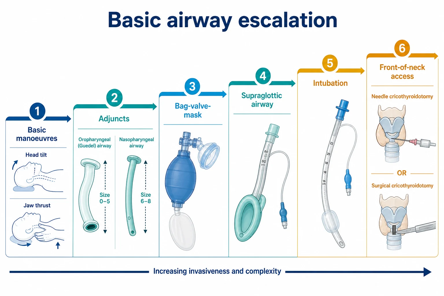
Basic airway skills sit at the base of a ladder that escalates only as needed:[1][1]
- Basic manoeuvres (jaw thrust, head-tilt/chin-lift) and high-flow oxygen.
- Adjunct (oropharyngeal or nasopharyngeal airway).
- Bag-valve-mask ventilation (two-handed, reservoir, PEEP).
- Supraglottic airway (e.g. i-gel, LMA) — a rescue if BVM fails.
- Endotracheal intubation (definitive, by a skilled operator).
- Front-of-neck access (e.g. scalpel-bougie cricothyroidotomy) — the last resort in "can't intubate, can't oxygenate" (CICO).
Excellent basic skills keep the patient oxygenated at every rung of this ladder. [1]
Pathophysiology — why the unconscious airway obstructs
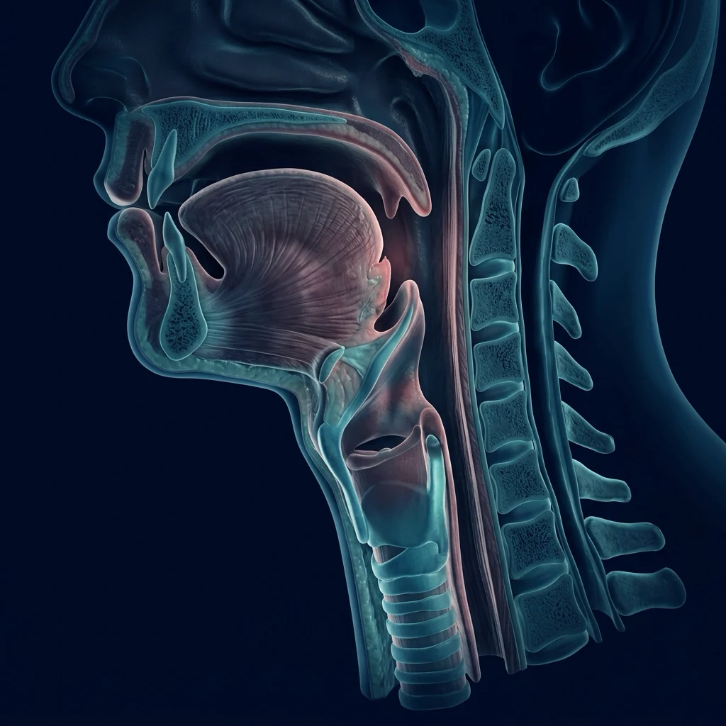
Airway patency in the awake patient is maintained by the pharyngeal dilator muscles (genioglossus, tensor palatini, stylopharyngeus) — tonic and phasic activity that pulls the tongue and soft tissues forward, resisting the negative pressure generated by inspiration. As consciousness is lost these muscles lose tone in a rostral-caudal pattern, and the tongue and epiglottis fall back against the posterior pharyngeal wall.[1] Obstruction is dynamic and inspiratory: each inspiratory effort sucks the floppy structures further into the lumen (a negative-pressure ball-valve), so the harder the patient tries to breathe, the more they occlude themselves — the basis of the paradoxical "see-saw" pattern.
The functional anatomy of obstruction maps onto the three classic levels: (1) nasopharyngeal/oropharyngeal — tongue and soft palate (the commonest, relieved by basic manoeuvres and adjuncts); (2) laryngeal — floppy epiglottis, laryngospasm, foreign body (may need suction, LMA, or definitive airway); and (3) lower airway — secretions, bronchospasm, pulmonary oedema. The first two are the domain of basic airway skills.[1]
Determinants of safe apnoea time. When a patient stops breathing, arterial saturation is maintained only while the alveolar oxygen reservoir lasts. The rate of desaturation is governed by the ratio of alveolar oxygen reserve to whole-body oxygen consumption (VO2 ~250 mL/min in the adult). Functional residual capacity (FRC) is the reservoir (~2500 mL seated, less when supine, much less in obesity and pregnancy). Preoxygenation replaces alveolar nitrogen with oxygen ("denitrogenation") and roughly triples the safe apnoea time; obesity, sepsis, pregnancy, paediatrics and shunt pathology all shorten it dramatically — a septic, obese, supine patient can desaturate in well under a minute.[8][9]
Detailed technique — basic manoeuvres
Head tilt / chin lift
The historical first-line manoeuvre for the unconscious patient without suspected cervical-spine injury. [1]
- Mechanism. Atlanto-occipital extension (head tilt) combined with forward displacement of the mandible (chin lift) lifts the tongue and hyoid apparatus forward off the posterior pharyngeal wall, breaking the ball-valve effect.
- Technique. Place one hand on the patient's forehead and apply backward pressure to extend the head at the atlanto-occipital joint (sniffing-the-morning-air position, ~30-45°). Place the fingertips of the other hand under the bony margin of the chin and lift the mandible forward. Avoid compressing the soft tissues of the submandibular floor, which can itself obstruct the airway.
- Contraindication. Suspected cervical-spine injury — the head-tilt component translates the cervical spine and may worsen cord injury. Use jaw thrust instead. (In cardiac arrest with an unprotected airway, the rescuer's hands are usually best used for chest compressions; minimal head tilt is acceptable if needed for ventilation, but jaw thrust is preferred.)[1]
- Pitfalls. (i) Incomplete extension in the obese or short-necked patient. (ii) Chin pressure on the submental soft tissue rather than the bony symphysis. (iii) Forgetting that this manoeuvre alone does not maintain the airway for long — add an adjunct and oxygen.
Jaw thrust
The definitive basic manoeuvre and the airway skill of choice whenever the cervical spine cannot be cleared (trauma, the ICU patient with unknown injury, the post-arrest patient, the deeply sedated patient).[1][1]
- Mechanism. Antero-superior displacement of the angle of the mandible lifts the mandible (and with it the tongue and epiglottis via the hyo-glossal and genio-hyoid attachments) forward, opening the pharynx. The cervical spine is not moved, so it is C-spine safe — the manoeuvre of choice in trauma and the intubated ICU patient being bagged pre-intubation.
- Technique. Stand at the head of the bed. Place the index (or middle) fingers of both hands behind the angle of the mandible (the gonial angle), and the thumbs on the chin/mask dome if you are bagging. Displace the angles upward (cephalad) and forward ("lift the face up to the mask"). Effective jaw thrust fatigues the operator's hands within minutes — call for a second person early and use a two-handed technique.
- Vice-versa of the C-E grip. When combined with a BVM, the jaw thrust is the three fingers of the "E" of the C-E grip: thumb-index form the "C" on the mask dome, three fingers form the "E" along the jaw and thrust it up into the mask.
- Pitfalls. (i) Placing fingers on the body rather than the angle of the mandible — ineffective. (ii) Allowing fatigue to let the jaw drop (swap operators every 2 minutes). (iii) Two-handed jaw thrust with no assistant to squeeze the bag — convert to a two-person technique. [1]
Head-tilt/chin-lift vs jaw thrust
| Feature | Head-tilt / chin-lift | Jaw thrust |
|---|---|---|
| Mechanism | Atlanto-occipital extension + chin elevation | Antero-superior mandibular displacement |
| C-spine safe? | No — moves the cervical spine | Yes — no cervical movement |
| Setting | Medical collapse, no trauma; BLS adult | Trauma, ICU, cardiac arrest, suspected C-spine injury, BVM seal |
| Operator fatigue | Low — sustained easily | High — fatigues within minutes, needs two hands / relief |
| Pairs with BVM | One-handed grip possible | Forms the "E" of the two-handed C-E grip |
| Position | Sniffing position | Supine, neutral C-spine |
| First-line in | Medical arrest (BLS) | Trauma, ICU, suspected C-spine injury |
Detailed technique — airway adjuncts
Oropharyngeal (Guedel) airway
A curved flanged plastic tube that stents the tongue off the posterior pharyngeal wall. It splints open only the oropharynx — it does not bypass obstruction at the larynx. [1]
- Sizing (the commonest exam point and the commonest error). Three acceptable methods:[1]
- Corner-of-mouth to angle of mandible (gonial angle) — the standard adult method.
- Corner-of-mouth to earlobe (pinna inferior border) — the alternative; gives a slightly longer size, useful in the long-jawed patient.
- Measure and hold the device against the patient's face before insertion to confirm. A size 3 (90 mm) suits most adult women, size 4 (100 mm) most adult men; sizes 2-5 cover the range.
- Errors of size. Too long — the tip pushes the epiglottis down onto the glottis and converts a patent airway into an obstruction (and may provoke laryngospasm). Too short — the device jams the tongue into the posterior pharynx, worsening obstruction. The wrong size is worse than no device.[1]
- Insertion (adult). Open the mouth (scissor technique). Insert the Guedel upside-down (concavity facing upward/cephalad) across the hard palate, advance until close to the posterior pharyngeal wall, then rotate 180° so the concavity faces caudally and slip it down behind the tongue. Never force against resistance — if rotation is blocked, withdraw, use a tongue depressor, or insert right-way-up over a depressor. A lubricated device passes more easily.[1]
- Contraindications. Intact gag reflex is the absolute contraindication — insertion provokes vomiting, laryngospasm, and a dangerous rise in intracranial and intra-ocular pressure. The Guedel is tolerated only in the deeply unconscious patient (GCS ≤8, no gag). Relative contraindications include dental instability, oral trauma and recent oral surgery.[1]
- Tip. If the patient gags, coughs or retches, remove it immediately and use a nasopharyngeal airway instead.
Nasopharyngeal airway
A soft, bevelled, flanged tube passed through the nose to lie with its tip in the pharynx behind the tongue base. The first-choice adjunct in the semi-conscious ICU patient because it is far better tolerated than a Guedel.[1]
- Sizing. Length: measure from the nostril (nares) to the tragus of the ear (the meatal-tragal measurement approximates the distance to the oropharynx). Diameter: roughly approximates the patient's little finger, or the nostril at rest. Adults commonly take sizes 6.0-7.0 (internal diameter in mm, often colour-coded). When in doubt choose the smaller of two sizes and the more patent nostril.[1]
- Insertion. Lubricate the tube. Insert the bevel-first end along the floor of the nose (inferior turbinate), directed toward the occiput (not cephalad/up toward the cribriform plate). Advance gently with a slow twisting motion until the flange sits at the nostril. If resistance is met, withdraw, rotate, and try the other nostril — never force, as the turbinates and nasopharyngeal mucosa bleed easily.
- Safety check. After insertion, confirm airflow through the tube (look, listen, feel), and that ventilation improves. If the tube is too long it may enter the oesophagus and insufflate the stomach; too short and it will not relieve obstruction.
- Advantages. Tolerated with a partial gag reflex (useful at GCS 9-13 where a Guedel is contraindicated); C-spine safe (no neck movement); can be left in place during seizures and post-ictal states; the airway of choice for the obtunded but not deeply unconscious patient.[1]
- Contraindications. Basilar (base-of-skull) fracture — the tube can pass through a fractured cribriform plate into the cranial vault (a catastrophic, avoidable complication). Also coagulopathy (epistaxis), significant midface/NOE (naso-ethmoid) trauma, and suspected nasal mass or CSF rhinorrhoea.[1]
Oropharyngeal (Guedel) vs nasopharyngeal airway
| Feature | Oropharyngeal (Guedel) | Nasopharyngeal (NPA) |
|---|---|---|
| Sizing — length | Mouth-corner to angle of jaw / earlobe | Nostril to tragus of ear |
| Sizing — calibre | N/A (rigid, sized by length) | Approximates little finger / nostril |
| Tolerated with gag reflex? | No — absolute contraindication | Yes (partial gag acceptable) |
| Level of consciousness | Deeply unconscious (GCS ≤8, no gag) | Semi-conscious to deeply unconscious (GCS 9-13 useful) |
| C-spine safe | Yes | Yes |
| Contraindications | Intact gag, oral trauma, dental instability | Base-of-skull fracture, coagulopathy, midface trauma |
| Bleeding risk | Low | Moderate (turbinates) |
| First-choice when | Arrest, deeply unconscious, cardiac arrest BVM | Obtunded ICU patient, post-ictal, partial gag, trismus |
Suctioning
Suction clears blood, saliva, vomit, and secretions that obstruct the airway or that a manoeuvre/adjunct cannot bypass. Suction before you ventilate if the airway is full of fluid — "gurgling" on inspiration is a positive indication to suction. [1]
- Equipment. A Yankauer (rigid) sucker for the oropharynx — wide-bore, angulated, designed for blood and particulate vomitus, and too large to pass beyond the oropharynx (so it will not induce laryngospasm at the cords). A soft (flexible) suction catheter (10-14 Fr) for the nasopharynx, the tracheal tube, and the conscious/semi-conscious patient who tolerates the Yankauer poorly.
- Technique (Yankauer). Suction on the way in (orally, along the cheek, into the posterior pharynx) and on the way out, never probing blindly past the tonsillar pillars (gag and laryngospasm). Limit each pass to <10-15 seconds in the spontaneously breathing patient to avoid hypoxia and vagally-mediated bradycardia. Pre-oxygenate before and after.
- Soft catheter via NPA/tracheal tube. Advance without suction, apply suction on withdrawal, rotate the catheter. For a tracheal tube, the catheter outer diameter must be <half the tube's inner diameter to avoid negative-pressure lung collapse.
- Indications. Audible/visible secretions, blood or vomit; suspected aspiration; before intubation to clear the view; to check patency of a tracheal tube or NPA.
- Complications. Hypoxia (suctioning evacuates alveolar oxygen), bradycardia (vagal), mucosal trauma and bleeding, laryngospasm, gagging and vomiting, and — with prolonged blind suction — infection transmission to the operator. Always pre-oxygenate and limit suction time.
- Pearl. In the crashing patient with copious vomitus, tip the head down (Trendelenburg) and turn to the side, suction aggressively, and have suction running before the airway is instrumented — "suction in hand, ready to go" is a core crisis behaviour.[1]
Bag-valve-mask technique in detail
The two-handed C-E (EC) grip
The single most important airway skill. A poor seal is the commonest reason for failed BVM ventilation.[1][4]
- Form the "C". Thumb and index finger of each hand form two "C" shapes over the dome of the mask, pressing the cushion down onto the face — the squeeze that seals.
- Form the "E". The three remaining fingers of each hand (middle, ring, little) lie along the mandibular ramus (the bony jaw) on each side and lift the face up into the mask — this is simultaneously a bilateral jaw thrust. Effective technique pulls the patient to the mask, not pushes the mask to the patient.
- Fit. The mask should span from the nasal bridge to the mental crease (chin) without leaks over the eyes or the bony chin. A sideways-leaking mask is usually too large or the jaw not thrust enough.
- The "two-handed" rule. Whenever possible use a two-handed grip with a second operator squeezing the bag — this delivers consistently better seal, tidal volume and oxygenation than any one-handed technique.[1] Reserve the one-handed "EC" grip for the single-rescuer cardiac-arrest scenario where the other hand cannot be spared.
Two-person BVM technique
The gold standard for the unprotected airway when a second rescuer is available.[1]
- Operator 1 (airway) stands at the head of the bed and performs a two-handed C-E grip, combining mask seal with bilateral jaw thrust. Both hands are on the mask — no hand on the bag.
- Operator 2 (ventilator) squeezes the bag at the appropriate rate and volume (see below), watching for chest rise, and watches the capnograph.
- Why it works. Mask ventilation is mechanically limited by leak and by upper-airway obstruction; a dedicated two-handed airway operator solves both simultaneously. Studies of difficult-mask patients show that two-handed technique succeeds where one-handed fails.[4][5]
- Predictors of difficult BVM (Langeron / Kheterpal): beard, obesity (high BMI), edentulism, age >55, short thyromental distance, snoring/obstructive sleep apnoea, limited jaw protrusion, male sex, history of neck radiation. When these are present, start two-handed, two-person from the outset and call for senior help early.[4][5]
PEEP valve
A PEEP valve on the BVM exhalation port applies positive end-expiratory pressure (typically 5-10 cmH2O) throughout the respiratory cycle. [1]
- Benefits. Recruits collapsed alveoli, increases functional residual capacity, improves V/Q matching and arterial oxygenation, and counteracts the loss of PEEP that occurs when a spontaneously breathing patient with pulmonary oedema or ARDS is bagged.[1]
- Indications. Any hypoxic patient: pulmonary oedema, ARDS, pneumonia, obesity, atelectasis. Increasingly recommended as a default attachment in the ICU/emergency BVM set.
- Caution. PEEP raises intrathoracic pressure, reducing venous return — in hypovolaemic or obstructed-shock states it can drop cardiac output. It also increases gastric insufflation if tidal volume is too large. Titrate to SpO2, not to a fixed number.
Oxygen delivery, rate and volume
- Reservoir + 15 L/min O2 → 90-100% FiO2. Without reservoir → ~40-60%. Always attach the reservoir.[1]
- Rate 10-12 breaths/min (one every 5-6 s) for the intubated/arrest patient; one every 6 s in BLS. Avoid hyperventilation.[1]
- Volume: just enough for visible chest rise (~500 mL / 6-7 mL/kg). Over-vigorous bagging → gastric insufflation, regurgitation, aspiration, and reduced venous return.[1]
One-handed vs two-handed BVM grip, and one-person vs two-person technique
| Technique | Seal quality | Tidal volume | Best use | Limitation |
|---|---|---|---|---|
| One-handed C-E (single rescuer) | Poor-moderate | Variable | Single-rescuer BLS cardiac arrest | Commonest cause of failed BVM; cannot combine with strong jaw thrust |
| Two-handed C-E (single rescuer) | Good | Good | Anticipated difficult mask, no assistant available | No hand free to squeeze the bag |
| Two-person (two-handed + assistant bags) | Best | Best | Gold standard — any ICU/emergency BVM | Needs an assistant |
| Three-person ("triple airway manoeuvre") | Excellent | Excellent | Morbidly obese / beard / oedematous difficult mask | Needs two assistants; underused |
Confirming ventilation
Visible bilateral chest rise plus waveform capnography (CO2 trace) is the standard. The presence of a capnograph waveform during BVM confirms gas exchange, not just mask movement, and is the earliest detector of oesophageal intubation or tube displacement. Colourimetric CO2 detectors are an acceptable fallback. In cardiac arrest a flat capnograph during BVM may simply reflect absent pulmonary perfusion — assess chest rise and compressions.[1]
The laryngeal mask airway as a rescue device
A supraglottic airway (LMA, i-gel, ProSeal) sits in the hypopharynx with its cuff around the laryngeal inlet, providing a hands-free airway that bypasses tongue and oropharyngeal obstruction and supports spontaneous or gently-ventilated breathing. It is the first rescue device when BVM fails in the CICO pathway, and an option in the "can't intubate, can ventilate" situation.[2][3]
- Role in basic airway management. When basic manoeuvres, adjuncts and BVM cannot oxygenate (the difficult-mask patient), the next step is a supraglottic airway, not repeated attempts at intubation. NAP4 found that poor basic airway skills and delayed supraglottic rescue were recurring themes in airway-related death and brain injury.[2]
- Insertion. Deflate/lubricate the cuff, blind-insert with the aperture facing the patient's feet, advance until resistance is felt, inflate the cuff (or, for i-gel, no cuff). Confirm with bilateral chest rise and a capnograph waveform.
- Advantages. Rapid (inserted in seconds by most operators without laryngoscopy), hands-free, less invasive than intubation, useful as a bridge to definitive airway, and a conduit for fibreoptic intubation. i-gel is cuff-free and especially quick.
- Limitations. It does not protect against aspiration of gastric contents (the ProSeal/i-gel with drain channel is better), and it is not a definitive airway. Positive-pressure ventilation should be gentle (low peak pressure) to avoid gastric insufflation.
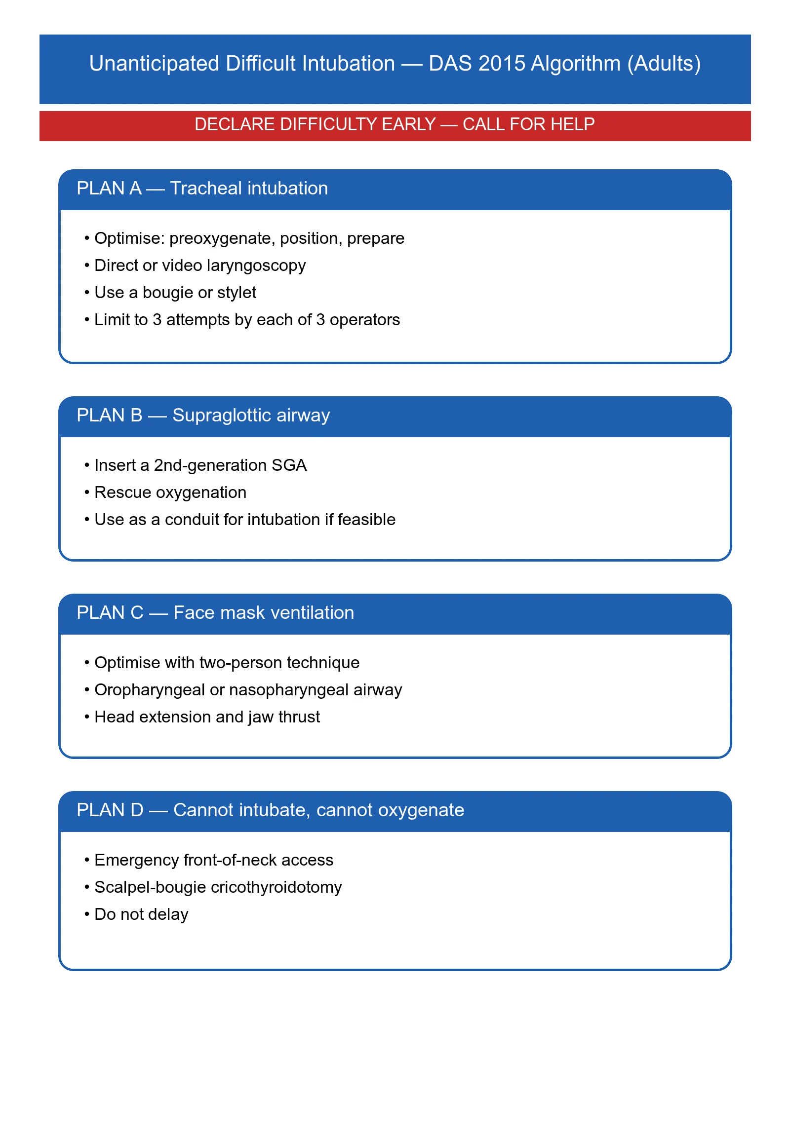
- Exam point. The DAS 2015 algorithm for unanticipated difficult intubation: Plan A (intubation) fails → Plan B (supraglottic airway rescue) → if ventilation adequate, "wake the patient up"; if ventilation fails, Plan C (CICO) → front-of-neck access.[3] Excellent BVM keeps the patient alive through Plans A, B and C.
Preoxygenation and optimising oxygenation before intubation
The single most effective defence against peri-intubation hypoxia is thorough preoxygenation before the induction of apnoea. The aim is to maximise the alveolar oxygen reservoir (denitrogenation) and, in the critically ill, to recruit lung and maintain oxygenation during the apnoeic period.[8][9]
- Why it matters. Critically ill patients desaturate far faster than elective patients — sepsis, shunt, low FRC, obesity and high VO2 collapse the safe apnoea time to under a minute. A well-preoxygenated patient may tolerate 5-8 minutes of apnoea; a poorly prepared one may desaturate before the tube is even passed.[8]
- Targets. End-tidal oxygen >85-90% (the most reliable bedside index of denitrogenation) or SpO2 ≥97-99% on 100% oxygen for 3 minutes of tidal breathing, or 8 vital-capacity breaths of 100% O2 (the latter is faster but less reliable in the dyspnoeic).[8]
- Methods.
- Non-rebreather mask + BVM (15 L/min, good mask seal) — the traditional method.
- Non-invasive ventilation (NIV/CPAP) for preoxygenation — the PREOxygenise trial (NEJM 2024) showed that NIV for preoxygenation reduced hypoxaemia during intubation of critically ill adults compared with high-flow oxygen by mask. NIV is preferred in the hypoxaemic, obese, or ARDS patient.[7]
- High-flow nasal cannula (HFNC) / THRIVE — 30-70 L/min humidified O2 by nasal cannula. Provides apnoeic oxygenation through the apnoeic period and some PEEP-like recruitment, but the FANS trial found that nasal apnoeic oxygenation alone did not reduce desaturation in the critically ill when added to face-mask preoxygenation — so it is an adjunct to, not a replacement for, good mask preoxygenation.[1][6]
- PREOPTI-DAM (eClinicalMedicine 2023) found HFNC comparable to facemask preoxygenation in anticipated difficult airway management — HFNC is a reasonable alternative when mask seal is poor.[11]
Apnoeic oxygenation and THRIVE
During apnoea, oxygen continues to diffuse from the patent upper airway into the alveoli and is taken up by blood (an entirely passive mass-flow phenomenon driven by the alveolar-to-arterial oxygen gradient, ~250 mL/min), while CO2 accumulates (~6-8 mmHg/min) because it is far less diffusible. Delivering high-flow oxygen into the pharynx during apnoea therefore sustains oxygenation long after a patient on room air would have desaturated.[1][9]
- THRIVE (Transnasal Humidified Rapid-Insufflation Ventilatory Exchange) — humidified nasal O2 at 70 L/min — extended mean apnoea time from ~7 minutes to over 20 minutes in carefully selected, appropriately-positioned patients in the original series (Patel & Nouraei, Anaesthesia 2015).[1]
- Caveats. THRIVE is an oxygenation technique, not a ventilatory one: PaCO2 rises throughout, eventually causing acidosis; it does not work well in the obstructed, obese, or "full-stomach" patient where the airway is not patent; and in the critically ill the FANS trial tempered expectations of the naso-apnoeic component. Use it to buy time, not to replace ventilation.[1][6]
The PEEP valve during preoxygenation and BVM
In the hypoxic, shunting patient (ARDS, pulmonary oedema, pneumonia), adding 5-10 cmH2O PEEP during preoxygenation and BVM recruits alveoli and improves oxygenation — combine HFNC/NIV with PEEP-valve BVM, target SpO2 ≥95% before induction, and apply PEEP-valve BVM during the apnoeic period of an RSI. The principle: denitrogenate, recruit, and oxygenate, then maintain oxygenation through apnoea.[7][8]
Opening the obstructed airway — the first 60 seconds
- RECOGNISE obstruction — look (see-saw, recession, cyanosis), listen (snore, gurgle, stridor, or ominous silence), feel (no airflow). Any unconscious patient with respiratory effort but no air movement has a threatened airway.[1]
- CALL FOR HELP AND OXYGEN — high-flow O2 via a non-rebreather mask at 15 L/min while you work. Get the BVM, suction and adjuncts to the bedside.
- MANOEUVRE — if no C-spine concern, head-tilt/chin-lift; if C-spine cannot be cleared (trauma, ICU, arrest), jaw thrust with both hands behind the angles of the mandible.
- RE-ASSESS — chest movement? airflow? If not, SUCTION any fluid, then reposition.
- ADJUNCT — if the patient is deeply unconscious (no gag), insert a sized oropharyngeal (Guedel) airway; if semi-conscious or a Guedel is contraindicated, insert a nasopharyngeal airway (nostril-to-tragus; not if base-of-skull fracture).
- BVM — attach reservoir + 15 L/min O2 + PEEP valve; two-handed C-E grip (or two-person). Squeeze at 10-12/min to visible chest rise.
- CONFIRM — bilateral chest rise + waveform capnography.
- ESCALATE as needed: BVM → supraglottic airway (LMA/i-gel) → definitive intubation → front-of-neck access (CICO).[1][3]
Two-person bag-valve-mask ventilation — step by step
- POSITION AND EQUIPMENT — patient supine, operator 1 at the head of the bed (sniffing position if no C-spine concern). Select an appropriately-sized mask (spanning nasal bridge to chin); attach the reservoir bag, run 15 L/min O2, and fit a PEEP valve (5-10 cmH2O) in the hypoxic patient.[1]
- OPEN THE AIRWAY — operator 1 performs a bilateral jaw thrust; insert an oropharyngeal or nasopharyngeal adjunct if appropriate.
- SEAL — two-handed C-E grip — thumbs and index fingers of both hands form "C"s over the mask dome; the three remaining fingers of each hand form "E"s along the mandible and lift the face up into the mask (jaw thrust + seal in one).
- VENTILATE — operator 2 squeezes the bag — one breath every 5-6 seconds (10-12/min) with just enough volume for visible chest rise (~500 mL). Avoid hyperventilation and over-inflation.
- CONFIRM — bilateral chest rise, good colour, and a capnograph waveform. If no chest rise: recheck seal, reposition head/jaw, suction, consider a different mask size, add the third assistant ("triple airway manoeuvre"), and if still failing escalate to a supraglottic airway.[1]
- ROTATE — the airway operator's hands fatigue within minutes; swap operators every 2 minutes, like chest compressions.
Preoxygenation strategies for the critically ill patient about to be intubated
| Method | Delivered FiO2 | Provides apnoeic O2 | Evidence / role |
|---|---|---|---|
| Non-rebreather mask | 60-90% | No | Quick, ubiquitous; inferior seal |
| Bag-valve-mask (good seal) + reservoir + 15 L/min | 90-100% | No (unless left flowing over face) | Traditional standard preoxygenation |
| NIV / CPAP for preoxygenation | ~100% | No | PREOxygenise (NEJM 2024): reduced hypoxaemia in the critically ill — preferred when hypoxaemic[7] |
| HFNC / THRIVE (nasal, 30-70 L/min) | Up to 100% | Yes | Extends apnoea time; FANS: added nasal apnoeic O2 did not reduce desaturation alone; PREOPTI-DAM: comparable to mask[1][6][11] |
| BVM + PEEP valve | 90-100% | During BVM only | Recruits alveoli, improves oxygenation in pulmonary oedema/ARDS/obesity[1] |
Clinical pearls
Key evidence
NAP4 — the Fourth National Audit Project of the Royal College of Anaesthetists and the Difficult Airway Society (PMID 21447489)
Design. A year-long national census of major airway complications in the UK (~2.9 million general anaesthetics, ~13,900 ICU admissions, ~5000 ED intubations), reporting 184 cases of death/brain damage/ergency surgical airway. [1]
Headline findings. (i) Poor basic airway management and unrecognised oesophageal intubation contributed to harm; (ii) the supraglottic airway was under-used as a rescue device — repeated attempts at intubation and failure to "declare failure" were recurring themes; (iii) ICU and ED airway events had worse outcomes than theatre events; (iv) aspiration of gastric contents was a leading cause of death; (v) obesity was over-represented. [1]
Practice change. Reinforced: master basic BVM; have a supraglottic airway and capnography immediately available in ICU/ED; limit intubation attempts; declare failure and move to rescue early; routine capnography for every intubated ICU patient.[2]
PREOxygenise — Noninvasive Ventilation for Preoxygenation during Emergency Intubation (Gibbs/Semler, NEJM 2024; PMID 38869091)
Design. Pragmatic multicentre randomised trial of NIV for preoxygenation vs usual care (high-flow O2 by mask) in 1301 critically ill adults undergoing emergency intubation across US ICUs/EDs. [1]
Population. Adults with respiratory failure (mostly hypoxaemic) needing emergency intubation. [1]
Primary outcome. Lowest SpO2 during the peri-intubation period. [1]
Headline finding. NIV preoxygenation reduced the incidence and severity of hypoxaemia (fewer patients desaturating to SpO2 <80%) compared with usual-care mask preoxygenation. No signal of increased aspiration despite the non-invasive positive pressure. [1]
Practice change. In the hypoxaemic critically ill patient, prefer NIV (or NIV + nasal apnoeic oxygenation) for preoxygenation over a simple high-flow mask.[7]
FANS — Apneic Oxygenation during Intubation of the Critically Ill (Semler, AJRCCM 2016; PMID 26426458)
Design. Single-centre randomised trial of nasal apnoeic oxygenation (15 L/min nasal cannula) vs no apnoeic oxygenation, added to face-mask preoxygenation, in 150 critically ill adults. [1]
Headline finding. No significant difference in the lowest SpO2 during intubation. Apnoeic oxygenation by low-flow nasal cannula did not prevent desaturation in the critically ill when added to mask preoxygenation. [1]
Practice change. Tempered enthusiasm for low-flow nasal apnoeic oxygenation alone; high-flow (THRIVE) and NIV preoxygenation are the techniques with the strongest evidence in the critically ill.[6]
THRIVE — Patel & Nouraei, Anaesthesia 2015 (PMID 25388828)
Design. Proof-of-concept physiological study: 25 patients at risk of difficult airway preoxygenated, then given transnasal humidified rapid-insufflation ventilatory exchange (70 L/min) during apnoea. [1]
Headline finding. Mean safe apnoea time rose from ~7 minutes (historical) to over 20 minutes without desaturation, in appropriately selected, well-positioned patients. [1]
Practice change. Established THRIVE as a powerful apnoeic-oxygenation adjunct for elective and selected emergency cases — but it is an oxygenation technique, not a ventilatory one (PaCO2 rises), and does not work when the airway is obstructed or in the morbidly obese/obstructed "full-stomach" patient.[1]
Prognosis
Basic airway competence prevents the hypoxic brain injury and cardiac arrest that follow an unrecognised or poorly managed obstructed airway. In every difficult-airway algorithm, the difference between a manageable crisis and a disaster is the quality of BVM ventilation and oxygenation while help arrives.[1][1]
[1]SAQ — The unconscious patient with a noisy then silent airway
10 minutes · 10 marks
You are called to a 75-year-old man on the ward who has become unresponsive after receiving morphine for hip fracture pain. On arrival he has see-saw chest-wall movement, sonorous snoring respirations at 8/min, SpO2 84% on room air, and then abruptly the snoring stops while the see-saw effort continues. Describe your immediate stepwise management of the airway.
SAQ — Preoxygenation strategy before emergency intubation of the critically ill
10 minutes · 10 marks
A 55-year-old woman with severe pneumonia (SpO2 88% on a non-rebreather mask at 15 L/min, RR 32, BP 95/60) needs emergency intubation. She is obese (BMI 38). Outline your preoxygenation and apnoeic-oxygenation strategy, citing the relevant trials.
Red flags
References
- [1]Patel A, Nouraei SA. Transnasal Humidified Rapid-Insufflation Ventilatory Exchange (THRIVE): a physiological method of increasing apnoea time in patients with difficult airways Anaesthesia, 2015.PMID 25388828
- [2]Cook TM, Woodall N, Frerk C (eds); Fourth National Audit Project. Major complications of airway management in the UK: results of the Fourth National Audit Project of the Royal College of Anaesthetists and the Difficult Airway Society. Part 2: intensive care and emergency departments Br J Anaesth, 2011.PMID 21447489
- [3]Frerk C, Mitchell VS, McNarry AF, Mendonca C, Bhagrath R, Patel A, O'Sullivan EP, Woodall NM, Ahmad I; Difficult Airway Society intubation guidelines working group. Difficult Airway Society 2015 guidelines for management of unanticipated difficult intubation in adults Br J Anaesth, 2015.PMID 26556848
- [4]Langeron O, Masso E, Huraux C, Guggiari M, Bianchi A, Coriat P, Riou B. Prediction of difficult mask ventilation Anesthesiology, 2000.PMID 10781266
- [5]Kheterpal S, Martin L, Shanks AM, Tremper KK. Prediction and outcomes of impossible mask ventilation: a review of 50,000 anesthetics Anesthesiology, 2009.PMID 19293691
- [6]Semler MW, Janz DR, Lentz RJ, et al.; FELLOW Investigators and the Pragmatic Critical Care Research Group. Randomized Trial of Apneic Oxygenation during Endotracheal Intubation of the Critically Ill Am J Respir Crit Care Med, 2016.PMID 26426458
- [7]Gibbs KW, Semler MW, Driver BE, et al.; Pragmatic Critical Care Research Group. Noninvasive Ventilation for Preoxygenation during Emergency Intubation N Engl J Med, 2024.PMID 38869091
- [8]Tanoubi I, Drolet P, Donati F. Optimizing preoxygenation in adults Can J Anaesth, 2009.PMID 19399574
- [9]El-Orbany M. Apneic oxygenation during emergency airway management Can J Anaesth, 2019.PMID 30112712
- [10]Chrimes N, Higgs A, Rehak A, et al. Lost in transition: the challenges of getting airway clinicians to move from the upper airway to the neck during an airway crisis Br J Anaesth, 2020.PMID 32475685
- [11]Vourc'h M, Huard D, Le Penndu M, et al. High-flow oxygen therapy versus facemask preoxygenation in anticipated difficult airway management (PREOPTI-DAM): an open-label, single-centre, randomised controlled phase 3 trial EClinicalMedicine, 2023.PMID 37251624
- [12]Heidegger T, Gerig HJ. Algorithms for management of the difficult airway Curr Opin Anaesthesiol, 2004.PMID 17031079