ICU · Airway management
Extubation & Its Complications — Stridor, Laryngospasm, Pulmonary Oedema
Also known as Extubation · Extubation failure · Post-extubation stridor · Laryngeal oedema · Laryngospasm · Larson manoeuvre · Cuff leak test · Rapid shallow breathing index · Negative-pressure pulmonary oedema · Post-obstructive pulmonary oedema · Reintubation · Post-extubation dysphagia · Unplanned extubation · Airway-exchange catheter
Extubation is the planned removal of the endotracheal tube once a spontaneous breathing trial is tolerated and airway protection is adequate; it carries a 10-20 per cent reintubation rate. The serious complications are post-extubation stridor (laryngeal oedema — predicted imperfectly by the cuff-leak test, reduced by prophylactic methylprednisolone or dexamethasone in high-risk patients), laryngospasm (treat with 100 per cent oxygen, CPAP, the Larson manoeuvre and jaw thrust; a small dose of suxamethonium if refractory), negative-pressure pulmonary oedema from forceful inspiration against a closed glottis, aspiration of subglottic secretions, post-extubation dysphagia (40-60 per cent), and unplanned extubation. Plan high-risk extubations with senior staff and an airway-exchange catheter.
On this page & tools
Your progress
Saved locally on this device.
Target exams
Overview & definition
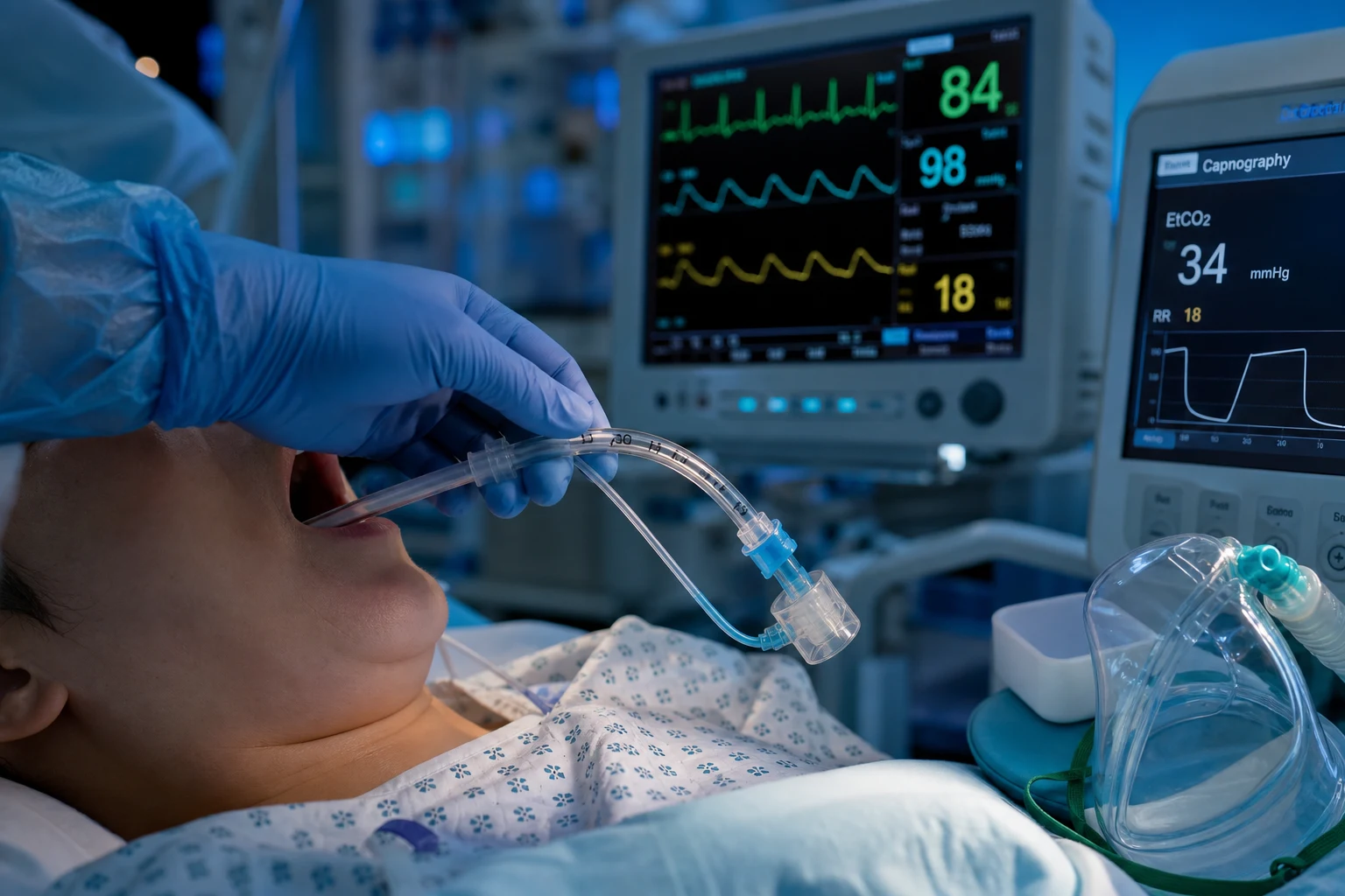
Extubation is the planned removal of the endotracheal tube once the patient can protect their airway and breathe spontaneously without the tube. It is the final step of weaning, and — like intubation — it is a high-risk airway event: roughly 10-20 per cent of ICU extubations require reintubation within 48-72 hours, and the immediate complications include laryngospasm, post-extubation stridor (laryngeal oedema), negative-pressure pulmonary oedema, aspiration, and — over the longer term — post-extubation dysphagia. Anticipate and prepare for them as deliberately as for intubation. In international audits (the ICUs-of-the-future ICE-ARI study and the multicentre VAP/NIV cohorts), nearly half of all extubations are unplanned-complication-free, but around one in twelve is associated with a serious adverse event; mortality rises steeply when reintubation is required.[1]
Readiness to extubate
Before extubation, confirm both liberation from the ventilator and the ability to protect the airway:[1]
- A passed spontaneous breathing trial with no distress, stable respiratory rate, and adequate gas exchange.
- The patient awake and cooperative (able to follow commands, protect the airway, cough).
- Low ventilator support (low PEEP, low FiO2) and minimal secretions that can be cleared.
- Haemodynamic stability and a manageable (not "difficult") airway for reintubation if needed.
- Adequate cough and a positive cuff-leak test in the patient intubated for a prolonged time or at high risk of oedema.[1][1]
The decision to extubate is therefore a two-gate decision: (1) the patient is liberated from the ventilator (the weaning gate, demonstrated by a successful spontaneous breathing trial), and (2) the patient can protect and maintain their airway without the tube (the airway-protection gate). The weaning indices below address the first gate; the cuff-leak test and the clinical criteria of cough, secretions, and consciousness address the second. [1]
Weaning indices and predictors of extubation success
The classical Yang and Tobin (1991) study compared a basket of weaning indices in a prospective cohort and found that the rapid shallow breathing index (RSBI) — the ratio of respiratory rate to tidal volume (f/Vt) — was the single best discriminator of weaning outcome. An RSBI under 105 breaths/min/L during a brief trial of minimal support predicted successful liberation with a sensitivity and specificity around 0.78.[3] It is a useful rule-in test: an RSBI well below 105 in a relaxed, un-distressed patient supports the decision to extubate. It is, however, a poor rule-out test — more than one in four failures are not flagged by it — and a single number never overrides the clinical picture of distress, accessory-muscle use, diaphoresis, or rising CO2.[1]
The other commonly cited indices carry the same limitation: each is statistically associated with success, but no single index is sufficiently accurate to gate extubation on its own. [1]
Weaning indices — what each measures, the threshold, and its usefulness
| Index | What it measures | Threshold favouring success | Sensitivity / specificity | Practical use |
|---|---|---|---|---|
| RSBI (f/Vt) | Rapid shallow breathing | <105 breaths/min/L | ~0.78 / ~0.78 (Yang & Tobin) | Best single index; rule-in only |
| Maximal inspiratory pressure (MIP / NIF) | Respiratory-muscle force | More negative than −20 to −30 cmH2O | Moderate | Strength of the diaphragm/accessory muscles |
| P0.1 (airway occlusion pressure) | Central respiratory drive | 0.5-3.0 cmH2O is normal; >4-6 suggests high load/fatigue | Moderate | High P0.1 = impending fatigue |
| Glasgow Coma Scale (GCS) | Airway-protection capacity | GCS >8 (able to follow commands, cough) | Moderate | Cognition < airway-protection gate |
| Cough strength (qualitative 0-5) | Secretion clearance | Strong / able to cough on command | Moderate | Cough peak flow >60 L/min favours success |
| Cuff-leak test | Laryngeal patency | Leak >110 mL or >15-24% of Vt | Modest sensitivity, higher specificity | High-risk patient only |
| Secretion burden (qualitative) | Load to clear | Low / manageable | Moderate | Excess secretions predict failure |
The P0.1 (airway-occlusion pressure at 100 ms) deserves a viva-ready explanation. Occluding the airway at end-expiration and measuring the negative pressure the patient generates in the first 100 ms quantifies the neural drive to breathe — the first 100 ms is too short for conscious correction or for reflex adjustments, so it reflects the brainstem's command. A high P0.1 (above 4-6 cmH2O) means the respiratory centre is being driven hard to overcome load, and is a marker of impending fatigue; a low or normal P0.1 with a good tidal volume suggests comfortable, sustainable breathing. Modern ventilators display it non-invasively.[1]
The maximal inspiratory pressure (MIP, also NIF) measures the most negative pressure the patient can generate against an occluded airway. A MIP more negative than −30 cmH2O (some units use −20 to −40) suggests adequate respiratory-muscle reserve. MIP is effort-dependent and falls with poor cooperation, so the most negative value obtained over several one-minute attempts is the one to record. Both MIP and P0.1 are commonly asked in the equipment and respiratory physiology viva.[1]
The cuff-leak test and prophylactic steroids
The cuff-leak test quantifies the air leak when the cuff is deflated: a small or absent leak indicates laryngeal narrowing and predicts post-extubation stridor. The classical cut-offs are a leak under 110 mL or under 15-25 per cent of the delivered tidal volume; a leak under 24 per cent of tidal volume is the threshold used by Jaber and adopted in many ICUs.[4] However, the most rigorous synthesis — the Kuriyama 2020 systematic review and meta-analysis — showed the cuff-leak test has only modest sensitivity and specificity, with a marked risk-stratifying role but an unacceptably high false-positive rate if used as the sole gatekeeper. A positive test (low leak) doubles to triples the odds of stridor, but most patients with a low leak do not develop stridor. Use the cuff-leak test to stratify high-risk patients — those who warrant prophylactic steroids and senior presence at extubation — not to delay extubation in a patient who is otherwise ready.[4][5][9]
In the high-risk patient (prolonged intubation, large tube, traumatic intubation, female, obese, high cuff pressure, reintubation, or a low cuff leak), prophylactic corticosteroid given before extubation reduces post-extubation laryngeal oedema and stridor. The strongest single trial is François (Lancet 2007), in which a 12-hour, four-dose methylprednisolone pretreatment (20 mg every 6 hours, starting 12 hours before the planned extubation) reduced post-extubation laryngeal oedema from 22 per cent to 3 per cent and the need for reintubation from 8 per cent to 4 per cent in patients with a low cuff leak.[2] A randomised trial showed dexamethasone given before extubation reduces post-extubation airway obstruction in adults at risk (Lee, Critical Care 2007).[1] The Cochrane meta-analysis (Markovitz/Randolph) supports a benefit in adults and children but emphasises heterogeneity, and the question of whether steroids should be given to all patients (universal prophylaxis) versus only high-risk patients remains debated; most guidelines favour selective use in the high-risk group because universal prophylaxis exposes many patients who would never develop stridor to hyperglycaemia, infection, and the other steroid burdens.[1][6]
The extubation technique
- Explain to the patient; pre-oxygenase; position semi-upright.
- Suction the mouth and above the cuff (subglottic secretions pool above the cuff and are aspirated when it is deflated) before deflation.
- Deflate the cuff fully; remove the tube at peak inspiration (the cords are widest), with a cough and a breath out.
- Apply oxygen (high-flow nasal cannula or a face mask) and reassess.[1]
The safe extubation sequence — the structured approach the examiner wants
- Confirm readiness — passed spontaneous breathing trial, RSBI under 105 (or clinically appropriate), GCS sufficient to protect airway, cough adequate, secretions manageable, cuff leak adequate (or steroid pretreatment given if low).
- Plan the disposition — high-risk patient? Senior present at bedside. Difficult-airway drone? Airway-exchange catheter ready. Back-up plan verbalised before the tube comes out.
- Position and pre-oxygenate — semi-upright, head of bed 30-45 degrees, 100 per cent oxygen for 3-5 minutes; suction the mouth and oropharynx.
- Suction above the cuff — subglottic suction (or a catheter above the cuff) clears the pooled secretions that would otherwise drop into the trachea when the cuff is deflated.
- Deflate the cuff fully — verify it is fully down (no residual air).
- Remove at peak inspiration — the cords are widest at peak inspiration; the patient takes a breath in, the tube is withdrawn in a smooth single motion, the patient coughs and breathes out.
- Apply oxygen immediately — high-flow nasal cannula or face mask at the planned FiO2; in high-risk patients, HFNC reduces reintubation versus conventional oxygen (the post-extubation HFNC trials).
- Reassess — work of breathing, voice, stridor, oxygenation, ability to clear secretions. Stay at the bedside for the first 10-15 minutes; the complications cluster in this window.
- Document — time, technique, complications, plan for the next 24 hours. NBM for at least 4 hours (or until a swallow assessment); elevate head of bed.
Complications
Laryngospasm
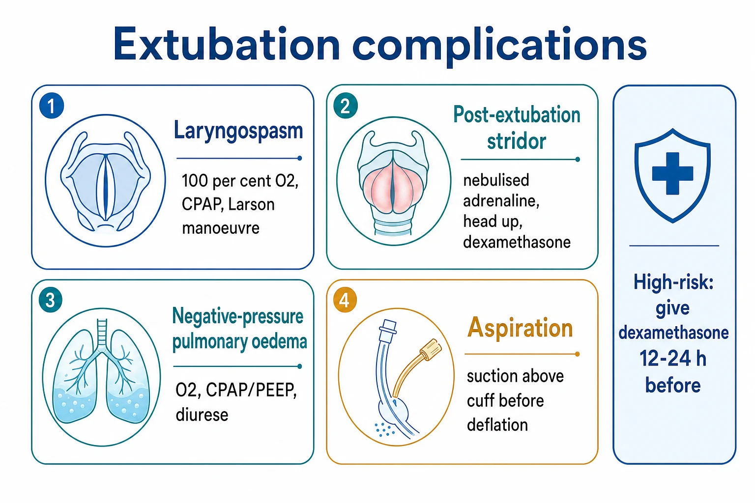
The vocal cords close reflexively, obstructing the airway. It is the most feared immediate complication because it progresses within 30-90 seconds to hypoxia and — if the patient inspires forcefully against the closed glottis — negative-pressure pulmonary oedema. The reflex is driven by irritation of the superior laryngeal nerve (blood, secretions, or the tube itself) and is commonest at emergence from anaesthesia and in the first minutes after extubation.[1]
Treat immediately and in a fixed sequence. The first move is 100 per cent oxygen and forward jaw thrust — pull the mandible forward and open the airway, which stretches the thyrohyoid membrane and mechanically distracts the laryngeal inlet, and apply continuous positive airway pressure (5-10 cmH2O via a bag-mask or Mapleson circuit) to push the cords apart. The Larson manoeuvre — firm bilateral pressure at the "laryngospasm notch" (behind the ear lobe, between the ascending ramus of the mandible and the mastoid process, inward and forward toward the styloid process) — is both diagnostic and therapeutic; firm, sustained pressure often breaks the spasm within seconds. If refractory, a small dose of propofol (0.5-1 mg/kg) deepens anaesthesia and relaxes the cords; if that fails, a small dose of suxamethonium (0.1-0.5 mg/kg IV, or 2-4 mg/kg IM if no IV access) is the definitive breaker of refractory laryngospasm — be prepared to bag-mask, and rarely to reintubate. Untreated laryngospasm causes hypoxia and negative-pressure pulmonary oedema.[1]
Post-extubation stridor (laryngeal oedema)
Inspiratory stridor within minutes to hours of extubation, from laryngeal (glottic and subglottic) oedema. The oedema is caused by ischaemic pressure injury from the cuff against the mucosa, by traumatic intubation, or by repeated intubation attempts; it narrows the glottic chink and produces turbulent, audible, inspiratory airflow obstruction. The classical incidence is 2-22 per cent, higher in the high-risk patient.[4]
Risk factors (the examiner wants these verbatim): female sex (narrower larynx), traumatic or repeated intubation, large tube relative to laryngeal calibre, prolonged intubation (over 36-72 hours, especially over 5-7 days), high cuff pressure, reintubation, obese or short neck, and a low cuff leak at extubation. Jaber (2003) found female sex, a longer intubation, and a cuff leak under 24 per cent to be the independent predictors in ICU adults.[4]
Post-extubation stridor — risk factors, mechanism, and what to do
| Risk factor | Mechanism | Relative contribution | Pre-extubation response |
|---|---|---|---|
| Female sex | Narrower laryngeal calibre | Moderate-high | Use the smallest effective tube; consider steroid pretreatment |
| Traumatic / repeated intubation | Mucosal injury, oedema at intubation | High | Senior operator; minimise attempts |
| Reintubation | Cumulative mechanical injury | High | Steroid pretreatment; consider early tracheostomy |
| Prolonged intubation (>36-72 h) | Sustained cuff pressure ischaemia | High | Cuff-leak test; 12-h methylprednisolone if low leak |
| Large tube | Excess cuff-to-larynx pressure | Moderate | Right-size the tube |
| High cuff pressure (>25-30 cmH2O) | Mucosal ischaemia | High | Measure cuff pressure daily; keep <25 cmH2O |
| Low cuff leak | Indicates laryngeal narrowing | High | Steroid pretreatment; senior at extubation |
| Obesity / short neck / OSA | Narrowed upper airway, reflux | Moderate | Position upright; consider NIV/HFNC post-extubation |
Treatment is a graded ladder. Sit the patient up (head of bed 30-45 degrees to reduce venous congestion), give humidified oxygen, and administer nebulised adrenaline (racemic epinephrine 0.5 mL of 2.25% in 2.5 mL saline, or simple L-adrenaline 5 mg) — the vasoconstriction reduces oedema within minutes. Give systemic dexamethasone (8 mg IV, then 8 mg every 8 hours for 3-4 doses, or equivalent) — the onset is delayed by 4-6 hours, so it does not rescue the immediate crisis but reduces the second wave of oedema. Heliox (helium-oxygen 80:20 or 70:30) reduces the work of breathing through the narrowed airway because helium is less dense than nitrogen, lowering turbulent-flow resistance. Severe stridor with respiratory distress, fatigue, rising CO2, or failure to maintain oxygenation needs reintubation — re-intubate early rather than allow a periarrest deterioration, and have the airway trolley ready.[1][1][2]
Post-extubation stridor — the graded treatment ladder
- Recognise — inspiratory stridor, increased work of breathing, desaturation within minutes-hours of extubation.
- Position — head of bed up 30-45 degrees; sit the patient forward if tolerated.
- Oxygen — humidified 100 per cent via face mask; consider HFNC.
- Nebulised adrenaline (racemic epinephrine) — 0.5 mL of 2.25% racemic in 2.5 mL saline, or 5 mg L-adrenaline; repeat every 1-2 hours as needed.
- Dexamethasone IV — 8 mg, then 8 mg every 8 hours for 3-4 doses (onset 4-6 h — for the second wave of oedema).
- Heliox (80:20 or 70:30) — reduces work of breathing through the narrowed glottis.
- Assess for intubation — fatigue, rising PaCO2, persistent desaturation, or worsening stridor despite the above → reintubate early; senior airway operator at bedside.
- Investigate — once stabilised: nasendoscopy to assess cord movement and oedema grade; consider laryngeal cause (ulceration, granuloma, vocal-cord paralysis).
Negative-pressure pulmonary oedema (post-obstructive)
Forceful inspiration against a closed glottis (laryngospasm or upper-airway obstruction) generates markedly negative intrathoracic pressure — down to −50 to −100 cmH2O in the distressed patient, against the normal −4 to −8 cmH2O. The negative pressure increases venous return, increases pulmonary capillary hydrostatic pressure, stresses the alveolar-capillary membrane, and floods the lungs with oedema fluid. It presents with hypoxia and pink frothy secretions after an obstructive event (most often laryngospasm, but also biting the tube, a kinked tube, or any cause of upper-airway obstruction). Treat with oxygen, positive pressure (CPAP/PEEP — NIV if the patient is awake, intubation and mechanical ventilation if severe), and diuresis; some patients need reintubation. It is largely self-limiting once the obstruction is relieved, with resolution over 12-24 hours, but can be life-threatening in the first hours. The lesson is prevention: break laryngospasm early, and pre-empt biting and obstruction.[1]
Aspiration
Subglottic secretions pooled above the cuff are aspirated when the cuff is deflated; gastric contents may also be aspirated if the stomach is full. Prevent it by suctioning above the cuff before deflation (subglottic suction catheter or a catheter passed above the cuff), by ensuring the stomach is as empty as feasible (defer extubation after a large feed, consider gastric decompression), and by extubating with the head of bed up. The aspirated material is typically a mix of oropharyngeal flora and gastric acid; the result ranges from a chemical pneumonitis (Mendelson, sterile, sterile infiltrate within hours) to a true aspiration pneumonia (bacterial, progressive over 24-72 hours). Antibiotics are not given routinely for immediate aspiration pneumonitis — observe and treat only if the pneumonia develops; give empiric antibiotics for aspiration pneumonia (clindamycin/amoxicillin-clavulanate/piperacillin-tazobactam, with anaerobic cover).[1]
Post-extubation dysphagia
Post-extubation dysphagia is common, under-recognised, and a major driver of post-extubation pneumonia, prolonged admission, and readmission. The systematic review and meta-analysis by McIntyre (2021) found an overall incidence of 41 per cent in critically ill adults (41 per cent pooled across 23 studies), with rates of 40-60 per cent in the longer-intubated, neurological, cardiac-surgical, and elderly populations. The mechanism is multifactorial: cuff-related laryngeal injury (ulceration, granuloma, vocal-cord paresis — documented endoscopically in 80-100 per cent of patients intubated over 24-48 hours in the LIFI cohort of Brodsky 2018), restricted laryngeal elevation from disuse and muscle weakness, sensory changes from the tube, and residual critical-illness weakness. Risk factors include intubation over 48 hours, age over 65, female sex, repeated intubation, a large tube, high cuff pressure, nasogastric tube, and pre-existing neurological disease.[7][8]
Diagnosis: a high index of suspicion in every post-extubation patient. Bedside swallow screens (the volume-viscosity swallow test, the 3-oz water swallow test) are first-line; formal assessment is by videofluoroscopic swallow study (VFSS) (the radiological gold standard — modified barium swallow at varied consistencies) or fibreoptic endoscopic evaluation of swallowing (FEES). The patient should remain nil by mouth until a swallow assessment clears them; if dysphagia is present, speech-language-pathology-guided texture modification, swallowing rehabilitation, and (if severe) feeding-tube placement are the management. Most post-extubation dysphagia resolves within 2-6 weeks, but a substantial minority persist for months and contribute to post-intensive-care syndrome.[7][8]
Unplanned extubation
Unplanned extubation — the unintended removal of the endotracheal tube (deliberate self-extubation by the patient, or accidental removal during nursing, procedures, or transport) — occurs in 0.5-4 per cent of intubated ICU days. Roughly half are deliberate (self-extubation) and half accidental. The classical Epstein study (AJRCCM 2000) showed that the impact depends on whether the patient was ready: a patient who self-extubates and does not require reintubation (about 40-60 per cent of cases, often already weaning) has a benign or even favourable course, but a patient who self-extubates and requires reintubation has a markedly increased morbidity, mortality, prolonged ICU stay, and the risk of an hypoxic event during the urgent reintubation. Prevention is by adequate sedation and analgesia (avoid both under- and over-sedation), secure tube fixation, delirium prevention, communication with the patient, and protocols for safe turning, transport, and procedures. The response is immediate: assess the patient, oxygenate (bag-mask), call for senior airway help, and reintubate if the patient was not ready for extubation or cannot maintain their airway — do not be falsely reassured by an initially adequate saturation.[10][1]
Failed extubation
Failed extubation is the need for reintubation within 48-72 hours of a planned extubation. The rate is 10-20 per cent in general ICU cohorts, higher in specific groups (over 20 per cent in neuro/elderly/cardiac, up to 30 per cent in those intubated for more than 7 days or with pre-existing lung disease). Reintubation is associated with a two- to ten-fold increase in mortality, with prolonged ICU and hospital stay, and with a higher rate of ventilator-associated pneumonia. The reasons are heterogenous: respiratory failure (the commonest — fatigue, secretions, atelectasis, residual sedation, fluid overload, cardiac failure unmasked by the loss of positive pressure), upper-airway obstruction (stridor, laryngeal oedema), failure to protect the airway (aspiration, poor cough, depressed consciousness), and cardiovascular instability. The reintubation is often harder than the original intubation because of laryngeal oedema, secretions, reduced reserve, and an agitated or hypoxic patient — so every planned extubation should be staffed and equipped for the possibility of an immediate, more difficult reintubation. The post-extubation HFNC and prophylactic-NIV trials (notably in hypercapnic and cardiac-failure patients) reduce reintubation in selected groups.[1]
Other complications
Hoarseness and sore throat (common, transient — over 50 per cent in the first 24 hours, resolving over days), vocal-cord injury (ulceration, granuloma, or recurrent-laryngeal-nerve palsy — the LIFI cohort documents laryngeal injury in 80-100 per cent at 24-48 hours and persistent upper-airway symptoms in 30-50 per cent at weeks), and late tracheal stenosis from cuff injury (weeks to months, presents with progressive dyspnoea and stridor).[8]
High-risk extubation
Some extubations warrant senior staff, an airway-exchange catheter left in situ (a long, soft, hollow catheter passed through the endotracheal tube into the trachea before extubation, left in place after the tube is removed, and used to "railroad" a new tube over if reintubation is needed — it preserves the airway and the route back in), advanced airway devices ready, and a plan agreed in advance: the difficult airway (where intubation was hard the first time), the cardiac or neurovascular patient with little reserve (in whom a brief desaturation is catastrophic), and the neuromuscular or diaphragmatic weakness patient (GBS, myasthenia, high cervical cord injury, post-cardiac-surgery diaphragmatic palsy). The reintubation may be harder than the original intubation (oedema, secretions, reduced reserve), so plan for it. In the patient intubated for a prolonged period or with a low cuff leak, give prophylactic methylprednisolone 12 hours before the planned extubation (the François regimen — four doses of 20 mg every 6 hours, starting 12 hours pre-extubation), and have an airway-exchange catheter, a difficult-airway trolley, and a senior airway operator at the bedside.[1][2]
Pharmacology of extubation adjuncts
The pharmacological and device adjuncts for extubation — what each does, the dose, and when to use it
| Adjunct | Mechanism / role | Dose / setup | When to use | Caveat |
|---|---|---|---|---|
| Methylprednisolone (prophylactic) | Reduces laryngeal oedema if started before extubation | 20 mg IV every 6 h × 4 doses, starting 12 h pre-extubation (François regimen) | High-risk: low cuff leak, prolonged intubation, female, reintubation | Hyperglycaemia, infection; not for universal use |
| Dexamethasone (prophylactic) | As above | 4-8 mg IV every 6 h × 3-4 doses pre-extubation | Alternative to methylprednisolone | Same steroid burdens |
| Nebulised adrenaline (racemic epinephrine) | Vasoconstriction reduces laryngeal oedema acutely | 0.5 mL of 2.25% racemic in 2.5 mL saline, or 5 mg L-adrenaline nebulised | Active post-extubation stridor | Rebound oedema when it wears off — observe 2-4 h |
| Dexamethasone (treatment) | Reduces the second wave of oedema | 8 mg IV, then 8 mg every 8 h × 3-4 doses | Active post-extubation stridor | Slow onset (4-6 h) — not the immediate rescue |
| Heliox (helium-oxygen) | Lower density reduces turbulent-flow resistance | 80:20 or 70:30 helium:oxygen via non-rebreather | Moderate stridor with high work of breathing | Not if FiO2 needed >40%; transitional |
| Suxamethonium | Breaks refractory laryngospasm | 0.1-0.5 mg/kg IV (or 2-4 mg/kg IM if no IV) | Refractory laryngospasm after propofol fails | Apnoea; bradycardia; prepare to bag/reintubate |
| Propofol | Deepens anaesthesia, relaxes cords | 0.5-1 mg/kg IV | Early refractory laryngospasm | Hypotension, apnoea |
| Airway-exchange catheter | Preserves the route back into the trachea | Sized to ETT; passed into trachea before extubation, left in situ | High-risk extubation (difficult airway, low cuff leak) | Cough, discomfort; remove once clearly safe |
Key trials and evidence
François 2007 (12-h methylprednisolone)
Lancet
Multicentre RCT, 698 intubated adults with a cuff leak <25% of tidal volume — 4 doses of methylprednisolone 20 mg IV every 6 h starting 12 h before extubation vs placebo
Key finding
Post-extubation laryngeal oedema 3% vs 22% (p<0.0001); reintubation for laryngeal oedema 4% vs 8% (p<0.02). No excess infection or hyperglycaemia-related harm.
Practice change
12-h, 4-dose methylprednisolone pretreatment is the standard prophylaxis for high-risk (low cuff leak) adults
Lee 2007 (dexamethasone)
Critical Care
Single-centre RCT in adults at risk — dexamethasone vs placebo before extubation
Key finding
Reduced post-extubation airway obstruction and stridor with dexamethasone
Practice change
Dexamethasone is a reasonable alternative to methylprednisolone for prophylaxis
Yang & Tobin 1991 (RSBI)
NEJM
Prospective cohort of 36 intubated adults — f/Vt (rapid shallow breathing index) compared with other weaning indices to predict weaning outcome
Key finding
RSBI <105 breaths/min/L was the single best predictor (sensitivity 0.78, specificity 0.78); outperformed minute ventilation, MIP, P0.1 alone
Practice change
RSBI became the standard weaning index at the bedside
Jaber 2003 (cuff-leak test)
Intensive Care Medicine
Prospective observational ICU cohort — cuff-leak test performed before extubation, stridor tracked
Key finding
Female sex, longer intubation, and a cuff leak <24% of tidal volume were independent predictors of post-extubation stridor
Practice change
Cuff-leak test adopted to stratify high-risk patients for steroid prophylaxis
Kuriyama 2020 (cuff-leak meta-analysis)
Critical Care
Systematic review and meta-analysis of the cuff-leak test in adults to predict post-extubation airway complications
Key finding
Modest sensitivity and specificity; useful for risk stratification, not as a sole gatekeeper (high false-positive rate)
Practice change
Use the cuff-leak test to select high-risk patients for steroids and senior presence, not to delay extubation alone
Markovitz/Randolph 2008 (Cochrane)
Cochrane Database Syst Rev
Cochrane review of corticosteroids to prevent and treat post-extubation stridor in neonates, children, and adults
Key finding
Benefit in adults and children for prevention; heterogeneity limits the universal-prophylaxis recommendation
Practice change
Selective prophylactic steroids in high-risk patients; universal prophylaxis debated
McIntyre 2021 (dysphagia meta-analysis)
Australian Critical Care
Systematic review and meta-analysis of post-extubation dysphagia incidence in critically ill adults
Key finding
Pooled incidence 41% across 23 studies; higher in prolonged, neuro, cardiac, and elderly cohorts
Practice change
Routine swallow assessment before oral intake in all post-extubation patients
Brodsky 2018 (LIFI cohort)
Critical Care Medicine
Prospective cohort (Laryngeal Injury Following Intubation) — endoscopic assessment of laryngeal injury after intubation 24-48 h
Key finding
Laryngeal injury in 80-100% within 24-48 h; persistent upper-airway symptoms in 30-50% at weeks
Practice change
Recognise the near-universality of laryngeal injury and the burden of post-extubation upper-airway symptoms
Epstein 2000 (unplanned extubation)
Am J Respir Crit Care Med
Prospective cohort — impact of unplanned extubation on outcome of mechanical ventilation
Key finding
~half of unplanned extubations did not need reintubation (benign course); those needing reintubation had prolonged stay and increased mortality
Practice change
Stratify the response to unplanned extubation by readiness; never falsely reassured by an initial saturation
SAQ — High-risk extubation planning and post-extubation stridor
10 minutes · 10 marks
A 71-year-old woman has been intubated in ICU for 8 days for severe Guillain-Barré syndrome with respiratory failure. She has now recovered sufficiently for a spontaneous breathing trial, which she passes. She is female, obese (BMI 34), and was a difficult intubation at admission (Cormack-Lehane grade 3, three attempts). The cuff-leak test shows a leak of 15% of tidal volume. The nurse asks whether she can be extubated.
SAQ — Unplanned (accidental) extubation in the ICU
10 minutes · 10 marks
You are called to the bedside of a 64-year-old man, day 5 of ventilation for ARDS (PaO2/FiO2 180 on PEEP 10, FiO2 0.5), who has pulled out his own endotracheal tube. He was receiving low-dose noradrenaline for septic shock, now resolving. On arrival he is agitated, speaking in short phrases, SpO2 90% on 15 L/min mask, RR 28, and his secretion load is moderate.
Clinical pearls
[1] [1]Red flags
References
- [1]Lee CH, Peng MJ, Wu CL. Dexamethasone to prevent postextubation airway obstruction in adults: a prospective, randomized, double-blind, placebo-controlled study Crit Care, 2007.PMID 17605780
- [2]François B, Bellissant E, Gissot V, Desachy A, et al. 12-h pretreatment with methylprednisolone versus placebo for prevention of postextubation laryngeal oedema: a randomised double-blind trial Lancet, 2007.PMID 17398307
- [3]Yang KL, Tobin MJ. A prospective study of indexes predicting the outcome of trials of weaning from mechanical ventilation N Engl J Med, 1991.PMID 2023603
- [4]Jaber S, Chanques G, Matecki S, Ramonatxo M, Souche B, Perrigault PF, Eledjam JJ. Post-extubation stridor in intensive care unit patients. Risk factors evaluation and importance of the cuff-leak test Intensive Care Med, 2003.PMID 12528025
- [5]Kuriyama A, Jackson JL, Kamei J. Performance of the cuff leak test in adults in predicting post-extubation airway complications: a systematic review and meta-analysis Crit Care, 2020.PMID 33160405
- [6]Markovitz BP, Randolph AG, Khemani RG. Corticosteroids for the prevention and treatment of post-extubation stridor in neonates, children and adults Cochrane Database Syst Rev, 2008.PMID 18425866
- [7]McIntyre M, Doeltgen S, Dalton N, Klagges D, Toomey C. Post-extubation dysphagia incidence in critically ill patients: A systematic review and meta-analysis Aust Crit Care, 2021.PMID 32739246
- [8]Brodsky MB, Levy MJ, Jedlanek E, Pandian V, Blackford B, Price C, et al. Laryngeal Injury and Upper Airway Symptoms After Oral Endotracheal Intubation With Mechanical Ventilation During Critical Care: A Systematic Review Crit Care Med, 2018.PMID 30096101
- [9]Schnell D, Planquette B, Berger A, Mercier E, Antonelli M, Mayaux J, et al. Cuff Leak Test for the Diagnosis of Post-Extubation Stridor: A Multicenter Evaluation Study J Intensive Care Med, 2019.PMID 28343416
- [10]Epstein SK, Nevins ML, Chung J. Effect of unplanned extubation on outcome of mechanical ventilation Am J Respir Crit Care Med, 2000.PMID 10852766