ICU · Airway management
Airway Management
Also known as Endotracheal intubation · Rapid sequence induction (RSI) · Difficult airway · Front-of-neck access (FONA)
Tracheal intubation in the critically ill patient is one of the highest-risk procedures in intensive care: the INTUBE study of nearly 3000 intubations across 29 countries found that 45.2% of patients suffered a major adverse peri-intubation event, led by cardiovascular instability. Unlike the fasted, monitored anaesthetic patient, the ICU patient is hypoxaemic, hypotensive, often shocked, and frequently difficult — so the airway is managed not as an isolated technical act but as a physiological resuscitation around a planned procedure. This topic covers airway assessment (MACOCHA, LEMON), the physiology of preoxygenation and desaturation, rapid sequence induction and its pharmacology, the Difficult Airway Society 2015 algorithm (Plans A to D), rescue oxygenation with supraglottic airways and face-mask ventilation, the cannot-intubate-cannot-oxygenate emergency and front-of-neck access, cardiovascular collapse and its prevention, capnographic monitoring, and extubation — built on the verified landmark trials and the DAS/NAP4 evidence.
On this page & tools
Your progress
Saved locally on this device.
Target exams
Red flags
Overview & definition
Airway management in intensive care is the set of techniques used to secure, oxygenate and ventilate the trachea of a critically ill patient — most often by endotracheal intubation, but extending through face-mask ventilation, supraglottic airway devices and, in extremis, a surgical airway. The defining feature of the ICU airway is that it is performed on a patient who is already physiologically deranged: hypoxaemic, hypercapnic, shocked, acidotic, and frequently with the full stomach, obtunded consciousness and limited physiological reserve of acute critical illness.[1][1]
The single most important idea for the exam is that intubation in the ICU is a resuscitation that happens to include a procedure, not a procedure bolted onto a stable patient. The INTUBE study found that nearly half of all ICU intubations were complicated by a major adverse event, and that cardiovascular instability — not the technical difficulty of placing the tube — was the dominant problem.[2] Preparation, preoxygenation, cardiovascular resuscitation and a senior team matter as much as laryngoscopy.
Airway assessment: predicting the difficult airway
The difficult airway is best managed by predicting it. Two scoring systems dominate the bedside. [1]
MACOCHA is the ICU-specific score, developed and validated in over a thousand consecutive ICU intubations. Seven factors predict difficulty: patient factors (Mallampati grade III or IV, obstructive sleep apnoea, reduced cervical-spine mobility, limited mouth opening), pathology factors (coma, severe hypoxia) and an operator factor (non-anaesthetist). In validation it had an area under the curve of 0.86 with a negative predictive value of 98% — so a low score reliably excludes difficulty, while a score of 3 or above flags a patient for senior help, a planned strategy and a difficult-airway kit.[3]
LEMON is the broader emergency-airway assessment: Look externally (facial trauma, beard, obesity), Evaluate the 3-3-2 rule (mouth opening, hyoid–chin, thyroid–floor of mouth), Mallampati, Obstruction, Neck mobility.[1]
Pathophysiology of peri-intubation
Two physiological problems govern the peri-intubation period: the speed of desaturation and cardiovascular collapse. [1]
Desaturation is rapid in the critically ill. A fasted, healthy adult preoxygenated to denitrogenate the functional residual capacity can tolerate around eight minutes of apnoea before desaturating; the hypoxaemic, shunting ICU patient with reduced functional residual capacity and high oxygen consumption desaturates within one to two minutes.[1] The aims of preoxygenation are therefore to maximise the alveolar oxygen reservoir (three minutes of tidal breathing or eight vital-capacity breaths of 100% oxygen via a tightly sealed mask) and to apply apnoeic oxygenation throughout the attempt (continued oxygen delivery to the pharynx by nasal cannulae or high flow, which exploits the pressure gradient that carries oxygen down the airway even without ventilation).[1]
The PROTRACH randomised trial tested high-flow nasal cannulae against standard bag-mask preoxygenation in non-severely-hypoxaemic ICU patients and found no significant improvement in the lowest oxygen saturation reached, but a real reduction in intubation-related adverse events — so high flow is a reasonable adjunct, particularly in the hypoxaemic patient, but it is not a licence to abandon careful mask preoxygenation.[5]
Cardiovascular collapse is driven by the loss of sympathetic tone under induction, the unmasking of hypovolaemia once the struggling patient is sedated, and the rise in intrathoracic pressure that reduces venous return. It is the leading major adverse event of ICU intubation and is largely preventable by pre-emptive resuscitation (see below).[2]
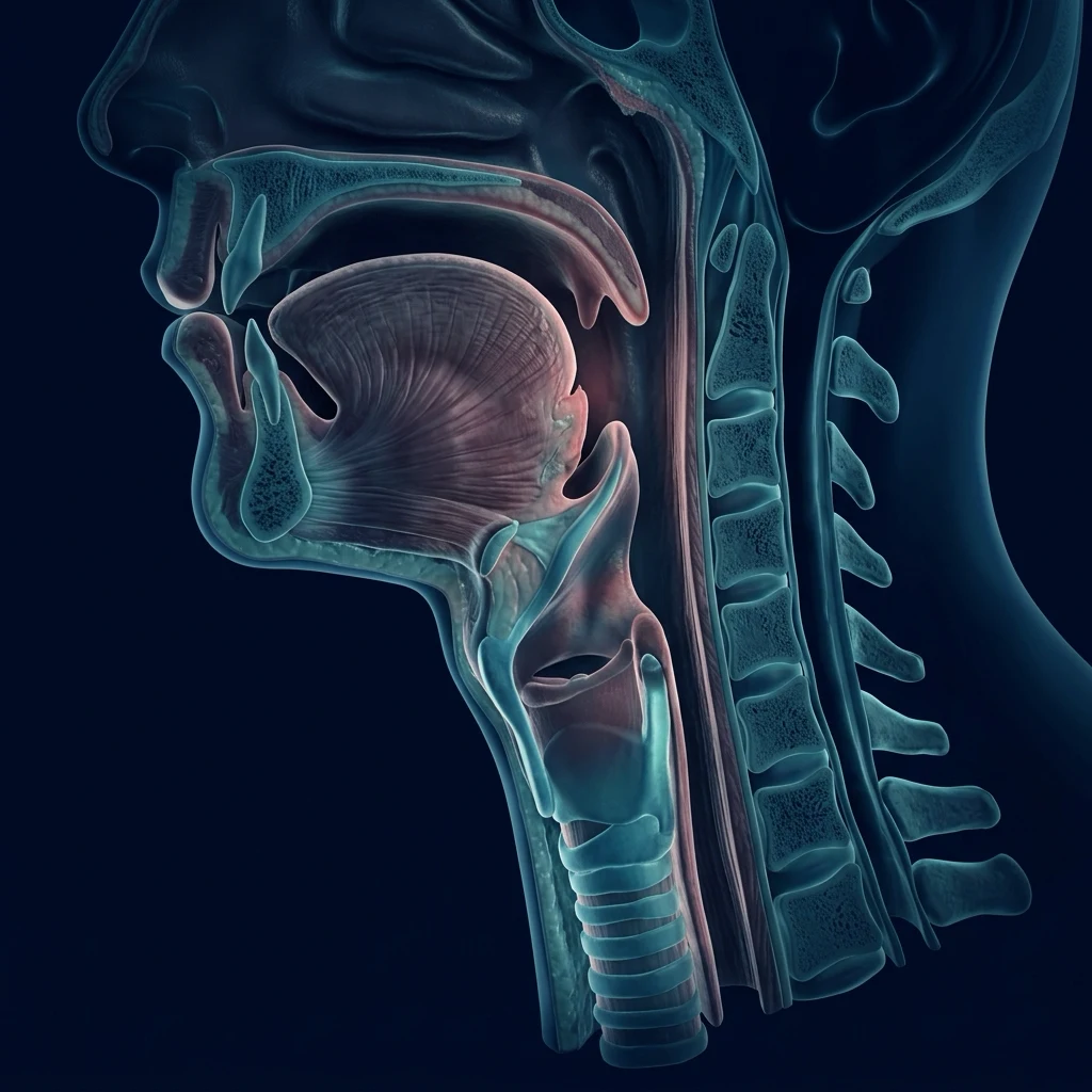
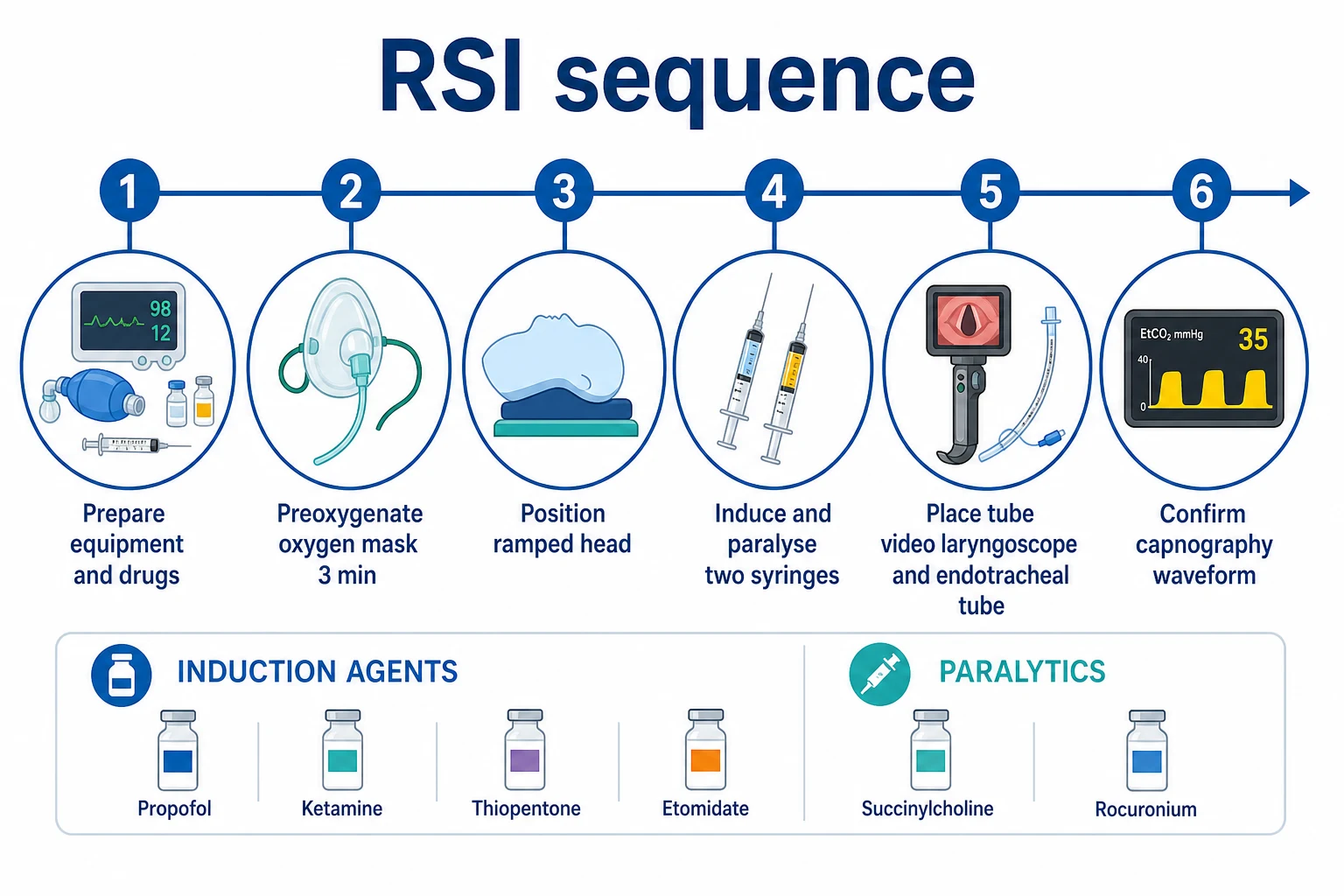
Preoxygenation in detail
Preoxygenation is the single most effective defence against peri-intubation hypoxaemia and buys the operator a window of safe apnoea. Its goal is to replace the nitrogen in the functional residual capacity (FRC) with oxygen — building an alveolar reservoir of around 2500 mL of oxygen in the healthy adult, less in the critically ill whose FRC is reduced by recumbency, atelectasis, obesity, pregnancy and pulmonary oedema. Two equally effective bedside regimens achieve denitrogenation in the cooperative patient: three minutes of tidal-volume breathing of 100% oxygen via a tightly sealed mask (Mapleson C, anaesthetic circuit or non-rebreather with a tight seal), or eight vital-capacity breaths of 100% oxygen, which achieves near-equivalent denitrogenation in around 60 seconds.[1]
The critically ill patient demands more than a bag and mask. The endpoints of adequate preoxygenation are an etO2 (end-tidal oxygen) ≥ 85–90% confirming effective denitrogenation, and an SpO2 as close to 100% as the underlying shunt allows. Where the patient remains hypoxaemic despite high-flow mask oxygen, add non-invasive positive pressure (CPAP or NIV) at 5–10 cmH2O during the preoxygenation phase to recruit collapsed alveoli and improve the reservoir — a strategy shown to reduce the rate of severe hypoxaemia during ICU intubation.[5]
Apnoeic oxygenation and THRIVE
Apnoeic oxygenation continues to deliver oxygen to the alveoli during the apnoeic period of laryngoscopy by exploiting the pressure gradient between the pharynx and the alveoli: alveolar oxygen is taken up into the blood faster than carbon dioxide is delivered, generating a sub-atmospheric alveolar pressure that draws pharyngeal oxygen down the airway. Standard low-flow nasal cannulae (2–5 L/min) provide this gradient, but the modern ICU technique is transnasal humidified rapid-insufflation ventilatory exchange (THRIVE) — high-flow nasal cannulae at 50–70 L/min with humidified oxygen, which extends the safe apnoea time of the healthy adult from around 8 minutes to over 20 minutes, and to over 10 minutes in the obese patient. Patel and Nouraei introduced THRIVE for the difficult airway; it is now widely used as a preoxygenation and apnoeic-oxygenation adjunct in ICU, particularly in the predicted-difficult or hypoxaemic patient.[7]
The important caveat, taught hard by the PROTRACH trial: THRIVE and standard apnoeic oxygenation are adjuncts, not substitutes for rigorous mask preoxygenation, and they do not prevent hypercapnia — CO2 rises at 1–2 mmHg per minute of apnoea, eventually causing acidosis. In the patient with raised intracranial pressure, hyperkalaemia or severe metabolic acidosis, apnoeic time must be short and ventilation resumed as soon as the tube is secured.[5]
| Feature | 3 min TVB | 8 VCB |
|---|---|---|
| Time | 3 minutes | ~1 minute |
| Equipment | Tight-seal mask + 100% O2 | Tight-seal mask + 100% O2 |
| Denitrogenation | etO2 ≥ 85–90% | Near-equivalent to TVB |
| Patient cooperation | Tidal breathing, low cooperation threshold | Needs deep vital-capacity breaths |
| Best for | Cooperative, stable ICU patient | Time-critical RSI, periarrest |
Management: rapid sequence induction and intubation
Rapid sequence induction (RSI) is the standard technique for the ICU patient: it sequences preoxygenation, a fast-acting induction agent and a fast-acting neuromuscular blocker, with cricoid pressure (debated) and no mask ventilation between induction and intubation, to minimise the risk of regurgitation and aspiration in the unfasted patient.[1][1]
A practical pre-intubation checklist captures the resuscitation around the procedure: [1]
- Prepare the patient, team, equipment (tube, suction, bougie, supraglottic airway, front-of-neck-access kit) and a back-out plan.
- Preoxygenate to the highest achievable saturation, with apnoeic oxygenation running.
- Position — ear-to-sternal-notch alignment (ramped position), especially in the obese.
- Resuscitate — correct hypovolaemia, start or up-titrate vasopressors, and reduce the induction dose in shock.
- Induce and paralyse, then intubate and confirm with capnography immediately. [1]
The difficult airway and the failed intubation algorithm
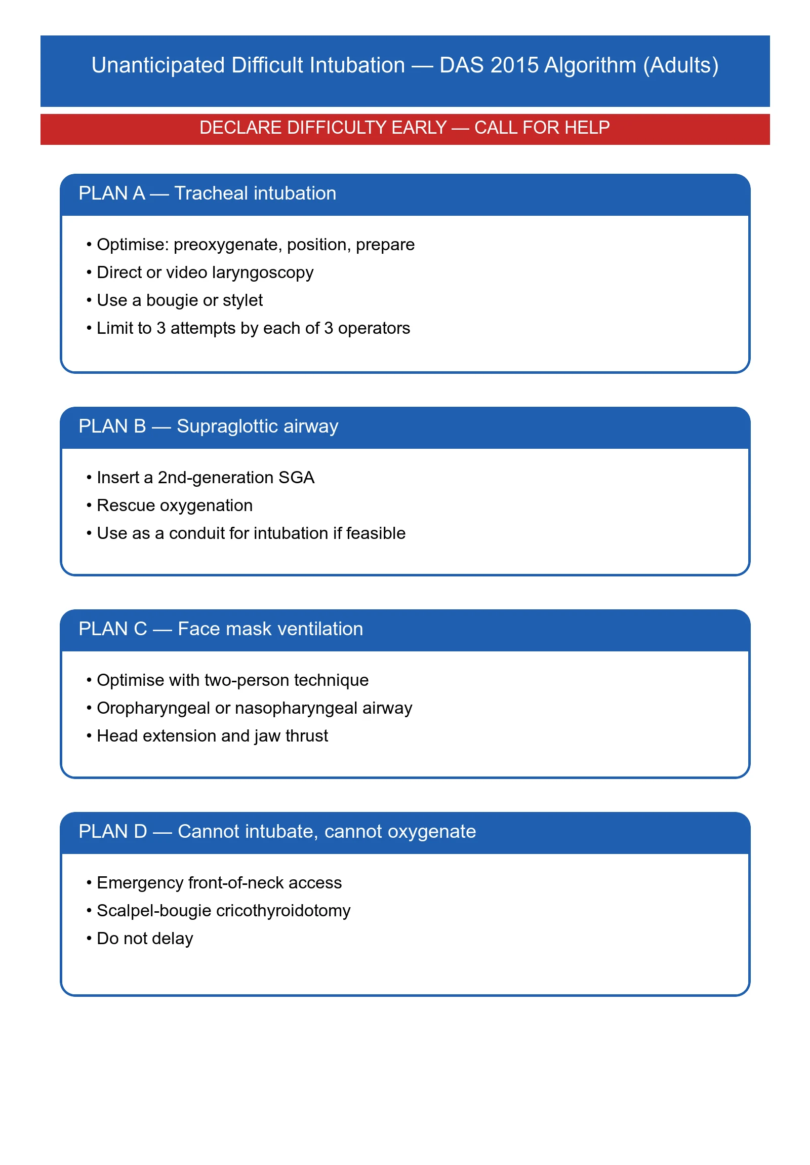
The Difficult Airway Society 2015 guidelines frame the response to unanticipated difficult intubation as a sequence of plans, each with a defined trigger to move on.[1]
- Plan A — tracheal intubation. Optimise the first attempt: preoxygenation, positioning, the best operator, and adjuncts — video laryngoscopy to improve the view, and a bougie or stylet. The BEAM trial showed that routine bougie use during direct laryngoscopy improved first-attempt success in patients with at least one difficult-airway characteristic (96% versus 82%). Limit attempts to three by each of three operators, and call for help early.[1][6]
- Plan B — supraglottic airway. If tracheal intubation fails, insert a second-generation supraglottic airway device; it both rescues oxygenation and, if it sits well, can serve as a conduit for intubation.[1]
- Plan C — face-mask ventilation. If the supraglottic airway fails, return to face-mask ventilation, optimised with a two-person technique, oral and nasal airways, and head extension and jaw thrust.[1]
- Plan D — cannot intubate, cannot oxygenate. If oxygenation cannot be maintained, perform emergency front-of-neck access without delay.[1]
The cardinal principle is to declare difficulty and call for help the moment the airway proves harder than expected, and to oxygenate between every attempt.[1]
The Vortex Approach: a unified cognitive tool
While DAS Plans A–D sequence the devices, the Vortex Approach sequences the decision-making in a crisis. It frames the airway as three lifelines — face-mask ventilation, supraglottic airway, and front-of-neck access — arranged as concentric rings around a central "green zone" of best-fit effort. The operator makes a single best effort at each of the three lifelines (up to three optimised attempts at each), declaring the attempt aloud and moving on as soon as oxygenation fails. The Vortex does not replace DAS but complements it: where DAS is a device-by-device algorithm, the Vortex is a shared mental model that aligns the whole team under stress, forces an explicit declaration of failure at each lifeline, and accelerates the decision to perform front-of-neck access before the patient arrests.[10]
The Vortex's two examinable principles: (1) declare failure and move on after one best effort (or up to three optimised attempts) at each lifeline, rather than persisting with a failing technique; and (2) the best lifeline is the one the team knows and has ready — the Vortex rewards preparation and team rehearsal over gadgetry.[10]
| Feature | DAS 2015 | Vortex |
|---|---|---|
| Structure | Linear device sequence (A→B→C→D) | Three concentric lifelines |
| Best suited | Unanticipated difficult intubation | Any airway crisis, any setting |
| Focus | Which device next | When to declare failure and move on |
| CICO trigger | Explicit Plan D | Central "green zone" reached after best effort at all 3 lifelines |
| Team role | Implicit | Explicit shared mental model |
CICO: scalpel-bougie technique
The adult cannot-intubate-cannot-oxygenate (CICO) rescue is the scalpel-bougie (or "scalpel-finger-bougie") cricothyroidotomy: palpate the cricothyroid membrane (stabilise the larynx with the non-dominant hand), make a transverse stab incision through skin and membrane, confirm entry by feeling the tracheal rings and the cock of the bougie, pass a bougie into the trachea, and railroad a 6.0 mm cuffed tracheal tube over it. Inflate the cuff, ventilate, and confirm with ETCO2. The technique is fast (under 60 seconds when drilled), uses the same equipment as a difficult intubation, and avoids the failures of cannula cricothyroidotomy (kinking, displacement, impossible jet ventilation against obstruction).[1][1]
Rescue oxygenation: supraglottic airways and face-mask ventilation
The supraglottic airway device is the workhorse of rescue. Second-generation devices (with a gastric drainage channel) are preferred because they protect against regurgitation and aspiration and provide a better seal for ventilation; they are the recommended Plan B device.[1] In the cannot-intubate patient who is still oxygenatable through a supraglottic airway, the device buys time to wake the patient (if the agent is short-acting) or to arrange a definitive airway by an expert.
Face-mask ventilation is the safety net beneath every airway. It is optimised by a two-person technique (one lifts the jaw with both hands while the other squeezes the bag), an oropharyngeal and/or nasopharyngeal airway to bypass soft-tissue obstruction, and head extension with jaw thrust. Difficult mask ventilation is predicted by obesity, beard, edentulism, older age and a history of snoring or sleep apnoea.[1]
Cannot intubate, cannot oxygenate: front-of-neck access
When tracheal intubation, a supraglottic airway and face-mask ventilation have all failed and oxygenation is being lost, the situation is cannot-intubate-cannot-oxygenate — a rare but time-critical emergency in which the patient dies within minutes without a surgical airway. The recommended adult technique is the scalpel-bougie cricothyroidotomy: a transverse stab through the cricothyroid membrane, a bougie passed into the trachea, and a cuffed tube railroaded over the bougie.[1][1]
NAP4 — the Fourth National Audit Project of major airway complications in the United Kingdom — identified delayed front-of-neck access and repeated fruitless attempts at intubation as recurring themes in airway deaths, and stressed that the technique must be practised, the equipment immediately available, and the decision made early rather than after the patient has arrested.[1]
Pharmacology of induction and paralysis
The ICU patient is hypotensive, so the induction agent is chosen and dosed to avoid cardiovascular collapse: a reduced dose of a haemodynamically stable agent (ketamine or etomidate) is preferred to the full dose of propofol that would be used in the stable anaesthetic patient.[1][1]
For paralysis, the choice is between succinylcholine (suxamethonium, 1 mg/kg) and rocuronium (1 mg/kg, or 1.2 mg/kg). The Cochrane review of 50 trials and over 4000 participants found that succinylcholine produced superior excellent intubating conditions to rocuronium (risk ratio 0.86, 95% confidence interval 0.81 to 0.92); there was no difference against the higher 1.2 mg/kg rocuronium dose, but succinylcholine remained clinically superior because of its short duration.[4]
The practical trade-off: succinylcholine has the fastest onset and an ultrashort duration (safe if the airway proves impossible, because the patient resumes breathing), but carries the risks of hyperkalaemia (in burns, crush, denervation, renal failure), malignant hyperthermia and bradycardia. Rocuronium at 1.2 mg/kg has a comparable onset to succinylcholine but a long duration that can be reversed by sugammadex — so in the patient at risk of hyperkalaemia or with contraindications to succinylcholine, rocuronium with sugammadex available is the safer choice.[1][1]
| Feature | Succinylcholine | Rocuronium |
|---|---|---|
| Onset | Very fast (30–60 s) | Fast at 1.2 mg/kg (≈60 s) |
| Duration | Ultrashort (5–10 min) | Long (≈30–60 min) |
| Excellent conditions | Superior (Cochrane RR 0.86) | Slightly fewer excellent conditions |
| Key risks | Hyperkalaemia, malignant hyperthermia, bradycardia | Anaphylaxis, prolonged paralysis |
| Reversal | None (spontaneous) | Sugammadex (rapid) |
The RSI drug sequence in practice
A reproducible RSI for the haemodynamically fragile ICU patient layers an opioid, an induction agent and a neuromuscular blocker, given in rapid succession after preoxygenation, with cricoid pressure (optional) and no positive-pressure ventilation until the tube is confirmed. The pragmatic ICU doses are summarised below. [1]
Opioid — fentanyl 1–3 mcg/kg (or alfentanil 10–30 mcg/kg): blunts the sympathetic response to laryngoscopy, attenuates the rise in intracranial pressure and reduces the dose of induction agent needed. Give it first, before induction, so that the peak effect coincides with laryngoscopy. Avoid full doses in the shocked or bradycardic patient.[1]
Induction agent — chosen for the patient's physiology:
- Propofol 1.5–2.5 mg/kg (reduced to 0.5–1 mg/kg or avoided in shock): rapid, smooth, anti-epileptic; profound vasodilator — the most common cause of induction hypotension in the ICU.
- Ketamine 1–2 mg/kg (reduced to 0.5–1 mg/kg in shock): preserves sympathetic tone and bronchodilates — preferred in asthma, bronchospasm, shock and burns. May transiently raise intracranial and intra-ocular pressure (the relevance of which is now debated).
- Etomidate 0.2–0.3 mg/kg: haemodynamically neutral and the agent of choice in severe shock; but it inhibits 11-beta-hydroxylase and causes adrenal suppression for 24–48 hours. The KETASED multicentre RCT in 655 acutely ill patients intubated with etomidate vs ketamine found no difference in 28-day mortality despite more adrenal insufficiency with etomidate (OR 6.7, 95% CI 1.4–27), so etomidate remains acceptable for single-dose RSI in shock — but avoid repeated dosing and consider stress-dose steroids in septic shock.[9]
- Midazolam 0.05–0.1 mg/kg: a useful adjunct for sedation but slower and less reliable for RSI; not a first-line induction agent in ICU.
Neuromuscular blocker:
- Succinylcholine 1.5 mg/kg (range 1–1.5 mg/kg): the textbook RSI dose, with onset 30–60 seconds and duration 5–10 minutes. Ultrashort duration is its safety advantage — if the airway is impossible, the patient resumes breathing.
- Rocuronium 1.2 mg/kg (the RSI dose, higher than the 0.6 mg/kg standard intubating dose): onset comparable to succinylcholine at ≈60 seconds, duration 30–60 minutes, fully and rapidly reversible by sugammadex 16 mg/kg.[4]
| Feature | Propofol | Ketamine |
|---|---|---|
| RSI dose | 1.5–2.5 mg/kg (0.5–1 in shock) | 1–2 mg/kg (0.5–1 in shock) |
| Onset | 30–60 s | 30–60 s |
| Haemodynamics | Vasodilation, bradycardia — hypotension | Sympathetic stimulation — preserves BP |
| Best for | Stable, raised ICP, status epilepticus | Shock, asthma, bronchospasm, burns |
| Risks | Hypotension, pain on injection, propofol infusion syndrome | Emergence phenomena, (transient ↑ICP) |
Cricoid pressure: a tradition under scrutiny
Cricoid pressure (the Sellick manoeuvre) — downward force on the cricoid cartilage to occlude the oesophagus against the vertebral column and prevent passive regurgitation — has been a feature of RSI since 1961. The evidence base is now deeply contested: cadaver and imaging studies show that the oesophagus is frequently lateral to the cricoid (not behind it), cricoid pressure can distort the airway and worsen the laryngoscopic view, and it can impede mask ventilation and supraglottic-airway placement. Current ICU practice is to apply it at the discretion of the intubating clinician, to release or relax it if it impedes laryngoscopy, mask ventilation or tube passage, and never to let it delay life-saving oxygenation. The 2015 DAS guideline no longer mandates cricoid pressure.[1][1]
The 30-second RSI sequence
- Confirm preoxygenation complete — etO2 ≥ 85%, SpO2 maximised, suction on, ramped position, monitoring attached.
- Fentanyl 1–3 mcg/kg IV — blunts the pressor response.
- Induction agent IV over 5–10 s — propofol, ketamine (shock) or etomidate (severe shock).
- Cricoid pressure applied (optional, operator judgement).
- Neuromuscular blocker IV immediately — succinylcholine 1.5 mg/kg or rocuronium 1.2 mg/kg.
- Wait 45–60 s for paralysis (loss of jaw tone, fasciculations settled).
- Laryngoscopy and intubation, no mask ventilation if oxygenation allows.
- Confirm with ETCO2 waveform over six breaths before releasing cricoid pressure.
Cardiovascular collapse and peri-intubation complications
Cardiovascular instability was the dominant major adverse event in INTUBE, occurring in 42.6% of intubations, followed by severe hypoxaemia in 9.3% and cardiac arrest in 3.1%; overall ICU mortality was 32.8%.[2] These are not rare events — they are the expected complication of intubating the critically ill — and they are largely preventable.
The prevention of cardiovascular collapse is pre-emptive: resuscitate before inducing — give fluid to the hypovolaemic, start or increase noradrenaline to the vasodilated, correct severe acidosis, and reduce the induction dose in shock (a third to a half of the usual dose of ketamine or etomidate). Aim for the highest tolerable blood pressure before pushing the induction agent, because it will fall the moment sympathetic tone is lost.[1][1]
Monitoring during and after intubation
Waveform capnography is mandatory for confirming tracheal placement and for ongoing monitoring. The misplaced oesophageal tube remains a leading cause of airway death; a sustained waveform of exhaled carbon dioxide over six breaths confirms tracheal placement, while chest movement, condensation and the absence of a waveform do not.[1][1]
After intubation, secure the tube, confirm depth (the tip 2 to 4 cm above the carina on a chest radiograph), and monitor continuously: continuous waveform capnography, pulse oximetry, arterial blood pressure (ideally invasive), and the electrocardiogram. Re-assess blood pressure immediately after induction — the first ten minutes are when cardiovascular collapse declares itself.[2]
Verification of tracheal tube placement
Confirmation that the tube is in the trachea — not the oesophagus or a main-stem bronchus — is the most safety-critical step of the intubation. The misplaced oesophageal tube remains a leading cause of airway death in NAP4, and a single missed confirmation has killed patients. Verification is layered across four domains. [1]
1. Continuous waveform capnography (the gold standard)
Waveform ETCO2 is mandatory and definitive. A sustained, square-wave capnograph trace of exhaled CO2 over at least six breaths confirms tracheal placement. The trace differentiates the tracheal tube from an oesophageal intubation (which may show a single, decaying trace as gastric CO2 is washed out, then disappears) and from a main-stem bronchial intubation (persistent trace, but with unilateral breath sounds and eventual desaturation). Colour-change CO2 detectors (the yellow-to-purple capnometer) are an inferior alternative where waveform is unavailable — they confirm CO2 but not waveform shape, and can give false positives in oesophageal intubation after bag-mask ventilation or antacid ingestion.[1][1]
2. Video laryngoscopy for direct visual confirmation
The modern ICU intubation is increasingly performed with a video laryngoscope (GlideScope, C-MAC, McGrath), which offers a magnified, angled view of the glottis on a screen visible to the whole team. The MACMAN multicentre RCT in 374 ICU patients compared Macintosh direct laryngoscopy with the Airway Scope video laryngoscope and found no improvement in first-pass success overall, but video laryngoscopy improved the Cormack-Lehane grade and is now first-line for the predicted-difficult airway, cervical-spine immobilisation, obesity, blood/secretions in the airway, and any case where the operator wants the team to share the view. Direct visual passage of the tube through the cords is the strongest confirmatory evidence, alongside ETCO2.[8]
3. The bougie as a primary adjunct
The bougie (gum elastic or disposable polymer) is the cheapest and most effective intubation adjunct. In the BEAM trial, routine bougie use during direct laryngoscopy improved first-attempt success from 82% to 96% in patients with at least one difficult-airway characteristic. The bougie is held in a curved ("hockey-stick") tip, passed through the cords when only the artytenoids are visible, and confirmed in the trachea by the "clicks" of the tip passing over the tracheal rings and the "hold-up" at the carina on deep insertion. Railroad a lubricated tube over the bougie with a 90° anti-clockwise twist to bring the bevel through the cords.[6]
4. Clinical adjuncts (supportive, never definitive)
Chest rise, bilateral air entry on auscultation, absence of gastric insufflation, and tube misting are supportive signs only — they are misleading in obesity, bronchospasm, pulmonary oedema and the moribund patient. Never rely on them in preference to ETCO2. After confirmation, check the tube depth (typically 21–23 cm at the lips in women, 23–25 cm in men) and confirm the tip sits 2–4 cm above the carina on a chest radiograph.[1]
| Feature | Waveform capnography | Colour-change capnometer |
|---|---|---|
| Specificity | Definitive | Inferior — false positives |
| Tube placement | Differentiates tracheal vs oesophageal | Detects CO2 only |
| Waveform shape | Yes (square-wave) | No |
| Use after cardiac arrest | Trace falls with low output — interpret with care | May be falsely negative in low-flow states |
| Recommendation | Mandatory for every intubation | Adjunct only, where waveform unavailable |
Post-intubation care: the first thirty minutes
The period immediately after tube confirmation is the second-highest-risk window of the procedure. Cardiovascular collapse declares itself in the first 10 minutes; tube displacement, ventilator dys-synchrony and sedation failure cluster in the first 30. A structured post-intubation bundle closes these gaps. [1]
Immediate (0–2 minutes)
- Confirm and secure the tube at the correct depth (tape or commercial holder), note the depth at the teeth/lips, and apply a soft cervical collar or tube-securing device to prevent accidental extubation during transfers.
- Release cricoid pressure once ETCO2 confirmed.
- Auscultate both axillae and the epigastrium; attach the ventilator.
- Re-check blood pressure immediately — the moment cricoid pressure and the pressor response to laryngoscopy wear off and the induction agent peaks, the patient is at greatest risk of collapse. Have noradrenaline, metaraminol or adrenaline drawn up.[2]
Early (2–30 minutes)
- Establish mechanical ventilation with a lung-protective strategy: tidal volume 6–8 mL/kg predicted body weight, plateau pressure below 30 cmH2O, PEEP titrated (typically 5–10 cmH2O), and a respiratory rate to target a physiological PaCO2 (or permissive hypercapnia in ARDS/raised ICP).
- Sedate and analgesedate to tolerance — propofol (1–3 mg/kg/h) or midazolam with fentanyl/morphine, avoiding long-acting benzodiazepines where possible (PADIS guidelines). Avoid under-sedation (ventilator dys-synchrony, extubation, recall) and over-sedation (hypotension, prolonged ventilation, delirium).
- Send a post-intubation arterial blood gas to verify oxygenation, ventilation (PaCO2), acid-base status and the effect of resuscitation (lactate clearance).
- Chest radiograph to confirm tube tip 2–4 cm above the carina, exclude main-stem intubation and pneumothorax, and assess the underlying pathology.
- Continue vasopressors — the patient who needed noradrenaline before induction will need it after; the loss of sympathetic tone under sedation unmasks profound vasodilation.
- NG tube placement to decompress the stomach (especially after bag-mask ventilation) and reduce the risk of aspiration and ventilator-related intra-abdominal hypertension.[2][1]
Documentation and handover
- Time of induction, drugs and doses, operator, number of attempts, lowest SpO2, lowest/highest blood pressure, any adverse event (cardiac arrest, hypoxaemia, regurgitation).
- Complications declared and acted on — peri-intubation cardiac arrest, oesophageal intubation, dental or airway trauma, hypotension requiring vasopressor escalation.
- Plan for ongoing ventilation, sedation, blood-pressure target and the next 4 hours.
Post-intubation bundle (the first 30 minutes)
- Confirm tube with ETCO2 waveform over six breaths; release cricoid pressure.
- Secure the tube, note depth at teeth, auscultate both axillae and epigastrium.
- Attach ventilator — lung-protective (Vt 6–8 mL/kg PBW, Pplat < 30, PEEP 5–10).
- Re-check blood pressure — escalate vasopressors if MAP < 65 mmHg.
- Sedate to tolerance (propofol or midazolam + opioid); start analgesia.
- Arterial blood gas — verify PaO2, PaCO2, pH, lactate.
- Chest radiograph — tube tip 2–4 cm above carina, exclude pneumothorax.
- NG tube to decompress the stomach; document the intubation.
Extubation and the difficult extubation is an airway emergency in waiting: the patient must maintain their own airway, breathe, protect against aspiration, and clear secretions. Plan extubation as deliberately as intubation — assess readiness (awake, cooperative, adequate oxygenation on minimal positive end-expiratory pressure, low respiratory rate, weak cough or secretion load corrected), have reintubation equipment and a senior operator present, and position the patient sitting up.[1][1]
The difficult extubation — the patient who was difficult to intubate, or who has airway oedema (anaphylaxis, prolonged prone positioning, angioedema), obesity or a guarded airway — warrants an extubation strategy: an airway exchange catheter left in situ through which the tube can be railroaded back if reintubation is needed, or a deliberate, planned extubation in a controlled setting.[1]
Specific situations
- The shocked patient: resuscitate aggressively and reduce the induction dose; ketamine is the haemodynamically stable induction agent of choice. Expect and pre-empt cardiovascular collapse.[1]
- The severely hypoxaemic patient: preoxygenate with high-flow oxygen and continuous positive airway pressure if tolerated, use apnoeic oxygenation, intubate in a head-up position, and accept the lowest effective dose of induction agent. In the hypoxaemic patient, peri-intubation deterioration into cardiac arrest is a recognised and feared event; the airway is managed as a resuscitation, not a procedure.[2]
- Raised intracranial pressure: avoid coughing, bucking and hypertension — preoxygenate, use a smooth induction with an opioid (fentanyl) and a generous paralysis dose, and avoid hypoxaemia and hypercapnia which raise intracranial pressure.[1]
- Obstetric: the gravid uterus raises intra-abdominal pressure, functional residual capacity is reduced and desaturation is rapid; intubate with a smaller tube, a senior operator and a low threshold for a supraglottic airway rescue.[1]
Prognosis & outcomes
The prognosis of the intubated ICU patient is driven less by the act of intubation than by the underlying illness — but the peri-intubation period is a high-risk window. INTUBE's 32.8% ICU mortality reflects a sicker population (over half intubated for respiratory failure), and the major adverse events that cluster around intubation — cardiovascular collapse, severe hypoxaemia, cardiac arrest — independently worsen outcome.[2] A well-conducted intubation (resuscitated, preoxygenated, senior, capnography-confirmed) reduces this harm; a poorly conducted one adds to it.
NAP4's lasting lesson is that most major airway complications are avoidable and stem from failures of planning, recognition and decision-making rather than from a lack of technical gadgets — the airway is managed by a prepared team with a plan and the willingness to call for help.[1]
Approach to the exam
The CICM airway question tests structured, physiology-led thinking, not gadget recall. A framework that scores well: [1]
- Assess before you induce — MACOCHA/LEMON, and identify the physiological derangements (shock, hypoxaemia, raised intracranial pressure).
- Prepare and preoxygenate — team, kit, back-out plan, ramped position, maximal preoxygenation with apnoeic oxygenation.
- Resuscitate around the induction — fluids, vasopressors, reduced induction dose; cardiovascular collapse is the enemy.
- Perform a planned RSI — induction agent and paralytic chosen for the patient; intubate and confirm with capnography.
- Have the algorithm ready — Plans A to D, declare difficulty early, call for help, and do not delay front-of-neck access in the cannot-intubate-cannot-oxygenate patient. [1]
Defend every step with the evidence: INTUBE for the scale of harm, MACOCHA for prediction, BEAM for the bougie, the Cochrane review for the paralytic, PROTRACH for preoxygenation, and DAS 2015 / NAP4 for the algorithm and the avoidability of harm.[1][2][3]
SAQ — Airway management in the critically ill patient: structured RSI
10 minutes · 10 marks
A 66-year-old woman is admitted to the emergency department with severe community-acquired pneumonia and septic shock. She is hypoxaemic (SpO2 89% on 15 L/min via non-rebreather, RR 32), hypotensive (MAP 58 on noradrenaline 0.25 mcg/kg/min), and has a lactate of 4.0 mmol/L. Her Glasgow Coma Score is 12 and she is tiring. A decision is made to intubate. She is obese (BMI 33), has a short neck, and an overbite.
SAQ — Failed intubation and the difficult-airway algorithm in ICU
10 minutes · 10 marks
During an RSI for a 60-year-old trauma patient with severe head injury and cervical spine immobilisation, the registrar makes three attempts at video laryngoscopy, all failing to pass the tube. The patient's SpO2 has fallen from 95% to 78%. A second-generation i-gel is inserted but ventilates poorly, with no colour change on the CO2 detector.
Red flags
- Peri-intubation cardiovascular collapse is the commonest major complication (42.6% in INTUBE) and the most preventable — resuscitate first, start or increase vasopressors, and reduce the induction dose in shock; a patient intubated in uncorrected shock arrests on the syringe.[2]
- A low preoxygenation reserve means rapid desaturation (one to two minutes in the critically ill) — preoxygenate to 100% and use apnoeic oxygenation; never begin laryngoscopy on an already-hypoxaemic patient.[5]
- Capnography is mandatory — the misplaced tube remains a leading cause of airway death (NAP4); confirm tracheal placement with a sustained exhaled-carbon-dioxide waveform on every intubation.[1]
- A MACOCHA score of 3 or above predicts difficult intubation (validation NPV 98%) — assess every ICU patient before induction, prepare the difficult-airway kit, and call senior help early.[3]
- Cannot-intubate-cannot-oxygenate is time-critical — if face-mask ventilation and a supraglottic airway both fail, perform emergency scalpel-bougie cricothyroidotomy without delay; repeated fruitless attempts kill the patient.[1][1]
References
- [1]Frerk C, Mitchell VS, McNarry AF, Mendonca C, et al. Difficult Airway Society 2015 guidelines for management of unanticipated difficult intubation in adults Br J Anaesth, 2015.PMID 26556848
- [2]Russotto V, Myatra SN, Laffey JG, et al. (INTUBE Study Investigators) Intubation Practices and Adverse Peri-intubation Events in Critically Ill Patients From 29 Countries JAMA, 2021.PMID 33755076
- [3]De Jong A, Molinari N, Terzi N, et al. (AzuRéa Network; Frida-Réa Study Group) Early identification of patients at risk for difficult intubation in the intensive care unit: development and validation of the MACOCHA score in a multicenter cohort study Am J Respir Crit Care Med, 2013.PMID 23348979
- [4]Tran DT, Newton EK, Mount VA, Lee JS, Wells GA, Perry JJ. Rocuronium versus succinylcholine for rapid sequence induction intubation Cochrane Database Syst Rev, 2015.PMID 26512948
- [5]Guitton C, Ehrmann S, Volteau C, et al. Nasal high-flow preoxygenation for endotracheal intubation in the critically ill patient: a randomized clinical trial Intensive Care Med, 2019.PMID 30666367
- [6]Driver BE, Prekker ME, Klein LR, et al. Effect of Use of a Bougie vs Endotracheal Tube and Stylet on First-Attempt Intubation Success Among Patients With Difficult Airways Undergoing Emergency Intubation: A Randomized Clinical Trial JAMA, 2018.PMID 29800096
- [7]Patel A, Nouraei SAR. Transnasal Humidified Rapid-Insufflation Ventilatory Exchange (THRIVE): a physiological method of increasing apnoea time in patients with difficult airways Anaesthesia, 2015.PMID 25388828
- [8]Lascarrou JB, Boisrame-Helms J, Bailly A, et al. Video Laryngoscopy vs Direct Laryngoscopy on Successful First-Pass Orotracheal Intubation Among ICU Patients: A Randomized Clinical Trial JAMA, 2017.PMID 28118659
- [9]Jabre P, Combes X, Lapostolle F, et al. (KETASED Collaborative Study Group) Etomidate versus ketamine for rapid sequence intubation in acutely ill patients: a multicentre randomised controlled trial Lancet, 2009.PMID 19573904
- [10]Chrimes N. The Vortex: a universal 'high-acuity implementation tool' for emergency airway management Br J Anaesth, 2016.PMID 27440673