ICU · Airway management
Supraglottic Airway Devices — LMA, i-gel & Second-Generation Devices
Also known as Supraglottic airway device · SAD · Laryngeal mask airway · LMA · i-gel · ProSeal · LMA Supreme · Second-generation supraglottic airway · Rescue airway · Airway exchange conduit
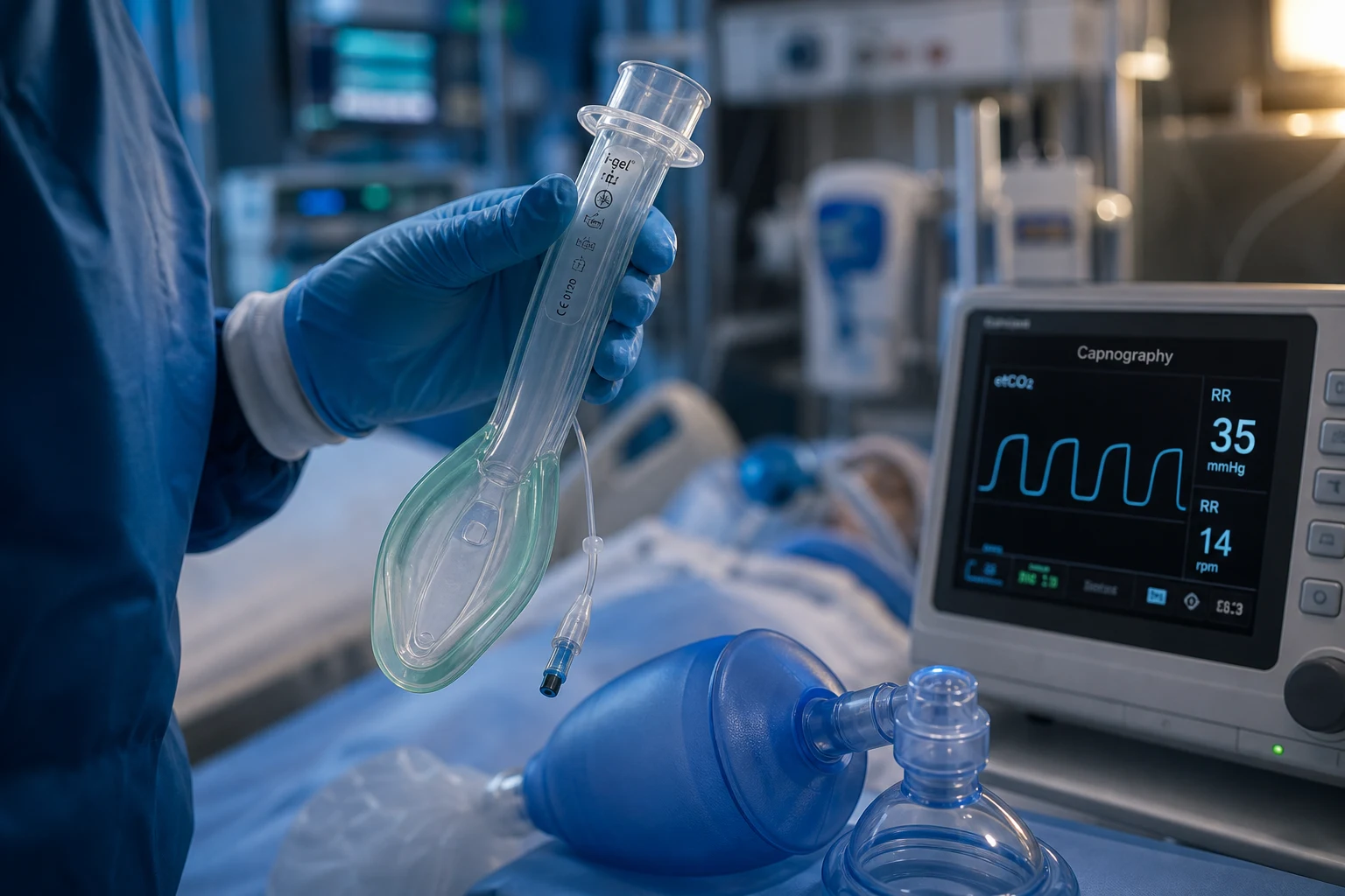
A supraglottic airway device (SAD) sits in the pharynx and seals around the laryngeal inlet, providing a hands-free airway without entering the trachea — bridging the gap between bag-valve-mask and endotracheal intubation. First-generation devices (classic LMA) have a single lumen and no gastric drainage and failed as rescue devices in NAP4; second-generation devices (i-gel, ProSeal, Supreme) add a gastric drainage channel and a higher seal pressure, giving partial aspiration protection and an intubation conduit. In the difficult-airway algorithm the SAD is the Plan B rescue; it is not a definitive airway, because it does not isolate the trachea.
On this page & tools
Your progress
Saved locally on this device.
Target exams
Overview & definition
A supraglottic airway device (SAD) sits in the pharynx and forms a seal around the laryngeal inlet, allowing hands-free ventilation without entering the trachea. It bridges the gap between the bag-valve-mask and endotracheal intubation — faster and less invasive than a tube, with a better seal and less gastric insufflation than a mask. In the difficult-airway algorithm the SAD is the Plan B rescue device; it can also serve as a conduit through which a definitive endotracheal tube is placed.[1][1]
A SAD is not a definitive airway: it sits above the glottis and does not isolate the trachea, so it gives only partial protection against aspiration. For prolonged ICU ventilation, an endotracheal tube remains the standard.[1]
First versus second generation
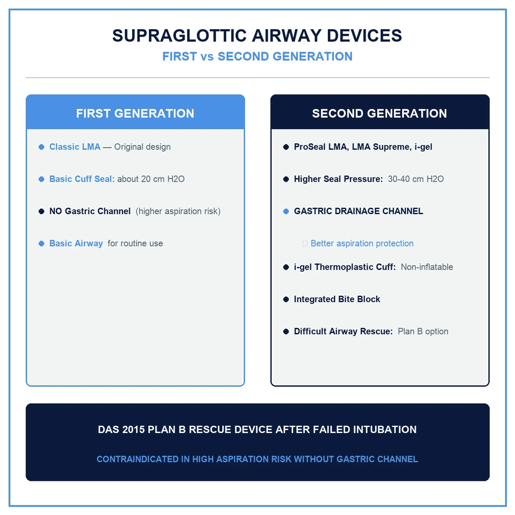
The distinction matters and drives modern practice.[1][1]
-
First-generation devices (classic LMA) — a single lumen with an inflatable cuff, no gastric drainage channel, and a relatively low seal pressure. They provide no protection against aspiration. NAP4 found that first-generation laryngeal masks failed as rescue devices in major airway events (regurgitation, aspiration, displacement). They are no longer recommended for emergency rescue.[1]
-
Second-generation devices (i-gel, LMA ProSeal, LMA Supreme, Baska) — add a gastric drainage channel (which vents regurgitated stomach contents away from the airway), a higher seal pressure, and an intubation conduit. They give partial aspiration protection and tolerate higher airway pressures. The DAS 2018 ICU guideline specifies a second-generation device as the Plan B rescue.[1]
The i-gel
The i-gel is the most widely used second-generation SAD in the ICU. Its features:[1]
- A non-inflatable cuff made of a thermoplastic elastomer (gel) that warms to body temperature and moulds to the perilaryngeal anatomy — there is no cuff pressure to manage.
- Very rapid insertion (often a few seconds) and a shallow learning curve.
- A gastric drainage channel and an integrated bite block.
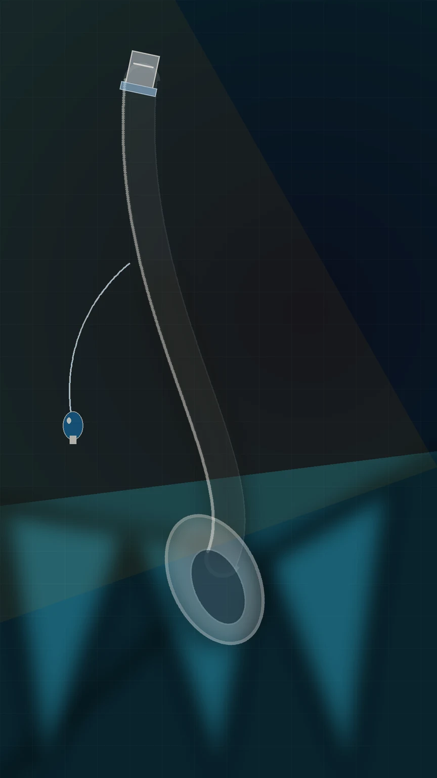
- A wide-bore, short, slightly curved tube that can accept an endotracheal tube (railroaded) or an Aintree airway exchange catheter for fibre-optic-guided intubation through the device.
- Sized 3 to 5 by patient weight.
Other second-generation devices
- LMA ProSeal — an inflatable cuff with a posterior cuff and a drain tube; a high seal pressure, but cuff pressure must be managed.
- LMA Supreme — a disposable double-lumen device with a gastric channel and a fixed curve; easy to insert, single use.
- Baska mask — a non-inflatable cuff with a self-sealing membranous design and gastric channels.[1]
Mechanism of the seal — perilaryngeal anatomy
The SAD works by forming a seal around the laryngeal inlet, not by entering the trachea. Understanding where the device sits explains both its strengths and its failure modes.[1]
- Where it sits. The cuff or bowl of the device lodges in the hypopharynx, with its tip against the upper oesophageal sphincter (cricopharyngeus) and its bowl over the laryngeal inlet. The anterior surface of the cuff faces the base of tongue and vallecula; the posterior surface faces the posterior pharyngeal wall.
- What seals. A first-generation cuff seals anteriorly against the laryngeal inlet with a single inflatable mask; a second-generation device adds a second seal at the upper oesophageal sphincter (the tip of the drain tube), which separates the airway from the gastrointestinal tract and allows venting of regurgitated contents.
- Why seal pressure matters. The oropharyngeal leak pressure (OLP) is the airway pressure at which the seal fails and gas escapes. First-generation devices leak around 15-20 cm H2O; second-generation devices achieve 25-40 cm H2O, tolerating the higher peak pressures of obesity, ARDS, or bronchospasm.
- The gel cuff (i-gel). The thermoplastic elastomer warms from ambient to body temperature over ~3-4 minutes, softening and moulding to the irregular perilaryngeal tissues — seal pressure rises after insertion as the cuff warms. No cuff pressure to monitor is both the advantage (no over-inflation nerve injury) and a limitation (you cannot "tighten" a leaking seal).
- The integrated bite block. A rigid channel through the stem of the i-gel (and others) prevents the patient from occluding the airway by biting, and provides a hand-grip for insertion. In the ICU it is especially valuable in the semi-conscious or post-ictal patient who may clench. [1]
Second-generation supraglottic devices compared for ICU practice
| Device | Cuff | Gastric drain channel | Bite block | Typical leak pressure | Intubation conduit | ICU role |
|---|---|---|---|---|---|---|
| i-gel | Non-inflatable thermoplastic gel (warming) | Yes | Integrated | 28-32 cm H2O (rises as cuff warms) | Yes — accepts Aintree catheter / ETT | Default Plan B rescue; fastest insertion, no cuff to manage |
| LMA ProSeal | Inflatable silicone, double cuff (anterior + posterior) | Yes (drain tube) | No | 30-35 cm H2O | Limited | High seal pressure; reusable; cuff pressure must be checked |
| LMA Supreme | Inflatable, single-use, fixed curve | Yes | No | 24-28 cm H2O | Limited | Easy first-time insertion; single use; lower seal than ProSeal |
| LMA Protector | Inflatable (fixed or adjustable cuff pressure) | Yes | No | 30-35 cm H2O | Yes (large bore) | Newer device; good conduit for fibreoptic intubation |
| Baska mask | Non-inflatable, self-sealing membranous cuff | Yes (dual channels) | Integrated | 30-40 cm H2O | Limited | High seal; non-inflatable; less widely adopted |
| AuraGain | Inflatable, pre-curved | Yes | No | 28-32 cm H2O | Yes | Good conduit; reusable and single-use variants |
| Classic LMA (1st gen) | Inflatable, single lumen | No | No | 15-20 cm H2O | Poor | Not recommended for emergency rescue (NAP4) |
The recurring theme: every device that survives modern emergency practice has a gastric drainage channel and a higher seal pressure than the classic LMA. The single distinguishing feature that drove the move from first to second generation was the recognition — crystallised in NAP4 — that a rescue airway which cannot vent regurgitated stomach contents is not a rescue airway.[1]
Advantages over bag-valve-mask
- Hands-free ventilation, freeing the operator.
- A better seal and less gastric insufflation (less regurgitation and aspiration).
- Allows continuous waveform capnography through the device.
- Can be used as a conduit for intubation.[1]
Advantages over endotracheal intubation
- Faster, less invasive, requires less skill, less haemodynamic disturbance, and is better tolerated at lighter planes of anaesthesia — but at the cost of tracheal protection.[1]
Indications — where the SAD earns its place
The SAD spans several distinct roles in critical care, each with a different risk/benefit profile.[1][2]
- Rescue airway (Plan B, failed intubation). The defining ICU role. When laryngoscopy fails and oxygenation is threatened, a second-generation SAD is inserted to restore oxygenation; it then either stays as a bridge, is used as a conduit for a definitive tube, or buys time while the patient wakes. The DAS 2018 algorithm specifies the SAD as Plan B after failed Plan A laryngoscopy.[1]
- Difficult-airway conduit. When the airway is anticipated to be difficult (MACOCHA positive, known front-of-neck distortion), a SAD may be placed electively and a fibre-optic scope passed through it to intubate under vision — "intubating through the SAD".[2]
- NIV / CPAP bridge. A second-generation SAD can deliver positive-pressure ventilation short-term while preparations for a definitive airway are made; it is not a durable NIV interface. The gastric drain limits the insufflation that a mask would force into the stomach, and the airway is now "tubed" rather than masked.
- Cardiac arrest. ERC 2021 accepts a SAD as an alternative to bag-valve-mask or endotracheal intubation for providers trained in its use; a second-generation device is preferred because of the gastric channel.[3]
- Procedural sedation / anaesthesia. In theatre and for short ICU procedures (cardioversion, endoscopy) in the fasted patient, the SAD is the standard airway — a role it earns back from intubation by being less invasive and better tolerated at light planes of anaesthesia.
- Pre-hospital airway. Paramedic systems increasingly use a second-generation SAD (or laryngeal tube) as the primary airway when intubation skills are not immediately available; the AIRWAYS-2 and PART trials inform this debate.[4][5]
The SAD inside the difficult-airway algorithm
In both the DAS 2015 (perioperative) and DAS 2018 (ICU) algorithms, the second-generation SAD occupies the same structural slot: it is the Plan B that follows a failed best-effort laryngoscopy (Plan A), and it precedes the front-of-neck-access eFONA (Plan D).[1][2] If the SAD oxygenates, the team stops and thinks: wake the patient, intubate through the device, or continue with the SAD as a temporary airway. If the SAD fails to oxygenate, the team declares CICO and moves to eFONA without further attempts at the upper airway. The SAD is therefore a cognitive hinge — its success or failure determines whether the next step is salvage or scalpel.
Insertion technique — the bedside sequence
Inserting a second-generation SAD (i-gel technique) — the bedside sequence
Choose the device and size
For the i-gel, size by WEIGHT: size 3 (30-60 kg), size 4 (50-90 kg), size 5 (90 kg and above). For inflatable devices (ProSeal, Supreme), size by sex/build (size 4 adult female, size 5 adult male is conventional). Open the correct size, plus one smaller as backup. Lubricate the posterior surface of the cuff with water-based gel — keep the drain tube outlet clear.
Position the patient
Sniffing position (head extension, slight neck flexion) aligns the axes — the same position as for laryngoscopy. In suspected C-spine injury, keep neutral and rely on jaw thrust. Have working SUCTION at the bedside — Yankauer under the pillow, switched on.
Insert along the hard palate
Grasp the i-gel by the integral bite block / stem. Introduce the leading tip into the mouth against the hard palate and slide it back in ONE smooth curve, following the contour of the palate and posterior pharynx. Jaw thrust from an assistant opens the hypopharynx. Do NOT rotate or force — resistance at the upper oesophageal sphincter signals correct depth.
Seat and confirm the "give"
For the i-gel, advance until gentle resistance is felt — the tip lodges in the upper oesophageal inlet and the bowl seats over the larynx. For inflatable devices, inflate the cuff to the stated volume (never over-inflate to "fix" a leak — it causes nerve injury). The drain tube should now align with the oesophagus; a lubricated suction catheter should pass freely down it (a check that the tip is in the oesophagus, not folded back).
Confirm with chest rise and waveform capnography
Attach the circuit and give a breath. Look for BILATERAL chest rise and a WAVEFORM CO2 trace — the only reliable confirmation. Auscultate the stomach and lung apices. A flat capnography line = oesophageal malposition: remove and reinsert. Never accept chest rise alone.
Secure and document
Tape or tie the device in the midline; document size, number of attempts, and any airway leak. If it is to stay in the ICU, attach continuous capnography and check a chest X-ray. Note the time — prolonged SAD ventilation should trigger plans for a definitive airway.
The integrated bite block and gastric drain channel — what they do in practice
The two features that separate a second-generation device from a classic LMA both have a specific mechanical job: [1]
- The gastric drainage channel terminates at the upper oesophageal sphincter. When a nasogastric tube or suction catheter passes freely down it, that is confirmation the tip is correctly seated over the oesophageal inlet. If regurgitation occurs, the channel vents liquid away from the larynx and out through the device — turn on suction, place an NG tube, and aspirate. The channel does not actively prevent regurgitation; it manages it after the fact.
- The integrated bite block sits in the stem of the i-gel. It prevents occlusion of the airway by biting (a real risk in the semi-conscious, post-ictal, or emerging patient), stiffens the device for insertion, and gives the operator a hand-grip. It is one reason the i-gel tolerates the lighter planes of anaesthesia common in ICU procedural sedation. [1]
Limitations and contraindications
- Not a definitive airway — no reliable tracheal isolation; aspiration risk remains, especially in the non-fasted or at-risk stomach.
- Limited seal pressure — may leak at the high airway pressures of a stiff lung (obesity, ARDS, asthma), where it cannot ventilate effectively.
- Risk of displacement, malposition (oesophageal placement), and cuff-related nerve injury.
- Contraindications: restricted mouth opening, upper-airway obstruction or trauma, and a high aspiration risk where time allows a definitive airway instead (the RODS assessment: restricted mouth opening, obstruction, disrupted upper airway, stiff lungs).[1][1]
Contraindications in detail — the RODS test
The bedside screen for whether a SAD is the wrong choice is the RODS mnemonic:[1]
- R — Restricted mouth opening. The device must pass between the teeth; an inter-incisor distance <2 cm (trismus, temporomandibular joint ankylosis, facial trauma, post-ictal clenching) precludes insertion. The integrated bite block needs the teeth to clear the stem.
- O — Obstruction. A supraglottic or glottic mass, infection (deep neck space infection, retropharyngeal or peritonsillar abscess, epiglottitis), or foreign body at the laryngeal inlet cannot be bypassed by a device that sits above the glottis. The SAD will seal around, not past, the obstruction.
- D — Disrupted or distorted upper airway. Pharyngeal, laryngeal, or oesophageal surgery or trauma; friable tumour; a large pharyngeal pouch; previous pharyngeal or laryngeal radiotherapy — the seal fails or insertion provokes bleeding.
- S — Stiff lungs / high airway pressure. Obesity, ARDS, severe bronchospasm, pregnancy — the high peak inspiratory pressure exceeds the device seal pressure and the device leaks, failing to ventilate. This is the most common reason a SAD fails in the ICU. [1]
High aspiration risk is a relative contraindication: the non-fasted patient, bowel obstruction, active upper GI bleed, or late pregnancy. The second-generation gastric channel gives partial protection — it is not a licence to abandon tracheal intubation when there is time and skill. In a true cardiac arrest or CICO situation, however, a SAD with a gastric channel is still far better than no airway. [1]
Insertion technique
- Size the device (i-gel by weight) and lubricate the cuff.
- Head extension (or neutral in C-spine), jaw thrust if needed.
- Insert along the hard palate and advance in a smooth curve until resistance is felt; for an inflatable device, inflate the cuff to the recommended volume.
- Confirm with bilateral chest rise and waveform capnography (a CO2 trace).
- Secure with tape or a tie; check a chest X-ray if the device is to stay.[1]
The SAD as an intubation conduit (rescue value)
When intubation fails but oxygenation is achieved through a second-generation SAD, the SAD can be converted into a definitive airway: pass an Aintree airway exchange catheter (loaded with a fibre-optic scope) through the SAD into the trachea, railroad an endotracheal tube over the catheter, then remove the SAD. This is the safe way to exchange a rescue SAD for a definitive tube.[1][1]
Why the Aintree catheter is the safe exchange tool
Blindly pushing an endotracheal tube through a SAD has a poor success rate and risks laryngeal trauma, because the SAD lumen rarely aligns perfectly with the glottis. The Aintree intubation catheter solves this: it is a long, hollow, semi-rigid catheter (internal diameter 4.7 mm) that accepts a fibre-optic bronchoscope through its lumen.[7][8]
- Load the bronchoscope through the Aintree catheter so the scope tip protrudes.
- Pass the scope–Aintree assembly through the SAD lumen, identify the glottis, and advance into the trachea to the carina.
- Withdraw the scope, leaving the Aintree catheter in the trachea.
- Railroad a small endotracheal tube (6.0-7.0 mm) over the Aintree catheter into the trachea, then remove the SAD over the tube.
- Confirm with capnography and chest rise; secure the definitive airway. [1]
The alternative is a blind intubating LMA (Fastrach), where a specialised curved ETT is railroaded through the device without a scope — faster but less reliable, and not a vision-guided technique. Comparative work on conduit efficiency shows the i-gel and LMA Protector both function well as Aintree conduits, with first-attempt success in the majority when the operator is trained.[7][8] In the ICU, where blood and secretions are common, the fibre-optic/Aintree route is preferred once oxygenation is secured — it converts a blind gamble into a visualised intubation.
Role in cardiac arrest
Resuscitation councils (ILCOR/ERC) accept a supraglottic airway as an alternative to bag-valve-mask or endotracheal intubation for providers trained in its use, with a second-generation device preferred. It does not replace definitive intubation by a skilled operator when available.[1]
Evidence in cardiac arrest — AIRWAYS-2 and PART
Two large pragmatic cluster-randomised trials reshaped the cardiac-arrest airway debate:[3]
- AIRWAYS-2 (Benger, 2018) randomised 9,296 out-of-hospital cardiac arrests to an initial i-gel versus initial endotracheal intubation by UK paramedics. There was no difference in the primary outcome of good functional outcome at 30 days (modified Rankin 0-3): 6.4% (i-gel) vs 6.8% (intubation). Regurgitation and aspiration were more frequent with the i-gel, while first-attempt insertion success was higher with the i-gel.[4]
- PART (Wang, 2018) randomised 3,004 out-of-hospital cardiac arrests to an initial laryngeal tube versus endotracheal intubation by US paramedics and found a survival benefit with the laryngeal tube (18.3% vs 15.4% survival to hospital discharge) — driven partly by higher first-attempt success and fewer chest-compression interruptions with the supraglottic device.[5]
- A 2024 systematic review and meta-analysis (Forestell) reconciled these: across out-of-hospital cardiac arrest trials, an initial supraglottic airway was associated with comparable or improved short-term survival versus intubation, but long-term favourable neurological outcomes did not clearly favour either strategy.[6]
Bottom line for the ICU: in cardiac arrest the airway of the trained operator matters more than the device brand. The SAD is an acceptable primary airway where intubation skills are absent or delayed; it does not replace definitive intubation by an intubation-skilled team once return of spontaneous circulation is achieved and the patient moves to ongoing ICU care. [1]
Complications
- Aspiration (the key risk, especially with a first-generation device or a misplaced/malpositioned device).
- Oesophageal placement (unrecognised without capnography).
- Displacement or failure to seal.
- Sore throat, and nerve injury (recurrent laryngeal, hypoglossal, lingual) from an over-inflated or malpositioned cuff.[1]
Complications in detail
- Aspiration. The cardinal and most feared complication. Risk is highest with a first-generation device (no gastric channel), a malpositioned device, or in the non-fasted or high-risk stomach. Even a second-generation device only vents regurgitated material — it does not prevent it from reaching the airway if the seal is breached or the device is displaced. NAP4 documented aspiration deaths from unrecognised SAD failure.[1]
- Nerve injury. An over-inflated or malpositioned cuff compresses nerves against the pharyngeal mucosa. The recurrent laryngeal and hypoglossal nerves are the classic victims, but lingual and glossopharyngeal injuries are reported — presenting as tongue numbness, weakness, or (if bilateral) upper-airway obstruction. The mechanism is pressure neuropraxia, preventable by using the correct cuff volume (or a non-inflatable i-gel) and avoiding prolonged use.
- Laryngospasm. A stimulatory device sitting at the laryngeal inlet can trigger reflex closure of the cords in the lightly anaesthetised patient — the i-gel is less stimulating than an inflatable cuff, but laryngospasm still occurs. Treat by deepening anaesthesia, 100% oxygen, continuous positive airway pressure, and — if unresponsive — a small dose of suxamethonium.
- Oedema and sore throat. Minor and common (10-40%); usually resolves within 24 hours. Significant pharyngeal or glottic oedema is rare but reported with an oversized cuff or prolonged use.
- Oesophageal / pharyngeal trauma and perforation. Rare but catastrophic; a forced insertion or oversized device can tear the piriform fossa or oesophagus, causing surgical emphysema, mediastinitis, or retropharyngeal abscess.
- Device failure and displacement. The device can rotate, fold, or be pushed too deep (into the oesophagus) or not deep enough (leak). Continuous capnography detects the failure; chest rise alone does not.
- Component allergy. Modern devices are latex-free, but silicone sensitivity and reactions to lubricants are reported.
Special populations
- Obesity. High airway pressure, low functional residual capacity, and difficult mask ventilation make a second-generation SAD an attractive rescue — but the seal may be insufficient at the peak pressures needed. Choose a device with the highest leak pressure (ProSeal, Baska), pass a nasogastric tube down the drain channel, and keep a low threshold to escalate to a definitive airway. Position head-up and ramped.
- Pregnancy. Reduced lower oesophageal sphincter tone and increased intra-abdominal pressure make aspiration the dominant risk — a SAD is a rescue device only, never a planned airway for general anaesthesia in the full-term obstetric patient. The second-generation gastric channel helps but does not negate the need for a cuffed endotracheal tube at induction.
- Paediatrics. SADs are widely used in paediatric anaesthesia and as rescue devices in PICU; size by body weight. The i-gel is available in sizes 1 to 5 (neonate to adult). Children desaturate fast — the SAD is a bridge, not a destination.
- Restricted C-spine. Manual in-line stabilisation is compatible with SAD insertion (the device does not require the cervical extension that laryngoscopy does); jaw thrust opens the hypopharynx. This is one reason the SAD succeeds in the trauma patient where laryngoscopy fails.
- Trismus / clenched jaw (post-ictal, post-ROSC). The i-gel's slim profile and integrated bite block may allow insertion where a laryngoscope blade cannot pass; if the jaw is truly locked, this becomes a front-of-neck-access decision, not a SAD decision. [1]
Comparison with other airway adjuncts
SAD versus other airway devices — the trade-offs
| Feature | Bag-valve-mask | SAD (2nd generation) | Endotracheal tube |
|---|---|---|---|
| Hands-free | No (requires seal) | Yes | Yes |
| Seal pressure | Low (mask leak) | Moderate (25-35 cm H2O) | High (cuff isolates trachea) |
| Gastric insufflation | High | Low (drain channel vents) | None (cuffed) |
| Aspiration protection | None | Partial | Definitive |
| Insertion speed / skill | Seconds / low | Seconds / low-moderate | Slower / high |
| Waveform capnography | Yes (with adapter) | Yes | Yes |
| Intubation conduit | No | Yes (Aintree / fibreoptic) | n/a |
| Broncho-pulmonary toilet | No | No | Yes (suction the trachea) |
| Definitive airway | No | No | Yes |
SAQ — Second-generation supraglottic airway as Plan B rescue in the failed intubation
10 minutes · 10 marks
A 65-year-old man is being intubated in ICU for severe community-acquired pneumonia. After induction and paralysis, three attempts at laryngoscopy (including video laryngoscopy) fail to intubate. He is desaturating (SpO2 84%). Face-mask ventilation with two-person technique and airway adjuncts produces only marginal chest rise.
SAQ — SAD in special populations: obesity, paediatrics, and the predicted difficult airway
10 minutes · 10 marks
A 55-year-old woman (BMI 42) is admitted to ICU with diabetic ketoacidosis and reduced consciousness (GCS 8). She is maintaining her airway but is predicted to be a difficult intubation (Mallampati 4, short neck, limited mouth opening, previous difficult intubation recorded). The team plans an awake fibreoptic intubation, but as sedation is given she becomes agitated and the airway is lost.
Clinical pearls
Red flags
Key trials and evidence
NAP4 — Fourth National Audit Project of airway management (PMID 21447488)
Document type
National prospective audit of major airway complications — Royal College of Anaesthetists and Difficult Airway Society, UK
Population
All major complications of airway management reported over 1 year across UK NHS hospitals (anaesthesia, ICU, ED)
Key findings — SADs
First-generation laryngeal masks failed as rescue devices in emergencies (regurgitation, aspiration, displacement). ICU and ED airway events were over-represented and often involved inadequate planning, missing equipment, and unrecognised misplacement.
Clinical bottom line
NAP4 drove the global shift to second-generation supraglottic devices for rescue and mandated capnography, planning, and equipment availability. The foundational document for modern SAD practice.
DAS 2018 — intubation in the critically ill (PMID 29406182)
Document type
Joint guideline — Difficult Airway Society, Intensive Care Society, Faculty of Intensive Care Medicine, Royal College of Anaesthetists
Scope
Tracheal intubation in critically ill adults (ICU and ED)
Plan B
A second-generation supraglottic airway (i-gel preferred) is the Plan B rescue after failed best-effort laryngoscopy; if it oxygenates, the options are wake the patient, intubate through the SAD, or continue temporarily.
Clinical bottom line
The definitive ICU airway guideline. Names the second-generation SAD as the structured rescue between Plan A (laryngoscopy) and Plan D (eFONA), and insists on pre-induction briefing of all four plans.
AIRWAYS-2 — i-gel vs intubation in OHCA (PMID 30167701)
Study design
Pragmatic, cluster-randomised controlled trial — 9,296 out-of-hospital cardiac arrests across 4 UK ambulance services
Population
Adults in non-traumatic out-of-hospital cardiac arrest managed by paramedics
Intervention
Initial i-gel vs initial endotracheal intubation
Primary outcome
Modified Rankin Scale 0-3 (good functional outcome) at 30 days — no significant difference (6.4% i-gel vs 6.8% intubation)
Safety
Regurgitation and aspiration were more frequent with the i-gel; first-attempt insertion success was higher with the i-gel
Clinical bottom line
In the hands of UK paramedics, an initial i-gel was not superior to intubation for functional outcome. It established the SAD as an acceptable primary airway where intubation skills are absent, with a trade-off of higher aspiration events.
PART — laryngeal tube vs intubation in OHCA (PMID 30167699)
Study design
Cluster-randomised controlled trial — 3,004 out-of-hospital cardiac arrests across 27 US EMS agencies
Population
Adults in non-traumatic out-of-hospital cardiac arrest managed by paramedics
Intervention
Initial laryngeal tube vs initial endotracheal intubation
Primary outcome
72-hour survival; key secondary survival to hospital discharge — favoured laryngeal tube (18.3% vs 15.4%)
Proposed mechanism
Higher first-attempt success and fewer chest-compression interruptions with the supraglottic device
Clinical bottom line
A supraglottic device bested intubation for survival in this US paramedic system, partly through haemodynamic/minimising-interruption effects. Apparent conflict with AIRWAYS-2 is explained by operator skill, device (laryngeal tube vs i-gel), and system factors — the device of the trained operator wins.
Forestell 2024 — SGA vs intubation in OHCA meta-analysis (PMID 37962112)
Study design
Systematic review and meta-analysis (Critical Care Medicine)
Population
Adults with out-of-hospital cardiac arrest across randomised and observational studies
Comparison
Initial supraglottic airway vs initial endotracheal intubation
Key finding
An initial supraglottic airway was associated with comparable or improved short-term (return of spontaneous circulation, survival to admission/discharge) outcomes; favourable long-term neurological outcomes did not clearly favour either strategy.
Clinical bottom line
Reconciles AIRWAYS-2 and PART: the SAD is a legitimate primary OHCA airway, but the evidence does not yet establish neurological superiority over intubation. Supports ERC 2021 equipoise.
ERC 2021 — Adult advanced life support (PMID 33773825)
Document type
European Resuscitation Council guideline (Resuscitation 2021;161:115-151)
Airway recommendation
A supraglottic airway or endotracheal tube are acceptable advanced airways for providers trained in their use; a second-generation device is preferred to a first-generation device because of the gastric channel and higher seal pressure.
Clinical bottom line
The current European standard for cardiac-arrest airway management. Endorses the second-generation SAD as a first-tier option while reserving definitive intubation for the intubation-skilled rescuer.
Michalek 2010 — i-gel as an intubation conduit (PMID 19926388)
Study design
Comparative manikin / clinical study (Resuscitation)
Comparison
The i-gel as a conduit for tracheal intubation versus the intubating laryngeal mask airway (Fastrach)
Key finding
The i-gel functions as an effective conduit for fibreoptic-guided intubation, with a wide-bore short tube that accepts an Aintree catheter and an ETT; it compares favourably with the dedicated intubating LMA.
Clinical bottom line
Establishes the i-gel as a legitimate intubation conduit — the basis for the modern Aintree-through-i-gel rescue exchange taught in DAS 2018.
Saracoglu 2022 — LMA Protector vs i-gel as Aintree conduit (PMID 35413787)
Study design
Comparative study of conduit efficiency (Anaesthesiology Intensive Therapy)
Comparison
LMA Protector vs i-gel as a conduit for Aintree catheter-guided fibreoptic tracheal intubation
Key finding
Both second-generation devices function well as Aintree conduits for fibreoptic-guided intubation, with acceptable first-attempt success in trained hands.
Clinical bottom line
Supports the second-generation SAD as a reliable intubation conduit and informs device choice when a conduit exchange is planned.
Quick reference — the exam-viva checklist
Before you finish a SAD viva, make sure you have covered: [1]
- Definition: a pharyngeal airway sealing around the laryngeal inlet; not a definitive airway.
- Generations: first (classic LMA, no gastric channel, low seal) vs second (i-gel, ProSeal, Supreme, Protector, Baska — gastric channel, higher seal, conduit). NAP4 retired the first generation from emergency rescue.
- Mechanism: hypopharyngeal bowl over the larynx, tip at the upper oesophageal sphincter; second seal + drain channel = partial aspiration protection.
- Indications: Plan B rescue, difficult-airway conduit, NIV/CPAP bridge, cardiac arrest, procedural sedation, pre-hospital airway.
- Contraindications (RODS): Restricted mouth opening, Obstruction, Disrupted/distorted upper airway, Stiff lungs; plus relative — high aspiration risk.
- Technique: size by weight, sniffing position, slide along the hard palate, seat at the "give", confirm with chest rise + waveform capnography, secure, document.
- Conduit: Aintree catheter over a fibreoptic scope, railroad a 6.0-7.0 ETT, remove the SAD.
- Complications: aspiration, nerve injury (recurrent laryngeal / hypoglossal / lingual), laryngospasm, sore throat, oesophageal trauma, displacement.
- Evidence: NAP4 (retired first generation), DAS 2018 (Plan B), AIRWAYS-2 and PART (cardiac arrest equipoise), ERC 2021 (second-generation SAD accepted). [1]
References
- [1]Higgs A, McGrath BA, Goddard C, et al.; Difficult Airway Society et al. Guidelines for the management of tracheal intubation in critically ill adults Br J Anaesth, 2018.PMID 29406182
- [2]Frerk C, Mitchell VS, McNarry AF, et al.; Difficult Airway Society Difficult Airway Society 2015 guidelines for management of unanticipated difficult intubation in adults Br J Anaesth, 2015.PMID 26556848
- [3]Soar J, Bottiger BW, Carli P, et al.; Adult Advanced Life Support Collaborators European Resuscitation Council Guidelines 2021: Adult advanced life support Resuscitation, 2021.PMID 33773825
- [4]Benger JR, Kirby K, Black S, et al.; AIRWAYS-2 trial collaborators Effect of a Strategy of a Supraglottic Airway Device vs Tracheal Intubation During Out-of-Hospital Cardiac Arrest on Functional Outcome: The AIRWAYS-2 Randomized Clinical Trial JAMA, 2018.PMID 30167701
- [5]Wang HE, Schmicker RH, Daya MR, et al.; PART investigators Effect of a Strategy of Initial Laryngeal Tube Insertion vs Endotracheal Intubation on 72-Hour Survival in Adults With Out-of-Hospital Cardiac Arrest: A Randomized Clinical Trial JAMA, 2018.PMID 30167699
- [6]Forestell B, Mancini ME, Coombs HC, et al. Supraglottic Airway Versus Tracheal Intubation for Airway Management in Out-of-Hospital Cardiac Arrest: A Systematic Review, Meta-Analysis, and Trial Sequential Analysis of Randomized Controlled Trials Crit Care Med, 2024.PMID 37962112
- [7]Michalek P, Hodgkinson P, Donaldson W A comparison of the I-gel supraglottic airway as a conduit for tracheal intubation with the intubating laryngeal mask airway: a manikin study Resuscitation, 2010.PMID 19926388
- [8]Saracoglu KT, Eris F, Turan AZ, et al. Efficiency of laryngeal mask airway ProtectorTM and i-gel® as a conduit in Aintree catheter-guided fibreoptic tracheal intubation: a randomised clinical trial Anaesthesiol Intensive Ther, 2022.PMID 35413787