ICU · Resuscitation
Acute severe community-acquired pneumonia: outpatient to ICU care pathway
Also known as CAP care pathway · Pneumonia severity triage · Emergency department to ICU pathway · CAP management algorithm · Door-to-antibiotic pathway · Pneumonia discharge bundle
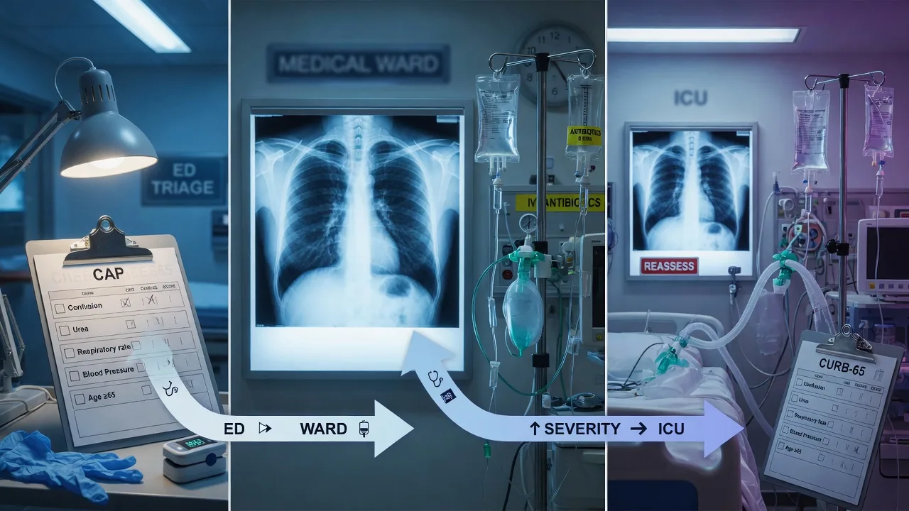
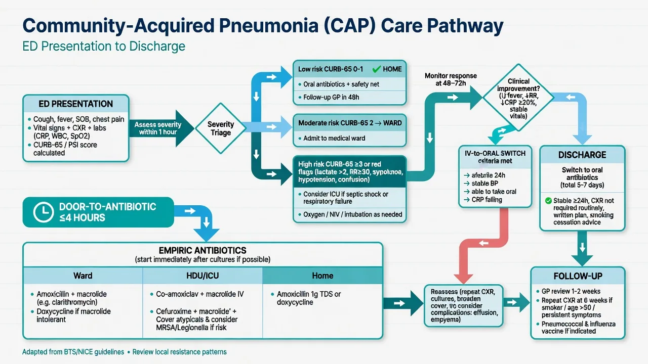
The CAP care pathway spans from community presentation through ED triage, ward management, HDU, and ICU. ED: assess severity (CURB-65/PSI), chest X-ray, bloods, blood cultures, antibiotics within 4h (1h if severe). Ward: continue antibiotics, monitor, daily review, oxygen, physiotherapy, VTE prophylaxis, early mobilisation. HDU: escalating oxygen needs, NIV, single vasopressor. ICU: mechanical ventilation, septic shock (multiple vasopressors), RRT, multi-organ failure. Discharge planning begins on admission: estimated length of stay, home support, follow-up CXR at 6 weeks, vaccination. Integrated care: seamless transitions between care levels — avoid 'silo' management. The pathway is governed by auditable quality metrics — door-to-antibiotic time, appropriate empiric antibiotic selection, length of stay, 30-day readmission, and risk-adjusted mortality — that close the audit loop from one admission to the next.
On this page & tools
Your progress
Saved locally on this device.
Target exams
Red flags
SAQ — Severe CAP: severity scoring and the site-of-care decision
10 minutes · 10 marks
A 68-year-old man presents to ED with a 3-day history of fever, productive cough and pleuritic chest pain. He is confused (GCS 13), RR 32, BP 92/55, SaO2 88% on room air. Bloods: urea 11 mmol/L, Na 128, lactate 2.4. CXR shows right lower-lobe consolidation. The registrar asks whether he should be admitted to ICU.
SAQ — IV-to-oral switch and the discharge bundle
10 minutes · 10 marks
A 55-year-old woman with right middle-lobe pneumococcal pneumonia has been on IV amoxicillin for 5 days. She is now afebrile for 36 hours, RR 18, BP 118/72, SaO2 96% on room air, alert and eating normally. Blood cultures (day 1) grew Streptococcus pneumoniae sensitive to penicillin. The team wishes to switch to oral and discharge her.
Clinical pearls
The integrated CAP care pathway — eight handoffs from ED to discharge
The fellowship candidate is expected to describe CAP not as a single encounter but as a continuous pathway with explicit handoffs between teams, locations, and phases of care. Each handoff is a moment of risk: miscommunication, an undocumented allergy, a missed culture, an unwritten antibiotic plan, or a discharge without follow-up booked converts a recoverable illness into a prolonged or fatal one. The pathway below is organised by the eight handoffs, each tied to a process metric that makes it auditable.[1][2]
The cardinal principle is that no stage stands alone. A poor ED assessment (CURB-65 not calculated) makes the site-of-care decision wrong; a delayed first antibiotic converts curable pneumonia into septic shock; missing a pre-antibiotic blood culture prevents de-escalation and prolongs broad-spectrum therapy; failure to reach clinical-stability criteria before the IV-to-oral switch causes early readmission; a discharge without vaccination or follow-up booked wastes the entire admission's investment. Integration is the point. [1]
flowchart LR
  A[Community / pre-hospital] --> B[ED triage & recognition]
  B --> C[Severity scoring: CURB-65 / PSI]
  C --> D[Door-to-antibiotic ≤4h / ≤1h]
  D --> E[Site-of-care decision]
  E --> F[Ward]
  E --> G[HDU]
  E --> H[ICU]
  F --> I[48-72h reassessment]
  G --> I
  H --> I
  I --> J{Clinical stability?}
  J -->|No| K[Reassess Dx / complication / host]
  K --> I
  J -->|Yes| L[De-escalate + IV-to-oral switch]
  L --> M[Discharge bundle]
  M --> N[6-week CXR + GP + ICU clinic]
  N --> O[Audit metrics → next admission]
  O --> B
Handoff 1 — ED triage and recognition
The first handoff is from the community (patient, family, ambulance, or GP) to the emergency department. The ED team's job is threefold and simultaneous: recognise that this is pneumonia, risk-stratify it, and start the clock for the door-to-antibiotic metric. Recognition is syndromic — most CAP is treated empirically because the organism is never identified.[2]
[1]ED triage of suspected CAP — the first 15 minutes
TRIAGE AND ABC
Airway patent, assess breathing (RR, SpO2, accessory muscle use, work of breathing), circulation (HR, BP, capillary refill, perfusion), disability (GCS/confusion — new confusion is a CURB-65 point and a marker of severe CAP). Assign an early-warning score (MEWS/NEWS2) and trigger a rapid response if deranged.
OXYGEN
Target SpO2 92-96% for most adults; 88-92% for COPD retainers. Use nasal prongs → simple mask → HFNC → NIV → invasive ventilation in escalating fashion. A rising oxygen requirement is an escalation trigger — do not let a patient silently climb the oxygen ladder on a general ward.
IV ACCESS AND BLOODS
FBC, U&E (urea for CURB-65), LFTs, CRP, procalcitonin, lactate, coagulation, troponin (myocardial infarction and AF are common CAP complications), VBG/ABG. Lactate >2 mmol/L indicates tissue hypoperfusion and pushes toward the severe/sepsis pathway.
CHEST X-RAY
PA ± lateral film. Look for lobar consolidation (typical), multilobar (severe), interstitial (atypical/viral/Pneumocystis), cavitation (Staph, Klebsiella, anaerobes, TB), pleural effusion (parapneumonic). A normal CXR with strong clinical suspicion = repeat in 24-48 h or CT.
CALCULATE CURB-65 AT THE BEDSIDE
30 seconds: Confusion, Urea >7, RR ≥30, BP (SBP <90 or DBP ≤60), age ≥65. Document the score explicitly — a score that is calculated but not documented and not acted upon is useless. This is the first auditable metric of the pathway.
START THE ANTIBIOTIC CLOCK
If severe CAP is recognised (IDSA/ATS criteria or clinical judgement), the door-to-antibiotic target is 1 hour. Take cultures within 45 minutes if possible, then give the empiric antibiotic immediately. Do NOT send the patient to imaging before the antibiotic is given if sepsis is present.
Elderly/immunocompromised CAP presents atypically — lower your threshold
| Presentation | Classic CAP | Elderly / immunocompromised |
|---|---|---|
| Symptoms | Cough, purulent sputum, pleuritic pain, fever, rigors | Fall, acute confusion/delirium, new incontinence, anorexia, 'off legs', lethargy |
| Fever | Usually >38°C | Absent in up to 30%; hypothermia is a poor sign |
| Most sensitive sign | Productive cough | Tachypnoea (RR >22) — often the only sign |
| Action | Standard pathway | Low threshold for CXR, blood cultures, and admission; calculate CURB-65 but apply clinical over-ride (scores under-perform here) |
Handoff 2 — Door-to-antibiotic time: the 4-hour / 1-hour quality metric
The door-to-antibiotic time is the single most studied and audited process metric in CAP. It is the time from ED arrival (or from recognition of severe CAP) to the first dose of an appropriate antibiotic. The evidence base is substantial: early antibiotic administration in severe CAP and septic shock is one of the strongest temporal-outcome relationships in critical care medicine.[3][8]
Door-to-antibiotic targets by CAP severity
| CAP severity | Target time-to-antibiotic | Rationale / source |
|---|---|---|
| Severe CAP / septic shock (IDSA/ATS criteria) | ≤ 1 hour from recognition | Surviving Sepsis Campaign Hour-1 bundle; each hour of delay in septic shock increases mortality ~7.6%[8] |
| Moderate-severity ward CAP | ≤ 4 hours from arrival | Meehan 1997 (JAMA) showed lower 30-day mortality when antibiotics given within 4 h in elderly CAP; embedded in older CMS/Joint Commission core measures[3] |
| Low-risk outpatient CAP (CURB-65 0-1) | Same day (before discharge) | Oral antibiotic prescribed and first dose given in ED/clinic before the patient leaves |
Meehan 1997 — Quality of care, process, and outcomes in elderly CAP (PMID 9403422)
Design
Retrospective cohort of 14,069 Medicare patients aged ≥65 hospitalised with CAP in Connecticut, USA
Aim
To determine whether previously recommended process-of-care measures (including timing of antibiotic administration) were associated with 30-day mortality
Key process measures
Antibiotic initiation within 8 hours of arrival; blood cultures before antibiotics; oxygenation assessment within 24 h
Key result
Antibiotic administration within 8 hours of arrival was associated with lower 30-day mortality (OR 0.86, 95% CI 0.76-0.98). Blood cultures drawn before antibiotics predicted a lower 30-day mortality. The within-8-hour and (in subsequent analyses) within-4-hour findings drove the adoption of door-to-antibiotic as a quality metric
Bottom line
This is the foundational study linking process-of-care timing to CAP outcomes and the origin of the door-to-antibiotic quality metric. Later work refined the threshold (1 h for severe CAP) and emphasised antibiotic appropriateness over raw speed, but the principle — treat early — is intact. The single most-cited CAP quality study
Meehan 2001 — The Connecticut Pneumonia Pathway Project (PMID 11530031)
Design
A statewide, multifaceted quality-improvement intervention across 32 Connecticut hospitals, applying a critical pathway for hospitalised CAP to ≥5,800 patients
Intervention
A coordinated pathway: standardised risk stratification, early antibiotic timing targets, blood culture before antibiotics, early switch to oral therapy, and discharge planning. Supported by audit, feedback, and clinician education
Key result
Intervention hospitals improved on all process measures (more blood cultures before antibiotics, more guideline-concordant antibiotic selection, faster IV-to-oral switch, shorter length of stay) without increasing readmission or mortality
Bottom line
Demonstrates that a structured CAP care pathway — with audit and feedback — changes clinician behaviour and improves process metrics. The model for institutional CAP pathways worldwide: pathway + measurement + feedback = improvement
Driving door-to-antibiotic compliance — system levers, not just individual effort
STOCK THE ED WITH PRE-MIXED EMPIRIC ANTIBIOTICS
Ceftriaxone and azithromycin available as ready-to-administer infusions. The single biggest cause of delay is pharmacy preparation time, not clinician decision time.
EMBED THE SEVERITY SCORE IN THE ED ELECTRONIC RECORD
Auto-calculate CURB-65 from the observation chart data; prompt the clinician to act on a score ≥3. Forced-function documentation of the score.
USE A SEPSIS/CAP ALERT PATHWAY
A predefined order-set triggered by sepsis criteria that bundles cultures, lactate, antibiotic, and fluids into one click. Reduces cognitive load in a busy ED.
AUDIT AND FEEDBACK MONTHLY
Every CAP case reviewed for time-to-antibiotic, culture-before-antibiotic rate, and antibiotic appropriateness. Feed back to individuals and the team. Behaviour change requires measurement.
DO NOT COMPROMISE DIAGNOSIS FOR SPEED
In ward CAP, a brief assessment (CXR, CURB-65) before antibiotics is acceptable and reduces misdiagnosis. In severe CAP/sepsis, treat first and complete the workup in parallel.
Handoff 3 — Severity scoring and the site-of-care decision
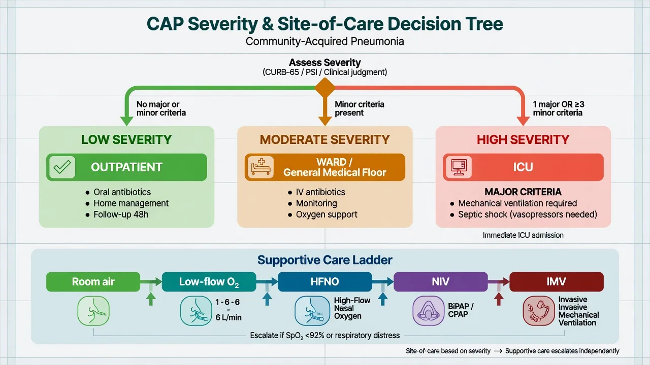
The single most important early decision in CAP is where to treat the patient — home, ward, HDU, or ICU. Three validated scores answer three different questions and are used in concert. The fellowship candidate must know all three and their limitations — no score replaces clinical judgement, especially in the young sick patient (under-scored by age-weighted tools), the elderly, the pregnant, and the immunocompromised.[1][2][4][5]
CURB-65 vs PSI vs IDSA/ATS — three scores, three questions
| Feature | CURB-65 | PSI (PORT score) | IDSA/ATS criteria |
|---|---|---|---|
| Question answered | Who needs hospital? | Who is low-risk (can go home safely)? | Who needs ICU? |
| Components | Confusion, Urea >7, RR ≥30, BP (SBP <90 or DBP ≤60), age ≥65 | Demographics, nursing home, 5 comorbidities, 5 exam findings, 7 lab/imaging findings → 5 risk classes (I-V) | 2 MAJOR: invasive ventilation, septic shock on vasopressors. 9 MINOR: RR ≥30, PaO2/FiO2 <250, multilobar, confusion, uraemia, WCC <4, platelets <100, hypothermia <36, hypotension needing fluids |
| Interpretation | 0-1 outpatient; 2 ward; ≥3 severe — consider ICU | I-II outpatient; III brief inpatient; IV-V inpatient (V often ICU) | 1 major OR ≥3 minor = severe CAP = ICU |
| Strengths | Fast, memorable, reproducible, bedside | Most accurate mortality prediction; reference standard for outpatient safety | Specifically designed for ICU triage |
| Weaknesses | Age dominates; under-scores young sick patients; omits oxygenation | Cumbersome (needs app/website); age dominates | Scoring all 9 minors can delay ICU; some use ≥2 minor in high-risk groups |
| When to use | ED triage, ambulance, first contact | Formal admission decision, discharge planning | Once admitted: who needs ICU vs ward/HDU |
CURB-65 — the bedside CAP severity score
IDSA/ATS severe CAP criteria — invasive/septic, or 3 minor
Fine 1997 — The Pneumonia Severity Index / PORT score (PMID 8995086)
Design
Prospective observational cohort study; derivation in 14,199 inpatients with CAP, validation in 38,039 patients (the PORT cohort)
Aim
Derive a prediction rule to identify patients with CAP at LOW risk of 30-day mortality who could be safely treated as outpatients
The rule
Two-step: step 1 screens for Class I (age <50, no comorbidity/derangement); step 2 assigns points for age, nursing home, 5 comorbidities (neoplastic, liver, CHF, cerebrovascular, renal), 5 exam findings (altered mental status, RR >30, SBP <90, temp <35 or >40, pulse >125), 7 labs/imaging (pH <7.35, BUN >30, Na <130, glucose >250, haematocrit <30, PaO2 <60 or SpO2 <90, pleural effusion)
Five risk classes
Mortality: I 0.1%, II 0.6%, III 0.9%, IV 9.3%, V 27.0%
Bottom line
PSI is the reference standard for CAP mortality prediction. Classes I-II can be managed as outpatients; IV-V require inpatient care. Cumbersome at the bedside (use an app), and age dominates so it under-scores severe physiology in young patients. Used alongside CURB-65
Lim 2003 — CURB-65 derivation and validation (PMID 12728155)
Design
Prospective study of 1,068 patients with suspected CAP across three UK hospitals; international derivation and validation of a simple severity score
The score
Five equally weighted points: **C**onfusion, **U**rea >7 mmol/L, **R**espiratory rate ≥30, **B**lood pressure (SBP <90 or DBP ≤60), age ≥65
Mortality by score
Score 0: 1.5%; score 1: 2.7%; score 2: 6.8%; score 3: 14.0%; score 4: 27.8%; score 5: 27-40%
Recommended use
0-1: low risk — consider outpatient. 2: moderate — inpatient (ward). ≥3: severe — consider ICU/HDU
Bottom line
CURB-65 is the bedside workhorse — fast, memorable, reproducible, validated internationally. Weakness: age dominates (an unwell 25-year-old scores 0) and it omits oxygenation. Use with clinical judgement and the PSI/IDSA/ATS criteria
Site-of-care decision — the practical sequence
CALCULATE CURB-65 AT THE BEDSIDE
Score 0-1: consider outpatient with safety-net advice and 48 h review. Score 2: admit to ward. Score ≥3: severe — admit, calculate PSI, assess for ICU.
IF ADMITTED, CALCULATE PSI AND CHECK IDSA/ATS CRITERIA
PSI refines the mortality prediction; IDSA/ATS answer the ICU question. **1 major criterion (invasive ventilation OR septic shock on vasopressors) = ICU immediately** — no further scoring needed.
ASSESS THE 9 IDSA/ATS MINOR CRITERIA
If ≥3 minor criteria present, manage in ICU/HDU. Some experts use ≥2 in the elderly or immunocompromised. Do NOT delay antibiotics while counting.
APPLY CLINICAL OVER-RIDE
Admit/upgrade if: poor social circumstances, unable to tolerate oral, immunocompromise, pregnancy, suspected sepsis, hypoxia not corrected by oxygen, recent influenza, end-organ dysfunction. The score is a guide, not a verdict.
REASSESS FREQUENTLY
A patient can deteriorate rapidly in the first 24 h. Repeat CURB-65 and clinical assessment at every nursing handover and on any change. Run a continuous MEWS/NEWS2 score.
Handoff 4 — Empiric antibiotic selection: right drug, right time
The empiric antibiotic is chosen before the organism is known, covering the likely pathogens for the patient's severity, comorbidities, and local resistance patterns. The principle is cover broadly and early, then narrow once the pathogen is identified. Antibiotic appropriateness — choosing a guideline-concordant regimen — is itself a quality metric, because inappropriate initial therapy is associated with higher mortality and longer stay.[1][2]
Empiric antibiotic regimens by site of care
| Scenario | Empiric regimen | Rationale |
|---|---|---|
| Outpatient, CURB-65 0-1, no comorbidity | Amoxycillin 1 g PO tds (or doxycycline 100 mg bd if penicillin-allergic) ± macrolide (clarithromycin 500 mg bd) if atypical suspected | S. pneumoniae remains the commonest pathogen and is penicillin-sensitive in most of ANZ/UK. Macrolide adds atypical cover |
| Outpatient with comorbidity (COPD, CHF, diabetes, CKD, malignancy, immunosuppression) | Amoxycillin-clavulanate 875/125 mg PO tds + macrolide OR doxycycline; or respiratory fluoroquinolone monotherapy | Broader cover for H. influenzae, Moraxella, enteric Gram-negatives, atypicals |
| Inpatient ward, CURB-65 2-3 | Benzylpenicillin 1.2 g IV q6h (or ceftriaxone 1-2 g IV od) + azithromycin 500 mg IV/PO od | IV β-lactam + macrolide covers typicals + atypicals |
| Severe CAP / ICU (IDSA/ATS criteria) | Ceftriaxone 2 g IV od + azithromycin 500 mg IV od; or piperacillin-tazobactam 4.5 g IV q6h + azithromycin if Pseudomonas risk | Broad β-lactam + macrolide is the standard; macrolide adds anti-Legionella and immunomodulatory benefit |
| Severe CAP + MRSA risk (post-influenza, ESRD, IVDU, recent healthcare, known colonisation) | ADD vancomycin 15-25 mg/kg q8-12h (AUC 400-600) or linezolid 600 mg q12h | Post-viral S. aureus CAP is necrotising and rapidly progressive; linezolid has better lung penetration and toxin suppression |
| Severe CAP + Pseudomonas risk (bronchiectasis, severe COPD, repeated antibiotics, recent hospitalisation) | Piperacillin-tazobactam (or ceftazidime, cefepime, meropenem) + macrolide | Standard ceftriaxone does NOT cover Pseudomonas |
| Suspected/confirmed influenza | ADD oseltamivir 75 mg PO bd, ideally within 48 h of symptom onset | Reduces influenza mortality even when given late in critically ill patients |
Combination vs monotherapy in severe CAP — the macrolide question
| Regimen | Role | Evidence |
|---|---|---|
| β-lactam + macrolide | Standard severe CAP regimen | Observational data and meta-analyses show lower mortality vs β-lactam alone in severe CAP; covers atypicals; immunomodulatory effect |
| Respiratory fluoroquinolone monotherapy (moxifloxacin, levofloxacin) | Alternative; covers typicals + atypicals in one drug | Adequate cover but some concern about lower anti-Legionella efficacy and QT prolongation; reserve for penicillin-allergic or when macrolide contraindicated |
| β-lactam + β-lactam (e.g., ceftriaxone + ampicillin) | Not standard; used in some regions for broad Gram-negative cover | No mortality advantage over β-lactam + macrolide; greater resistance pressure |
| β-lactam monotherapy (no macrolide) | Not acceptable in severe CAP — misses Legionella and loses immunomodulatory benefit | Higher mortality in severe CAP cohorts vs combination |
Garin 2014 — β-lactam monotherapy vs β-lactam + macrolide in moderately severe CAP (PMID 25286173)
Design
Multicentre, randomised, open-label, non-inferiority trial across 6 European hospitals; 580 adults with moderately severe CAP (PSI class III-V) without need for ICU
Intervention
β-lactam monotherapy (amoxicillin-clavulanate or ceftriaxone) vs β-lactam + macrolide combination
Primary outcome
Day-7 clinical stability — the β-lactam monotherapy arm was found to be NON-INFERIOR for clinical stability and several secondary outcomes
Key caveat
Patients with severe CAP requiring ICU, septic shock, or proven/likely Legionella or atypical infection were EXCLUDED. The trial therefore informs the WARD / moderately-severe CAP question, NOT the severe-CAP/ICU question where combination therapy remains standard
Bottom line
In NON-severe hospitalised CAP without septic shock or suspected atypicals, β-lactam monotherapy is non-inferior to combination. This supports de-escalation/stewardship in ward CAP. In severe CAP / ICU, β-lactam + macrolide remains the standard — do NOT extrapolate this trial to the intubated patient
Handoff 5 — ICU admission criteria: who needs intensive care
Not every CAP needs the ICU, but the patients who do need it need it immediately. The IDSA/ATS criteria are the operational rule: one major criterion (invasive ventilation or septic shock requiring vasopressors) OR three or more minor criteria mandates ICU-level care. The delay between meeting a criterion and arriving in ICU is itself a predictor of mortality — a deteriorating patient on a general ward is at greater risk than the same patient in a high-dependency environment.[1][8]
Level of care — ward vs HDU vs ICU for CAP
| Domain | Ward | HDU / high-dependency | ICU |
|---|---|---|---|
| Respiratory support | Low-flow nasal prongs / simple mask | HFNC, single-modality NIV (e.g., COPD exacerbation) | Invasive mechanical ventilation, dual NIV failure, escalating PEEP/FiO2 |
| Haemodynamics | Stable, no vasopressors | Single vasopressor at low dose, stable on fluids | Multiple vasopressors, refractory septic shock, need for central monitoring |
| Monitoring | NEWS2 q4h, SpO2 continuous | Continuous SpO2, hourly obs, arterial line, close nursing (1:2) | Invasive monitoring, 1:1 nursing, arterial/CVC, cardiac output monitoring |
| Organ support | None | One organ system supported | Two or more organ systems supported (ventilation + vasopressors + RRT) |
| Indication from CAP | CURB-65 2, stable | CURB-65 3 with single evolving organ failure | IDSA/ATS major criterion, or ≥3 minor criteria, or 2+ organ failures |
The ICU admission decision for CAP — when to escalate
CHECK THE 2 MAJOR IDSA/ATS CRITERIA FIRST
Does the patient need invasive mechanical ventilation? Are they in septic shock requiring vasopressors to keep MAP ≥65 after adequate fluid resuscitation? If YES to either → ICU immediately.
IF NO MAJOR CRITERION, COUNT THE 9 MINOR CRITERIA
RR ≥30, PaO2/FiO2 <250, multilobar infiltrates, confusion, uraemia (BUN >7 mmol/L), WCC <4, platelets <100, hypothermia <36, hypotension needing aggressive fluids. ≥3 minor criteria → ICU/HDU.
FACTORS THAT LOWER THE ICU THRESHOLD
Elderly, immunocompromised, pregnant, neuromuscular disease (weak respiratory muscles), severe comorbidity (CKD, cirrhosis, heart failure), poor social backup, post-influenza, suspected necrotising Staph/Klebsiella.
ESCALATION TRIGGERS ON THE WARD
Any of: rising RR, falling SpO2, rising lactate, new confusion, falling urine output, hypotension unresponsive to fluids, escalating oxygen requirement beyond low-flow, need for NIV. These trigger a rapid-response call and reassessment of the ICU decision.
DO NOT LET NIV HAPPEN UNMONITORED ON A GENERAL WARD
A patient who needs NIV for CAP needs HDU/ICU-level monitoring — NIV can mask deterioration, cause gastric insufflation/aspiration, and require rapid intubation if it fails. NIV on a general ward is suboptimal care.
REASSESS THE DECISION EVERY FEW HOURS
The trajectory matters as much as the snapshot. A patient trending toward multiple minor criteria will likely soon meet a major criterion — get them to ICU early, not after they arrest on the ward.
NIV in CAP — when it works and when it fails
| Scenario | NIV role | Caution |
|---|---|---|
| CAP with type I respiratory failure, no severe acidosis | Trial of HFNC first; NIV if HFNC fails | Must be in HDU/ICU; define a clear intubation trigger (e.g., persisting RR >30, pH <7.25, SpO2 <90% on FiO2 0.6) |
| CAP in COPD with type II failure / hypercapnia | NIV often effective (treats the COPD component) | COPD-NIV response does not equal CAP resolution; still need antibiotics, physiotherapy, escalation if worsening |
| Immunocompromised CAP | NIV may avoid intubation (lower mortality vs intubation in this group) | Close monitoring; early intubation if failing (delayed intubation in immunocompromised is harmful) |
| Severe CAP with ARDS physiology | NIV has a HIGH failure rate (~50-60%); intubate early | Do not persist with NIV in a deteriorating patient; each hour of failed NIV delays definitive airway protection |
Handoff 6 — Monitoring response and de-escalation
Once empiric therapy is underway, the daily question is: is the patient getting better, and can I narrow the antibiotic? The classical teaching is that clinical response is assessed at 48-72 hours. Improvement triggers de-escalation and the IV-to-oral switch; failure to improve by day 3-5 triggers a structured re-evaluation (not a knee-jerk antibiotic escalation). De-escalation is the antimicrobial-stewardship backbone of the pathway.[1][6]
Markers of clinical response in CAP — day by day
| Marker | Improving | Worrying / non-response |
|---|---|---|
| Fever | Defervescence over 48-72 h (typical) | Persistent or recurrent fever at day 3-5 — reassess |
| RR / oxygenation | RR falling, SpO2 rising, FiO2 weaning | Persisting tachypnoea, escalating FiO2 — ARDS, effusion, PE, wrong organism |
| Haemodynamics | Off vasopressors, MAP stable, lactate clearing | Persisting/rebounding shock — wrong drug, source uncontrolled (empyema) |
| WCC / CRP | CRP falls by >50% in 4-5 days | CRP static or rising — resistant organism, complication |
| Procalcitonin | Falls >80% from baseline, or <0.25 | Persistently elevated — ongoing bacterial infection |
| Mental state | Confusion resolving | Worsening confusion — hypoxia, sepsis, hyponatraemia, withdrawal |
| Radiology | CXR improvement lags clinical | Progressive multilobar infiltrates — ARDS, necrotising organism |
| Functional status | Mobilising, eating | Bed-bound, not eating — frailty, complication, prolonged recovery |
Halm 1998 — Time to clinical stability in hospitalised CAP (PMID 9600479)
Design
Prospective multicentre cohort of 680 hospitalised CAP patients; defined and measured the time to reach objective clinical stability criteria
Stability criteria
Improvement in five parameters: temperature (≤37.8°C / ≤99°F for ≥24 h); heart rate ≤100; respiratory rate ≤24; systolic BP ≥90; oxygen saturation ≥90% (or baseline) on room air; ability to tolerate oral intake; normal mental status
Key result
Median time to overall stability was 3 days. By day 3, ~50% were stable; by day 7, ~80%. Temperature and heart rate normalised first (1-2 days); hypoxia took longest to resolve. Stability predicted safe IV-to-oral switch and discharge
Bottom line
These are the 'Halm stability criteria' — the objective foundation of the IV-to-oral switch and the discharge decision. A patient who meets all five criteria for 24 h is, in most cases, safe to switch to oral therapy and consider discharge
De-escalation of CAP antibiotics — step by step
DAILY MICROBIOLOGY REVIEW
As cultures and PCR return (typically 48-72 h), identify the causative organism and its sensitivities.
NARROW THE SPECTRUM
Pneumococcus sensitive to penicillin → switch ceftriaxone to benzylpenicillin or amoxycillin. Legionella → stop β-lactam, continue macrolide/fluoroquinolone. Influenza only → stop antibacterials once bacterial co-infection excluded.
STOP REDUNDANT COVER
Discontinue MRSA cover (vancomycin/linezolid) if no MRSA at 48-72 h. Discontinue Pseudomonas cover if no Pseudomonas. Discontinue oseltamivir once influenza PCR negative (outside season).
USE PROCALCITONIN AS A STEWARDSHIP ADJUNCT
When PCT falls >80% from peak OR is <0.25 ng/mL and the patient is clinically stable, stop antibiotics. Clinical judgement integrates the biomarker; it does not obey it.
SET A STOP DATE AT THE START
Every antibiotic prescription has a documented indication, review date, and stop date. Stewardship is a system, not an afterthought.
DAILY ANTIBIOTIC TIME-OUT ON ROUNDS
"Does this patient still need this antibiotic, at this dose, for this duration?" If no — stop, narrow, or switch. De-escalation rates in real-world ICU practice remain 40-60%; fellowship-level care means de-escalating every day that cultures allow.
Differential diagnosis of non-resolving CAP at 72 hours
| Category | Causes | Action |
|---|---|---|
| Wrong organism / resistant | MRSA, Pseudomonas, ESBL, atypical covered inadequately (Legionella missed), TB, fungal (Aspergillus, Pneumocystis) | Review cultures, extend empiric cover, send induced sputum/BAL; bronchoscopy |
| Wrong diagnosis | Pulmonary embolism, pulmonary oedema, vasculitis (GPA), organising pneumonia, malignancy, alveolar haemorrhage, drug-induced pneumonitis | CTPA, echo, autoimmune screen, bronchoscopy ± biopsy |
| Complication | Empyema, lung abscess, ARDS, metastatic infection (endocarditis), venous thromboembolism, superimposed HAP/VAP | Ultrasound/CT chest, thoracentesis, chest tube, echocardiogram |
| Host factors | Immunocompromise (undiagnosed HIV, neutropenia), untreated comorbidity (diabetes, malnutrition, alcohol), recurrent aspiration | HIV test, immune workup, optimise comorbidity, swallow assessment |
| Pharmacological | Inadequate dose, poor lung penetration, drug fever | Review dosing, therapeutic drug monitoring (vancomycin AUC) |
Handoff 7 — IV-to-oral switch and clinical stability
The intravenous-to-oral switch is a key milestone in CAP recovery: it marks clinical stability, enables ward/step-down care, and is a prerequisite for discharge. The principle is pharmacological — once the patient can absorb oral medication and is clinically improving, oral antibiotics achieve equivalent serum concentrations to IV for the common CAP pathogens (good oral bioavailability: fluoroquinolones, linezolid, clindamycin, metronidazole, doxycycline, fluconazole, and amoxycillin).[2][6]
Criteria for the IV-to-oral switch in CAP
| Criterion | Threshold | Comment |
|---|---|---|
| Clinical improvement | Cough, dyspnoea, RR improving | Subjective + objective improvement over 24 h |
| Afebrile | Temperature <37.8°C for 24-48 h | Two consecutive readings >8 h apart. Persistent fever → investigate before switching |
| Haemodynamic stability | Off vasopressors, SBP >90, MAP >65, lactate normal | No active shock |
| Tolerating oral intake | Eating and drinking, no vomiting, no ileus | Essential — oral antibiotics are useless if not absorbed |
| Mental status | Improving, able to take oral meds reliably | Confusion precludes reliable oral dosing |
| Respiratory support weaning | Stable on low-flow nasal prongs or room air; not escalating FiO2 | A patient on HFNC/NIV/IMV is NOT ready |
| Inflammatory markers | Falling CRP and PCT | CRP fall >50% from peak supports the switch |
| No uncontrolled complication | No new empyema, abscess, endocarditis | Source control complete |
IV-to-oral switch — the practical sequence
CHECK ALL SWITCH CRITERIA DAILY FROM DAY 2
When criteria met, switch from IV to oral. Use a drug with high oral bioavailability: amoxycillin, amoxycillin-clavulanate, clarithromycin/azithromycin, doxycycline, moxifloxacin/levofloxacin, linezolid. Match the IV organism and sensitivities.
MONITOR FOR 24 HOURS AFTER SWITCH
Confirm clinical stability on oral therapy before discharge. Most failures occur in this window — if the patient spikes fever or desaturates after the switch, reassess.
CONFIRM DISCHARGE READINESS
Afebrile 24-48 h, clinical stability for 24 h on oral antibiotics, tolerating oral, SpO2 stable on room air (or baseline), able to mobilise safely, no new complications, home support available, follow-up arranged.
MATCH THE ORAL AGENT TO THE ORGANISM
Sensitive pneumococcus → amoxycillin. Legionella → clarithromycin or doxycycline. Staph (MSSA) → flucloxacillin ± rifampicin (consult ID). Do not switch to an agent with poor oral bioavailability (e.g., oral cephalosporins have variable absorption).
DEFINE TOTAL DURATION
Total (IV + oral) course: 5 days for uncomplicated CAP with good response; 7-14 days for Legionella, Pseudomonas, S. aureus; longer for bacteraemia with metastatic infection or empyema.
Antibiotic duration in CAP — pathogen-specific
| Scenario | Recommended duration | Rationale |
|---|---|---|
| Uncomplicated CAP, good response | 5 days (stop if afebrile 48-72 h AND clinically stable) | Short courses (≤5-7 days) are non-inferior to longer; many patients are over-treated |
| S. pneumoniae, H. influenzae (typical, sensitive) | 5-7 days | Afebrile 48-72 h + clinically stable = stop |
| Legionella pneumophila | 7-14 days (14-21 if immunocompromised) | Intracellular pathogen; macrolide or fluoroquinolone |
| Mycoplasma / Chlamydia | 7-14 days | Macrolide, doxycycline, or fluoroquinolone |
| Pseudomonas aeruginosa | 7-14 days (longer if bacteraemic/necrotising) | Re-assess daily; longer if slow response or complicated |
| S. aureus CAP (MSSA/MRSA) | ≥7-14 days; longer if bacteraemia, endocarditis, cavitation | Always look for endocarditis (echo); bacteraemia often ≥14 days |
| Empyema / lung abscess | 4-6 weeks (until radiological resolution) | Source control (drain) more important than duration |
| PCT-guided stopping | Stop when PCT >80% drop OR <0.25 ng/mL | Non-inferior to fixed-duration; stewardship adjunct |
Handoff 8 — Discharge planning, readmission prevention, and the audit loop
CAP is not 'cured' at discharge. Recovery is prolonged — fatigue for 3-6 months, cognitive impairment for 6-12 months, and some patients never fully recover (post-intensive-care syndrome, PICS). The discharge phase is where long-term outcomes are won or lost, and where the audit loop closes: the metrics of this admission (door-to-antibiotic, LOS, readmission) feed the quality improvement that shapes the next patient's pathway.[1][2][10]
Discharge bundle for CAP — the complete checklist
CLINICAL STABILITY
Afebrile 24-48 h, stable SpO2 on room air (or baseline), tolerating oral antibiotics for 24 h, mobilising safely, no active complications. This is the Halm stability criteria sustained.
ANTIBIOTIC PLAN
Oral course to complete planned duration; written plan; clear advice on adherence and what to do if symptoms recur. Document the total intended course (IV + oral).
VACCINATION
Pneumococcal, influenza (seasonal), COVID-19 booster, and any overdue vaccines (Tdap, herpes zoster, RSV) given BEFORE discharge. In-hospital vaccination has the highest uptake of any setting.
SMOKING CESSATION
Brief intervention, nicotine replacement therapy, referral to quitline — the single most effective prevention of recurrent CAP and the underlying COPD/lung cancer risk. The CAP admission is a powerful teachable moment.
FOLLOW-UP ARRANGED
GP at 1 week; CXR at 6 weeks (resolution — non-resolving infiltrate mandates investigation); ICU follow-up clinic at 2-3 months for those ICU-admitted (PICS screening); pulmonary rehabilitation referral for persisting breathlessness.
PATIENT EDUCATION
Teach-back on warning signs (worsening dyspnoea, recurrent fever, haemoptysis, pleuritic chest pain), recovery timeline, medication management, written materials at year-6 reading level, contact details for worsening symptoms.
SOCIAL / COMMUNITY SUPPORT
Home help, meals, transport, carer involvement as needed; early discharge coordinator involvement for complex patients. Address functional decline (common in elderly post-CAP).
DOCUMENT QUALITY METRICS
Time-to-antibiotic, blood culture rate, de-escalation rate, length of stay, 30-day readmission — feed into the audit cycle. A pathway that is not audited is not a pathway.
Dharmarajan 2013 — Patterns of 30-day readmission after pneumonia (PMID 24259033)
Design
Retrospective cohort of 3,278,252 Medicare hospitalisations (heart failure, pneumonia, or acute MI) across the USA; analysed 30-day readmission patterns
Key finding (pneumonia)
Of pneumonia patients readmitted within 30 days, the readmission was most often NOT for recurrent pneumonia. The majority were for comorbidity exacerbation — heart failure, COPD, other respiratory illness — or unrelated conditions. Only a minority of readmissions were directly for pneumonia again
Implication
Post-discharge readmission risk is driven as much by uncontrolled comorbidity and the social/functional state as by the pneumonia itself. The discharge bundle must therefore address comorbidity optimisation, functional recovery, and early follow-up — not just the antibiotic course
Bottom line
30-day readmission after CAP is common (up to ~20%) and is a penalised quality metric in many health systems. Reducing it requires treating the WHOLE patient at discharge — comorbidity, function, social support, follow-up — because most readmissions are NOT antibiotic failure
Risk factors for 30-day readmission after CAP — and how to address them
| Domain | Risk factor | Discharge intervention |
|---|---|---|
| Comorbidity | Heart failure, COPD, CKD, diabetes, liver disease | Optimise each before discharge; medication reconciliation; clear GP handover |
| Functional/social | Poor mobility, frailty, lives alone, low health literacy | Discharge coordinator; home help; carer education; written simple instructions |
| Microbiological | Severe pathogen (Staph, Pseudomonas, Legionella), bacteraemia | Ensure adequate duration; early follow-up; low threshold to represent |
| Process | Long LOS, ICU admission, late antibiotics, no follow-up booked | Book follow-up before discharge; GP review at 1 week; CXR at 6 weeks |
| Behavioural | Smoking, alcohol misuse, non-adherence | Smoking cessation (NRT + quitline), alcohol liaison, adherence packaging |
| Vaccination | Not vaccinated against pneumococcus/influenza | Vaccinate before discharge — halves recurrent CAP and influenza risk |
Preventing readmission — what actually works
OPTIMISE COMORBIDITY BEFORE DISCHARGE
Up-titrate heart failure therapy, check inhaler technique in COPD, optimise diabetes control, address AF rate control and anticoagulation. Most readmissions are for comorbidity, not pneumonia recurrence.
EARLY STRUCTURED FOLLOW-UP
GP or nurse-led review within 7 days. A telephone follow-up within 48-72 h catches early deterioration. Provide the GP with a clear discharge summary (diagnosis, organism, antibiotic plan, pending results).
VACCINATE BEFORE DISCHARGE
Pneumococcal (PCV/PPSV23 per schedule), influenza (seasonal), COVID-19 booster. In-hospital vaccination uptake is the highest of any setting.
FUNCTIONAL REHABILITATION
Early mobilisation in hospital; referral to pulmonary rehabilitation for those with persisting breathlessness; address deconditioning and PICS (especially post-ICU).
MEDICATION RECONCILIATION
Reconcile pre-admission, in-hospital, and discharge medications. Stop unnecessary drugs; ensure the antibiotic course is clearly documented; review inhaler technique; provide adherence packaging if needed.
PATIENT/CARER EDUCATION WITH TEACH-BACK
Warning signs (worsening dyspnoea, fever, haemoptysis), recovery timeline (fatigue common for weeks), when and how to seek help. Written materials at year-6 reading level.
Care bundles — the operational backbone of the pathway
A care bundle is a small, explicit set of evidence-based interventions (usually 3-6 elements) that must ALL be completed for every eligible patient — the principle being that bundled care is more reliable than individual actions. Bundles turn guidelines into behaviour. The CAP pathway is built on four overlapping bundles.[2][8]
The four care bundles of the CAP pathway
| Bundle | When applied | Elements |
|---|---|---|
| Severe CAP / sepsis Hour-1 bundle | On recognition of severe CAP or septic shock | (1) Measure lactate; (2) Blood cultures before antibiotics; (3) Broad-spectrum antibiotics within 1 h; (4) 30 mL/kg crystalloid if hypotensive/lactate ≥4; (5) Vasopressors for MAP <65 after fluids |
| CAP empiric-therapy bundle | ED, all admitted CAP | (1) CURB-65 documented; (2) Blood cultures before antibiotics (if severe); (3) Urinary pneumococcal + Legionella antigens (severe); (4) Guideline-concordant antibiotic within target time; (5) Reassessment documented |
| ICU ventilator bundle (applies to intubated CAP) | Intubated, daily | (1) Head of bed 30-45°; (2) Sedation interruption + SAT/SBT daily; (3) PUD prophylaxis; (4) DVT prophylaxis; (5) (add) glucose control, oral chlorhexidine debatable — prevents VAP superinfection |
| CAP discharge bundle | At discharge | (1) Clinical stability documented; (2) Antibiotic plan complete; (3) Vaccinated; (4) Smoking cessation delivered; (5) Follow-up booked (GP 1 wk, CXR 6 wk, ICU clinic if applicable); (6) Patient educated |
Severe CAP / sepsis Hour-1 bundle — the resuscitative handoff
MEASURE LACTATE
Lactate >2 mmol/L indicates tissue hypoperfusion; >4 mmol/L defines severe sepsis/lactate-related septic shock. Serial lactate guides resuscitation (aim >10% clearance in 2-6 h).
BLOOD CULTURES BEFORE ANTIBIOTICS
Two sets from separate sites, aerobic + anaerobic, ~20 mL per bottle. Do NOT delay antibiotics >45 min for cultures if shocked. Urinary pneumococcal + Legionella antigens unaffected by prior antibiotics — always send.
BROAD-SPECTRUM ANTIBIOTICS WITHIN 1 HOUR
Ceftriaxone 2 g IV + azithromycin 500 mg IV. Add vancomycin/linezolid if MRSA risk; switch to piperacillin-tazobactam if Pseudomonas risk; add oseltamivir in flu season. The single most important intervention.
30 mL/kg CRYSTALLOID IF HYPOTENSIVE OR LACTATE ≥4
Balanced crystalloid preferred. Reassess after each bolus — clinical exam, lactate, point-of-care ultrasound (IVC, B-lines). Over-aggressive fluids cause pulmonary oedema in severe CAP with impaired alveolar-capillary membrane.
VASOPRESSORS FOR MAP <65 AFTER FLUIDS
Noradrenaline first-line. Add vasopressin if refractory. Consider hydrocortisone 200 mg/day for refractory septic shock (and for severe CAP per adjunct evidence). Target MAP ≥65, urine output >0.5 mL/kg/h.
Quality metrics — closing the audit loop
The CAP care pathway is governed by auditable process and outcome metrics. Process metrics (door-to-antibiotic, culture-before-antibiotic, bundle compliance) measure whether the pathway was followed; outcome metrics (mortality, length of stay, readmission) measure whether it worked. The audit loop is: measure → compare to benchmark → identify gaps → intervene (PDSA) → re-measure.[3][9][10]
CAP quality metrics — process vs outcome
| Metric | Type | Benchmark / target | Why it matters |
|---|---|---|---|
| Door-to-antibiotic time | Process | ≤1 h severe CAP; ≤4 h ward CAP | Strongest temporal-outcome link in severe CAP/sepsis; the canonical CAP process metric[3] |
| Blood culture rate (before antibiotics, severe CAP) | Process | >90% of severe CAP | Enables de-escalation, surveillance, public health; mandatory before antibiotics if possible[1] |
| Appropriate / guideline-concordant antibiotic selection | Process | >90% concordance with local guideline | Inappropriate initial therapy → higher mortality, longer stay, resistance[2] |
| CURB-65 documentation rate | Process | >95% of admitted CAP | A score not documented is a score not acted upon; gateway to the site-of-care decision |
| Bundle compliance (all elements, all-or-nothing) | Process | >80% of eligible | Bundled care outperforms a la carte care; reliability metric |
| De-escalation rate | Process/stewardship | >80% when organism identified | Reduces C. diff, resistance, toxicity, cost; under-used (40-60% in practice) |
| IV-to-oral switch rate / timeliness | Process | Switch within 24 h of meeting criteria | Shortens LOS; delayed switch is the commonest avoidable extra bed-day |
| Length of stay (LOS) | Outcome/process | Local benchmark (e.g., median 5-7 days) | Proxy for pathway efficiency; prolonged LOS increases nosocomial risk and cost |
| ICU and hospital mortality (risk-adjusted, SMR) | Outcome | SMR <1.0 | The bottom-line effectiveness metric; risk-adjustment essential (PSI, APACHE) |
| 30-day readmission | Outcome | <15-20% | Penalised quality metric; most readmissions are comorbidity-driven, not antibiotic failure[10] |
| Vaccination rate at discharge | Process/outcome | >90% of eligible | Prevents recurrent CAP and influenza; highest uptake setting |
| Smoking cessation referral rate | Process | >90% of smokers | Most effective secondary prevention; teachable moment |
Running the CAP audit loop — the quality-improvement cycle
DEFINE THE METRICS AND BENCHMARKS
Agree locally on door-to-antibiotic target, culture rate, antibiotic concordance, bundle compliance, LOS, SMR, 30-day readmission. Make them visible on a dashboard.
MEASURE CONTINUOUSLY
Extract from the electronic record: time-to-antibiotic (auto-captured from medication chart), culture-before-antibiotic (timestamp delta), bundle elements (checklist). Monthly audit of a sample for completeness.
COMPARE TO BENCHMARK AND PEERS
Plot monthly trends; compare to national/international benchmarks and to peer units. Identify outliers and trends (worsening or improving).
IDENTIFY THE GAPS
Is door-to-antibiotic failing? Is de-escalation low? Is readmission high? Drill into the specific failure point — usually a handoff (ED→ward, ward→discharge) rather than a single clinician.
INTERVENE (PDSA CYCLE)
Plan-Do-Study-Act: test a change (e.g., pre-mixed antibiotics in ED, auto-calculating CURB-65, pharmacist-led IV-to-oral switch), measure the effect, refine, spread.
FEED BACK TO THE TEAM
Monthly multidisciplinary meeting: share the data, celebrate wins, discuss failures without blame. Behaviour change requires feedback, not just data.
Transitions of care and handoffs — where pathways break
Most pathway failures occur at handoffs between teams or locations, not within them. The patient moved from ED to ward without a documented antibiotic plan; the ward patient transferred to ICU without a structured handover; the patient discharged to the community without follow-up booked — these are the failure points. Structured handover tools (ISBAR, IPASS) and standardised discharge summaries reduce these errors.[2]
Structured handoff tools — ISBAR and the discharge summary
| Tool | When | Elements |
|---|---|---|
| ISBAR | Any verbal/clinical handoff (ED→ward, ward→ICU, shift change) | Identify (patient), Situation (current problem), Background (history, CAP severity, organism), Assessment (current status, pending results), Recommendation (plan, escalation triggers) |
| IPASS | Verbal handoff (especially in training settings) | Illness severity, Patient summary, Action list, Situation awareness + contingency planning, Synthesis by receiver |
| Discharge summary | Hospital → community | Diagnosis + organism + sensitivities, antibiotic course (IV + oral, total duration), pending results (cultures, CXR follow-up at 6 weeks), comorbidity changes, vaccinations given, follow-up booked, warning signs |
| Antibiotic plan card | With the patient | Drug, dose, duration, indication, review/stop date, what to do if symptoms recur |
The high-risk handoffs in the CAP pathway — and how to make them safe
ED → WARD/HDU/ICU
Structured ISBAR handover. Document: severity score, time of first antibiotic, cultures taken, antibiotic plan with review/stop date, escalation triggers. A verbal handover alone is insufficient — the plan must be written.
WARD → ICU (escalation)
Triggered by deterioration (rising RR, falling SpO2, rising lactate, new confusion, hypotension). Use a rapid-response system. The ICU handover includes what has been tried (fluids, antibiotics, oxygen) and the working differential.
ICU → WARD (step-down)
Often a high-risk handoff (loss of 1:1 nursing, monitoring). Document: ventilator settings/weaning, current antibiotics and stop date, pending cultures, rehabilitation needs, PICS risk, family communication.
WARD → HOME (discharge)
The complete discharge bundle (see above). The single commonest failure is no follow-up booked; the second is an unwritten antibiotic plan. Both are preventable with a checklist.
HOSPITAL → GP / COMMUNITY
Discharge summary sent within 24 h (electronically where possible). Includes the 6-week CXR request, vaccination record, and a clear 'represent if...' instruction.
ANY HANDOFF — CLOSE THE LOOP
The receiver must acknowledge and synthesise (read-back for critical information). Handoff is not complete until the receiver owns the plan.
Additional clinical pearls
Additional red flags
Mnemonics and exam summary
The CAP discharge bundle — 'SAFE DISCHARGE'
The one-minute exam answer — severe CAP from ED to discharge
| Stage | Key action | Time / target / metric |
|---|---|---|
| 1. Recognise | New respiratory symptoms + CXR infiltrate + fever/inflammatory markers; CURB-65 | Minutes; CURB-65 documented (metric) |
| 2. Door-to-antibiotic | Cultures before antibiotics (if severe), then empiric regimen | ≤1 h severe CAP; ≤4 h ward CAP (canonical metric)[3] |
| 3. Site-of-care | CURB-65 (bedside) + PSI (admission) + IDSA/ATS (ICU); 1 major OR ≥3 minor = ICU | <30 min |
| 4. Empiric antibiotic | Ceftriaxone 2 g + azithromycin 500 mg IV (+ vanc/linezolid if MRSA; + pip-tazo if Pseudomonas; + oseltamivir in flu season) | Guideline-concordant (metric)[2] |
| 5. Monitor / de-escalate | Clinical + CRP/PCT at 48-72 h; narrow to pathogen; set stop date | Daily; de-escalation rate (metric) |
| 6. IV-to-oral switch | When Halm stability criteria met (afebrile 48 h, tolerating oral, stable SpO2) | Typically day 3-5 |
| 7. Discharge bundle | Stable + completed/will complete oral course + vaccinated + smoking cessation + follow-up booked | Discharge bundle compliance (metric) |
| 8. Audit | Door-to-antibiotic, culture rate, LOS, SMR, 30-day readmission | Monthly PDSA cycle |
References summary
The integrated CAP care pathway rests on landmark studies across severity scoring (Fine PSI 1997; Lim CURB-65 2003), process-of-care and quality measurement (Meehan 1997 door-to-antibiotic; Meehan 2001 Connecticut Pneumonia Pathway Project), clinical stability and IV-to-oral switching (Halm 1998 time-to-stability), empiric combination therapy (Garin 2014), severe CAP and sepsis resuscitation (Evans 2021 Surviving Sepsis; Niederman & Torres 2022), guideline structure (Lim 2009 BTS), and readmission epidemiology (Dharmarajan 2013). Mastery for the fellowship exam means connecting these into one auditable narrative — a patient traversing from ED presentation through ICU to long-term recovery, with every handoff measured, every bundle applied, and every readmission feeding the next improvement cycle. [1]
[1] [2] [3] [4] [5] [6] [7] [8] [9] [10]References
- [1]Niederman MS, Torres A. Severe community-acquired pneumonia Eur Respir Rev, 2022.PMID 36517046
- [2]Lim WS, Baudouin SV, George RC, et al. BTS guidelines for the management of community acquired pneumonia in adults: update 2009 Thorax, 2009.PMID 19783532
- [3]Meehan TP, Fine MJ, Krumholz HM, et al. Quality of care, process, and outcomes in elderly patients with pneumonia JAMA, 1997.PMID 9403422
- [4]Fine MJ, Auble TE, Yealy DM, et al. A prediction rule to identify low-risk patients with community-acquired pneumonia N Engl J Med, 1997.PMID 8995086
- [5]Lim WS, van der Eerden MM, Laing R, et al. Defining community acquired pneumonia severity on presentation to hospital: an international derivation and validation study Thorax, 2003.PMID 12728155
- [6]Halm EA, Fine MJ, Marrie TJ, et al. Time to clinical stability in patients hospitalized with community-acquired pneumonia: implications for practice guidelines JAMA, 1998.PMID 9600479
- [7]Garin N, Genne D, Carballo S, et al. β-Lactam monotherapy vs β-lactam-macrolide combination treatment in moderately severe community-acquired pneumonia: a randomized noninferiority trial JAMA Intern Med, 2014.PMID 25286173
- [8]Evans L, Rhodes A, Alhazzani W, et al. Surviving sepsis campaign: international guidelines for management of sepsis and septic shock 2021 Intensive Care Med, 2021.PMID 34599691
- [9]Meehan TP, Weingarten SR, Holmboe ES, et al. A statewide initiative to improve the care of hospitalized pneumonia patients: The Connecticut Pneumonia Pathway Project Am J Med, 2001.PMID 11530031
- [10]Dharmarajan K, Hsieh AF, Lin Z, et al. Hospital readmission performance and patterns of readmission: retrospective cohort study of Medicare admissions BMJ, 2013.PMID 24259033