ICU · Resuscitation
Critical care ultrasound (POCUS) in ICU
Also known as Point-of-care ultrasound (POCUS) · Focused cardiac ultrasound (FoCUS) · Bedside ultrasound · Lung ultrasound (BLUE protocol) · RUSH protocol · Critical care ultrasonography (CCUS) · FATE (Focused Assessed Transthoracic Echo) · FALLS (Fluid Administration Limited by Lung Sonography)
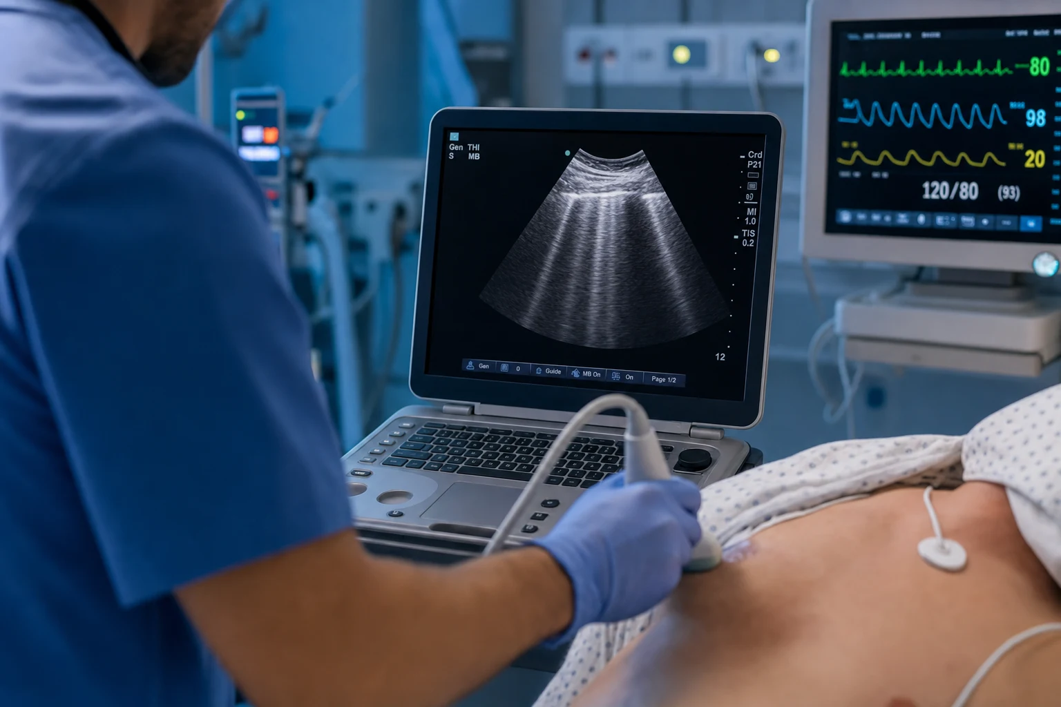
Critical care ultrasound (CCUS), or point-of-care ultrasound (POCUS), is a core ICU skill — a rapid, non-invasive, repeatable, bedside imaging modality performed and interpreted by the treating clinician at the moment of decision-making. It is an EXTENSION of the clinical examination, not a replacement for formal imaging. Applications span five domains: (1) Cardiac — focused cardiac ultrasound (FoCUS/FATE) for LV and RV function, pericardial effusion and tamponade, volume status, and gross valve pathology, with the RUSH protocol integrating the 'pump' into shock assessment; (2) Lung — the BLUE protocol using A-lines (normal air), B-lines (interstitial syndrome — oedema, ARDS, fibrosis), consolidation, pleural effusion, and absent lung sliding with a lung point for pneumothorax; (3) Abdominal — FAST/eFAST for free fluid (haemorrhage), gallbladder (cholecystitis), bladder volume, and AAA screening; (4) Vascular — two-point or whole-leg compression sonography for DVT, and real-time ultrasound guidance for central venous catheter (CVC) insertion; (5) Procedural — line placement, thoracentesis, paracentesis, drain insertion. Organising protocols include RUSH (Rapid Ultrasound in Shock — pump, tank, pipes), BLUE (Bedside Lung Ultrasound in Emergency), FATE (Focused Assessment with Transthoracic Echocardiography), and FALLS (using B-lines to titrate fluids). POCUS does NOT replace formal echocardiography, CT, or comprehensive departmental ultrasound: it is goal-directed, focused, repeatable, and integrated with the clinical picture. Competency is defined by international consensus statements (ACCP/SRLF competence statement, the WINFOCUS/ICM evidence-based lung ultrasound recommendations, and the EACVI focus cardiac ultrasound core curriculum) and assessed through structured certification (FATE, CEUS/ICS FoCCUS, FUSIC, ACEP emergency ultrasound).
On this page & tools
Your progress
Saved locally on this device.
Target exams
Red flags
Overview and rationale
Critical care ultrasound (CCUS) is the bedside use of ultrasound by the intensivist to answer focused, time-critical clinical questions at the point of care. Unlike a departmental study — performed by a sonographer, reported by a radiologist or cardiologist, and delivered hours later — CCUS is performed and interpreted in real time by the clinician responsible for the patient, and the result is immediately folded into the differential diagnosis and the next therapeutic step. For this reason CCUS is best understood not as an imaging modality but as an extension of the clinical examination — the modern equivalent of the stethoscope, used to confirm or refute a hypothesis formed at the bedside.[1][5]
The value of CCUS in the ICU is that it is rapid (a focused exam takes minutes), non-invasive, repeatable (serial exams track change in real time), free of ionising radiation, and transportable to the patient who is too unstable to move. Its limitations are equally important to hold in mind: it is operator-dependent, its accuracy is bounded by image quality (obesity, surgical emphysema, bowel gas, bandages all degrade it), and it is a focused study — it does not substitute for formal echocardiography, computed tomography, or comprehensive departmental ultrasound when those are indicated and the patient is stable enough to obtain them.[1][4]
[1]The scope of critical care ultrasound
CCUS is organised into five anatomical domains, each answering a defined set of questions, and several integrating protocols that combine domains to work through an undifferentiated presentation (shock, respiratory failure, trauma). The boundaries between a focused study (what the intensivist does at the bedside) and a comprehensive study (what the cardiology or radiology department does) are defined by international consensus and are a frequent exam point.[1][4][7]
The five CCUS domains at a glance
| Domain | Core questions answered | Typical exam/protocol |
|---|---|---|
| Cardiac (FoCUS/FATE) | Is there tamponade? Is LV function good or poor? Is the RV dilated? Is the heart small/hyperkinetic (empty) or full? Are there gross valve abnormalities? | FATE, FoCUS, RUSH (pump) |
| Lung | Is there a pneumothorax? Interstitial syndrome (B-lines)? Consolidation? Pleural effusion? | BLUE protocol |
| Abdominal | Is there free fluid (blood)? Gallstones/wall-thickening? Aortic aneurysm? Bladder volume (retention)? | FAST/eFAST |
| Vascular | Is there a DVT? Is the central vein patent and distinguishable from the artery? | Compression sonography, CVC guidance |
| Procedural | Where exactly is the target (vessel, effusion, bladder, abscess)? | Real-time guidance |
Ultrasound principles — probe selection, frequency and depth
Effective CCUS begins with choosing the right transducer and setting the machine correctly for the question. The wrong probe or the wrong depth makes a straightforward finding invisible.[5]
Probe (transducer) selection
Three transducers cover almost every CCUS application. The choice is governed by the trade-off between penetration (reaching deep structures) and resolution (resolving superficial detail), which in turn is governed by frequency — higher frequency gives better resolution but loses penetration because it is absorbed more rapidly by tissue.[5]
Probe selection — which transducer for which job
| Transducer | Footprint and frequency | Best for | Why |
|---|---|---|---|
| Curvilinear (convex) | Large curved footprint; low frequency (~2–5 MHz) | Abdomen (FAST, gallbladder, AAA, bladder), deep structures, lung, obstetric | Deep penetration (up to 25–30 cm); good field of view; lower resolution acceptable for large/deep targets |
| Linear (vascular) | Flat rectangular footprint; high frequency (~6–13 MHz) | Vascular access (CVC, arterial line), DVT compression, superficial nerve blocks, soft tissue/musculoskeletal, chest wall and pleura for pneumothorax | Superb near-field resolution for superficial structures (within 4–5 cm); poor penetration below ~6 cm |
| Phased array (cardiac) | Small flat square footprint; low frequency (~1–5 MHz) | Cardiac (FoCUS/FATE), between ribs, lung when rib spacing is tight | Small footprint fits between intercostal spaces; sector-shaped beam sweeps through a wide arc from a narrow acoustic window — ideal for the heart behind the ribs |
| Microconvex / endocavitous | Small curved / probe-tipped | Transoesophageal (formal, not POCUS), transvaginal/transrectal | Niche applications outside routine CCUS |
A practical rule: curvilinear for the abdomen and lung, linear for vessels and superficial structures, phased array for the heart. Many modern machines combine these into a single broadband or switchable transducer, but the principle of matching frequency to depth is unchanged.[5]
Frequency, depth and gain
- Frequency: select the highest frequency that still penetrates to the target. For a carotid or femoral vein 1 cm deep, use the linear probe at its highest frequency for maximal resolution. For an AAA at 12 cm depth, drop to the lowest frequency of the curvilinear probe to penetrate.[5]
- Depth: set the region of interest to fill roughly the middle two-thirds of the screen. Too shallow and you miss the target; too deep and the target is a tiny structure in a sea of tissue with the focal zone in the wrong place.
- Gain (overall brightness): adjust so that the image is not too dark (under-gained, structures missed) or too bright/saturated (over-gained, noise obscures contrast). Use time-gain compensation to even out brightness with depth, since deeper structures return weaker echoes.
- Focal zone: where present, set the focal zone at the depth of the structure of interest, because lateral resolution is best there.
Artefact recognition — what is not real
Ultrasound images are built from assumptions about how sound travels in tissue. Whenever those assumptions are violated (sound bends, reflects repeatedly, or attenuates unevenly), the machine draws structures that are not anatomically there — artefacts. Recognising artefacts is essential both to avoid misdiagnosis (calling an artefact a lesion) and because several artefacts are themselves diagnostic — B-lines, the lung point and the lung pulse are all artefacts that signify disease.[3][12]
Common ultrasound artefacts in CCUS
| Artefact | Mechanism | Appearance | Significance |
|---|---|---|---|
| Reverberation (A-lines) | Sound bounces back and forth between two strong reflectors (the pleural line and the probe skin surface); each round trip is plotted as another line at a multiple of the real distance | Horizontal hyperechoic lines at regular intervals below the pleural line in lung | A-lines = normal aerated lung (air reflects all sound). Part of the normal BLUE profile |
| B-lines (comet-tail artefact; ring-down) | Multiple tiny reverberations from thickened interlobular septa or fluid-filled alveoli | Vertical hyperechoic streaks arising from the pleural line, spreading to the bottom of the screen, erasing A-lines, moving with sliding | B-lines = interstitial syndrome (pulmonary oedema, ARDS, fibrosis, pneumonia). >3 in a zone is abnormal |
| Mirror-image artefact | Sound reflects off a strong smooth interface (diaphragm) back into the liver, is reflected again, and the machine plots a ghost copy of the liver above the diaphragm | Apparent liver/solid tissue 'above' the diaphragm where there should be lung | Can mimic consolidation or a subpulmonic effusion; recognised by the symmetry of the mirror |
| Acoustic shadowing | Sound is fully reflected or absorbed by a very dense/echogenic structure (gallstone, rib, calcified valve, gas); no signal returns from behind it | Dark anechoic band deep to the reflector, fanning out | A gallstone casts a clean shadow; ribs cast shadows that create the 'bat sign' landmarks in lung; bowel gas shadows limit abdominal views |
| Posterior (acoustic) enhancement | Sound travels through a fluid-filled (weakly attenuating) structure with less loss than through surrounding solid tissue, so the tissue deep to it appears artificially bright | Bright (hyperechoic) band directly behind a cyst or fluid collection | Helps confirm a structure is fluid-filled (gallbladder, bladder, simple cyst, effusion) |
| Side-lobe / slice-thickness | Off-axis beam energy is plotted as if it came from the main beam | Echogenic 'smoke' or curved lines within an anechoic structure (e.g. the gallbladder) | Can mimic sludge or debris; disappears with angle change |
| Refraction (edge) shadow | Sound bends at the curved edge of a rounded structure, creating a shadow from the edge | Thin dark lines projecting from the lateral margins of a cyst or gallbladder | A normal variant; do not confuse with a stone shadow |
| Lung point | At the junction of normal sliding lung and the non-sliding lung of a pneumothorax, the visceral pleura re-appears on the screen during respiration | A precise point where sliding resumes, seen in real time on M-mode as the 'seashore' giving way to the 'barcode/stratosphere' pattern | Specific for pneumothorax; locates it and roughly estimates its size |
| Lung pulse | The beating heart transmits vibrations through a lung that is fully apposed (no air between pleural layers), visible as pulsation of the pleural line | Regular cardiac-frequency movement at the pleural line, with absent B-lines | Excludes pneumothorax at that interspace; useful in apnoeic intubated patients where sliding is absent |
The cardinal rule of artefact interpretation: artefacts are reproducible with probe position, do not have distinct boundaries, and often disappear or change character when the angle or depth is changed — unlike real structures. The exceptions are the lung artefacts (A-lines, B-lines, lung point, lung pulse), which are reproducible precisely because they arise from the physics of the air–tissue interface and are the basis of lung ultrasound diagnosis.[3][12]
Cardiac ultrasound — focused (FoCUS) versus comprehensive
The distinction that defines scope
A focused cardiac ultrasound (FoCUS) is a limited, goal-directed bedside study performed by the non-cardiologist intensivist or emergency physician to answer a small number of yes/no or categorical questions: Is there a pericardial effusion? Is there tamponade physiology? Is global LV systolic function normal, moderately reduced, or severely reduced? Is the RV dilated or hypokinetic? Is the heart small and hyperkinetic (suggesting hypovolaemia) or full? Is there gross valvular pathology (e.g. a flail leaflet, a vegetation, a severely calcified or immobile valve)? FoCUS does not quantify (no ejection fraction by Simpson's biplane, no formal valve gradients, no detailed haemodynamics) and does not replace a comprehensive transthoracic or transoesophageal echocardiogram.[4][7]
A comprehensive echocardiogram is performed by a trained sonographer and reported by a cardiologist; it quantifies chamber sizes and function, measures valve gradients and areas, assesses diastolic function, estimates pulmonary artery pressure, and integrates Doppler haemodynamics. The intensivist must know the boundary: FoCUS is for the unstable patient who needs a decision now; comprehensive echo is for the stable patient who needs a definitive anatomical and haemodynamic answer.[4][7]
FoCUS (POCUS) versus comprehensive echocardiography
| Feature | Focused cardiac ultrasound (FoCUS) | Comprehensive echo (TTE/TOE) |
|---|---|---|
| Performer | Treating intensivist/ED physician at the bedside | Trained sonographer; reported by cardiologist |
| Timing | Real time, point of care | Elective; minutes to hours later |
| Goal | Answer focused categorical questions (effusion? RV dilated? LV good/poor?) | Quantify (EF, valve area/gradient, chamber volumes, PA pressure, diastology) |
| Views | 2–4 core views (parasternal long/short, apical 4-chamber, subcostal) | Full protocol with all standard views + Doppler |
| Modality | 2D ± colour Doppler (where available) | 2D, M-mode, colour/spectral/tissue Doppler |
| Output | Categorical, qualitative | Quantitative measurements and full report |
| Role | Extension of exam; guides immediate therapy | Definitive diagnosis; serial follow-up |
| Replaces formal echo? | No | — |
The core FoCUS views
Four views, remembered as the FATE / mnemonic windows, are sufficient for most focused questions. They are acquired in whatever order gives the best image; in the intubated patient the subcostal and apical views are often the most achievable.[4]
- Parasternal long-axis (PLAX): probe in the 3rd–4th left intercostal space, parasternal line, indicator toward the right shoulder. Shows the RV outflow, LV, mitral and aortic valves, aortic root and descending aorta in cross-section. Best for LV size and function, aortic and mitral valve motion, and a posterior pericardial effusion.
- Parasternal short-axis (PSAX): rotate 90 degrees from PLAX at the same window. Cross-sections of the LV at mitral, papillary and apical levels; RV crescent wraps around the LV. Septal flattening (D-shaped LV) here = RV pressure or volume overload. Best regional wall motion assessment.
- Apical four-chamber (A4C): probe at the point of maximal impulse, indicator toward the patient's left. All four chambers in one frame; the natural plane to compare RV and LV size (the RV is normally two-thirds of the LV; if the RV approaches the LV size it is dilated) and to detect an apical effusion.
- Subcostal / subxiphoid: probe just below the xiphisternum, indicator toward the patient's left, beam aimed at the heart under the liver. The most reliable view for a pericardial effusion (fluid tracks circumferentially) and for assessing the IVC as it enters the right atrium; often the only usable view in the obese, bandaged, or arrested patient. [1]
What FoCUS looks for in shock (the RUSH 'pump')
In the undifferentiated hypotensive patient, the cardiac FoCUS answers questions that map directly onto the type of shock.[4]
- Hyperkinetic, small, kissing LV cavity → hypovolaemic or distributive (septic) shock — the empty, hypercontractile heart.
- Poorly contracting, dilated LV with global hypokinesis → cardiogenic shock.
- Dilated RV with septal shift (D-sign), tricuspid regurgitation, RV hypokinesis (McConnell's sign) → obstructive shock from massive PE or severe pulmonary hypertension.
- Pericardial effusion with RV free-wall diastolic collapse, a distended IVC, and a small, underfilled LV → obstructive shock from cardiac tamponade.
- A dilated, poorly contracting LV with a pericardial effusion but no tamponade physiology may be myocarditis or end-stage cardiomyopathy. [1]
Pericardial effusion and tamponade
FoCUS is highly sensitive for a pericardial effusion, which appears as an anechoic (black) stripe between the visceral and parietal pericardium circumferentially around the heart. The key exam point is that an effusion is not the same as tamponade: tamponade is a clinical and haemodynamic diagnosis, of which the echocardiographic hallmarks are right atrial systolic collapse (early, sensitive) and right ventricular free-wall diastolic collapse (later, more specific), accompanied by a distended, non-collapsing IVC and, on Doppler, exaggerated respiratory variation in inflow. The intensivist's job at the bedside is to integrate the ultrasound with the clinical picture — tachycardia, hypotension, raised JVP, muffled heart sounds (Beck's triad) — and to act: a patient with clinical tamponade and an effusion with RV collapse needs emergency pericardiocentesis (often ultrasound-guided), not a wait for formal echo.[4][7]
Valve assessment — the limit of focused ultrasound
FoCUS can screen for gross valve pathology with 2D and, where available, colour Doppler: a flail or prolapsing mitral leaflet, a vegetation (especially on the aortic or mitral valve in a septic patient), a fixed/stuck leaflet suggesting severe calcific stenosis, or an obviously regurgitant valve on colour. It cannot grade valve disease reliably — stenosis severity requires continuous-wave Doppler to measure gradients and the continuity equation for area, and regurgitation severity requires quantitative Doppler. A vegetation seen on FoCUS is highly suggestive of infective endocarditis and mandates a formal study (and often TOE), but a normal FoCUS does not exclude endocarditis.[4][7]
Lung ultrasound — the BLUE protocol
Lung ultrasound is the domain in which POCUS most dramatically outperforms the bedside chest X-ray and approaches CT. Because air and water interact with ultrasound in predictable, opposite ways, the aerated lung, the oedematous lung, the consolidated lung, the pleural effusion and the pneumothorax each produce a characteristic sonographic signature — and these signatures are the basis of the BLUE protocol (Bedside Lung Ultrasound in Emergency), a decision tree that identifies the cause of acute respiratory failure in minutes.[2][3]
The eight BLUE points
Each hemithorax is examined at standardised points: the upper blue point and lower blue point on the anterior chest (lined up with the hands of BLUE — the operator's two hands placed side by side from the sternum), and the posterolateral alveolar and/or pleural syndrome (PLAPS) point on the lateral/posterior chest. Both lungs are examined, giving a profile that drives the diagnostic algorithm.[2]
The five elemental lung findings
- Lung sliding (the 'seashore' sign): the to-and-fro movement of the visceral against the parietal pleura with respiration, visible in 2D as a shimmering of the pleural line and in M-mode as a granular 'sandy' pattern below a stratified 'wave' pattern (the seashore sign). Present sliding = the pleural layers are apposed = no pneumothorax at that point.[3]
- A-lines: horizontal reverberation artefacts below the pleural line at multiples of the skin-to-pleura distance. A-lines + sliding = normal aerated lung (the 'A' profile).[2]
- B-lines (comet-tail artefacts): vertical, laser-like, hyperechoic artefacts arising from the pleural line, erasing A-lines, spreading to the bottom of the screen without fading, and moving with sliding. Three or more B-lines in a single interspace = interstitial syndrome. Bilateral diffuse B-lines = pulmonary oedema, ARDS, or interstitial fibrosis; focal B-lines = pneumonia, atelectasis, contusion, or infarction.[2][12]
- Consolidation (alveolar syndrome / C-profile): when alveoli fill with fluid or pus, the lung becomes tissue-like and transmits sound — appearing as a hypoechoic, liver- or spleen-like ('hepatatised') area with echogenic punctate or branching air bronchograms within it. A 'shred sign' marks the irregular deep border of a superficial consolidation.[3]
- Pleural effusion (PLAPS): an anechoic (or echoic/complex if exudative or haemorrhagic) collection above the diaphragm, with the lung floating in it; the volume can be estimated from the interpleural distance at end-expiration.[3]
The pneumothorax sequence
Pneumothorax is air in the pleural space, so the visceral pleura is separated from the parietal pleura by air — air that reflects all ultrasound and hides the lung beneath. The diagnosis is therefore made by the absence of normal findings and confirmed by one specific positive finding.[9]
Diagnosing pneumothorax by lung ultrasound
- Look for lung sliding. Absent sliding means the pleural layers are not moving against each other — either a pneumothorax, or any cause of absent ventilation on that side (apnoea, mainstem intubation, pleural symphysis, fibrosis, severe emphysema, contusion). Absent sliding is sensitive but not specific for pneumothorax.[3]
- Look for B-lines. B-lines arise from the visceral pleura, so any B-lines exclude pneumothorax at that interspace (the visceral pleura must be apposed to generate them). This is a powerful and rapid rule-out.[2]
- Look for the lung pulse. The cardiac impulse transmitted through apposed pleura; its presence also excludes pneumothorax, and is especially useful in the apnoeic intubated patient.[12]
- Search for the lung point. Move the probe laterally and posteriorally to find the exact place where the collapsed lung re-meets the chest wall — the point where sliding/B-lines/lung pulse abruptly reappear during respiration. The lung point is specific for pneumothorax (near 100 per cent) and, when found far posteriorally, indicates a small/occult pneumothorax; when absent or only found very anteriorly with a large area of absent sliding, the pneumothorax is large.[9]
- Integrate and act. If sliding and B-lines are present, pneumothorax is excluded. If they are absent and a lung point is found, pneumothorax is confirmed and its size gauged. If they are absent and no lung point is found (complete collapse), the diagnosis is very likely — confirm by CT if stable, or act on clinical grounds if not.[9]
The BLUE decision tree
The BLUE protocol combines the lung profile with a venous (DVT) assessment to predict the cause of acute respiratory failure with reported accuracy around 90 per cent.[2]
The BLUE profiles and the predicted cause of respiratory failure
| Lung profile | Definition | Most likely cause |
|---|---|---|
| A profile | A-lines + sliding, bilateral anterior | Normal lung; pulmonary embolism (if DVT found); COPD/asthma |
| A' profile (lung point) | A-lines + absent sliding + lung point | Pneumothorax |
| B profile | Bilateral diffuse B-lines + sliding | Pulmonary oedema (cardiogenic) |
| B' profile | Bilateral B-lines + absent sliding | Pneumothorax (rare) or posterior consolidation |
| A/B profile (unilateral B-lines) | B-lines on one side, A-lines on the other | Pneumonia (unilateral interstitial syndrome) |
| C profile | Anterior consolidation | Pneumonia |
| PLAPS (posterolateral alveolar/pleural) | Effusion and/or consolidation posterolaterally | Pneumonia, pleural effusion (any cause) |
| A profile + DVT | Normal A-profile lung + venous thrombosis on compression | Pulmonary embolism |
The FALLS protocol (Fluid Administration Limited by Lung Sonography) extends this idea to shock: the intensivist gives fluid boluses while watching the lung for the appearance of B-lines, using lung ultrasound as a safety check against iatrogenic pulmonary oedema — the moment B-lines appear, the lung has reached its threshold for interstitial oedema and further fluid is likely harmful.[2]
Abdominal ultrasound — FAST, gallbladder, IVC, bladder, aorta
FAST and eFAST
The Focused Assessment with Sonography in Trauma (FAST) examines four abdominal windows for free (anechoic) intraperitoneal fluid, which in the trauma context is presumed to be blood until proven otherwise:[5]
- Hepatorenal recess (Morison's pouch) — the most dependent supramesocolic space in the supine patient; the most sensitive single view.
- Splenorenal recess — the left upper quadrant mirror of Morison's pouch.
- Suprapubic / pouch of Douglas — the most dependent space in the pelvis; views the bladder (which should be full, acting as an acoustic window) and the rectovesical/rectouterine space behind it.
- Subxiphoid / pericardial — the cardiac view for haemopericardium (an effusion in trauma is tamponade until proven otherwise). [1]
The extended FAST (eFAST) adds lung views to detect a pneumothorax (often missed on supine chest X-ray) and, in some protocols, a more detailed aortic assessment. A positive FAST in a haemodynamically unstable trauma patient is an indication for immediate operative intervention; a negative FAST in an unstable patient does not exclude injury and warrants further assessment (CT if stable enough, or repeat/dynamic FAST). Outside trauma, the same FAST views detect free fluid from ruptured ectopic pregnancy, ruptured abdominal aortic aneurysm, haemorrhagic pancreatitis, ascites, and bowel perforation.[5]
The gallbladder
Bedside assessment of the gallbladder identifies stones (echogenic foci that cast clean acoustic shadows and move with position), wall thickening (greater than 3 mm, measured anterior wall), pericholecystic fluid, and a sonographic Murphy's sign (maximal tenderness as the probe presses directly over the sonographically localised gallbladder). These are the components of acute calculous cholecystitis and are especially relevant in the critically ill patient at risk of acalculous cholecystitis (prolonged fasting, TPN, sepsis, burns), where the finding is a distended, thick-walled gallbladder with sludge and pericholecystic fluid in the absence of stones.[5]
The inferior vena cava for volume and fluid responsiveness
The IVC is examined in the subcostal longitudinal plane as it enters the right atrium, in M-mode just below the hepatic vein inflow. Its absolute diameter and its respiratory variation (collapsibility) are used as surrogates for right atrial pressure and, with important caveats, for fluid responsiveness.[8]
IVC assessment and its interpretation
| IVC finding | Interpretation | Caveat |
|---|---|---|
| Small (< 1.5 cm) and collapses > 50% on inspiration | Low right atrial pressure; suggests the patient is volume-depleted and may respond to fluid | The most reliable 'fluid-consider' pattern |
| Large (> 2.5 cm) and does not collapse (< 50%) | High right atrial pressure; suggests volume overload, RV failure, tamponade, or severe TR | A non-collapsing distended IVC is a sign of right-heart dysfunction, not just volume overload |
| Collapsibility > 50% in a spontaneously breathing patient | Suggests fluid responsiveness | More useful in mechanically ventilated patients, where the physiology reverses (IVC distends with inspiration) |
| In the ventilated patient: distensibility index > 18% ([Dmax − Dmin]/Dmin) | Predicts fluid responsiveness | Requires fully controlled ventilation with a standard tidal volume (~8 mL/kg); invalid in spontaneous effort, arrhythmia, open chest, high PEEP |
The IVC is a better bedside guide than the central venous pressure, but inferior to a passive leg raise (which is itself a reversible, endogenous fluid challenge) and to dynamic indices (stroke volume variation, pulse pressure variation) for predicting fluid responsiveness. It is best used as one data point, not the sole determinant of fluid therapy.[8]
Bladder volume
Bladder volume is estimated by measuring the bladder in two perpendicular transverse and sagittal planes and applying the prolate ellipsoid formula (volume = length × width × height × 0.523), or by the machine's automated calculation. It is used to measure post-void residual (a residual over 100–150 mL suggests urinary retention and may warrant catheterisation) and to confirm a full bladder before suprapubic catheterisation or a suprapubic FAST window.[5]
Abdominal aortic aneurysm screening
The abdominal aorta is measured in the transverse plane from the outer wall to outer wall at the suprarenal (diaphragmatic hiatus), renal, and infrarenal levels. A diameter greater than 3 cm defines an aneurysm; greater than 5.5 cm marks the threshold at which elective repair is usually offered because rupture risk exceeds operative risk. In the unstable patient with abdominal/back pain and a pulsatile mass, finding an aortic diameter greater than 3 cm (especially with retroperitoneal fluid) supports the diagnosis of a ruptured AAA and mandates immediate vascular surgery involvement.[5]
Vascular ultrasound — DVT compression and CVC guidance
Compression sonography for deep vein thrombosis
Compression sonography exploits the fact that a normal vein is fully compressible under light probe pressure (its walls coapt and it disappears), whereas a vein containing thrombus is non-compressible. The test is therefore one of compressibility, not of directly seeing clot.[10][11]
Two-point versus whole-leg compression ultrasound for DVT
| Approach | Veins compressed | Sensitivity for proximal DVT | Role |
|---|---|---|---|
| Two-point | Common femoral vein (at the saphenofemoral junction) and popliteal vein (to its trifurcation) | ~90–95% for proximal DVT; misses isolated calf DVT | Standard in many systems; if negative, repeat at 5–7 days (or add D-dimer) to catch a calf DVT that has propagated proximally |
| Three-point | Adds the proximal femoral vein | Marginally higher sensitivity | Common modification |
| Whole-leg | Common femoral, femoral, popliteal, and the calf (posterior tibial, peroneal) veins to the ankle | ~96–100%; detects isolated calf DVT | Higher sensitivity, single exam; does not require repeat; slightly more operator-dependent; risks over-diagnosing clinically silent calf DVT |
A non-compressible common femoral or popliteal vein confirms proximal DVT with specificity around 96 per cent and warrants anticoagulation. In the ICU, where DVT prevalence is high and compression is rapid, a positive compression study in the right clinical context is sufficient to start treatment without waiting for a formal departmental study.[10][11]
The DVT assessment is also the final step of the BLUE protocol: an A-profile (normal lung) combined with a positive compression study strongly suggests pulmonary embolism as the cause of respiratory failure — a haemodynamically significant PE is, in effect, diagnosed by finding its source clot in the leg.[2]
Ultrasound-guided central venous catheterisation
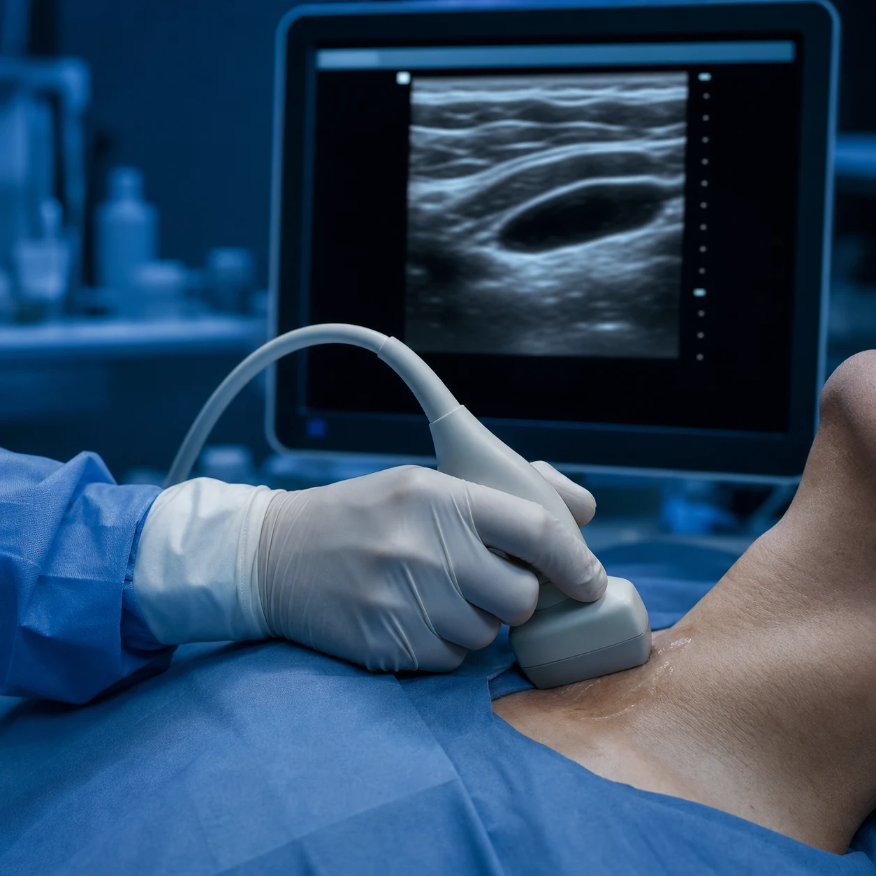
Ultrasound guidance for central venous catheter (CVC) insertion is now standard of care for the internal jugular, subclavian, and femoral routes. It is used in two ways: static (pre-procedural mapping — locate the vein, confirm patency, mark the site, then proceed 'blindly') and dynamic (real-time visualisation of the needle entering the vein throughout the procedure). Real-time dynamic guidance is preferred.[6]
The evidence is consistent: ultrasound guidance reduces mechanical complications (arterial puncture, haematoma, pneumothorax, haemothorax), increases first-pass and overall success, and reduces the number of attempts, with the benefit largest for the internal jugular vein. The Cochrane review by Brass and colleagues confirmed this benefit for subclavian and femoral routes as well.[6]
Ultrasound-guided internal jugular cannulation — safe practice
- Scan before you scrub. Use the linear probe in transverse plane to identify the internal jugular vein, confirm it is compressible and distinguishable from the carotid artery (the vein is lateral, larger, compressible, non-pulsatile; the artery is medial, smaller, pulsatile, non-compressible). Identify variant anatomy (the vein overlying the artery in ~10 per cent).[6]
- Set depth and gain so the vein fills the middle of the screen and the needle path is short and shallow.[5]
- Prepare and drape with a sterile probe cover and sterile gel; re-confirm the target under sterile conditions.[6]
- Insert under real-time guidance in the transverse or longitudinal/sagittal plane. Transverse gives the best view of vein-vs-artery but shows the needle as a bright dot; longitudinal shows the whole needle and its tip but a narrower vessel view. Most operators use transverse with deliberate tip visualisation, or an oblique hybrid.[6]
- Always visualise the needle tip. The single most common error is advancing the needle while seeing the shaft but not the tip — the tip may be deeper than it appears and can perforate the posterior wall or the artery. Tilt and slide the probe to keep the tip in view.[6]
- Confirm intravenous placement by free flow of non-pulsatile venous blood and/or by saline injection under ultrasound (microbubbles flush through the vein); the catheter can be seen in the vein in longitudinal view.[6]
- Post-procedure check: exclude a pneumothorax (lung ultrasound or chest X-ray) after any central line, especially supraclavicular or subclavian.[6]
Competency, training and certification
Because CCUS is operator-dependent, international consensus statements define what a competent practitioner must be able to do, and how that competency is acquired and assessed. The defining documents are the American College of Chest Physicians / Société de Réanimation de Langue Française (ACCP/SRLF) competence statement, the international evidence-based recommendations for point-of-care lung ultrasound (Volpicelli et al, WINFOCUS-affiliated, Intensive Care Medicine 2012), the international evidence-based recommendations for focused cardiac ultrasound (Via et al, 2014), and the European Association of Cardiovascular Imaging focus cardiac ultrasound core curriculum and syllabus (Neskovic et al, 2018).[1][3][4][7]
These consensus documents converge on a tiered competency model: a basic level (recognising the common life-threatening findings — effusion/tamponade, gross LV dysfunction, RV dilatation, B-lines, absent lung sliding, free intraperitoneal fluid, a non-compressible vein, ultrasound-guided vascular access) that all intensivists and emergency physicians should achieve, and an advanced level (comprehensive echocardiography, advanced lung and abdominal protocols) reserved for those with dedicated training. The boundary is emphasised repeatedly: basic competency is not a licence to call oneself an echocardiographer.[1][7]
Named certifications and pathways include: [1]
- FATE (Focused Assessed Transthoracic Echocardiography) — the Scandinavian protocol (Jensen and colleagues), widely taught and certified internationally; emphasises the four core views plus IVC and lung, for haemodynamic assessment.
- FoCCUS / CEUS (Comprehensive Focused Cardiac Ultrasound via the Intercollegiate Committee for Critical Care Echocardiography, UK) — the UK ICS/FICM pathway to focused echo competency and accreditation.
- FUSIC (Focused Ultrasound in Intensive Care) — the UK Intensive Care Society's one-stop, multi-domain POCUS accreditation (heart, lung, abdomen, vascular, procedural).
- ACCP / CHEST critical care ultrasonography — the North American College of Chest Physicians competency framework and certification.
- WINFOCUS (World Interactive Network Focused on Critical Ultrasound) — the international society that has driven the evidence-based recommendations and runs training and consensus globally.
- ACEP emergency ultrasound — the American College of Emergency Physicians guidelines and credentialing for point-of-care ultrasound in the emergency department. [1]
Competency is acquired through a combination of structured courses, supervised scanning, logbook accumulation, and assessment (often image-acquisition and interpretation reviews). The consensus statements emphasise that competency requires both the technical skill to acquire diagnostic images and the cognitive skill to interpret them and integrate them into clinical decisions — and that both must be maintained through continued practice and quality assurance.[1][7]
POCUS versus formal imaging
A recurring exam theme is when POCUS is sufficient and when it must defer to formal imaging. The principle is that POCUS answers a focused question in real time to guide immediate therapy; formal imaging answers the full question definitively. POCUS does not replace a comprehensive echocardiogram in the stable patient with suspected valve disease, nor a CT pulmonary angiogram in the haemodynamically stable patient with suspected PE, nor a departmental vascular or abdominal study when the question is anatomical and not time-critical.[1][4]
POCUS versus formal imaging — when to use each
| Dimension | Point-of-care ultrasound (POCUS) | Formal imaging (echo, CT, departmental US) |
|---|---|---|
| Performer | Treating clinician, bedside | Sonographer/technician + specialist reporter |
| Timing | Immediate, real time, repeatable | Scheduled; minutes to hours; not easily repeatable |
| Scope | Focused, goal-directed, categorical | Comprehensive, quantitative, definitive |
| Patient | Any, including the unstable and the arrested | Patient must be stable/transportable (for CT, departmental US) |
| Strength | Speed, repeatability, integration with the clinical picture at the bedside | Accuracy, quantification, complete anatomical/haemodynamic detail |
| Weakness | Operator-dependent; limited by image quality; cannot quantify | Time delay; transport risk (CT); ionising radiation (CT); cost |
| Replaces? | No — complements and triages | No — too slow for the moment of crisis |
| Examples it answers | 'Is there tamponade?' 'Is there a pneumothorax?' 'Is there free fluid in this trauma?' 'Is there a DVT?' | 'What is the EF and the valve gradient?' 'Is there a segmental PE on CTPA?' 'What is the AAA diameter on CT for surgical planning?' |
The correct relationship is sequential and complementary: POCUS makes the immediate decision (start fluids? pericardiocentesis? chest tube? anticoagulate? intubate?); formal imaging, when the patient is stabilised, makes the definitive diagnosis and plans definitive treatment. [1]
SAQ — RUSH and the differentiation of shock by ultrasound
10 minutes · 10 marks
A 65-year-old woman is admitted to ICU with hypotension (BP 80/45, MAP 55), HR 120, cool peripheries, oliguria and a lactate of 4. The cause is unclear. Describe how point-of-care ultrasound would clarify the shock type.
SAQ — Cardiac tamponade on POCUS
10 minutes · 10 marks
A 50-year-old man with metastatic lung cancer presents with breathlessness, hypotension (BP 85/50), HR 130, JVP distended to the ear, and muffled heart sounds. POCUS shows a 2 cm circumferential pericardial effusion. The registrar asks what features confirm tamponade.
Clinical pearls
Red flags
Key trials and evidence
Lichtenstein 2008 — the BLUE protocol (PMID 18403664)
Source
Chest 134(1):117-125 — prospective observational derivation and validation study
Design
Consecutive ICU patients with acute respiratory failure; standardised lung ultrasound performed by trained operators, blinded to the final diagnosis; the lung profile (A, B, A/B, C, PLAPS) combined with a venous (DVT) assessment was used to predict the cause
Key finding
The BLUE protocol correctly identified the cause of acute respiratory failure in 90.5 per cent of patients. The A-profile-plus-DVT combination was highly specific for pulmonary embolism; the B-profile for pulmonary oedema; the A/B- and C-profiles for pneumonia; A'-profile (lung point) for pneumothorax
Clinical bottom line
A rapid, repeatable, radiation-free lung-plus-vein protocol can diagnose the cause of acute respiratory failure at the bedside in minutes with high accuracy — the foundation of modern lung ultrasound in the ICU
Volpicelli 2012 — International evidence-based recommendations for point-of-care lung ultrasound (PMID 22392031)
Source
Intensive Care Medicine 38(4):577-591 — international expert consensus (WINFOCUS-affiliated)
Design
Multidisciplinary panel synthesis of the evidence on technique (probe, position, the eight BLUE points), artefact interpretation (A-lines, B-lines, lung sliding, lung point, lung pulse, consolidation, effusion), and clinical application across pneumothorax, interstitial syndrome, consolidation, pleural effusion, and procedural guidance
Key finding
Lung ultrasound is more sensitive than supine chest radiography for pneumothorax, consolidation, and pleural effusion; B-lines define interstitial syndrome and correlate with extravascular lung water; the lung point is highly specific for pneumothorax. Standardised technique and terminology are defined
Clinical bottom line
The reference standard for how lung ultrasound should be performed and interpreted at the point of care — the document that defines the eight examination points and the diagnostic artefacts
Via 2014 — International evidence-based recommendations for focused cardiac ultrasound (PMID 24951446)
Source
Journal of the American Society of Echocardiography 27(7):683.e1-683.e33 — international consensus
Design
Multispecialty panel defined the scope, indications, views, and reporting of focused cardiac ultrasound (FoCUS) as performed by non-cardiologists, and explicitly distinguished it from comprehensive echocardiography
Key finding
FoCUS is a limited, qualitative, goal-directed study answering categorical questions (effusion, tamponade physiology, LV/RV size and function, gross valve pathology, volume status); it is an extension of the clinical examination and does NOT replace comprehensive echo. Standard views (parasternal long and short, apical four-chamber, subcostal) and a standardised report are defined
Clinical bottom line
Defines what the intensivist's focused cardiac study can and cannot claim — the boundary between POCUS and formal echocardiography, and the questions FoCUS is competent to answer
Mayo 2009 — ACCP/SRLF competence statement on critical care ultrasonography (PMID 19188546)
Source
Chest 135(4):1050-1060 — joint American College of Chest Physicians and Société de Réanimation de Langue Française consensus
Design
Expert panel defined the competencies required for critical care ultrasonography (general CCUS, vascular access, thoracic, abdominal, and focused cardiac) and the training and assessment pathways to achieve them
Key finding
CCUS is a core competence for the intensivist; competency is tiered (basic versus advanced); image acquisition and interpretation must both be taught, supervised, logbook-recorded, and assessed; advanced echocardiography is a separate discipline
Clinical bottom line
The defining competency framework for critical care ultrasound — the document that makes basic POCUS (cardiac, lung, abdominal, vascular, procedural) an expectation of every intensivist
Neskovic 2018 — EACVI focus cardiac ultrasound core curriculum and syllabus (PMID 29529170)
Source
European Heart Journal Cardiovascular Imaging 19(5):475-506 — European Association of Cardiovascular Imaging
Design
EACVI panel defined a structured core curriculum and syllabus for focus cardiac ultrasound, specifying the knowledge, skills, and assessment required, and the relationship to comprehensive echocardiography and to the various national certifications
Key finding
A defined competency pathway with explicit learning outcomes, case-load and logbook expectations, and an assessment framework; FoCUS is positioned as the entry-level cardiac study, with a clear upgrade path to comprehensive echocardiography for those who seek it
Clinical bottom line
The European benchmark for FoCUS training and assessment — the document behind certifications such as FATE, FoCCUS, and FUSIC, and the curriculum against which training programmes are measured
Barbier 2004 — IVC diameter and fluid responsiveness in septic shock (PMID 15034650)
Source
Intensive Care Medicine 30(9):1740-1746 — prospective physiological study
Design
Mechanically ventilated septic patients; IVC diameter measured by transthoracic echo; the distensibility of the IVC during the respiratory cycle was correlated with the increase in cardiac output after a standardised fluid bolus
Key finding
A distensibility index (the change in IVC diameter with respiration) above approximately 18 per cent predicted fluid responsiveness with high sensitivity and specificity in ventilated patients with a standard tidal volume
Clinical bottom line
Established the IVC distensibility index as a bedside predictor of fluid responsiveness in the ventilated patient — a dynamic index superior to static measures such as the CVP, with the important caveats of tidal volume, spontaneous effort, and right-heart pathology
Volpicelli 2014 — Semi-quantification of pneumothorax volume by lung ultrasound (PMID 25056671)
Source
Intensive Care Medicine 40(10):1460-1461 — clinical study
Design
Patients with confirmed pneumothorax assessed by lung ultrasound and CT; the location of the lung point (how far from the sternum it was found) was correlated with the pneumothorax volume measured on CT
Key finding
The position of the lung point correlates with pneumothorax volume: a lung point found far posteriorally indicates a small pneumothorax, while an anterior lung point or a complete absence of the lung point indicates a large one
Clinical bottom line
Lung ultrasound not only diagnoses pneumothorax but estimates its size from the lung point position, helping decide between observation and drainage at the bedside
Brass 2015 — Ultrasound guidance versus landmarks for subclavian/femoral CVC (PMID 25575245)
Source
Cochrane Database of Systematic Reviews (1):CD011447 — systematic review and meta-analysis of randomised trials
Design
Pooled randomised trials comparing ultrasound-guided with landmark (anatomical) technique for subclavian and femoral vein catheterisation; endpoints were catheterisation failure, mechanical complications, and number of attempts
Key finding
Ultrasound guidance reduced the risk of catheterisation failure and of arterial puncture/haematoma compared with the landmark technique for both subclavian and femoral routes, with no significant effect on pneumothorax (low event rate). The evidence confirms and extends the established benefit seen for the internal jugular route
Clinical bottom line
Real-time ultrasound guidance is standard of care for central venous catheterisation at all three principal sites, reducing failure and mechanical complications
Heijboer/Lensing 1993 — Compression ultrasonography versus impedance plethysmography for DVT (PMID 8413431)
Source
New England Journal of Medicine 329(18):1365-1369 — prospective comparative cohort in symptomatic outpatients
Design
Consecutive patients with clinically suspected deep-vein thrombosis underwent both real-time compression ultrasonography and impedance plethysmography, with venography as the reference standard where needed
Key finding
Compression ultrasonography was more sensitive than impedance plethysmography for proximal DVT (around 95 per cent) with high specificity, establishing compression sonography as the superior first-line test
Clinical bottom line
A landmark study establishing real-time compression ultrasonography as the diagnostic standard for proximal DVT — the technique now used as the final step of the BLUE protocol
Cogo/Lensing 1998 — Single compression ultrasound + serial testing for suspected DVT (PMID 9451260)
Source
BMJ 316(7124):17-20 — prospective management cohort study
Design
Consecutive outpatients with suspected first DVT underwent a single two-point compression ultrasound; if negative, anticoagulation was withheld and the test repeated after 5–7 days, with follow-up for venous thromboembolism over months
Key finding
A negative single two-point compression study safely withheld anticoagulation in most patients; the serial repeat caught the small proportion with initially isolated calf DVT that propagated proximally, keeping the overall thromboembolic risk during follow-up very low
Clinical bottom line
Validated the two-point-plus-serial-repeat strategy as a safe, efficient diagnostic algorithm for suspected DVT, and quantified the small miss-rate that serial testing is designed to catch
Moore and Copel 2011 — Point-of-care ultrasonography (PMID 21345104)
Source
New England Journal of Medicine 364(8):749-757 — authoritative clinical review
Design
Narrative review of the principles, applications, and limitations of point-of-care ultrasound across cardiac, pulmonary, abdominal, vascular, and procedural domains
Key finding
POCUS is an extension of the physical examination, governed by correct probe selection, frequency/depth setting, and artefact recognition; it answers focused questions rapidly and repeatably but does not replace formal imaging when comprehensive assessment is required
Clinical bottom line
The widely-cited reference framing POCUS as a clinical skill integrated with the bedside assessment, and a clear summary of the principles every operator must master
Lichtenstein 2014 — Lung ultrasound in the critically ill (PMID 24401163)
Source
Annals of Intensive Care 4(1):1 — comprehensive clinical review
Design
Synthesis of the author's body of work (including the BLUE and FALLS protocols) into a unified framework for lung ultrasound in critical illness: the seven principles, the five elemental findings, and their integration into decision-making
Key finding
Lung ultrasound, interpreted through standardised artefacts (A-lines, B-lines, lung sliding, lung point, lung pulse, consolidation, effusion), provides a simple, radiation-free, repeatable bedside assessment of the lung that outperforms auscultation and supine chest radiography
Clinical bottom line
The consolidated reference for bedside lung ultrasound — the conceptual framework behind the BLUE and FALLS protocols and the day-to-day use of lung ultrasound in the ICU
Limitations and pitfalls
CCUS is powerful precisely because it is fast and repeatable, but its limitations are the mirror image of its strengths and must be held in mind whenever the probe is picked up.[1][5]
- Operator dependence. Every CCUS finding depends on the operator's ability to acquire and interpret the image. Inadequate training degrades both sensitivity and specificity, and a confident but wrong reading can be more dangerous than no reading at all.
- Image-quality barriers. Obesity, surgical emphysema (air reflects all ultrasound), bandages and dressings, poor acoustic windows, bowel gas (abdomen), and patient inability to cooperate (agitation, position) all reduce image quality. A non-diagnostic study must be reported as non-diagnostic.
- Focused, not comprehensive. CCUS answers the question asked; it does not exclude pathology outside that question. A negative FoCUS does not exclude endocarditis; a normal lung scan does not exclude a parenchymal nodule; a negative FAST does not exclude hollow-viscus or retroperitoneal injury.
- Artefact misinterpretation. A-lines, mirror artefact, side-lobe artefact, and shadowing can all masquerade as pathology. The operator must recognise the common artefacts and, where in doubt, change probe angle/depth to test whether the finding persists.
- The hard questions stay with formal imaging. Valve gradients and areas, ejection fraction quantification, pulmonary artery pressure, complex anatomy (CT), and tissue characterisation (CT/MRI) remain the province of formal studies in the stable patient.
- Documentation and quality assurance. Findings should be recorded (images saved, a focused report written) and reviewed, both for clinical communication and for the maintenance and improvement of competence over time.[1][7]
Putting it together — CCUS in the undifferentiated ICU patient
In practice, the intensivist does not perform 'a lung scan' or 'a cardiac scan' in isolation but integrates domains to work through the presentation. In the undifferentiated hypotensive patient, RUSH sequences the cardiac (pump), IVC (tank volume), aorta and venous (tank integrity), and lung/pleura (source and effect) assessments. In the undifferentiated breathless patient, BLUE sequences the lung profile with a venous assessment. In the trauma patient, eFAST adds lung to the abdominal and cardiac windows. The unifying habit is to form a focused question, choose the right probe and domain, acquire and interpret the image at the bedside, integrate it with the clinical picture, act, and then repeat to track the response — and to know when the question has outgrown POCUS and must be handed to formal imaging.[1][2][4][5]
Choosing the protocol by presentation
| Presentation | Primary CCUS protocol | What it answers |
|---|---|---|
| Undifferentiated hypotension/shock | RUSH (HI-MAP variant) | Pump (cardiac), tank (IVC volume, effusion), pipes (aorta, DVT, pneumothorax) → type of shock |
| Acute respiratory failure | BLUE | Lung profile + DVT → oedema, pneumonia, pneumothorax, PE, COPD/asthma |
| Blunt or penetrating trauma | FAST / eFAST | Free intraperitoneal fluid, haemopericardium, pneumothorax |
| Cardiac arrest | FATE / FoCUS as part of the reversible-causes search | Tamponade, massive PE (RV dilatation), severe hypovolaemia, tension pneumothorax |
| Suspected DVT/PE | Compression + FoCUS | Non-compressible vein; RV strain pattern |
| Oliguria / volume question | IVC + FoCUS + lung | Volume status and fluid responsiveness; interstitial oedema as a ceiling |
| Procedural | Real-time guidance | Vessel, effusion, bladder, abscess localisation |
CCUS is, in the end, a clinical skill: its value lies not in the image on the screen but in the decision it enables at the bedside. Performed within its scope, by a competent operator, and integrated with the clinical examination, it shortens the time to the right therapy in the ICU's most unstable patients — and, just as importantly, it tells the intensivist when to stop and obtain the definitive study. [1]
References
- [1]Mayo PH, Beaulieu Y, Doelken P, et al. American College of Chest Physicians/La Société de Réanimation de Langue Française statement on competence in critical care ultrasonography Chest, 2009.PMID 19188546
- [2]Lichtenstein DA, Mezière GA Relevance of lung ultrasound in the diagnosis of acute respiratory failure: the BLUE protocol Chest, 2008.PMID 18403664
- [3]Volpicelli G, Elbarbary M, Blaivas M, et al. International evidence-based recommendations for point-of-care lung ultrasound Intensive Care Med, 2012.PMID 22392031
- [4]Via G, Hussain A, Wells M, et al. International evidence-based recommendations for focused cardiac ultrasound J Am Soc Echocardiogr, 2014.PMID 24951446
- [5]Moore CL, Copel JA Point-of-care ultrasonography N Engl J Med, 2011.PMID 21345104
- [6]Brass P, Hellmich M, Kolodziej L, et al. Ultrasound guidance versus anatomical landmarks for subclavian or femoral vein catheterization Cochrane Database Syst Rev, 2015.PMID 25575245
- [7]Neskovic AN, Skinner H, Price S, et al. Focus cardiac ultrasound core curriculum and core syllabus of the European Association of Cardiovascular Imaging Eur Heart J Cardiovasc Imaging, 2018.PMID 29529170
- [8]Barbier C, Loubières Y, Schmit C, et al. Respiratory changes in inferior vena cava diameter are helpful in predicting fluid responsiveness in ventilated septic patients Intensive Care Med, 2004.PMID 15034650
- [9]Volpicelli G, Boero E, Sverzellati N, et al. Semi-quantification of pneumothorax volume by lung ultrasound Intensive Care Med, 2014.PMID 25056671
- [10]Heijboer H, Büller HR, Lensing AW, et al. A comparison of real-time compression ultrasonography with impedance plethysmography for the diagnosis of deep-vein thrombosis in symptomatic outpatients N Engl J Med, 1993.PMID 8413431
- [11]Cogo A, Lensing AW, Koopman MM, et al. Compression ultrasonography for diagnostic management of patients with clinically suspected deep vein thrombosis: prospective cohort study BMJ, 1998.PMID 9451260
- [12]Lichtenstein DA Lung ultrasound in the critically ill Ann Intensive Care, 2014.PMID 24401163