ICU · Resuscitation
Disseminated intravascular coagulation (DIC)
Also known as DIC · Consumptive coagulopathy · ISTH DIC score
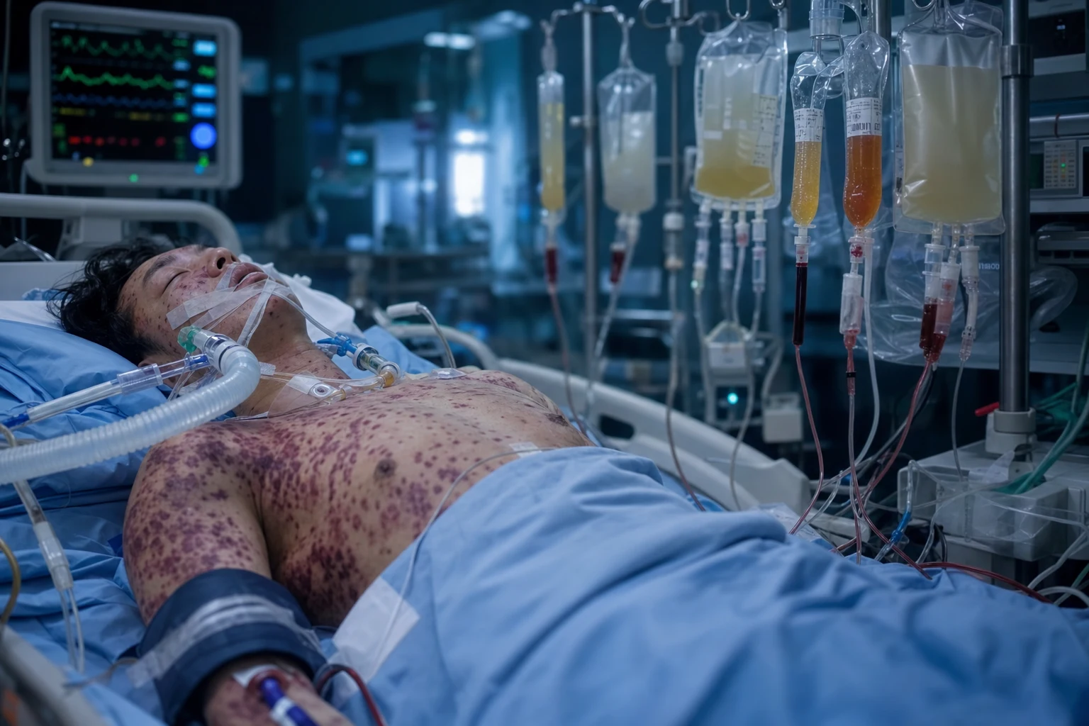
DIC is a syndrome of widespread intravascular coagulation causing consumption of platelets and clotting factors, microvascular thrombosis, and bleeding. It is NOT a primary disease — always secondary to a trigger: sepsis (1), trauma, malignancy, obstetric (amniotic fluid embolism, pre-eclampsia, retained dead fetus), transfusion reaction, snake bite. Diagnosis: ISTH DIC score (platelets, fibrinogen, FDP/D-dimer, PT). Score =5 = overt DIC. Management: treat the UNDERLYING CAUSE (most important). Supportive: platelets (<50 with bleeding), FFP (INR 1.5 + bleeding), cryoprecipitate (fibrinogen <1.5 g/L), heparin (thrombotic DIC only — rare). Do NOT give antifibrinolytics (tranexamic acid) in DIC (risk of fatal thrombosis).
On this page & tools
Your progress
Saved locally on this device.
10 MCQs with explanations
Target exams
Red flags
ISTH DIC score
[1] [2]Management
DIC management protocol
Treat the underlying cause — THE most important step
DIC is NEVER a primary diagnosis. It ALWAYS has a trigger. Sepsis: antibiotics + source control. Trauma: resuscitation + surgery. Malignancy: chemotherapy (APL — all-trans retinoic acid). Obstetric: delivery (pre-eclampsia), uterine evacuation (retained products). Without treating the cause, DIC will persist regardless of blood product support.
Platelet transfusion
Give platelets if: bleeding AND platelets <50,000/mm3 (or <30,000 in high bleeding risk). Also before invasive procedures. One adult dose raises platelets by ~30,000-40,000. Continue transfusion to keep >50 in active bleeding.
FFP (fresh frozen plasma)
Give FFP if: bleeding AND PT/INR prolonged (>1.5) or aPTT prolonged. Dose: 15 mL/kg (typically 4 units). Provides all clotting factors. Note: DIC has BOTH bleeding AND thrombosis — giving FFP does not worsen thrombosis (restores balance).
Cryoprecipitate (for fibrinogen)
Give cryoprecipitate if fibrinogen <1.5 g/L (1.0 g/L if ISTH criteria). Fibrinogen is the FIRST factor to drop in DIC (consumption). 10 units of cryo raises fibrinogen by ~0.5-1.0 g/L. Target fibrinogen >1.5 g/L. Fibrinogen concentrate is an alternative (more rapid, smaller volume).
Heparin (SELECTIVE use — thrombotic DIC)
Most DIC presents with BLEEDING — do NOT give heparin routinely. Give therapeutic heparin ONLY in THROMBOTIC DIC (purpura fulminans, acral ischaemia, venous thromboembolism). Thrombotic DIC is rare — most DIC is bleeding-dominant. Prophylactic heparin (LMWH for VTE prevention) is reasonable if no active bleeding.
DO NOT give antifibrinolytics
Tranexamic acid (TXA) and aprotinin are CONTRAINDICATED in DIC. DIC has concurrent thrombosis and bleeding — antifibrinolytics prevent clot breakdown → catastrophic thrombosis → organ failure → death. The exception: if there is PROVEN PRIMARY HYPERFIBRINOLYSIS (rare — e.g., prostate cancer, some snake bites) — but this requires expert haematology input.
SAQ — Sepsis-induced DIC: scoring and management
10 minutes · 10 marks
A 60-year-old man with perforated diverticulitis and septic shock has oozing from his intravenous sites, blood from his NG tube, and a petechial rash. Bloods: platelets 60 ×10^9/L, PT INR 2.1, aPTT 60 s, fibrinogen 1.2 g/L, D-dimer markedly raised. The registrar asks for the diagnosis and management.
SAQ — Thrombotic DIC and purpura fulminans
10 minutes · 10 marks
A 17-year-old girl is admitted with fever, headache, and a rapidly progressive purpuric rash on her limbs with necrotic patches and acral cyanosis. BP 80/40, lactate 6. Bloods: platelets 40, INR 2.4, fibrinogen 0.9 g/L. The diagnosis is meningococcaemia with purpura fulminans.
Clinical pearls
Red flags
Pathophysiology — the cascade
DIC is best understood as a loss of the checks and balances that keep thrombin generation local and self-limiting. A massive tissue-factor (TF) load overwhelms the natural anticoagulant pathways (antithrombin, protein C, TFPI) and fibrinolysis is shut down by PAI-1, so coagulation becomes systemic and sustained — simultaneously depositing fibrin (microvascular thrombosis, organ failure) and consuming platelets and factors (bleeding). [1]
The DIC cascade — from tissue factor to simultaneous bleeding AND thrombosis
| Stage | Event | Consequence | Laboratory signature |
|---|---|---|---|
| 1. Trigger | Tissue factor released from damaged tissue, cytokine-activated monocytes (sepsis), mucin (adenocarcinoma), amniotic fluid, leukaemic promyelocyte granules, or snake venom procoagulants | TF binds factor VIIa → extrinsic tenase → activation of IX and X | Trigger-specific (e.g. rising lactate in sepsis) |
| 2. Thrombin explosion | Factor Xa + Va → prothrombinase → massive thrombin generation. Thrombin is the central mediator — cleaves fibrinogen, activates V, VIII, XI, XIII and platelets | Diffuse fibrin formation + platelet activation throughout the microcirculation | Prolonging PT/APTT (factor consumption); falling fibrinogen |
| 3. Microvascular thrombosis | Fibrin strands + platelet aggregates occlude the capillaries; RBCs are sheared on the fibrin mesh | Organ dysfunction (AKI, ARDS, hepatic, CNS), acral ischaemia, purpura fulminans | Schistocytes (fragmented RBCs); rising lactate; organ markers |
| 4. Consumption | Platelets and clotting factors (especially V, VIII, XIII, fibrinogen) are consumed faster than the liver/marrow can replace them | The bleeding phenotype — oozing from venepuncture sites, mucosal and GI haemorrhage, intracranial bleed | Thrombocytopenia (falling trend); prolonged PT/APTT; low fibrinogen |
| 5. Failed brakes | Antithrombin consumed + degraded by neutrophil elastase; protein C pathway exhausted (and protein C is also consumed); TFPI overwhelmed; endothelial glycocalyx shed | Thrombin generation continues unchecked; the cycle self-perpetuates | Low antithrombin; low protein C (not routinely measured) |
| 6. Shut-down fibrinolysis | PAI-1 (plasminogen activator inhibitor-1) massively upregulated by cytokines and hypoxia; TAFI activated → clots become resistant to lysis | Microthrombi persist → progressive organ failure; bleeding is from consumption, NOT excess fibrinolysis | High D-dimer (some lysis still occurs) but TEG/ROTEM shows low/no lysis (LY30 <0.8%) |
STOP-MTB — the five DIC triggers (sepsis is #1)
Causes — trigger-specific mechanisms and management
DIC triggers — mechanism, time course, and trigger-specific definitive therapy
| Trigger | Mechanism of tissue factor / procoagulant release | Time course | Trigger-specific definitive therapy |
|---|---|---|---|
| Sepsis (#1 ICU cause) | Endotoxin + cytokines (IL-6, TNF) upregulate TF on monocytes and endothelium; thrombomodulin downregulated; PAI-1 upregulated | Hours–days | Antibiotics within 1 h, source control (drain / debride / remove line), fluid resuscitation |
| Severe trauma / burns | TF from devitalised tissue; endothelial glycocalyx disruption; acute traumatic coagulopathy (activated protein C, hyperfibrinolysis early) | Minutes–hours | Haemorrhage control (damage-control surgery/orthopaedics), permissive hypotension, warmed 1:1:1 transfusion |
| Obstetric — abruption / AFE | Decidual TF and trophoblast tissue enter maternal circulation; amniotic fluid contains TF + platelet-activating factor | Acute (minutes–hours) | Delivery / uterine evacuation (the definitive step); massive transfusion; resuscitate AFE cardiopulmonary collapse |
| Obstetric — retained dead fetus / pre-eclampsia / HELLP / septic abortion | Sustained TF exposure from necrotic retained products; endothelial dysfunction in pre-eclampsia | Days–weeks | Uterine evacuation; magnesium for severe pre-eclampsia; delivery |
| Malignancy — APL | Leukaemic promyelocyte granules are TF- and annexin-II-rich; also heightened fibrinolysis (annexin II) | Days (often present at diagnosis) | ATRA (all-trans retinoic acid) immediately on suspicion, before cytogenetic confirmation; add arsenic trioxide; aggressive product support |
| Malignancy — mucin-secreting adenocarcinoma (pancreas, gastric, prostate, lung) | Mucin activates factor X directly and triggers platelet aggregation; "Trousseau's migratory thrombophlebitis" | Weeks–months (chronic/smouldering DIC) | Treat the tumour; long-term therapeutic LMWH (warfarin is ineffective in Trousseau's) |
| Snake bite (viperids — Russell's, saw-scaled, etc.) | Venom prothrombin activators + metalloproteases consume factors and cleave fibrinogen → venom-induced consumption coagulopathy (VICC) | Minutes–hours after bite | Species-specific antivenom; do NOT give FFP prophylactically (spontaneous bleed rate is low once bite-site controlled; reserve for active bleeding); supportive |
| Severe pancreatitis | Free trypsin activates prothrombin and factor X; Ca²⁺ soap formation; hepatic dysfunction | Days | Treat the pancreatitis (fluid resuscitation, early enteral feeding, source control of necrosis) |
| ABO-incompatible transfusion / massive haemolysis | Intravascular haemolysis releases red-cell phospholipid + ADP → platelet activation and TF-pathway trigger | Minutes (acute) | Stop the transfusion immediately; supportive; treat acute kidney injury |
Pathophysiology in five steps — the 30-second viva answer
Trigger releases tissue factor
A massive TF load (sepsis-monocyte TF, damaged tissue, amniotic fluid, leukaemic granules, snake venom) floods the circulation and binds factor VIIa.
Widespread thrombin generation
The extrinsic tenase (TF-VIIa) activates IX and X; Xa with Va generates explosive thrombin production that the natural anticoagulants (antithrombin, protein C, TFPI) cannot contain.
Microvascular thrombosis → organ failure
Diffuse fibrin + platelet aggregates occlude capillaries → AKI, ARDS, hepatic dysfunction, CNS change, acral ischaemia, purpura fulminans. RBCs are fragmented on the fibrin mesh (schistocytes, MAHA).
Consumption → bleeding
Platelets and clotting factors (V, VIII, XIII, fibrinogen) are consumed faster than replaced → oozing from venepuncture sites and mucosae, GI/GU bleed, intracranial haemorrhage.
Fibrinolysis shut down → the cycle persists
PAI-1 is massively upregulated, so clots are not cleared and thrombin generation self-perpetuates. This is why antifibrinolytics (TXA) worsen DIC — fibrinolysis is already impaired, not excessive.
ISTH scoring — the world standard
The ISTH overt-DIC score is simple, reproducible, validated against expert-panel diagnosis, and independently predicts mortality — it is the score the examiners want. It requires a known underlying trigger (you cannot diagnose "idiopathic DIC"); without a trigger the score does not apply. [1]
ISTH scoring systems compared — know ISTH, name the others
| Score | Body / origin | Components | Overt-DIC threshold | Strength / weakness |
|---|---|---|---|---|
| ISTH overt-DIC (the standard) | International Society on Thrombosis and Haemostasis | Platelets, D-dimer/FDP, PT prolongation, fibrinogen + prerequisite trigger | ≥5 | Simple, world standard; rising score independently predicts 28-day mortality; D-dimer is semi-quantitative (0/2/3) |
| ISTH sepsis-induced coagulopathy (SIC) | Iba & Levi, 2017 | Platelets, PT, SOFA (organ dysfunction) | ≥4 | Simpler; integrates organ failure; complements ISTH-DIC in sepsis |
| JAAM | Japanese Association for Acute Medicine | SIRS, platelets, PT, D-dimer/fibrinogen | ≥4 | Useful in early/sepsis-DIC; captures the SIRS component |
| JMHW | Japanese Ministry of Health & Welfare | Older Japanese score | — | Less sensitive than ISTH; largely superseded |
| ASH 2018 | American Society of Hematology | Symptom-based; 2 points if bleeding or organ dysfunction present | — | Adds bleeding/organ symptoms; less widely adopted in ICU |
Worked example — applying the ISTH score at the bedside
Confirm an underlying trigger is present
e.g. a septic patient in ICU with Gram-negative bacteraemia. No trigger → the score does not apply (consider an alternative diagnosis such as TTP, liver disease, or drug effect).
Platelets 65 ×10⁹/L → score 1
Platelet count <100 but ≥50 = 1 point. (<50 = 2; >100 = 0.) The falling trend matters more than the absolute — a count dropping from 220 to 110 scores 0 but is alarming.
D-dimer strongly elevated → score 3
Marked rise in fibrin-related marker (D-dimer/FDP) = 3 points (moderate = 2; none = 0). D-dimer is sensitive but non-specific — it only counts in the context of the other three parameters.
PT prolonged 8 s above normal → score 2
PT prolongation >6 s = 2 points (3–6 s = 1; <3 s = 0). A normal PT does not exclude early DIC.
Fibrinogen 1.8 g/L → score 0
Fibrinogen >1.0 g/L = 0 points (<1.0 = 1). Fibrinogen is an acute-phase reactant — a "normal" value in sepsis is abnormally low; a falling trend is more informative.
Total = 1 + 3 + 2 + 0 = 6 → OVERT DIC
Score ≥5 = overt DIC. Repeat the full score every 24 h — a rising score predicts mortality and guides product support; a falling score confirms response to treatment of the trigger.
Clinical features
Bleeding-predominant vs thrombotic-predominant DIC — the two phenotypes
| Feature | Bleeding-predominant (more common in ICU) | Thrombotic-predominant |
|---|---|---|
| Typical triggers | APL, obstetric (abruption, AFE), trauma, severe liver failure, snake bite | Meningococcal/pneumococcal sepsis, catastrophic antiphospholipid syndrome (CAPS), solid-organ malignancy |
| Hallmark sign | Oozing from IV sites, venepuncture sites, surgical wounds, line sites | Purpura fulminans — confluent purpuric patches with central necrosis; acral (digits/nose/ears) ischaemia → gangrene |
| Bleeding sites | Mucosa (gums, nose), GI/GU, intracranial (often catastrophic), retroperitoneal | Minimal bleeding; bleeding may be absent |
| Organ impact | Hypovolaemic/haemorrhagic shock; multi-site bleeding | Skin necrosis; limb/digit amputation; AKI from cortical necrosis; ARDS |
| Key therapy beyond the trigger | Product support — cryoprecipitate (fibrinogen), platelets, FFP | Therapeutic heparin (UFH infusion or therapeutic LMWH) ± protein C concentrate |
Clinical features by system — what you will find at the bedside
| System | Bleeding phenotype | Thrombotic phenotype |
|---|---|---|
| Skin | Petechiae, ecchymoses, oozing from puncture/line sites, prolonged bleeding from IM/SC injection sites | Purpura fulminans, skin necrosis, livedo, acral cyanosis, digital gangrene |
| Bleeding pattern | Venepuncture, arterial-line, drain and surgical sites ooze continuously — the classic clue; also epistaxis, haematuria, melaena, haematemesis | Skin haemorrhage into necrotic areas (paradoxical bleeding into infarcted tissue) |
| Renal | Haematuria; AKI from hypoperfusion | Cortical necrosis → oliguric AKI (often irreversible) |
| Respiratory | Haemoptysis (rare) | ARDS from pulmonary microvascular thrombosis |
| CNS | Intracranial haemorrhage (catastrophic); confusion from hypoperfusion | Microthrombotic encephalopathy; seizures |
| GI | GI haemorrhage; hepatic dysfunction from ischaemia | Mesenteric ischaemia; hepatic infarction |
| Haematology | Anaemia from blood loss AND MAHA (schistocytes); falling platelets | Falling platelets; rising lactate; schistocytes |
The classic bedside clue — 'the patient who bleeds from everywhere they have been stuck'
Laboratory diagnosis — what each value tells you
Laboratory abnormalities in DIC — what each value tells you
| Test | Typical finding | Mechanism | Pitfall / caveat |
|---|---|---|---|
| Platelets | Low and falling (the trend matters more than the absolute) | Consumption in microthrombi | May be normal early; a single value is misleading — always trend it |
| PT / INR | Prolonged (rising trend) | Consumption of factors V, VII, X (VII has the shortest half-life — drops first) | Also prolonged in liver disease, warfarin, vitamin K deficiency; not DIC-specific |
| aPTT | Prolonged | Consumption of factors VIII, IX, XI, XII | May be normal early; also prolonged by heparin |
| Fibrinogen | Low and falling | Consumption; it is the first factor to drop | Acute-phase reactant — may be normal/high early in sepsis; a "normal" value in inflammation is relatively low; trend it |
| D-dimer / FDP | Markedly elevated | Breakdown of cross-linked fibrin (some lysis still occurs) | Sensitive but non-specific — elevated in thrombosis, surgery, pregnancy, malignancy, infection |
| Thrombin time | Prolonged | Low fibrinogen + circulating FDPs interfere with polymerisation | Also prolonged by heparin and dysfibrinogenaemia |
| Blood film | Schistocytes (fragmented RBCs), polychromasia | Microangiopathic haemolytic anaemia (MAHA) — RBCs sheared on fibrin mesh | Schistocytes also in TTP/HUS, malignant HTN, mechanical valve |
| Antithrombin / protein C | Low (consumption) | Exhausted natural anticoagulant pathways | Not routinely measured in ICU; confirmatory in research |
| LDH / haptoglobin | LDH high, haptoglobin low | Intravascular haemolysis of MAHA | Non-specific; supports the MAHA component |
TEG / ROTEM patterns in DIC — viscoelastic testing at the bedside
| Phase | TEG parameter | ROTEM parameter | Finding in DIC |
|---|---|---|---|
| Clot initiation (reaction time) | R time | CT (clotting time) EXTEM/INTEM | Prolonged (factor depletion) |
| Clot kinetics (clot build-up) | K time; alpha angle | CFT (clot formation time); alpha | Prolonged / shallow angle (low fibrinogen → slow polymerisation) |
| Clot strength (maximum amplitude) | MA | MCF (maximum clot firmness) | Reduced (thrombocytopenia; low factor XIII) |
| Clot stability / lysis | LY30 | ML (maximum lysis) | Low — fibrinolysis is shut down (PAI-1 upregulated); high LY30 suggests primary hyperfibrinolysis (early trauma, prostate cancer, some snake bites) — the only context TXA is appropriate |
| Fibrin contribution | Functional fibrinogen TEG | FIBTEM MCF | Low FIBTEM confirms fibrinogen deficiency — guides cryoprecipitate/fibrinogen concentrate dosing |
Differential diagnosis — the consumptive / thrombocytopenic mimics
DIC vs TTP / HUS vs severe liver disease vs massive-transfusion dilutional coagulopathy
| Feature | DIC | TTP / HUS | Severe liver disease | Dilutional (massive transfusion) |
|---|---|---|---|---|
| Pathology | Consumption + microthrombosis + impaired fibrinolysis | ADAMTS13 deficiency (TTP) → ultra-large vWF multimers → platelet microthrombi; HUS from complement (atypical) or Shiga-toxin | Reduced synthesis + consumption + portal HTN splenic sequestration | Dilution of factors + platelets + citrate + hypothermia/acidosis |
| PT / aPTT | Prolonged | Normal (no factor consumption) | Prolonged | Prolonged |
| Fibrinogen | Low (falling) | Normal | Low (reduced synthesis) | Low (dilution) |
| D-dimer | Markedly high | Normal / mildly high | Mildly high (reduced clearance) | Variable |
| Schistocytes | Present | Numerous (>4–5%) | Absent / few | Absent |
| ADAMTS13 | Normal | <10% (TTP) | Normal | Normal |
| Critical differentiator | Trigger present + falling fibrinogen + high D-dimer + schistocytes | Normal PT/aPTT + many schistocytes + thrombocytopenia → urgent plasma exchange | Factor VIII normal/high (endothelial origin); D-dimer only mildly raised | History of massive transfusion; corrects with factor/platelet repletion |
Management — the practical protocol
Transfusion thresholds in bleeding DIC (BCSH / ISTH guidance)
| Product | Dose | Threshold (active bleeding / pre-procedure) | Target | Rationale |
|---|---|---|---|---|
| Platelets | 1 adult therapeutic dose (~1 pool / 1 unit per 10 kg) | <50 ×10⁹/L (<100 if active bleeding, CNS, or perioperative) | >50 (>75–100 in CNS) | Replace consumed platelets |
| Cryoprecipitate | 2 pools (10–12 units), or fibrinogen concentrate 3–4 g | Fibrinogen <1.5 g/L | >1.5 g/L (some centres >2.0 in APL) | Fibrinogen is the first factor to drop; restores clot structure |
| Fibrinogen concentrate | 3–4 g IV (faster, smaller volume than cryo) | Fibrinogen <1.5 g/L | >1.5 g/L | Preferred where available; rapid, low-volume, pathogen-reduced |
| FFP | 15 mL/kg (~4 units / ~1 L) | PT/aPTT >1.5× normal with bleeding | Normalise PT/aPTT | Provides all clotting factors; does NOT worsen thrombosis |
| Prothrombin complex concentrate (PCC) | Specialist use only | — | — | Not first-line in DIC (contains no factor V or fibrinogen); consider only with expert advice |
Heparin in DIC — when, what, and what dose
| Scenario | Heparin strategy | Dose |
|---|---|---|
| Thrombotic-predominant DIC (purpura fulminans, acral ischaemia, APL, CAPS, skin necrosis) | Therapeutic heparin | UFH infusion ~18 U/kg/h (titrate to APTT 1.5–2.5×) OR LMWH enoxaparin 1 mg/kg BD |
| Most bleeding-dominant DIC | No therapeutic heparin | — (heparin would worsen bleeding) |
| VTE prophylaxis (no active bleeding) | Prophylactic LMWH is reasonable and recommended | Enoxaparin 40 mg SC OD (dose-adjust for renal function) |
| APL (high thrombotic risk early) | Prophylactic-to-therapeutic heparin during induction | Often added once bleeding controlled |
| Severe renal failure | Prefer UFH (rapidly reversible) | UFH infusion titrated to APTT |
Purpura fulminans management (meningococcal — the prototype)
Antibiotics immediately — do not wait for confirmation
Ceftriaxone 2 g IV (add vancomycin if pneumococcal suspected). Time-to-antibiotic is the single biggest determinant of survival in meningococcaemia.
Therapeutic heparin — even in the face of skin haemorrhage
UFH infusion (reversible, titratable). The gangrene is driven by ongoing microvascular thrombosis; heparin halts extension. Evidence is observational but consistent and guideline-supported.
Protein C concentrate (where available)
Restores the consumed natural anticoagulant; most evidence in congenital protein C deficiency and severe acquired deficiency. FFP provides some protein C if concentrate unavailable.
Product support as needed
Cryoprecipitate for fibrinogen <1.5 g/L, platelets if <50 and bleeding, FFP for prolonged PT/APTT — but do not let product support delay heparin and antibiotics.
Surgical debridement / fasciotomy / amputation
For established necrotic skin, compartment syndrome, or non-viable digits/limbs — usually delayed until demarcation is clear.
Treat the cause and support organs
ICU support for shock (vasopressors), ARDS (lung-protective ventilation), AKI (renal replacement therapy). Mortality remains 20–40%.
APL-associated DIC — the immediate management (a haematology emergency)
Suspect APL on the blood film / FBC
Hypergranular promyelocytes with Auer rods; the microgranular variant (M3v) may be missed — check coagulation in ANY new AML presentation. Coagulopathy + new leukaemia = APL until proven otherwise.
Start ATRA (all-trans-retinoic-acid) 45 mg/m²/day immediately on suspicion
Do NOT wait for cytogenetic/PCR confirmation. ATRA differentiates the leukaemic promyelocytes and halts tissue-factor release within 24–48 h. Delay costs lives.
Aggressive product support
Cryoprecipitate to keep fibrinogen >1.5–2.0 g/L, platelets >30 (>50 if febrile/bleeding), FFP to keep PT/APTT <1.5× normal. Monitor coagulation every 6–12 h.
Add arsenic trioxide (ATO) for high-risk disease
ATO combined with ATRA is standard for higher-risk APL; also has differentiating and pro-apoptotic effects. Differentiation syndrome (fever, dyspnoea, weight gain, pulmonary infiltrates) — treat with dexamethasone.
Avoid invasive procedures and intramuscular injections
Until the coagulopathy resolves — even a central line carries bleeding risk in active APL-DIC. Use ultrasound-guided access.
Continue support until ATRA effect (24–72 h)
The coagulopathy usually corrects within days of ATRA. Heparin is not routine but may be added for thrombosis; mortality from APL has fallen from ~30% to <10% with ATRA.
Obstetric DIC
Obstetric DIC syndromes — recognise and act on the trigger
| Syndrome | Mechanism | Presentation | Key management |
|---|---|---|---|
| Placental abruption | Retroplacental clot + decidual tissue factor enters maternal circulation; concealed abruption (couvelaire uterus) causes severe DIC | Painful vaginal bleeding, uterine tenderness, fetal distress; rigid, tender uterus | Delivery (vaginal if rapid, CS if fetal distress); uterotonics; evacuate retained products |
| Amniotic fluid embolism (AFE) | Amniotic fluid + foetal debris enter maternal circulation via disrupted veins → anaphylactoid reaction + catastrophic DIC | Peripartum sudden cardiovascular collapse (LV failure / right-heart strain) + hypoxia, followed within minutes–hours by massive coagulopathy | Dual resuscitation: (1) treat cardiopulmonary collapse (vasopressors, inotropes); (2) deliver the baby; massive transfusion; supportive |
| Severe pre-eclampsia / HELLP | Endothelial dysfunction + platelet consumption; HELLP adds haemolysis and liver ischaemia | Hypertension, proteinuria, right-upper-quadrant pain, low platelets, transaminitis | Magnesium sulphate (seizure prophylaxis), blood-pressure control, delivery |
| Retained dead fetus | Sustained TF release from necrotic retained products over days–weeks | Chronic/smouldering DIC; history of intrauterine fetal demise | Uterine evacuation; product support peri-procedure |
| Septic abortion / intra-amniotic infection | Infection-driven TF + endothelial activation | Fever, uterine tenderness, purulent discharge, septic shock | Broad-spectrum antibiotics + uterine evacuation + source control |
The trials — anticoagulant therapies tested in sepsis-DIC
The recurring lesson of sepsis-DIC trials: specifically blocking one node of the coagulation cascade does not improve survival in unselected septic patients, even when the agent corrects the laboratory abnormalities. Treat the cause; use products; do not reach for an anticoagulant "magic bullet." [1]
Taylor 2001 — ISTH SSC definition and scoring of DIC
Source
Thrombosis and Haemostasis — Scientific and Standardisation Committee of the ISTH
PMID
11816725
What it did
Defined DIC operationally and proposed the 5-component scoring system (platelets, D-dimer/FDP, PT prolongation, fibrinogen, underlying disorder) — the world standard
Threshold
Score ≥5 = overt DIC; repeat every 24 h
Why it matters
Replaced descriptive, operator-dependent diagnosis with a reproducible, validated score that predicts mortality independently
KyberSept — Warren 2001 — High-dose antithrombin III in severe sepsis
Source
JAMA — multicentre RCT, 2314 severe sepsis patients
PMID
11597289
Intervention
High-dose antithrombin III (30,000 U over 4 days) vs placebo
Result
NO mortality benefit at 28 days (38.9% vs 38.7%); trend to more bleeding, especially with concomitant heparin
Status
Antithrombin NOT recommended in sepsis-DIC
OPTIMIST — Abraham 2003 — Tifacogin (recombinant TFPI) in severe sepsis
Source
JAMA — multicentre RCT, 1754 severe sepsis patients
PMID
12851279
Intervention
Recombinant tissue factor pathway inhibitor (tifacogin) vs placebo — blocks the TF-VIIa complex at the origin of the cascade
Result
NO mortality benefit (34.2% vs 33.9%); significant increase in serious bleeding (especially with concomitant heparin)
Status
Tifacogin withdrawn — blocking the apex of the cascade is not the answer
PROWESS-SHOCK — Ranieri 2012 — Drotrecogin alfa (activated protein C) in septic shock
Source
New England Journal of Medicine — multicentre RCT, 1696 septic shock patients
PMID
22616830
Intervention
Drotrecogin alfa (activated) — recombinant activated protein C — vs placebo
Result
NEGATIVE — no difference in 28-day mortality (26.4% vs 24.2%, p=0.31)
Status
Drug withdrawn from the market (2011); activated protein C is not part of sepsis-DIC management
SCARLET — Vincent 2019 — Recombinant soluble thrombomodulin (ART-123) in sepsis-associated coagulopathy
Source
JAMA — multicentre RCT, 800 patients with sepsis-associated coagulopathy
PMID
31104069
Intervention
Recombinant human soluble thrombomodulin 0.06 mg/kg IV daily for 6 days vs placebo
Mechanism
Binds thrombin → activates protein C (switches thrombin from pro- to anti-coagulant) and inactivates HMGB1 (a late sepsis mediator)
Result
Primary endpoint NEGATIVE — 28-day mortality 26.4% vs 24.9% (p=0.32); pre-specified subgroup with baseline ISTH score ≥5 and organ dysfunction suggested possible benefit
Status
Not approved by FDA/EMA for sepsis-DIC; used in Japan; remains investigational in most of the world
CRASH-2 — 2010 — Tranexamic acid in trauma haemorrhage (the TXA exception)
Source
Lancet — multinational RCT, 20,211 trauma patients with significant haemorrhage
PMID
20554319
Intervention
Tranexamic acid 1 g IV loading then 1 g over 8 h vs placebo, within 8 h of injury
Result
All-cause mortality reduced (14.5% vs 16.0%, p=0.0035); benefit greatest when given WITHIN 3 HOURS of injury
Reconciling with DIC
Early traumatic coagulopathy has a primary-hyperfibrinolysis component (high LY30 on TEG) in which TXA helps; ESTABLISHED DIC with PAI-1 upregulation and low LY30 is different — TXA worsens it. Use viscoelastic testing to decide.
Prognosis
Prognostic factors in DIC
| Factor | Effect on prognosis | Detail |
|---|---|---|
| Underlying cause | Dominant determinant | Meningococcal purpura fulminans 20–40% mortality; APL now <10% with ATRA; sepsis-DIC ~40%; AFE 20–60% |
| ISTH score trajectory | Rising score predicts mortality | A rising score over 24–72 h independently predicts 28-day mortality; a falling score confirms response |
| Fibrinogen trend | Refractory low fibrinogen is ominous | Persistent fibrinogen <1.0 g/L despite replacement suggests ongoing consumption and poor prognosis |
| Organ failure (SOFA) | Drives mortality | Multi-organ failure (particularly AKI and ARDS) is the usual mode of death, not exsanguination |
| Time to source control / antibiotics | Critical in sepsis-DIC | Each hour's delay to antibiotics in septic shock increases mortality |
Common exam pitfalls
Common exam pitfalls in DIC
| Pitfall | The error | The correct answer |
|---|---|---|
| "Give TXA for the bleeding" | TXA worsens microvascular thrombosis (fibrinolysis is already impaired) | Products (cryoprecipitate first) + treat the trigger; TXA only if TEG/ROTEM shows primary hyperfibrinolysis (high LY30) |
| "Platelet count is normal so it isn't DIC" | Platelets may be normal early | The trend matters more than the absolute; repeat and trend; check fibrinogen, PT and D-dimer |
| "Fibrinogen is normal so it isn't DIC" | Fibrinogen is an acute-phase reactant | A "normal" fibrinogen in sepsis is relatively low; a falling trend is diagnostic |
| "Treat the INR with FFP" | Corrects a number but does not treat the disease | Treat the trigger; give FFP only for active bleeding or before procedures with PT/aPTT >1.5× |
| "Give heparin for all DIC" | Most DIC is bleeding-dominant | Therapeutic heparin only for thrombotic-predominant DIC; prophylactic LMWH reasonable if no bleeding |
| "Give antithrombin / protein C / thrombomodulin" | All failed in RCTs | No specific anticoagulant "magic bullet"; treat the cause + products |
| "Confuse with TTP" | Both give schistocytes + thrombocytopenia | TTP has normal PT/aPTT and many schistocytes → urgent plasma exchange, not products |
The 90-second viva answer for 'Discuss DIC in the ICU patient'
Define
DIC is an acquired syndrome of widespread intravascular coagulation causing simultaneous bleeding and thrombosis, ALWAYS secondary to a trigger.
Pathophysiology (one line)
Tissue factor release → massive thrombin generation → fibrin deposition (microthrombosis, organ failure) AND consumption of platelets and factors (bleeding), with impaired fibrinolysis (PAI-1 upregulated).
Triggers (mnemonic)
STOP-MTB: Sepsis, Trauma, Obstetric, Pancreatitis/Malignancy, Transfusion, Bites (snake).
Diagnosis
ISTH overt-DIC score (platelets + D-dimer + PT + fibrinogen, prerequisite trigger); ≥5 = overt DIC, repeat daily. Lab pattern: falling platelets, prolonged PT/APTT, low/falling fibrinogen, high D-dimer, schistocytes.
Management
1) Treat the underlying cause (definitive). 2) Product support for bleeding: cryoprecipitate/fibrinogen concentrate (fibrinogen <1.5 g/L), platelets (<50 + bleeding), FFP (PT/aPTT >1.5× + bleeding). 3) Therapeutic heparin for thrombotic-predominant DIC. 4) NO antifibrinolytics (TXA) unless documented primary hyperfibrinolysis.
Differential
TTP/HUS (normal PT/aPTT, many schistocytes, ADAMTS13 <10% → plasma exchange), liver disease (factor VIII normal/high), dilutional coagulopathy (massive transfusion history).
Prognosis
Dominated by the underlying cause; mortality 20–80% depending on trigger. Rising ISTH score predicts mortality independently.
Clinical pearls — the extended set
[1] [1] [1]References
- [1]Taylor FB, Toh CH, Hoots WK, Wada H, Levi M Towards definition, clinical and laboratory criteria, and a scoring system for disseminated intravascular coagulation Thromb Haemost, 2001.PMID 11816725
- [2]Levi M, Toh CH, Thachil J, Watson HG Guidelines for the diagnosis and management of disseminated intravascular coagulation. British Committee for Standards in Haematology Br J Haematol, 2009.PMID 19222477
- [3]Warren BL, Eid A, Singer P, et al. Caring for the critically ill patient. High-dose antithrombin III in severe sepsis: a randomized controlled trial JAMA, 2001.PMID 11597289
- [4]Abraham E, Reinhart K, Opal S, et al. Efficacy and safety of tifacogin (recombinant tissue factor pathway inhibitor) in severe sepsis: a randomized controlled trial JAMA, 2003.PMID 12851279
- [5]Ranieri VM, Thompson BT, Barie PS, et al. Drotrecogin alfa (activated) in adults with septic shock N Engl J Med, 2012.PMID 22616830
- [6]Vincent JL, Francois B, Zabolotskikh I, et al. Effect of a Recombinant Human Soluble Thrombomodulin on Mortality in Patients With Sepsis-Associated Coagulopathy: The SCARLET Randomized Clinical Trial JAMA, 2019.PMID 31104069
- [7]CRASH-2 trial collaborators Effects of tranexamic acid on death, vascular occlusive events, and blood transfusion in trauma patients with significant haemorrhage (CRASH-2): a randomised, placebo-controlled trial Lancet, 2010.PMID 20554319