ICU · Resuscitation
Extracorporeal membrane oxygenation (ECMO)
Also known as ECMO · Extracorporeal life support (ECLS) · VV-ECMO (venovenous) · VA-ECMO (venoarterial) · ECPR (extracorporeal CPR) · Extracorporeal CO2 removal (ECCO2R) · Centrifugal pump ECMO · Avalon dual-lumen cannula · North-south syndrome (Harlequin) · EOLIA trial
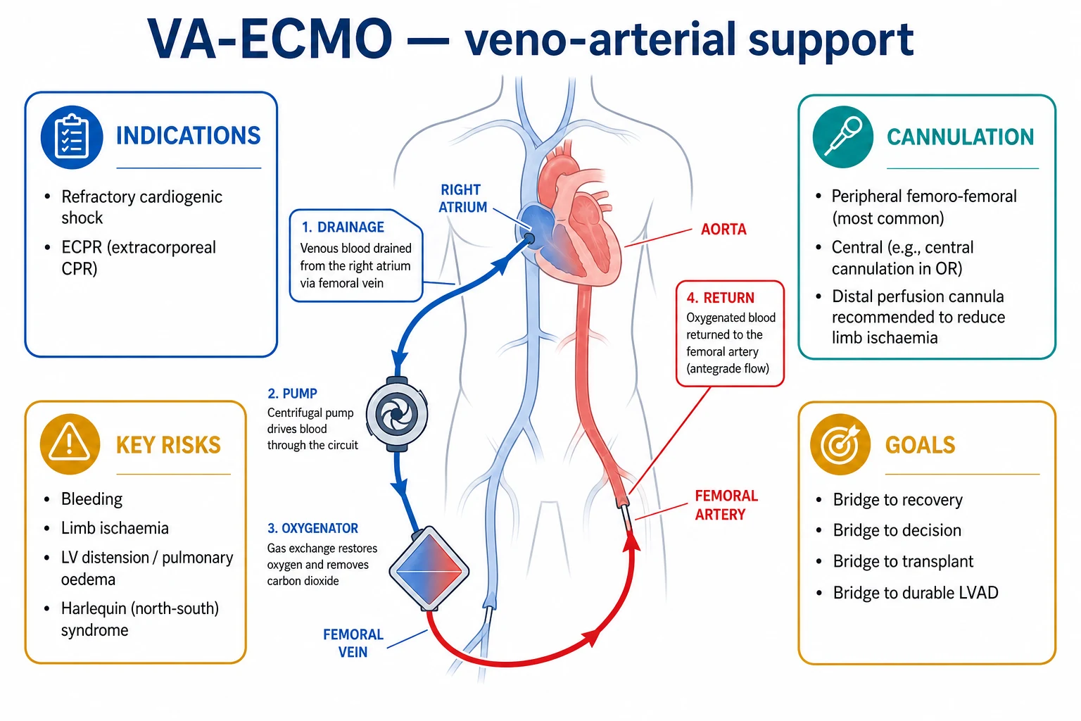
Extracorporeal membrane oxygenation (ECMO) is a modified cardiopulmonary bypass that provides temporary support for refractory cardiac and/or respiratory failure outside the operating theatre. VA-ECMO (venoarterial) drains venous blood from the right atrium via a femoral vein, pumps it through a membrane oxygenator and returns oxygenated blood to the femoral artery — providing BOTH circulatory (cardiac output) and respiratory (oxygenation/CO2 removal) support; indications: cardiogenic shock refractory to inotropes/IABP, ECPR, fulminant myocarditis, post-cardiotomy failure, severe drug toxicity with cardiac depression. VV-ECMO (venovenous) drains from the IVC (femoral vein) and returns oxygenated blood to the right atrium (IJ vein or dual-lumen single cannula) — provides RESPIRATORY support only (the heart must work); indications: severe ARDS (PaO2/FiO2 <80 despite optimised ventilation), bridge to lung transplant, near-fatal asthma (80-90% survival in case series), massive PE as bridge. Circuit: centrifugal (non-occlusive) pump → polymethylpentene hollow-fibre membrane oxygenator → heat exchanger. Percutaneous Seldinger femoro-femoral cannulation is standard. Systemic anticoagulation (UFH, ACT 1.5x baseline or anti-Xa 0.3-0.7) required. Complications: bleeding (1), haemolysis, thrombosis, infection, limb ischaemia (VA-femoral — needs distal perfusion cannula), Harlequin/north-south syndrome (differential hypoxia in VA-femoral), LV distension (afterload). Weaning: VV by reducing sweep gas; VA by reducing flow to 1-1.5 L/min with echo guidance. EOLIA trial (Combes, NEJM 2018): VV-ECMO for very severe ARDS was borderline positive on primary but positive on secondary composite endpoints and post-hoc Bayesian reanalysis. CESAR trial (Peek, Lancet 2009): transfer to ECMO centre improved survival in severe reversible adult respiratory failure.
On this page & tools
Your progress
Saved locally on this device.
Target exams
Red flags
VA-ECMO vs VV-ECMO
VV-ECMO (venovenous)
Lung support only
- Cannulation: femoral vein (drainage) → IJ vein (return). Or double-lumen single cannula (Avalon)
- Supports: oxygenation and CO2 removal (lungs only)
- Heart: must function normally — VV-ECMO does NOT provide circulatory support
- Indications: severe ARDS (PaO2/FiO2 <80 despite optimised ventilation), bridge to lung transplant, severe asthma, pulmonary embolism
- No arterial cannulation → no limb ischaemia risk
- No Harlequin syndrome
VA-ECMO (venoarterial)
Heart + lung support
- Cannulation: femoral vein (drainage) → femoral artery (return). Or central (RA → aorta)
- Supports: both circulation (cardiac output) AND oxygenation
- Indications: cardiogenic shock (refractory to inotropes/IABP), cardiac arrest (ECPR), myocarditis, post-cardiotomy, drug overdose with cardiac depression
- Risk: limb ischaemia (femoral artery cannulation — monitor perfusion)
- Risk: Harlequin syndrome (differential hypoxia)
- Risk: LV distension (blood returning to heart but cannot eject against ECMO pressure)
Key complications
ECMO complications and management
Bleeding (#1 complication)
Caused by systemic anticoagulation (heparin) + platelet dysfunction (circuit consumption). Target ACT 1.5x baseline or aPTT 50-70s (lower than cardiac surgery). Bleeding sites: cannulation sites, GI, intracranial, pulmonary. Management: reduce anticoagulation, transfuse platelets/FFP, surgical control if needed.
Thrombosis (circuit clotting)
Blood contacts artificial surface → clotting. Monitor circuit for clots. Anticoagulation (heparin infusion) to prevent. Circuit change if significant clot burden.
Limb ischaemia (VA-ECMO femoral)
Large arterial cannula obstructs femoral artery flow to leg. Monitor: distal pulses, capillary refill, temperature, pulse oximetry of cannulated limb. Prevention: distal perfusion cannula (small catheter from ECMO circuit to superficial femoral artery — antegrade leg perfusion). If ischaemia: surgical fasciotomy/embolectomy.
Harlequin syndrome (differential hypoxia)
In VA-ECMO via femoral approach: ECMO blood flows retrograde up aorta (from below). Native heart ejects blood from above. If native heart is hypoxaemic (lung failure): upper body (brain, coronaries, arms) receives DEOXYGENATED blood from native heart while lower body gets oxygenated ECMO blood. Monitor: right radial artery ABG (reflects brain/heart oxygenation). Treatment: convert to VA-VA (add venous return), improve native lung oxygenation, or use central cannulation.
Infection
Large cannulae + critical illness + immunosuppression → high infection risk. Surveillance cultures. Antibiotics for suspected infection. Aseptic cannula care.
Haemolysis
Mechanical destruction of RBCs in pump/circuit. Monitor: free haemoglobin, LDH, bilirubin, urine colour (haemoglobinuria). If severe: circuit change, investigate for obstruction/kinking.
Indications and contraindications
Indications
Reversible failure
- VV-ECMO: severe ARDS (PaO2/FiO2 <80 or hypercapnia despite optimal ventilation), bridge to lung transplant, severe asthma, massive PE
- VA-ECMO: cardiogenic shock (refractory to inotropes/IABP), ECPR (cardiac arrest), fulminant myocarditis, post-cardiotomy, severe drug toxicity
- Key principle: disease must be REVERSIBLE (or bridge to transplant)
- Duration: typically days-weeks (longer = more complications)
Contraindications
Do NOT cannulate
- ABSOLUTE: irreversible brain injury, terminal malignancy, severe chronic organ failure (cirrhosis, COPD, heart failure), unwitnessed cardiac arrest with prolonged CPR (>30min no ROSC)
- RELATIVE: age >75, severe peripheral vascular disease, severe obesity (cannulation difficulty), anticoagulation/bleeding diathesis, prolonged mechanical ventilation (>7-10 days — lung fibrosis)
- Contraindications are NOT absolute — individualised decision by ECMO team
Clinical pearls
Red flags
The ECMO circuit — components and physiology
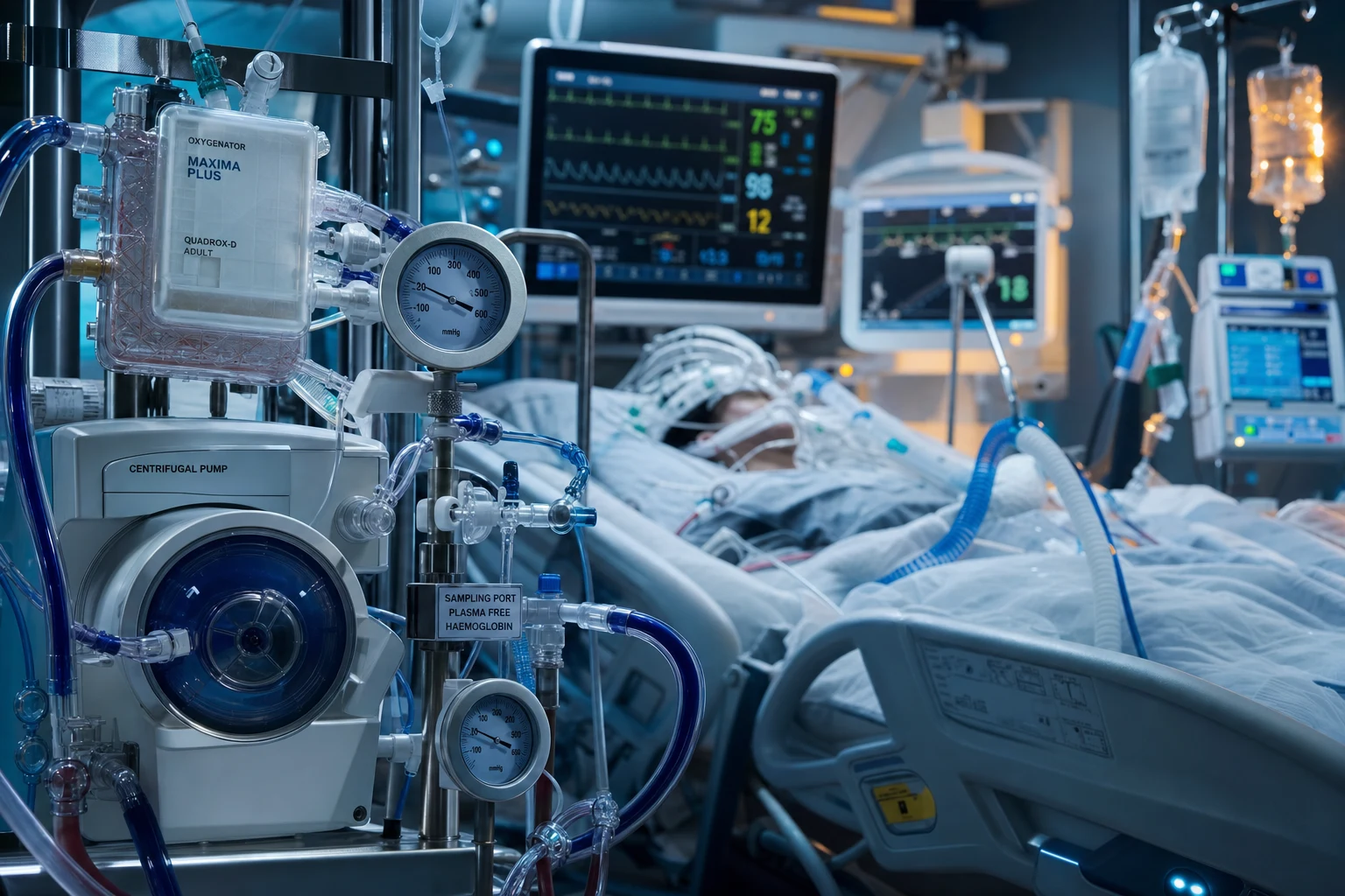
ECMO is a modified, miniaturised form of cardiopulmonary bypass engineered for prolonged use (days to weeks) rather than the hours typical of theatre bypass. Blood is drained from the venous system by a non-occlusive centrifugal pump, passed through a membrane oxygenator where gas exchange occurs, warmed by a heat exchanger, and returned to the patient. Understanding each component is high-yield because the physiology dictates the indications, the complications and the weaning logic.[5][2]
The ECMO circuit — blood path and component function
1. Venous drainage cannula
Drains deoxygenated blood by gravity/siphon (in older roller-pump systems) or active suction (modern centrifugal pumps). Drainage cannula is large-bore (21–29 Fr) to minimise resistance — flow is highly sensitive to cannula length and radius (Poiseuille). Drainage site: femoral vein (tip at IVC–RA junction) for VV and VA; RA directly for central VA. Negative suction >−100 mmHg risks "chattering" (vessel wall apposition) and haemolysis.
2. Centrifugal pump (the heart)
A magnetic impeller spinning at 3000–4000 rpm generates a pressure gradient that propels blood — kinetic energy pump, NON-occlusive (unlike roller pumps). Advantage: no dangerous positive pressure if the line downstream occludes, less haemolysis, compact, afterload-independent forward flow. This is the "artificial heart" of the circuit. Pump flow (cardiac output equivalent) is set in L/min (typically 2–6 L/min in adults; ~60–80 mL/kg/min).
3. Membrane oxygenator (the artificial lung)
Polymethylpentene (PMP) hollow-fibre membrane separates blood from a counter-current "sweep gas" (pure O2 ± added CO2). Gas transfer follows Fick law: O2 diffuses INTO blood, CO2 diffuses OUT. Modern PMP membranes are plasma-resistant and durable for weeks (older silicone membranes needed frequent change). The oxygenator determines oxygenation capacity (surface area ~1.5–2.5 m² in adults). Clots form here first — inspect for fibrin strands.
4. Heat exchanger
A water-bath warmer in the circuit rewarms blood to 36–37°C before return — critical because extracorporeal blood cools rapidly and hypothermia worsens coagulopathy, arrhythmia and the lethal triad. The warmer is integral to the oxygenator block in most modern devices.
5. Arterial (VA) or venous (VV) return cannula
Returns oxygenated blood. VA: femoral artery (15–23 Fr) — blood flows retrograde up the aorta; or axillary/central aorta. VV: IJ vein (tip in RA, directed across tricuspid valve) or via a dual-lumen single cannula (Avalon/OriCath) inserted via right IJ with one lumen draining the IVC and the other returning oxygenated blood toward the tricuspid valve.
6. Oxygenator sweep gas
Sweep gas = oxygen source blended with adjustable flow. SWEEP GAS FLOW controls CO2 CLEARANCE (more sweep → more CO2 removed). BLOOD FLOW (pump speed) controls OXYGEN DELIVERY (more flow → more oxygenated blood returned). This dissociation is the key to troubleshooting: low PaO2 → increase blood flow; high PaCO2 → increase sweep gas flow.
7. Monitoring and safety
Inline pre-/post-membrane blood gas sensors (venous SvO2, post-membrane PaO2), bubble detectors, pressure transducers (pre/post membrane ΔP rise signals clotting), flow probes, and an emergency hand-crank for pump failure. A bridge between venous and arterial limbs allows temporary recirculation if the patient must be disconnected.
The two levers every ICU candidate must know
PUMP BLOOD FLOW
Controls O2 delivery
- Determines how much oxygenated blood is delivered to the patient per minute
- Increase pump speed → more venous blood drained, more oxygenated blood returned → higher PaO2 / DO2
- In VA-ECMO, pump flow = the circulatory support (cardiac output supplement)
- Limited by: venous drainage (cannula size, intravascular volume), and in VA by afterload
SWEEP GAS FLOW
Controls CO2 clearance
- Determines how much CO2 is washed out of blood across the membrane
- Increase sweep flow → more CO2 removed → lower PaCO2
- VV-ECMO: CO2 removal is highly efficient — even low blood flow removes substantial CO2 (basis of ECCO2R)
- Reducing sweep to zero = oxygenator "off" for a VV-ECMO weaning trial
VV-ECMO — respiratory support in detail
VV-ECMO drains deoxygenated blood from the venous system (femoral vein, tip in IVC), oxygenates and decarbonylates it in the membrane lung, and returns it to the RIGHT ATRIUM (via the IJ vein, or through the return port of a dual-lumen Avalon cannula). The oxygenated blood then passes through the right heart and pulmonary circulation and is ejected by the patient's OWN left ventricle. The native heart does all the circulatory work — VV-ECMO provides no haemodynamic support.[5][2]
Why VV-ECMO works — and its single biggest limitation
Because the patient's heart is the only pump delivering systemic flow, two consequences follow: (1) any oxygen delivered by VV-ECMO is distributed by native cardiac output, so the systemic arterial oxygen content rises predictably; (2) if the heart fails, VV-ECMO cannot maintain the circulation — the patient collapses and must be converted to VA-ECMO or a V-AV hybrid. This is why VV-ECMO candidates must have, or be predicted to recover, adequate native cardiac function. [1]
Recirculation — the signature VV-ECMO problem
In femoro-femoral VV-ECMO, the return cannula in the IJ (or RA) lies close to the drainage cannula in the IVC. A fraction of the freshly oxygenated return blood can be sucked straight back into the drainage cannula without ever reaching the right heart — this is recirculation, and it is the commonest reason for "inadequate oxygenation" on VV-ECMO. Clues: post-membrane PaO2 is high but systemic PaO2 is low; high pump flow fails to improve oxygenation. [1]
Reducing recirculation in VV-ECMO
Recognise it
Systemic PaO2 lower than expected for the pump flow; post-oxygenator PaO2 normal/high; the gap between post-membrane and arterial oxygenation is large. Calculate recirculation fraction (normally <20%): R = (SpreO2 − SvO2) / (SpostO2 − SvO2).
Reposition cannulae
Increase separation between drainage (IVC) and return (RA) tips. The dual-lumen Avalon/OriCath cannula is engineered to direct the return jet across the tricuspid valve, minimising recirculation — its use is increasingly preferred for awake, mobile VV patients.
Optimise volume and pump speed
Hypovolaemia collapses the IVC around the drainage cannula → more suction → more recirculation. Give volume. Excessively high pump speed also increases recirculation — there is an optimal flow beyond which oxygenation paradoxically falls.
Add a second drainage cannula if needed
In large patients, a second femoral drainage cannula (dual-drainage VV) increases effective flow without more recirculation — "VV configuration 2-out, 1-in".
Indications for VV-ECMO — when to escalate
VV-ECMO in severe ARDS — escalation thresholds (EOLIA-style)
Cannulate now
PaO2/FiO2 <50 for >3 h, or pH <7.20 despite maximal optimised therapy, or refractory hypercapnia with rising plateau pressure. Time-critical — contact ECMO retrieval centre, mobilise cannulation team. Mortality benefit plausible if started before irreversible lung injury or multiorgan failure.
VV-ECMO for near-fatal asthma
Status asthmaticus causes death by dynamic hyperinflation (auto-PEEP, gas trapping → tension physiology) and profound respiratory acidosis, NOT primarily by hypoxaemia (early asthma is a low-PaCO2 disease; the late, peri-arrest phase is hypercapnic and acidotic). When a ventilated asthmatic develops a rising plateau pressure, falling blood pressure (breath-stacking → obstructive shock) and a pH approaching 7.0, conventional ventilation has failed. VV-ECMO bypasses the obstructed lungs entirely: it oxygenates the blood and, crucially, removes CO2 with extreme efficiency, buying time for bronchodilators and steroids to work.[5]
Why VV works in asthma
CO2 clearance + oxygenation
- Asthma death = hypercapnia/acidosis + dynamic hyperinflation → VV-ECMO removes CO2 independent of the lungs, allowing the ventilator to be slowed to prevent breath-stacking
- Reduces minute ventilation and plateau pressure → relieves gas trapping → improves venous return and cardiac output
- Heart is usually intact in pure asthma → VV suffices (no need for VA)
- Lung injury is REVERSIBLE (bronchospasm resolves over hours-days with treatment) → ideal bridge-to-recovery indication
Reported outcomes
80-90% survival
- Case series and registry data report survival to discharge of approximately 80-90% in near-fatal asthma rescued with VV-ECMO — among the highest survival of any ECMO indication
- Reflects the reversible, single-organ, previously-healthy-patient nature of the disease
- Typical run is shorter than ARDS (days rather than weeks)
- Establish VV-ECMO BEFORE cardiac arrest — arrest in asthma carries a much worse prognosis
VA-ECMO — circulatory (and respiratory) support in detail
VA-ECMO drains venous blood from the right atrium (via femoral vein) and returns oxygenated blood directly into the ARTERIAL system (femoral artery, axillary artery, or central aorta). It therefore provides both cardiac output (the pump does the heart's work) and oxygenation (the membrane lung does the lungs' work). It is the support of choice when the heart itself has failed.[7][2]
Indications for VA-ECMO
- Refractory cardiogenic shock — the dominant indication. Failure to achieve adequate perfusion despite adequate volume, two inotropes/vasopressors, and an IABP. Typical causes: acute MI with pump failure, fulminant myocarditis (often parvovirus/enterovirus; classically recovers over days-weeks), decompensated end-stage cardiomyopathy (bridge to VAD or transplant), post-cardiotomy failure (cannot wean from theatre bypass), takotsubo with haemodynamic collapse, drug overdose with severe myocardial depression (e.g. β-blocker, calcium-channel-blocker, tricyclic toxicity).
- ECPR (extracorporeal CPR) — cannulation during refractory in-hospital (and selected out-of-hospital) cardiac arrest, most benefit in witnessed, shockable (VF/VT) rhythms cannulated within ~60 min. Discussed in detail below.
- Peri-arrest malignant arrhythmia — refractory VF/VT storms (e.g. early post-MI, Brugada, recurrent torsades) where VA-ECMO breaks the cycle of low-output ischaemia-driven arrhythmia.
- Severe drug toxicity with cardiovascular collapse — bridge to toxin clearance (e.g. massive calcium-channel-blocker or β-blocker overdose). [1]
The two defining VA-ECMO hazards — Harlequin and LV distension
Femoral VA-ECMO returns oxygenated blood into the femoral artery, from where it flows RETROGRADE up the descending aorta against the native (failing) LV output. Two problems arise directly from this retrograde flow:[7]
North-south (Harlequin) syndrome — differential hypoxia
The mechanism
If the native LUNGS are failing (e.g. a cardiogenic-shock patient who ALSO has pulmonary oedema or ARDS), the blood the LV ejects into the ascending aorta is POORLY oxygenated. Meanwhile ECMO delivers well-oxygenated blood from BELOW (retrograde up the descending aorta). The two columns of blood meet somewhere in the aorta — the "watershed". Above it (brain, coronaries, arms) receives deoxygenated native blood; below it receives oxygenated ECMO blood.
Why it matters
The patient can have a normal-looking femoral arterial line (oxygenated ECMO blood) while the BRAIN and HEART are hypoxic. This is "north-south syndrome" — north (upper body) hypoxic, south (lower body) oxygenated. The first sign may be a falling right-radial or cerebral oximetry despite a good femoral trace.
Detection
ALWAYS monitor the RIGHT RADIAL artery (or a cerebral/cerebral oximetry NIRS) in femoral VA-ECMO — it reflects what the brain and coronaries are seeing, not the femoral line. A right-radial PaO2 <60 mmHg or SpO2 <90% on femoral VA-ECMO with lung failure = Harlequin.
Treatment
(1) Optimise native lung oxygenation (ventilator, PEEP, diuresis). (2) Increase pump flow to push the watershed higher (more retrograde oxygenated column). (3) Switch to central VA-ECMO (return blood to ascending aorta — no watershed). (4) Convert to a V-AV configuration (add a venous return limb so oxygenated blood also reaches the pulmonary circulation and thus the LV output). (5) Reposition the arterial return to axillary/subclavian.
LV distension — the afterload problem
The mechanism
Femoral VA-ECMO increases AFTERLOAD — the retrograde arterial column raises aortic pressure against which the failing LV must eject. A profoundly failing LV cannot open the aortic valve against this pressure → no ejection → blood stagnates in the LV → progressive LV distension, rising LA and pulmonary venous pressure → pulmonary oedema, and stasis within the LV cavity risks APICAL THROMBUS formation and subsequent embolism.
Why it matters
LV distension worsens subendocardial ischaemia (raised wall stress, falling coronary perfusion), causes pulmonary oedema, and prevents myocardial recovery — the very thing VA-ECMO is there to enable. A patient on VA-ECMO with a rising pulmonary artery pressure, new pulmonary oedema, or a sluggish, non-ejecting LV on echo has LV distension.
Detection
Echocardiography is the key tool: a dilated, non-opening aortic valve, sluggish LV apex (smoke / spontaneous echo contrast), rising PA pressures, pulmonary oedema on imaging. Continuous pulmonary artery catheter (if present) shows rising PA pressure.
Treatment — vent the LV
(1) IABP (intra-aortic balloon pump) — reduces afterload and improves native coronary perfusion, the cheapest LV vent. (2) Impella (percutaneous axial-flow LVAD) — actively unloads the LV by drawing blood from the LV apex to the ascending aorta; the most effective percutaneous vent. (3) Inotropes (low-dose milrinone/dobutamine) to augment native LV contractility IF the myocardium can respond. (4) Surgical atrial septostomy or direct LV apical vent in extreme cases. (5) Reduce pump flow slightly (trade off against systemic perfusion).
Cannulation technique — percutaneous Seldinger femoro-femoral
The standard adult ECMO configuration is percutaneous femoro-femoral cannulation, performed at the bedside under ultrasound and fluoroscopic guidance by a trained perfusionist/intensivist/surgeon team. The technique mirrors central-line insertion, scaled up.[2][7]
Percutaneous Seldinger femoro-femoral cannulation
1. Site selection and ultrasound
Ultrasound the common femoral vein (drainage) and common femoral artery (VA return) bilaterally; assess patency, size and calcification. Choose the side with the largest, most compressible vessels. Mark the femoral head (cannulate over the femoral head so the needle passes through compressible structure, enabling haemostasis if decannulated emergently).
2. Venous access (drainage cannula)
Seldinger technique: needle → guidewire → serial dilators over the wire to the final French size (typically 21–25 Fr). Advance the multi-stage drainage cannula over the stiff guidewire with fluoroscopy, positioning the tip at the IVC–RA junction (femoral approach) or mid-RA (IJ approach). Confirm position and free drainage. Administer heparin bolus (50–100 IU/kg) once guidewire is safely intravascular.
3. Arterial access — VA return cannula
Needle the common femoral artery → guidewire → serial dilation → 15–23 Fr arterial return cannula advanced to the iliac artery. Because a large-bore cannula in the common femoral artery can occlude downstream leg perfusion, ALWAYS place a DISTAL PERFUSION CANNULA (a 6–8 Fr sheath in the superficial femoral/popliteal artery, connected to a side-port of the ECMO arterial line) to provide antegrade flow to the cannulated limb.
4. Distal perfusion cannula (VA-ECMO)
Mandatory in femoral VA-ECMO. Connects the post-oxygenator arterial line to the superficial femoral artery, shunting oxygenated blood down the leg. Reduces limb ischaemia from >70% to <10%. Place it PROPHYLACTICALLY at cannulation, not reactively when the leg goes white. Monitor with continuous distal-limb pulse oximetry and Doppler.
5. De-air and connect
The circuit is primed with crystalloid (or blood prime in small patients), de-aired meticulously (any air embolus is catastrophic), and connected to the cannulae. Begin pump flow at low speed and titrate up to target (~60–80 mL/kg/min), confirming venous drainage, arterial return pressure and systemic perfusion.
6. Secure and confirm
Suture-secure all cannulae at multiple points, dress with chlorhexidine/occlusive dressing, repeat chest X-ray (cannula positions, lung inflation), and document access sites and the distal perfusion cannula clearly for the receiving team.
Cannulation configurations at a glance
VV-ECMO configs
Venous in / venous out
- Femoro-femoral: femoral vein drain (IVC) → IJ vein return (RA). Two-site, simplest.
- Femoro-IJ "2-in": two femoral drainage cannulae → one IJ return (for large patients needing high flow)
- Dual-lumen single cannula (Avalon Elite / OriCath): one right-IJ cannula with two lumens — drains IVC and SVC, returns oxygenated blood toward the tricuspid valve. Allows patient mobility, single insertion site, lowest recirculation. Requires skilled placement and fluoroscopy.
VA-ECMO configs
Venous in / arterial out
- Femoro-femoral (peripheral): femoral vein drain → femoral artery return. Bedside, rapid, default for ECPR. Risks: limb ischaemia, Harlequin, LV distension.
- Central (surgical): RA drain → ascending aorta return. Theatre only. Avoids Harlequin (return to ascending aorta) and reduces limb risk. Used post-cardiotomy.
- Axillary/subclavian return: graft sewn to axillary artery → avoids limb ischaemia, allows ambulation, reduces Harlequin. Increasingly used for longer VA runs.
- V-AV hybrid: venous drain → arterial AND venous return. Used when femoral VA-ECMO develops Harlequin: add a venous return limb to oxygenate the LV output.
Anticoagulation and daily circuit management
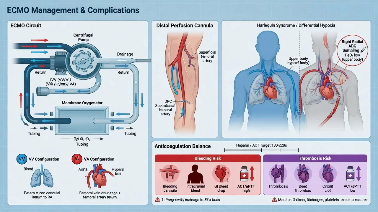
Blood contacting the artificial surfaces of the circuit activates clotting — without anticoagulation the oxygenator clots within hours. The counterweight is that bleeding is already the leading ECMO complication. The net result is a deliberately MODERATE anticoagulation target, lower than for surgical bypass.[2]
Daily ECMO anticoagulation and circuit care
Anticoagulant
Unfractionated heparin (UFH) infusion is standard — titratable, reversible with protamine, monitorable. Target anti-Xa 0.3–0.7 IU/mL OR aPTT ~50–70 s (~1.5x baseline). ACT (activated clotting time) ~1.5x baseline is a bedside surrogate. Direct thrombin inhibitors (bivalirudin, argatroban) reserved for heparin-induced thrombocytopenia.
Haemostasis monitoring
Daily anti-Xa AND aPTT (anti-Xa is heparin-specific and unaffected by the acute-phase fibrinogen rise that falsely prolongs aPTT). Full blood count (platelet count falls with circuit consumption and HIT), fibrinogen, PT/INR, D-dimer. Thromboelastography (TEG/ROTEM) where available to disentangle clotting-factor vs platelet contributions.
Transfusion thresholds
On ECMO the targets are higher than for ordinary ICU patients: Hb >80 g/L (DO2 is marginal; transfusion improves oxygen delivery), platelets >50 × 10⁹/L (and >80 if bleeding or post-procedure), fibrinogen >1.5–2.0 g/L. Avoid over-transfusion of plasma if not bleeding (volume, TRALI).
Circuit inspection
Inspect the oxygenator every shift for fibrin strands and clots (especially at the blood-inlet face), monitor pre-/post-membrane pressure GRADIENT (a rising ΔP signals clot build-up — plan an oxygenator/circuit change before it occludes), check for discolouration (dark pre-membrane venous blood is normal; pink post-membrane is oxygenated).
When bleeding occurs
Hold/reduce heparin, correct thrombocytopenia and coagulopathy (platelets, FFP, cryoprecipitate for fibrinogen), localise and surgically control the source (cannulation site, intrathoracic, GI). Antifibrinolytics (TXA) selectively. For life-threatening bleeding, stop heparin entirely and accept higher thrombosis risk, or switch to a heparin-free protocol with circuit flush.
Detailed complications — a systems approach
Beyond the headline bleeding and limb ischaemia, ECMO produces a recognisable constellation of complications driven by anticoagulation, the circuit, the cannulae and critical illness itself.[6][2]
Bleeding
#1 complication
- Reported in 30–50% of runs; intracranial haemorrhage 2–5% (often catastrophic)
- Mechanism: therapeutic heparin + circuit-induced platelet dysfunction + acquired von Willebrand (high shear in pump) + coagulopathy of critical illness
- Sites: cannulation (most common), surgical, GI, pulmonary, intracranial
- Mitigation: moderate (not full) heparin target; meticulous cannulation; minimise vascular access; transfuse to targets; surgical control of surgical bleeds
Thrombosis
Circuit + patient
- Oxygenator clotting (rising ΔP), pump-head thrombus, venous thromboembolism, intracardiac thrombus (with LV distension/stasis)
- Monitor ΔP, D-dimer trend, fibrinogen consumption; inspect oxygenator face
- Manage: increase heparin if safe, exchange oxygenator/circuit if obstructing
Haemolysis
Mechanical RBC injury
- Mechanical shear in the pump, high negative suction on drainage, kinking, clot in oxygenator
- Detect: rising plasma-free haemoglobin, LDH, indirect bilirubin, haemoglobinuria (dark urine), falling haemoglobin
- Manage: check for kinking/obstruction, reduce excessive negative suction, lower pump speed if possible, exchange circuit if severe — ongoing haemolysis causes acute kidney injury and drives transfusion
Limb ischaemia (VA-femoral)
Cannula obstructs leg
- Large femoral arterial cannula occludes common femoral artery → ischaemic leg
- Risk reduced from >70% to <10% by prophylactic distal perfusion cannula
- Monitor: distal pulses (Doppler), capillary refill, limb temperature, continuous distal pulse oximetry, limb lactate
- If ischaemia: check distal perfusion cannula patency, surgical exploration/embolectomy, fasciotomy if compartment syndrome, reconfigure cannulation
Infection
Line + critical illness
- Cannula-related bloodstream infection, ventilator-associated pneumonia, catheter-associated UTI
- Risk rises with run duration (especially >14 days)
- Surveillance cultures, antimicrobial stewardship, daily review of line necessity, aseptic cannula-site care, VAP bundle
Acute kidney injury
Common, multifactorial
- From the underlying shock/sepsis, haemolysis, microemboli, nephrotoxins
- 20–40% of ECMO patients need continuous renal replacement therapy (often run in-line with the ECMO circuit)
- Most AKI is reversible if the underlying illness resolves; persistent AKI portends a worse prognosis
Neurological
Stroke + bleed
- Ischaemic stroke (thromboembolism) and intracranial haemorrhage (anticoagulation) each 2–5%; combined neurological events are a leading cause of death on ECMO
- Daily neuro exam; low threshold for CT head if any change (sedation hold to assess)
- The trade-off between anticoagulation (prevents clot/stroke) and bleeding (ICH) is the central tension of ECMO management
Weaning from ECMO
Weaning is the deliberate, monitored reduction of ECMO support to test whether the native heart (VA) or lungs (VV) can sustain the patient alone. It is NOT simply "turning the machine off" — an abrupt stop in a non-recovered patient is fatal. Weaning VV and VA follow different logics.[2][7]
Weaning VV-ECMO — test the lungs
VV-ECMO weaning protocol
1. Establish lung recovery
Improving lung compliance on the ventilator, improving PaO2/FiO2, falling PEEP requirement, resolving infiltrates on imaging, improving driving pressure, and resolution of the underlying cause (e.g. pneumonia treated, fluid offloaded). The patient should be on lung-protective settings throughout.
2. Reduce sweep gas, NOT blood flow
The weaning lever for VV-ECMO is the SWEEP GAS (CO2 clearance). Stepwise reduce sweep flow while keeping pump blood flow constant. If PaCO2 and pH remain acceptable, continue. Reducing blood flow does not wean oxygenation reliably (recirculation, mixing) — sweep is the cleaner test.
3. The trial-off
When sweep is at minimal flow, disconnect the sweep gas (cap the oxygenator) for 30–60 min on full ventilator support. Maintain pump blood flow (no flow = stasis = clot). Serial arterial blood gases at baseline, 15, 30, 60 min. Pass criteria: PaO2 >60 mmHg, PaCO2 stable, pH >7.30, SpO2 >90% on FiO2 ≤0.5, tolerable ventilator pressures.
4. Decannulation
If the trial-off is tolerated, clamp the circuit, stop anticoagulation (reverse heparin if needed), remove cannulae with manual compression or percutaneous closure, and confirm haemostasis. Continue lung-protective ventilation and watch for rebound hypercapnia.
Weaning VA-ECMO — test the heart
VA-ECMO weaning protocol
1. Establish cardiac recovery
Improving native cardiac output (rising mixed venous saturation, rising pulse-pressure variation of native arterial trace, improving echocardiographic EF and aortic-valve opening), resolving arrhythmia, falling inotrope/vasopressor requirement, resolving lactataemia. The underlying cause (e.g. revascularised MI, treated myocarditis) must be addressed.
2. Stepwise flow reduction under echo
Reduce pump flow in 0.5 L/min steps from full support down to ~1–1.5 L/min (about 1–1.5 L/min is the minimum safe flow to avoid circuit stasis/clotting). At each step assess perfusion (MAP, lactate, SvO2, urine output) and perform echocardiography.
3. The trial-off
At low flow, clamp the circuit for a brief period (or run at minimal flow) and assess: native cardiac output, LV/RV function on echo (LV ejection fraction, aortic valve opening duration, RV size/function), haemodynamics (MAP, CVP, PA pressure), and perfusion markers. Atrial and ventricular function must be adequate with the circuit off. TEE is preferred for the weaning echo.
4. Pass criteria and decannulation
Pass: stable MAP without new inotropes, LV EF >20–25% with aortic valve opening, no worsening pulmonary hypertension, SvO2 >60%, lactate stable/falling, adequate urine output. Then remove the distal perfusion cannula and arterial cannula (surgical or percutaneous closure — femoral artery repair often needed), then the venous cannula. Reverse anticoagulation.
5. If failed trial
Return to full or intermediate support, optimise the failing ventricle (inotropes, afterload reduction, treat arrhythmia, consider VAD/transplant referral), and re-attempt in 24–72 h. Repeated failed weans → durable VAD or transplant evaluation, or palliation if the disease is irreversible.
Weaning criteria compared
VV-ECMO wean
Lung recovery
- Lever: reduce SWEEP gas flow (CO2 clearance)
- Pump blood flow kept ON during trial (prevents circuit clotting)
- Pass criteria: PaO2 >60 mmHg on FiO2 ≤0.5, stable PaCO2/pH, tolerable ventilator pressures over 30–60 min
- Decannulate after a sustained successful sweep-off trial
VA-ECMO wean
Heart recovery
- Lever: reduce PUMP BLOOD FLOW (circulatory support) stepwise to 1–1.5 L/min
- Echocardiography mandatory at each step (LV EF, aortic valve opening, RV function)
- Pass criteria: stable MAP, LV EF >20–25%, aortic valve opening, SvO2 >60%, lactate stable/falling
- Failed wean → durable VAD, transplant, or palliation decision
Key ECMO trials
EOLIA (Combes, NEJM 2018)
Multicentre RCT: 249 adults with very severe ARDS in 64 international ICUs
Population: Very severe ARDS (PaO2/FiO2 <50 for >3 h, or <80 for >6 h, or pH <7.25 with PaCO2 ≥60 for >6 h) despite optimised ventilation
Key finding
Stopped early for futility at interim analysis. 28-day mortality 35% (ECMO) vs 43% (control) — NOT statistically significant (RR 0.76, 95% CI 0.55–1.04, p=0.09). However, the pre-specified SECONDARY composite endpoint of death by day 60 or treatment failure FAVOURED ECMO, and a post-hoc Bayesian reanalysis (Goligher, JAMA 2018) estimated a high posterior probability (>85–99%) that ECMO was beneficial across a range of priors.
Practice change
The trial every candidate must defend in BOTH directions. On its primary endpoint EOLIA was borderline/negative (stopped early), but the secondary composite, the cross-over of 28% of control patients to rescue ECMO, and the Bayesian reanalysis collectively SUPPORT VV-ECMO for very severe ARDS. Modern practice: use VV-ECMO for the very severe band (PaO2/FiO2 <50–80 refractory) at a high-volume ECMO centre.
CESAR (Peek, Lancet 2009)
Multicentre RCT: 180 adults with severe but potentially reversible respiratory failure in the UK
Population: Adults with severe reversible adult respiratory failure (Murray score ≥3, or pH <7.20)
Key finding
Significantly better outcome with transfer-to-ECMO-centre: 37% dead/severely disabled vs 53% (RR 0.69, p=0.03). Importantly, only 75% of the transfer group actually RECEIVED ECMO — the benefit was partly that of being managed at a specialist centre.
Practice change
The original adult ECMO evidence base. Criticised for the 'transfer-to-centre' design (tests a service model, not the device) and single-arm nature of the intervention. Superseded by EOLIA for device-specific evidence, but still the only positive adult RCT and the basis for establishing ECMO services.
Noah H1N1 (Noah, JAMA 2011)
Observational cohort with propensity-matched controls: 80 ECMO-referred vs 80 matched severe H1N1 patients
Population: Severe H1N1 influenza A ARDS during the 2009-10 pandemic (UK)
Key finding
Hospital mortality 24% (ECMO-referred) vs 48% (matched controls) — although propensity matching is imperfect for this indication. ECMO was widely deployed for H1N1 with registry survival ~56% (ELSO data), driving a global expansion of adult ECMO capacity.
Practice change
Not an RCT, but the largest experience that established ECMO as a credible adult therapy during the H1N1 pandemic and underpinned capacity-building. Paired with CESAR, formed the pre-EOLIA evidence base for adult respiratory ECMO.
ELSO Registry (Thiagarajan, ASAIO J 2019)
International registry report: >78,000 adult and paediatric ECMO runs reported to the Extracorporeal Life Support Organization
Population: All ECMO runs reported worldwide (respiratory, cardiac, ECPR)
Key finding
Overall survival to discharge: adult respiratory ECMO ~59%, adult cardiac ECMO ~41%, adult ECPR ~29%. Paediatric survival higher. Bleeding the commonest complication; intracranial haemorrhage 2–5%.
Practice change
The benchmark epidemiology of ECMO. Survival numbers to quote in the exam: VV-ECMO ~50–60%, VA-ECMO ~40–50%, ECPR ~25–30%. Outcomes have improved modestly over time with centre volume and protocolisation.
Brodie & Bacchetta review (NEJM 2011)
Narrative review of the physiology, indications and evidence for adult respiratory ECMO
Population: Adults with severe respiratory failure
Key finding
Synthesised the post-H1N1, post-CESAR case for adult VV-ECMO, defined patient-selection criteria (reversibility, no terminal comorbidity), and introduced the modern concept of separating O2 delivery (blood flow) from CO2 clearance (sweep gas) and of ECCO2R for hypercapnic failure at low flow.
Practice change
The canonical reference for the physiology and selection logic of adult respiratory ECMO. Cite for circuit physiology, the blood-flow/sweep-gas dissociation, and the reversibility principle.
Abrams cardiac ECMO review (Intensive Care Med 2018)
Narrative review of VA-ECMO for cardiogenic shock and cardiac arrest
Population: Adults with refractory cardiogenic shock and refractory cardiac arrest (ECPR)
Key finding
Defined the modern indications (refractory cardiogenic shock, ECPR), the femoral configuration and its hazards (Harlequin/north-south syndrome, LV distension), and the rationale for LV venting (IABP, Impella). Emphasised that VA-ECMO is a BRIDGE (to recovery, VAD, transplant) and not a destination therapy.
Practice change
The canonical reference for adult VA-ECMO and ECPR physiology and complications. Cite for Harlequin, LV distension, the bridge concept, and ECPR selection.
ECPR — extracorporeal CPR
ECPR is the rapid institution of VA-ECMO during ongoing cardiopulmonary resuscitation in refractory cardiac arrest, most credibly in witnessed, shockable-rhythm in-hospital arrest cannulated within ~60 min of collapse. The rationale: ECMO restores systemic perfusion (especially cerebral and coronary) more effectively than manual chest compressions, and buys time to reverse the precipitant (PCI for MI, rewarm in hypothermia, correct toxin, ablate arrhythmia focus).[7][2]
ECPR — selection, insertion, and the evidence
Selection (who benefits)
Witnessed arrest, bystander/monitor CPR, initial shockable rhythm (VF/pVT), no-flow time <5 min (collapse to CPR), low-flow time <60 min (CPR to cannulation), reversible cause, no pre-existing terminal comorbidity, age typically <70. The largest experience is in refractory in-hospital arrest; out-of-hospital ECPR is more selective and resource-intensive.
Cannulation under CPR
Percutaneous femoro-femoral Seldinger cannulation performed during ongoing compressions (a dedicated cannulation team while the arrest team continues CPR). Ultrasound-guided venous puncture, distal perfusion cannula placed once circulation restored. Target flow 2–4 L/min as soon as possible.
Post-arrest management
Targeted temperature management (32–36°C), percutaneous coronary intervention if MI-driven, lung-protective ventilation, avoid hyperoxia (PaO2 targets to minimise reperfusion/oxidative injury), prognosticate at ≥72 h with multimodal testing. Avoid premature withdrawal of life-sustaining therapy — neurological recovery can lag.
The evidence
No definitive RCT (trials such as INCEPTION, Prague OHCA, EROCA are heterogeneous and largely negative or equivocal for overall survival). Best survival signals are in highly selected shockable-rhythm patients cannulated early (>30–40% neurologically intact survival in some centres vs near-zero for refractory arrest without ECPR). ECPR is resource-intensive and should be delivered in experienced centres with established protocols.
ECMO programme — referral, retrieval, and the centre-volume effect
ECMO is not a solo bedside procedure — it is a system. Outcomes correlate with centre volume (more runs/year → lower mortality), and the safe transfer of an unstable patient to an ECMO centre is itself a specialised skill. The CESAR and Noah data both captured a "centre effect" (better outcomes at specialist centres) that is entangled with the device effect.[1][4]
The ECMO referral and retrieval pathway
Early referral
Refer BEFORE the patient crashes — a referral made at the "consider ECMO" threshold (PaO2/FiO2 <80 refractory, or cardiogenic shock on two inotropes) is more useful than one made at cardiac arrest. The ECMO centre triages by phone using standard criteria (reversibility, comorbidity, contraindications).
Retrieval team
A specialist retrieval team (intensivist/perfusionist/nurse) brings the circuit and cannulae to the referring hospital, cannulates the patient, establishes ECMO, and transfers back by road/rotary-wing with ECMO running ("ECMO-to-go"). Avoids the risk of moving an unstable, non-ECMO patient.
Inter-hospital transfer
On ECMO the patient is stabilised (perfused, oxygenated) and transfers more safely than pre-cannulation. Requires a dedicated transport ventilator, pump (battery/vehicle power), oxygen supply, monitoring, and a team trained in mobile ECMO.
Daily multidisciplinary review
At the ECMO centre, daily MDT review (intensivist, perfusionist, surgeon, specialist nurse, pharmacist, physiotherapy, dietitian, ethics) sets the goal: bridge to recovery, bridge to VAD/transplant, or withdrawal if irreversible. The single most important question each day is "is the underlying disease recovering, and if not, what is the exit strategy?"
Contraindications — refined
The contraindication list is best understood through the question "is this a reversible, single-organ (or recoverable multi-organ) problem in a patient with a meaningful prognosis?" If no, ECMO is futile.[2]
Absolute
Do NOT cannulate
- Irreversible brain injury (severe hypoxic-ischaemic encephalopathy after prolonged arrest with poor prognostic signs)
- Terminal malignancy or end-stage disease with no prospect of meaningful recovery
- Severe irreversible chronic organ failure (end-stage COPD not a transplant candidate, decompensated cirrhosis, NYHA IV heart failure not a VAD/transplant candidate)
- Unwitnessed out-of-hospital cardiac arrest with prolonged downtime and asystole (ECPR futile)
- Patient/advance directive declining life-sustaining therapy
Relative
Case-by-case
- Age >75 (weigh with physiological rather than chronological age)
- Severe peripheral vascular disease (cannulation difficulty — consider central/axillary)
- Morbid obesity (cannulation and ventilation difficulty, transport challenges)
- Established severe coagulopathy or active major bleeding (high bleeding mortality on ECMO)
- Prolonged high-pressure mechanical ventilation (>7–10 days — barotrauma, lung fibrosis, lower reversibility)
- Mechanical ventilation >7 days plus multiorgan failure (prognosis falls sharply)
- Immunocompromise (weigh against reversibility and transplant candidacy)
Exam practice
SAQ — VV-ECMO for severe ARDS (EOLIA scenario)
10 minutes · 10 marks
A 45-year-old previously well man is admitted to the ICU with influenza A pneumonia and severe ARDS. Despite lung-protective ventilation (Vt 6 mL/kg PBW, plateau pressure 32 cmH2O, PEEP 14, FiO2 1.0), proning for 18 h/day, and neuromuscular blockade, his PaO2/FiO2 is 65, pH 7.21, PaCO2 68. He is on noradrenaline 0.25 mcg/kg/min for MAP 68, lactate 2.4, in sinus rhythm, and his echocardiogram shows a normal LV with no pulmonary hypertension. The regional ECMO centre has been contacted and is sending a retrieval team.
SAQ — VA-ECMO complications (Harlequin and LV distension)
8 minutes · 8 marks
A 60-year-old man is on femoral VA-ECMO (5 L/min) for cardiogenic shock after a large anterior STEMI, with concurrent pulmonary oedema. His femoral arterial line shows SpO2 99%, but his right-radial arterial line shows SpO2 86% and the cerebral NIRS has fallen. His pulmonary artery pressure has risen from 45 to 62 mmHg and new pulmonary oedema is visible on chest X-ray.
Clinical pearls — the exam-exhaustive set
Red flags — the lethal pitfalls
Prognosis
ECMO outcomes are dominated by the indication and the underlying disease's reversibility. The ELSO registry reports survival to discharge of approximately 59% for adult respiratory (VV) ECMO, 41% for adult cardiac (VA) ECMO, and 29% for adult ECPR.[6] Within respiratory ECMO, near-fatal asthma is the standout indication (~80–90% survival) because the disease is single-organ and reversible, whereas severe ARDS sits around 50–60% and pulmonary fibrosis is rarely worth cannulating. Within cardiac ECMO, fulminant myocarditis (often recoverable over days-weeks) fares better than post-cardiotomy failure or ECPR. Mortality on ECMO is driven by bleeding (especially intracranial), neurological injury, uncontrolled sepsis, irreversible multiorgan failure, and failure to recover the index organ (the bridge-to-nowhere problem). The two modifiable determinants of outcome are early referral to a high-volume specialist centre and rigorous daily reassessment of the exit strategy (recovery, durable VAD, transplant, or palliation).[1][4][2]
ECMO — the numbers that matter
References
- [1]Peek GJ, Mugford M, Tiruvoipati R, et al. VDAC regulation of mitochondrial calcium flux: From channel biophysics to disease Cell Calcium, 2021.PMID 33529977
- [2]Extracorporeal Life Support Organization (ELSO). Notum palmitoleoyl-protein carboxylesterase regulates Fas cell surface death receptor-mediated apoptosis via the Wnt signaling pathway in colon adenocarcinoma Bioengineered, 2021.PMID 34402722
- [3]Combes A, Hajage D, Capellier G, et al. A Single Nucleotide Mutation of the IspE Gene Participating in the MEP Pathway for Isoprenoid Biosynthesis Causes a Green-Revertible Yellow Leaf Phenotype in Rice Plant Cell Physiol, 2018.PMID 29893915
- [4]Noah MA, Peek GJ, Finney SJ, et al. Three-dimensional architecture of grana and stroma thylakoids of higher plants as determined by electron tomography Plant Physiol, 2011.PMID 21224341
- [5]Brodie D, Bacchetta M. Mycophenolate versus azathioprine as maintenance therapy for lupus nephritis N Engl J Med, 2011.PMID 22087680
- [6]Thiagarajan RR, Barbaro RP, Rycus PT, et al. Retraction Chem Biol Drug Des, 2019.PMID 31342669
- [7]Abrams D, Combes A, Brodie D. Novel hits for acetylcholinesterase inhibition derived by docking-based screening on ZINC database J Enzyme Inhib Med Chem, 2018.PMID 29651876