ICU · Haematology & coagulation
Haematology and Coagulation in the ICU
Also known as DIC · Thrombocytopenia · Coagulopathy · VTE prophylaxis · Massive transfusion · Thromboelastography · Anticoagulation reversal
The haematology and the coagulation in the ICU span the physiology of the haemostasis (the cascade — the intrinsic, the extrinsic, the common; the cell-based model — the initiation, the amplification, the propagation), the platelet function (the adhesion, the activation, the aggregation), the natural anticoagulants (the antithrombin, the protein C/S, the TFPI), the fibrinolysis (the plasmin, the D-dimer), the laboratory tests (the PT, the aPTT, the TT, the fibrinogen, the D-dimer, the TEG/ROTEM), the thrombocytopenia (the DIC, the HIT, the TTP), the coagulopathy (the liver, the dilutional, the anticoagulant), the venous thromboembolism prophylaxis, the massive transfusion, and the anticoagulation reversal. This topic builds the examiner's framework on the cascade and the cell-based model, the bleeding disorders by the test pattern, the DIC (the overconsumption), the HIT (the heparin-induced), the TTP (the urgent plasma exchange), the VTE prophylaxis (the LMWH), and the reversal (the warfarin, the DOAC).
On this page & tools
Your progress
Saved locally on this device.
Target exams
Overview & definition
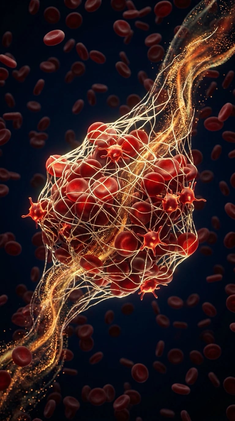
The haematology and the coagulation in the ICU span the thrombocytopenia, the coagulopathy, the venous thromboembolism (the VTE), and the massive transfusion. Each is common, each carries the risk (the bleeding OR the thrombosis), and each has a specific, the evidence-based management.[1][1]
The coagulation cascade: the classical "waterfall" model
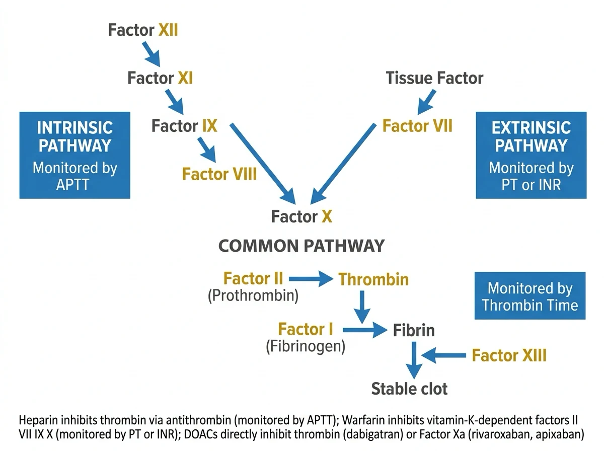
The classical cascade (Macfarlane 1964; Davie and Ratnoff 1964) divides the coagulation into the intrinsic, the extrinsic, and the common pathways — each a chain of the zymogen-to-serine-protease activations converging on the thrombin. The model is an IN-VITRO artefact of how the laboratory tests are performed (the PT, the aPTT), NOT a faithful description of the in-vivo haemostasis — but it remains the framework the examiners expect, and it maps cleanly onto the laboratory screen.[1]
The intrinsic pathway (the contact activation, the aPTT). Triggered in vitro by the surface contact. The Factor XII (the Hageman factor) auto-activates on the negatively-charged surface to the XIIa; the XIIa activates the XI to the XIa; the XIa activates the IX to the IXa; and the IXa, with its cofactor the VIIIa, assembles on the activated platelet surface to activate the X to the Xa. The deficiencies present with an isolated prolonged aPTT — but the XII deficiency bleeds NOT (it is a redundant contact activator), while the XI deficiency bleeds mildly and the VIII (the haemophilia A) and the IX (the haemophilia B) bleed severely. [1]
The extrinsic pathway (the tissue factor, the PT). The physiologic initiator. The tissue factor (the TF — an integral membrane protein on the subendothelial fibroblasts, the smooth muscle, and the adventitial cells, normally separated from the blood by the intact endothelium) binds the trace circulating VIIa, and the TF–VIIa complex activates both the X to the Xa and the IX to the IXa. This is the ONLY pathway that initiates the coagulation in vivo. Its defects (the VII deficiency, the warfarin, the liver failure, the vitamin K deficiency, the early DIC) present with an isolated prolonged PT. [1]
The common pathway. The Xa, with its cofactor the Va, forms the prothrombinase complex that converts the prothrombin (II) to the thrombin (IIa). The thrombin then cleaves the fibrinogen (I) to the fibrin, AND it activates the XIII (which cross-links the fibrin to stabilise the clot against the premature lysis). Critically, the thrombin is also the master POSITIVE FEEDBACK activator — it activates the platelets, the V, the VIII, and the XI, amplifying its own generation. The common-pathway defects (the X, the V, the II, the I) prolong BOTH the PT and the aPTT — except the XIII deficiency, which has NORMAL routine labs and a DELAYED bleeding (the umbilical stump, the delayed wound oozing). [1]
Intrinsic pathway
Surface/contact — the aPTT
- Factors XII → XI → IX → VIII → X (with the cofactor the VIIIa on the platelet surface).
- Initiated in vitro by the contact (the glass, the kaolin, the ellagic acid).
- Measured by the aPTT (the activated partial thromboplastin time).
- Factor XII deficiency: the prolonged aPTT but NO bleeding (redundant contact activator).
- Factor XI deficiency: the mild bleeding. Factor VIII (haemophilia A) and IX (haemophilia B): the severe bleeding.
Extrinsic pathway
Tissue factor — the PT/INR
- Factor VII + the tissue factor (the TF) → Xa (and the IXa).
- The physiologic initiator of the in-vivo haemostasis (Hoffman cell-based model).
- Measured by the PT and reported as the INR.
- The VII has the shortest half-life (6 hours) — the first to fall in the warfarin and the vitamin K deficiency.
- Prolonged by the warfarin, the liver failure, the vitamin K deficiency, the early DIC.
Common pathway
Prothrombinase → thrombin → fibrin
- X + Va (the prothrombinase) → II → thrombin (IIa); thrombin → fibrinogen (I) → fibrin.
- Thrombin activates the XIII (the cross-linking) AND the V, the VIII, the XI, the platelets (the positive feedback).
- Measured by BOTH the PT and the aPTT (and the thrombin time for the final step).
- Factor XIII deficiency: the NORMAL PT and aPTT with the delayed bleeding.
- Prolonged by the DIC, the massive transfusion, the liver failure, the anticoagulants.
The classical coagulation cascade (the examiner's diagram)
Intrinsic (the contact) pathway
XII → XIIa → XI → XIa → IX → IXa → (with the VIIIa) → X. The aPTT measures this limb. Triggered in vitro by the surface contact; in vivo the XII is redundant (the XII deficiency does not bleed).
Extrinsic (the tissue factor) pathway
The TF (on the subendothelium) + the VIIa → activates the X to the Xa AND the IX to the IXa. The PT measures this limb. This is the physiologic initiator in vivo.
Common pathway — the prothrombinase
The Xa + the Va convert the prothrombin (II) to the thrombin (IIa) on the activated platelet phospholipid surface. The PT and the aPTT both measure this convergence.
Thrombin generation and the positive feedback
The thrombin cleaves the fibrinogen (I) to the fibrin, activates the XIII (the cross-linking for the clot stability), AND activates the V, the VIII, the XI, and the platelets — amplifying its own generation in a positive-feedback burst.
Fibrin cross-linking and the clot
The XIIIa cross-links the fibrin monomers into the stable polymer resistant to the premature lysis. The clot incorporates the platelets (the white thrombus) and, in the venous system, the red cells (the red thrombus).
The cell-based model of haemostasis
The cascade model fails clinically — it cannot explain why the XII deficiency does not bleed, nor why the VII deficiency bleeds despite a robust intrinsic system. The cell-based model of Hoffman and Monroe (2001) replaces the fluid-phase cascade with a cell-surface process in three overlapping phases, each on a different cell type. The thrombin is generated in the small amounts by the TF-bearing cells, then in the BURST on the activated platelet surface.[2]
1. Initiation (the TF-bearing cell). The vascular injury exposes the TF-bearing cells (the subendothelial fibroblasts, the smooth muscle, the adventitial cells). The trace circulating VIIa binds the TF, generating the small amounts of the IXa and the Xa on the TF-cell surface. The Xa, with the Va, makes a TRACE amount of the thrombin — insufficient to form a clot, but sufficient to activate the platelets, the V, the VIII, the XI, and the XIII. The TFPI then shuts the TF–VIIa complex down (the Xa-dependent feedback) — the TF pathway is an INITIATOR and not a sustained generator. [1]
2. Amplification (the platelet surface). The trace thrombin activates the platelets that have adhered to the site of the injury. The activated platelets undergo the granule release and the shape change, expose the negatively-charged phosphatidylserine (the procoagulant surface), and bind the Va and the VIIIa. The cofactors are now loaded onto the platelet surface, ready for the propagation. [1]
3. Propagation (the activated platelet surface). The IXa (generated in the initiation, and reinforced by the XIa on the platelet surface) binds the VIIIa on the activated platelet; the intrinsic tenase (the IXa–VIIIa) activates the X to the Xa far more efficiently than the TF–VIIa ever did. The Xa then binds the Va to form the prothrombinase complex, generating the BURST of thrombin that converts the fibrinogen to the fibrin and stabilises the clot. This propagation phase is where the haemophilias fail — without the VIIIa or the IXa, the thrombin burst is attenuated, the clot is friable, and the patient bleeds (late, into the joints and the muscles, NOT immediately). [1]
Why this model matters clinically. The factor XII deficiency does not bleed because the XII is not on the propagation pathway. The haemophilias bleed because the VIII and the IX are essential for the propagation on the platelet surface. The recombinant VIIa and the activated PCC (the FEIBA) bypass the blocked intrinsic tenase and load the X directly — the basis of the bypass therapy in the haemophilias with the inhibitors. The von Willebrand disease impairs the initial platelet adhesion AND the carriage of the VIII (the low VIII). The model explains the viscoelastic testing: the TEG/ROTEM R-time reflects the initiation, the K and the alpha angle the propagation and the fibrinogen, the MA/MCF the platelet contribution to the clot strength.[2]
Cascade model
The classical fluid-phase description
- The intrinsic (XII→XI→IX→VIII→X) and the extrinsic (VII→TF→X) converge on the common X→thrombin→fibrin.
- An in-vitro description of the laboratory tests (the PT, the aPTT).
- Cannot explain the non-bleeding XII deficiency nor the bleeding VII deficiency.
- Useful for the exam diagram and the laboratory interpretation.
Cell-based model
The Hoffman–Monroe in-vivo reality
- Three phases: the initiation (the TF cell), the amplification (the platelet), the propagation (the activated platelet).
- The TF pathway INITIATES; the intrinsic pathway PROPAGATES (sustains) the thrombin burst.
- Explains the non-bleeding XII and the bleeding VII/VIII/IX.
- Explains the bypass therapy (the rVIIa, the FEIBA) for the haemophilias with the inhibitors.
Platelet structure and function
The platelet is the first responder to the vascular injury. The primary haemostasis (the formation of the platelet plug) precedes the secondary haemostasis (the coagulation cascade). The platelet function has the three stages: the adhesion, the activation, and the aggregation.[1]
The platelet structure. The discoid anucleate fragment (2 to 3 micrometres), derived from the megakaryocyte cytoplasm. The surface bears the glycoprotein receptors (the GPIb-IX-V for the adhesion, the GPIIb/IIIa for the aggregation, the GPVI and the GPIa-IIa for the collagen). The cytoplasm holds the alpha-granules (the vWF, the fibrinogen, the factor V, the PF4, the P-selectin) and the dense-granules (the ADP, the ATP, the serotonin, the calcium). The canalicular system and the microtubules maintain the shape and provide the surface area for the reactions. [1]
Adhesion (the vWF and the GPIb). At the high shear (the arterioles, the capillaries), the von Willebrand factor (the vWF — released from the endothelial Weibel-Palade bodies and the platelet alpha-granules, and stored in the subendothelial matrix) binds the exposed subendothelial collagen and unfolds under the shear. The platelet surface GPIb-IX-V complex binds the vWF, tethering the platelet to the site of injury (the rolling, then the firm arrest). This is the Bernard-Soulier defect (the GPIb deficiency — the giant platelets, the thrombocytopenia, the bleeding) and the von Willebrand disease (the vWF deficiency or the dysfunction). At the low shear, the platelet also binds directly via the GPVI and the GPIa-IIa (the collagen receptors). [1]
Activation (the granule release, the shape change, the TxA2). The adhesion triggers the intracellular signalling (the calcium mobilisation, the G-protein-coupled-receptor activation, the phosphatidylserine externalisation). The platelet undergoes the granule exocytosis — the alpha-granules release the vWF, the fibrinogen, the factor V, the PF4; the dense-granules release the ADP, the serotonin, the calcium. The cyclo-oxygenase-1 converts the arachidonic acid to the thromboxane A2 (the target of the aspirin — the irreversible COX-1 inhibition for the life of the platelet). The ADP and the TxA2 are the secondary agonists that recruit and activate the passing platelets. The shape change (the disc to the sphere with the long pseudopodia) increases the surface area for the reactions. [1]
Aggregation (the GPIIb/IIIa and the fibrinogen bridge). The activation transforms the GPIIb/IIIa from the low-affinity to the high-affinity receptor for the fibrinogen (and the vWF). The bivalent fibrinogen bridges the GPIIb/IIIa of the adjacent platelets, cross-linking them into the primary haemostatic plug. This is the Glanzmann thrombasthenia (the GPIIb/IIIa deficiency — the normal count, the absent aggregation, the severe mucocutaneous bleeding) and the pharmacologic target of the abciximab, the tirofiban, and the eptifibatide (the IV GPIIb/IIIa antagonists in the PCI). [1]
Adhesion (the GPIb–vWF)
The primary tethering
- The vWF binds the exposed collagen; the platelet GPIb-IX-V binds the vWF.
- Critical at the high shear (the arterioles, the capillaries).
- Bernard-Soulier syndrome: the GPIb deficiency (the giant platelets, the thrombocytopenia, the bleeding).
- Von Willebrand disease: the vWF deficiency or the dysfunction (the most common inherited bleeding disorder).
Activation (the granules, the TxA2)
The recruitment and the surface exposure
- The granule release: the alpha (the vWF, the fibrinogen, the factor V, the PF4) and the dense (the ADP, the serotonin, the calcium).
- The COX-1 generates the thromboxane A2 — the target of the aspirin (the irreversible inhibition).
- The P2Y12 receptor for the ADP — the target of the clopidogrel, the ticagrelor, the prasugrel.
- The phosphatidylserine externalisation provides the procoagulant surface for the tenase and the prothrombinase.
Aggregation (the GPIIb/IIIa)
The fibrinogen cross-linking
- The high-affinity GPIIb/IIIa binds the bivalent fibrinogen that bridges the adjacent platelets.
- The final common pathway of the platelet aggregation.
- Glanzmann thrombasthenia: the GPIIb/IIIa deficiency (the normal count, the absent aggregation, the severe bleeding).
- The target of the abciximab, the tirofiban, the eptifibatide (the IV antagonists in the PCI).
The natural anticoagulant pathways
The coagulation, once initiated, must be LOCALISED and TEMPORAL — unchecked, it would consume the factors and occlude the vasculature (the DIC, the microvascular thrombosis, the organ failure). The three natural anticoagulant systems keep the clot at the site of the injury and confine its spread.[3]
The antithrombin (the ATIII, the serpin). The principal inhibitor of the thrombin (IIa) and the Xa (and the IXa, the XIa, the XIIa). The ATIII binds the active-site serine of the target protease and traps it in a 1:1 complex cleared by the liver. The heparin (and the endogenous heparan sulphate on the endothelium) amplifies the ATIII activity a THOUSAND-FOLD by the conformational change that exposes the reactive site — the molecular basis of the therapeutic heparin (the unfractionated, the LMWH) and the reason the ATIII deficiency causes the heparin resistance. The ATIII deficiency (the congenital, or the acquired — the nephrotic syndrome, the liver failure, the pre-eclampsia, the DIC, the sepsis) is a classical thrombophilia. The sepsis consumes the ATIII, contributing to the microvascular thrombosis of the septic coagulopathy.[3]
The protein C and the protein S (the thrombomodulin–protein C pathway). The thrombin, when it binds the endothelial thrombomodulin, FLIPS from a procoagulant to an anticoagulant enzyme. The thrombin–thrombomodulin complex (with the endothelial protein C receptor, the EPCR) activates the protein C; the activated protein C (with its cofactor the protein S) proteolytically inactivates the Va and the VIIIa, braking the prothrombinase and the intrinsic tenase. The protein C and the protein S are the vitamin-K-dependent factors — hence the warfarin is transiently PROTHROMBOTIC at the initiation (the protein C falls first, before the II, the VII, the IX, and the X), the mechanism of the warfarin-induced skin necrosis in the undiagnosed protein C deficiency. The protein C deficiency and the protein S deficiency are the inherited thrombophilias; the factor V Leiden resists the activated protein C inactivation (the most common inherited thrombophilia in the European populations).[3]
The tissue factor pathway inhibitor (the TFPI). The endothelium-bound and the lipoprotein-bound TFPI directly inhibits the TF–VIIa complex — but ONLY after it has bound and inhibited the Xa (the Xa-dependent, the two-step feedback). The TFPI is the brake on the extrinsic pathway and the reason the TF pathway is an INITIATOR and not a sustained generator: once the Xa is made, the TFPI quenches the TF–VIIa, and the sustained thrombin generation must come from the intrinsic tenase on the activated platelet surface. The TFPI deficiency (rare) causes a thrombophilia; the recombinant TFPI (the tifacogin) was tested in the sepsis but did not improve the survival. [1]
Antithrombin (ATIII)
The serpin — the heparin cofactor
- Inhibits the thrombin and the Xa (and the IXa, the XIa, the XIIa) by the 1:1 trapping.
- The heparin/heparan sulphate amplifies the activity a thousand-fold.
- The ATIII deficiency: the congenital thrombophilia, or the acquired (the nephrotic, the liver, the DIC).
- The ATIII consumption in the sepsis contributes to the microvascular thrombosis and the heparin resistance.
Protein C and S
The thrombomodulin pathway
- The thrombin–thrombomodulin (with the EPCR) activates the protein C; the APC + the protein S inactivate the Va and the VIIIa.
- The vitamin-K-dependent — hence the warfarin is transiently prothrombotic (the skin necrosis in the protein C deficiency).
- The factor V Leiden resists the APC (the commonest inherited thrombophilia in the Europeans).
- The protein C/S deficiency: the inherited thrombophilias (the venous thrombosis, the purpura fulminans in the homozygote).
TFPI
The TF–VIIa brake
- Inhibits the TF–VIIa only AFTER binding the Xa (the Xa-dependent, the two-step feedback).
- Confines the TF pathway to the initiation; the propagation is taken over by the intrinsic tenase.
- The TFPI deficiency: a rare thrombophilia.
- The recombinant tifacogin tested in the sepsis without the survival benefit.
The fibrinolytic system
The clot, once formed, must be remodelled and ultimately removed for the vessel to recanalise. The fibrinolysis is the plasmin-mediated digestion of the fibrin, balanced against the inhibitors that prevent the runaway clot lysis.[1]
The plasmin. The plasminogen (the circulating zymogen, the liver-synthesised) is converted to the plasmin by the tissue plasminogen activator (the tPA, the endothelial release in response to the venous occlusion and the exercise) and the urokinase (the uPA, the urinary and the extravascular). The plasmin cleaves the fibrin (and, less specifically, the fibrinogen) into the fibrin degradation products — the most clinically important of which is the D-dimer, a cross-linked fragment whose presence PROVES that the thrombin AND the XIII AND the plasmin have all acted, i.e., that a real cross-linked clot was made and then broken. [1]
The inhibitors. The alpha-2-antiplasmin (the rapid serpin inhibitor of the FREE plasmin — once the plasmin is bound to the fibrin, the alpha-2-antiplasmin is slower to inhibit it, the fibrin protects the plasmin). The PAI-1 (the plasminogen activator inhibitor-1, the rapid inhibitor of the tPA — the raised in the obesity, the sepsis, the malignancy, contributing to the prothrombotic state). The TAFI (the thrombin-activatable fibrinolysis inhibitor, the carboxypeptidase that removes the C-terminal lysines from the partially-degraded fibrin and so reduces the plasminogen binding, down-regulating the fibrinolysis). The tranexamic acid (the TXA) and the aprotinin inhibit the fibrinolysis — the TXA by blocking the plasminogen (and the plasmin) binding to the fibrin via the lysine-binding sites; the aprotinin by the direct plasmin inhibition.[5]
The hyperfibrinolysis and the hypofibrinolysis. The pathological excess of the fibrinolysis (the trauma, the prostate and the uterine surgery, the cirrhosis with the impaired clearance of the tPA, the obstetric haemorrhage, the snake envenomation, the amyloidosis) causes the delayed bleeding from the dissolved clots — recognised on the TEG/ROTEM as the rapid clot lysis (the LY30 above 7.5 per cent, or the maximum lysis above 15 per cent). This is the rationale for the early TXA in the trauma (the CRASH-2) — the benefit is greatest within the first hour and reverses to harm after the third hour. The pathological deficiency of the fibrinolysis (the high PAI-1) contributes to the atherothrombosis and the recurrent VTE.[5][6][12]
The endothelium and the haemostatic balance
The endothelium is not a passive pipe — it is the active gatekeeper of the haemostatic balance, tilting between the antithrombotic (the healthy state) and the prothrombotic (the injury, the inflammation, the sepsis, the COVID-19). The healthy endothelium is constitutively ANTITHROMBOTIC: it expresses the thrombomodulin (the protein C activation), the heparan sulphate (the ATIII potentiation), the TFPI, the nitric oxide and the prostacyclin (the vasodilation and the platelet inhibition), and the CD39 (the ecto-ADPase that degrades the ADP). The injured or the inflamed endothelium FLIPS to the prothrombotic: it expresses the tissue factor, releases the vWF (the high-molecular-weight multimers from the Weibel-Palade bodies), exposes the subendothelial collagen, and sheds the glycocalyx. The sepsis and the systemic inflammation (the SIRS, the COVID-19) tip the balance toward the diffuse microvascular thrombosis (the sepsis-induced coagulopathy, the DIC).[11]
The thrombocytopenia in the ICU
The thrombocytopenia (the platelet count below 150 x 10^9/L) is common in the ICU (the sepsis, the drugs, the haemodilution, the consumption). The key question: is the patient bleeding or thrombosing?[1]
The DIC (the disseminated intravascular coagulation). The systemic activation of the coagulation by the sepsis, the trauma, the malignancy — the consumption of the platelets and the factors (the bleeding) AND the microvascular thrombosis (the organ failure). The labs: the low platelets, the prolonged PT/APTT, the low fibrinogen, the raised D-dimer. The treatment: the TREAT THE CAUSE, the supportive (the platelet and the FFP for the bleeding or the procedure, the cryoprecipitate for the fibrinogen below 1.5), and the heparin is NOT routinely used.[1][1]
The HIT (the heparin-induced thrombocytopenia). The IgG antibody against the heparin-PF4 complex — the platelet drop 5 to 10 days after the heparin exposure (or sooner if the previous exposure), with the THROMBOSIS (not the bleeding). The 4Ts score (the Thrombocytopenia, the Timing, the Thrombosis, the oTher cause) screens. The management: STOP ALL HEPARIN, switch to the argatroban or the bivalirudin (the direct thrombin inhibitor), confirm with the ELISA and the serotonin release assay, and the warfarin is NOT started until the platelet recovery.[1]
The TTP (the thrombotic thrombocytopenic purpura). The ADAMTS13 deficiency (the congenital or the autoimmune) — the microvascular platelet thrombi, the thrombocytopenia, the haemolysis, the organ failure. The treatment: the URGENT PLASMA EXCHANGE (the mortality if delayed). The steroids for the autoimmune. The caplacizumab (the anti-vWF) for the acquired.[1][1]
The coagulopathy
The coagulopathy in the ICU arises from the liver failure (the reduced synthesis), the massive transfusion (the dilution), the anticoagulant (the warfarin, the DOAC), and the vitamin K deficiency.[1]
The warfarin reversal. The vitamin K (10 mg IV, the slow infusion, the onset in 6 to 12 hours), the prothrombin complex concentrate (the PCC — the 4-factor, the 25 to 50 IU/kg, the immediate INR correction), and the FFP (the 15 mL/kg, the slower and the volume-loaded). The PCC is preferred over the FFP for the rapid reversal. The DOAC reversal: the idarucizumab for the dabigatran, the andexanet alfa for the anti-Xa (the apixaban, the rivaroxaban), the PCC as the alternative.[1][1]
The VTE prophylaxis
The ICU patient is the HIGH-RISK for the VTE (the immobility, the inflammation, the endothelial injury). The prophylaxis is the LMWH (the enoxaparin 40 mg SC daily, dose-adjusted for the renal function), or the unfractionated heparin (the 5000 units SC TDS) for the renal impairment. The mechanical (the IPC, the TEDS) for the contraindication to the anticoagulant. The early mobilisation. The intermittent pneumatic compression if the pharmacological is contraindicated (the active bleeding, the recent surgery).[1]
The massive transfusion
The massive transfusion (the over 10 units of the RBCs in 24 hours, or the over 1 blood volume) follows the damage-control principles (the 1:1:1 ratio of the plasma to the red cell to the platelet), the tranexamic acid, the calcium replacement (the citrate chelation), the warming, and the viscoelastic testing (the TEG or the ROTEM) to guide the component therapy.[1][1]
Management: the integrated approach
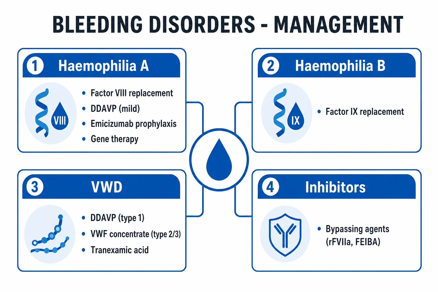
- The thrombocytopenia — classify (the DIC, the HIT, the TTP, the drugs); treat the cause.[1]
- The coagulopathy — the PCC for the warfarin; the idarucizumab/andexanet for the DOAC; the vitamin K; the component for the bleeding.[1]
- The VTE prophylaxis — the LMWH for all (unless contraindicated); the mechanical for the contraindication.[1]
- The massive transfusion — the 1:1:1, the TXA, the calcium, the warming, the TEG-guided.[1]
Monitoring
- The full blood count, the PT/INR, the APTT, the fibrinogen, the D-dimer — the routine.
- The TEG or the ROTEM — the viscoelastic for the active bleeding.
- The anti-Xa — for the LMWH monitoring (the high-risk, the renal impairment).[1]
Laboratory tests of haemostasis
The coagulation laboratory divides into the SCREENING tests (the PT, the aPTT, the TT), the QUANTITATIVE assays (the fibrinogen, the platelet count, the individual factor assays), the MARKERS (the D-dimer), the INHIBITOR screens (the mixing studies, the lupus anticoagulant), and the GLOBAL/viscoelastic assays (the TEG, the ROTEM). Each test interrogates a different part of the system.[1]
The prothrombin time (the PT, the INR). The tissue thromboplastin (the recombinant TF) and the calcium are added to the citrated plasma; the test measures the EXTRINSIC plus the COMMON pathway — the VII, the X, the V, the II, and the I. The normal is 11 to 13 seconds. Prolonged by the warfarin, the liver failure, the vitamin K deficiency, the DIC, the massive transfusion, and the direct Xa inhibitors (the apixaban, the rivaroxaban). The INR (the international normalised ratio) corrects the PT for the reagent sensitivity (the ISI) so the result is comparable across the laboratories — designed for the warfarin monitoring, NOT a reliable measure of the liver-synthetic function or the coagulopathy in the non-warfarin patient. The VII has the shortest half-life (6 hours) and is the first factor to fall in the warfarin and the vitamin K deficiency.[1]
The activated partial thromboplastin time (the aPTT). The phospholipid, the activator (the kaolin, the celite, the ellagic acid), and the calcium are added to the citrated plasma; the test measures the INTRINSIC plus the COMMON pathway — the XII, the XI, the IX, the VIII, the X, the V, the II, and the I. The normal is 25 to 35 seconds. Prolonged by the heparin (the UFH and the LMWH, though the LMWH affects the aPTT less), the haemophilias (the VIII, the IX), the von Willebrand disease, the DIC, the lupus anticoagulant, the direct thrombin inhibitor (the dabigatran, the argatroban, the bivalirudin), and the massive transfusion. The XII deficiency prolongs the aPTT but does NOT cause the bleeding. The aPTT is the basis of the heparin monitoring (the target of 1.5 to 2.5 times the control for the therapeutic UFH) — though the anti-Xa assay is preferred in the critical illness.[1]
The thrombin time (the TT). The thrombin is added directly to the plasma; the test measures ONLY the fibrinogen-to-fibrin conversion. The normal is 14 to 16 seconds. Prolonged by the heparin (the thrombin is inhibited — the single most sensitive test for the heparin contamination), the low fibrinogen (below 1 g/L) and the dysfibrinogenaemia, the DIC (the elevated FDPs interfere), and the dabigatran (the direct thrombin inhibitor — the TT is the most sensitive test for the dabigatran activity). The reptilase time (the batroxobin) is unaffected by the heparin and distinguishes the heparin contamination from the true hypofibrinogenaemia.[1]
The fibrinogen (the Clauss assay). A functional assay (the dilute thrombin added to the plasma, the clot time inversely proportional to the fibrinogen concentration). The normal is 2 to 4 g/L. The fibrinogen is an acute-phase reactant (the normal or the raised in the inflammation, the pregnancy, the sepsis) — so a "normal" fibrinogen in the sick patient may still be inappropriately low. A genuinely LOW fibrinogen in the critically-ill patient is the DIC (the consumption) or the massive transfusion (the dilution) until proven otherwise. The transfusion threshold: the cryoprecipitate or the fibrinogen concentrate for the fibrinogen below 1.5 g/L in the bleeding (below 2.0 g/L in the obstetric and the cardiac surgery).[1]
The D-dimer. The cross-linked fibrin degradation product — the marker of the thrombus formation AND the lysis (the thrombin made the fibrin, the XIII cross-linked it, the plasmin broke it). The high sensitivity and the low specificity — raised in the VTE, the DIC, the sepsis, the malignancy, the pregnancy, the postoperative state, the trauma, the inflammation, the advancing age, the renal failure. Useful primarily for the RULE-OUT (a normal D-dimer makes the VTE and the DIC unlikely in the low-probability patient) — never diagnostic alone. The trend (the rising D-dimer in the sepsis) tracks the coagulation activation. The ISTH DIC score incorporates the D-dimer as one of its four components.[10]
The thromboelastography (the TEG) and the rotational thromboelastometry (the ROTEM). The viscoelastic, whole-blood point-of-care assays that measure the CLOTTING (the clot initiation), the CLOT STRENGTH (the clot firmness), and the CLOT LYSIS (the fibrinolysis) in the real time on a small blood sample in a cuvette/pin. The conventional tests (the PT, the aPTT) measure only the plasma clotting in a tube; the viscoelastic tests measure the WHOLE-BLOOD clot formation including the platelet and the fibrinolysis contribution — faster and more relevant to the bleeding patient. The output parameters (the TEG names / the ROTEM names): the R-time / the CT (the reaction time / the clotting time — the clotting-factor function, the time to the first clot), the K-time / the CFT (the clot formation time — the fibrinogen and the clot kinetics), the alpha angle (the rate of the clot build-up — the fibrinogen and the platelets), the MA / the MCF (the maximum amplitude / the maximum clot firmness — the platelet count and the platelet function), and the LY30 / the ML (the lysis at 30 minutes / the maximum lysis — the fibrinolysis, the hyperfibrinolysis if raised). The viscoelastic testing guides the goal-directed component therapy in the massive transfusion, the cardiac surgery, the liver transplant, and the postpartum haemorrhage — the FFP for the prolonged R/CT, the cryoprecipitate or the fibrinogen concentrate for the low alpha angle and the prolonged K/CFT, the platelets for the low MA/MCF, and the TXA for the raised LY30/ML.[1][1]
TEG (the native)
The thromboelastography parameters
- R-time: the clotting-factor function (the time to the first clot). Prolonged → the FFP.
- K-time and the alpha angle: the fibrinogen and the clot kinetics. Abnormal → the cryoprecipitate or the fibrinogen concentrate.
- MA (the maximum amplitude): the platelet count and the function. Low → the platelets.
- LY30 (the lysis at 30 min): the fibrinolysis. Raised above 7.5 per cent → the TXA.
ROTEM (the rotational)
The thromboelastometry parameters
- CT (the clotting time): the clotting-factor function. Prolonged → the FFP.
- CFT and the alpha angle: the fibrinogen and the clot kinetics. Abnormal → the fibrinogen concentrate.
- MCF (the maximum clot firmness): the platelet and the fibrin contribution. Low → the platelets and/or the fibrinogen.
- ML (the maximum lysis): the fibrinolysis. Raised → the TXA.
- EXTEM (the tissue factor activation) and INTEM (the contact activation) map to the PT and the aPTT respectively; the FIBTEM (the cytochalasin D) isolates the fibrinogen contribution.
The conventional tests
The PT, the aPTT, the fibrinogen, the platelets
- Performed on the PLATELET-POOR PLASMA — they exclude the cellular contribution.
- Measure only the plasma clotting, the endpoint of the clot initiation.
- Slow (the transport to the lab, the centrifugation, the analysis — 30 to 60 minutes).
- Do not reflect the platelet function or the fibrinolysis.
The viscoelastic tests (the TEG/ROTEM)
The whole-blood, the point-of-care
- Performed on the WHOLE BLOOD — capture the cellular and the plasma contributions.
- Measure the clot initiation, the strength, AND the lysis in the real time.
- Fast (the bedside, the 10 to 30 minutes).
- Guide the goal-directed component therapy in the massive transfusion and the surgery.
Interpreting the viscoelastic trace in the bleeding patient
Step 1 — is the R-time / CT prolonged?
A prolonged R-time (TEG) or CT (ROTEM) means the clotting-factor deficiency — give the FFP (15 mL/kg) or, in the rapid reversal, the prothrombin complex concentrate. A normal R/CT means the clotting factors are adequate.
Step 2 — is the alpha angle low and the K-time / CFT prolonged?
A low alpha angle and a prolonged K/CFT mean the fibrinogen deficiency (or the dysfibrinogenaemia) — give the cryoprecipitate (10 units) or the fibrinogen concentrate (target above 1.5 to 2.0 g/L). The fibrinogen is the FIRST factor to fall in the massive transfusion and the DIC.
Step 3 — is the MA / MCF low?
A low MA or MCF means the platelet deficiency or the platelet dysfunction — give the platelets (one adult dose). Aim for the MA above 50 mm in the active bleeding.
Step 4 — is the LY30 / ML raised?
A raised LY30 (above 7.5 per cent) or ML (above 15 per cent) means the hyperfibrinolysis — give the tranexamic acid (1 g IV). The hyperfibrinolysis is the classic pattern of the early trauma and the obstetric haemorrhage.
Step 5 — repeat and re-target
Re-run the viscoelastic test after the intervention (every 30 to 60 minutes in the active bleeding) and re-target the therapy. The goal is the normalisation of the trace, not the normalisation of the conventional labs (which lag).
Bleeding disorders by the laboratory pattern
The approach to the abnormal coagulation screen is the PATTERN recognition — which tests are prolonged, which are normal, and what the platelet count and the fibrinogen show. The mixing study (the 1:1 mix with the normal plasma) then separates the factor deficiency (the mixing corrects) from the inhibitor (the mixing does not correct).[1]
Isolated prolonged PT
The extrinsic pathway
- The factor VII deficiency (rare).
- The EARLY warfarin (the VII falls first — the short half-life).
- The early vitamin K deficiency and the early liver failure.
- The mild DIC.
Isolated prolonged aPTT
The intrinsic pathway
- The heparin contamination (the drawn-from-the-line sample).
- The haemophilia A (the VIII) and the haemophilia B (the IX).
- The von Willebrand disease (the low VIII carried by the vWF).
- The factor XI deficiency (the mild bleeding).
- The factor XII deficiency (the prolonged aPTT but NO bleeding — the redundant contact activator).
- The lupus anticoagulant (a CLOTTING inhibitor in the tube, but THROMBOSIS in the patient).
- The dabigatran and the direct thrombin inhibitors.
Both PT and aPTT prolonged
The common pathway or the multifactor
- The DIC (the consumption — with the low platelets, the low fibrinogen, the raised D-dimer).
- The massive transfusion (the dilutional coagulopathy).
- The severe liver failure (the reduced synthesis — with the normal or the high D-dimer).
- The severe vitamin K deficiency, the late warfarin.
- The factor X, V, II, or I deficiency (rare).
- The supratherapeutic heparin or the direct oral anticoagulants.
Normal PT and aPTT with bleeding
The tests miss these
- The factor XIII deficiency (the normal routine labs, the delayed bleeding, the poor wound healing).
- The platelet function defects (the Bernard-Soulier, the Glanzmann, the vWD, the aspirin, the uraemia).
- The mild von Willebrand disease (the borderline — check the vWF antigen and the ristocetin cofactor).
- The alpha-2-antiplasmin deficiency (the hyperfibrinolysis — seen on the TEG, not the PT/aPTT).
- The vascular disorders (the Ehlers-Danlos, the Osler-Weber-Rendu).
Thrombocytopenia with the pattern
Classify by the smear and the labs
- The DIC: the low platelets, the prolonged PT/aPTT, the low fibrinogen, the raised D-dimer, the schistocytes.
- The TTP: the low platelets, the NORMAL PT and fibrinogen, the haemolysis, the schistocytes, the ADAMTS13 below 10 per cent.
- The HIT: the low platelets 5 to 10 days after the heparin, the THROMBOSIS (the normal PT and aPTT).
- The ITP: the isolated low platelets, the otherwise normal labs, the large platelets on the smear.
- The post-transfusion purpura: the severe thrombocytopenia 5 to 10 days after the transfusion.
The mixing study (the 1:1 mix) — the prolonged PT or aPTT
Step 1 — the prolonged PT or aPTT
Confirm the prolongation is real (not the heparin contamination — check the thrombin time or draw from a fresh peripheral stick). Repeat the test if the sample was difficult or the volume was low.
Step 2 — the 1:1 mix with the normal plasma
Mix the patient plasma 1:1 with the pooled normal plasma and repeat the PT or the aPTT. The normal plasma supplies any missing factor.
Step 3a — the mix CORRECTS
A corrected mix means a FACTOR DEFICIENCY (the haemophilia, the vWD, the liver failure, the warfarin, the vitamin K deficiency). Proceed to the individual factor assays.
Step 3b — the mix does NOT correct
An uncorrected mix means an INHIBITOR. Either a specific factor inhibitor (the acquired haemophilia — the anti-VIII) or a non-specific antiphospholipid (the lupus anticoagulant — a THROMBOTIC tendency despite the prolonged aPTT).
Step 4 — the clinical correlation
The lupus anticoagulant corrects to the clinical picture: the prolonged aPTT with the THROMBOSIS (the antiphospholipid syndrome), not the bleeding. Confirm with the lupus-sensitive and the lupus-insensitive aPTT reagents, the dRVVT, and the hexagonal-phase phospholipid neutralisation.
The ISTH overt-DIC score
The International Society on Thrombosis and Haemostasis (the ISTH) score quantifies the overt DIC: the platelet count (above 100 = 0; below 100 = 1; below 50 = 2), the fibrinogen (above 1 = 0; below 1 = 1), the D-dimer (no increase = 0; moderate = 2; strong = 3), and the PT prolongation (below 3 s = 0; 3 to 6 s = 1; above 6 s = 2). A score of 5 or more (with the trend over time) indicates the overt DIC; a score below 5 is suggestive and warrants the repeat in 24 to 48 hours. The score correlates with the mortality in the sepsis and the trauma, and it guides the component replacement (the platelet, the FFP, the cryoprecipitate) and the consideration of the anticoagulation in the thrombotic DIC.[10][11]
SAQ — ICU thrombocytopenia: the differential and heparin-induced thrombocytopenia
10 minutes · 10 marks
A 70-year-old woman is admitted to ICU with septic shock from cholangitis. On day 7 of her admission she has a platelet count of 65 ×10^9/L (was 280 on admission). She is on unfractionated heparin for VTE prophylaxis and has been on a meropenem infusion. The registrar asks for the differential and the next investigations.
SAQ — Reversal of anticoagulants in the bleeding ICU patient
10 minutes · 10 marks
An 80-year-old man on warfarin for atrial fibrillation (INR 3.2) presents with an intracerebral haemorrhage and a GCS of 9. The neurosurgical team will evacuate the clot in 60 minutes. Outline the immediate anticoagulation reversal strategy.
Clinical pearls
Prognosis
The DIC mortality is 40 to 80 per cent (the sepsis, the multi-organ failure). The HIT mortality (the thrombosis, the limb amputation) is 10 to 20 per cent untreated. The TTP mortality is under 10 per cent with the early plasma exchange. The VTE prophylaxis reduces the DVT rate by 50 to 60 per cent.[1][1]
Red flags
Key trials and the evidence
CRASH-2 (Lancet 2010)
Multicentre, placebo-controlled RCT: 20,211 adult trauma patients with or at risk of the significant bleeding across 274 hospitals in 40 countries.
Population: Adult trauma patients with the significant haemorrhage
Key finding
The TXA reduced the all-cause mortality (14.5 per cent vs 16.0 per cent, p = 0.0035) and the bleeding death (4.9 per cent vs 5.7 per cent, p = 0.0077), with NO increase in the vascular occlusive events.
Practice change
The TXA 1 g IV ASAP in the trauma bleeding — ideally within 1 hour, MUST be within 3 hours (after 3 hours it increases the bleeding death).
CRASH-2 exploratory analysis (Lancet 2011)
Exploratory analysis of the CRASH-2 database by the time-to-treatment.
Population: Trauma patients stratified by the interval from the injury to the TXA
Key finding
The benefit was greatest within 1 hour (the bleeding death halved). Given AFTER 3 hours, the TXA INCREASED the bleeding death. The treatment earlier than 3 hours from the injury reduced the mortality.
Practice change
The TXA is a TIME-CRITICAL drug. The rule of the golden hour: the give within 1 hour, the never after 3 hours.
PROPPR (Holcomb, JAMA 2015)
Multicentre RCT: 680 severely injured adult trauma patients predicted to require the massive transfusion.
Population: Trauma patients predicted to need the massive transfusion (the activation of the MTP)
Key finding
No significant difference in the 24-hour mortality (12.7 per cent vs 17.0 per cent, p = 0.09) nor the 30-day mortality — BUT the 1:1:1 achieved the earlier haemostasis and the fewer exsanguination deaths at 24 hours.
Practice change
The 1:1:1 is safe and may benefit the patients at the risk of the exsanguination. Use it empirically during the active massive bleeding, then transition to the viscoelastic-guided therapy.
PROTECT (Cook, NEJM 2011)
Multicentre blinded RCT: 3,764 critically ill adults in 67 ICUs.
Population: The medical-surgical ICU patients expected to stay beyond 72 hours
Key finding
The dalteparin reduced the proximal DVT (5.1 per cent vs 5.8 per cent; relative risk 0.85, p = 0.001 superiority adjusted, not the primary endpoint) but did NOT reduce the PE or the mortality. The dalteparin reduced the PE (1.3 per cent vs 2.3 per cent) and the heparin-induced thrombocytopenia.
Practice change
The LMWH is the preferred pharmacologic VTE prophylaxis in the critically ill. The UFH is the alternative for the renal failure.
Rock et al — Canadian Apheresis Group (NEJM 1991)
Multicentre RCT: 102 patients with the TTP.
Population: Adults with the clinically diagnosed TTP
Key finding
The plasma exchange reduced the mortality (the 22 per cent vs the 37 per cent at 6 months, p = 0.035) and the clinical response was superior. The plasma exchange became the standard of care.
Practice change
The plasma exchange is the first-line therapy for the TTP — the plasma infusion is inferior. Do not wait for the ADAMTS13 to start.
Peyvandi et al — HERMES / TITAN (NEJM 2016)
Randomised, double-blind, placebo-controlled trial: 75 patients with the acquired TTP.
Population: Adults with the acquired (immune) TTP, treated with the plasma exchange
Key finding
The caplacizumab reduced the time to the response (2.9 vs 4.7 days; p less than 0.01) and the composite outcome of the death, the recurrence, and the major thromboembolism (12 per cent vs 49 per cent).
Practice change
The caplacizumab, the anti-vWF nanobody that blocks the platelet adhesion, accelerates the recovery and reduces the TTP complications. Add it to the plasma exchange and the steroids for the acquired TTP.
The antifibrinolytic timing meta-analysis (Lancet 2018)
Individual-patient-data meta-analysis of the CRASH-2 and the WOMAN trials (40,000 patients).
Population: The trauma and the postpartum haemorrhage patients
Key finding
The benefit diminished with the delay — the maximal within the first hour, reduced at 1 to 3 hours, and reversed to HARM after 3 hours (the increased bleeding death).
Practice change
The TXA is the time-critical antifibrinolytic. The earlier the better; the after 3 hours it harms. This is the rationale for the prehospital and the ED administration.
The anticoagulation reversal
Warfarin (the vitamin K antagonist)
The II, VII, IX, X, the protein C/S
- The sustained reversal: the vitamin K 10 mg IV over 10 minutes (the onset 6 to 12 hours, the peak 24 hours).
- The rapid reversal for the bleeding: the 4-factor PCC 25 to 50 IU/kg (the immediate INR correction, the minimal volume) PLUS the vitamin K.
- The FFP 15 mL/kg (about 4 units) if the PCC is unavailable — slower and the volume-loaded.
- The PCC carries a small thrombosis risk; weigh the reversal against the indication.
Dabigatran (the direct thrombin inhibitor)
The specific antidote
- The idarucizumab 5 g IV (two 2.5 g boluses) — the monoclonal antibody fragment that binds and neutralises the dabigatran, the immediate reversal.
- No effect on the endogenous thrombin — the targeted reversal.
- The renal clearance of the dabigatran may prolong the effect; re-dose if the bleeding recurs.
- Monitor with the thrombin time (the most sensitive) or the ecarin clotting time.
Apixaban and rivaroxaban (the anti-Xa DOACs)
The specific antidote or the PCC
- The andexanet alfa — the recombinant modified factor Xa decoy that sequesters the anti-Xa inhibitors, the specific reversal.
- The andexanet dosing: the high-dose (800 mg/h bolus then infusion) for the high-dose DOAC within 8 hours, the low-dose otherwise.
- The andexanet carries a thrombosis risk (it transiently lowers the TFPI).
- If the andexanet is unavailable: the 4-factor PCC 50 IU/kg (the off-label but the guideline-supported).
- The anti-Xa level confirms the reversal; the PT and the aPTT are unreliable.
Heparin (the UFH and the LMWH)
The pharmacologic and the specific
- The UFH is reversed by the protamine sulphate 1 mg per 100 units of the UFH (within the last 2 to 3 hours) — the complete neutralisation.
- The LMWH is partially reversed by the protamine (about 60 to 75 per cent) — the protamine 1 mg per 100 anti-Xa units of the enoxaparin (within the 8 hours).
- The fondaparinux is NOT reversed by the protamine — use the rVIIa if the life-threatening bleeding.
- The HIT (if the platelets are falling): STOP all heparin, switch to the argatroban or the bivalirudin — NEVER the protamine alone for the bleeding in the HIT (the protamine does not address the thrombosis).
The emergency reversal of the anticoagulant in the life-threatening bleeding
Step 1 — the STOP the anticoagulant and the activate the protocol
Hold the next dose. Identify the drug, the dose, the last intake time, and the renal function (the DOAC effect is prolonged in the renal failure). Send the PT, the aPTT, the thrombin time (the dabigatran), the anti-Xa (the apixaban/rivaroxaban), the fibrinogen, the platelet count, and the TEG/ROTEM.
Step 2 — the warfarin reversal
The 4-factor PCC 25 to 50 IU/kg IV PLUS the vitamin K 10 mg IV over 10 minutes. Re-check the INR at 15 to 30 minutes. Use the FFP 15 mL/kg only if the PCC is unavailable. The PCC reverses the INR immediately; the vitamin K sustains it.
Step 3 — the dabigatran reversal
The idarucizumab 5 g IV (two 2.5 g boluses, 15 minutes apart). The immediate reversal. Confirm with the normalising thrombin time. Re-dose if the bleeding recurs (the dabigatran redistributes from the extravascular compartment).
Step 4 — the apixaban and the rivaroxaban reversal
The andexanet alfa (the high-dose or the low-dose bolus-infusion per the timing and the dose) — OR, if unavailable, the 4-factor PCC 50 IU/kg. The andexanet is specific but carries the thrombosis risk; the PCC is the off-label alternative. Confirm with the anti-Xa level.
Step 5 — the heparin reversal
The UFH: the protamine sulphate 1 mg per 100 units of the UFH in the last 2 to 3 hours. The LMWH: the protamine (the partial reversal). Rule OUT the HIT (the falling platelets after the heparin) — if the HIT is present, switch to the argatroban, do NOT rely on the protamine.
Step 6 — the general measures and the re-bleeding prevention
The haemodynamic support, the source control (the surgery, the interventional radiology), the avoidance of the further anticoagulants until the bleeding is controlled, the transfusion to the haemostatic targets (the platelets above 50, the fibrinogen above 1.5), and the TXA in the hyperfibrinolysis (the TEG-guided).
Summary: the integrated framework
The haematology and the coagulation in the ICU reduce to four questions. (1) Is the patient bleeding or thrombosing? — the DIC and the TTP do both; the HIT thromboses; the haemophilia and the anticoagulants bleed. (2) What is the laboratory pattern? — the isolated PT (the extrinsic), the isolated aPTT (the intrinsic, the heparin, the dabigatran, the lupus anticoagulant), both prolonged (the common pathway, the DIC, the liver, the massive transfusion), or the normal labs with the bleeding (the XIII, the platelet function, the vWD). (3) What is the mechanism? — the reduced synthesis (the liver, the vitamin K), the consumption (the DIC), the dilution (the massive transfusion), the drugs (the warfarin, the DOAC, the heparin), the loss (the massive bleeding), or the destruction (the TTP, the HUS, the ITP). (4) What is the targeted therapy? — the treat the cause, replace the missing component (the platelets, the FFP, the cryoprecipitate, the PCC), reverse the drug (the idarucizumab, the andexanet, the protamine, the vitamin K), and guide the therapy with the viscoelastic testing.[1][1][10]
References
- [1]Davie EW, Ratnoff OD WATERFALL SEQUENCE FOR INTRINSIC BLOOD CLOTTING Science, 1964.PMID 14173416
- [2]Hoffman M, Monroe DM A cell-based model of hemostasis Thromb Haemost, 2001.PMID 11434702
- [3]Esmon CT Role of coagulation inhibitors in inflammation Thromb Haemost, 2001.PMID 11487041
- [4]Rock GA, Shumak KH, Buskard NA, et al Comparison of plasma exchange with plasma infusion in the treatment of thrombotic thrombocytopenic purpura. Canadian Apheresis Study Group N Engl J Med, 1991.PMID 2062330
- [5]CRASH-2 trial collaborators Effects of tranexamic acid on death, vascular occlusive events, and blood transfusion in trauma patients with significant haemorrhage (CRASH-2): a randomised, placebo-controlled trial Lancet, 2010.PMID 20554319
- [6]CRASH-2 collaborators The importance of early treatment with tranexamic acid in bleeding trauma patients: an exploratory analysis of the CRASH-2 randomised controlled trial Lancet, 2011.PMID 21439633
- [7]Cook D, Meade M, Guyatt G, et al Dalteparin versus unfractionated heparin in critically ill patients N Engl J Med, 2011.PMID 21417952
- [8]Holcomb JB, Tilley BC, Baraniuk S, et al (PROPPR Investigators) Transfusion of plasma, platelets, and red blood cells in a 1:1:1 vs a 1:1:2 ratio and mortality in patients with severe trauma: the PROPPR randomized clinical trial JAMA, 2015.PMID 25647203
- [9]Cuker A Clinical and laboratory diagnosis of heparin-induced thrombocytopenia: an integrated approach Semin Thromb Hemost, 2014.PMID 24363239
- [10]Iba T, Levy JH, Levi M, Thachil J, Wada H Diagnosis and management of sepsis-induced coagulopathy and disseminated intravascular coagulation J Thromb Haemost, 2019.PMID 31410983
- [11]Levi M Coagulation and sepsis Thromb Res, 2017.PMID 27886531
- [12]The WOMAN and CRASH-2 collaborators Effect of treatment delay on the effectiveness and safety of antifibrinolytics in acute severe haemorrhage: a meta-analysis of individual patient-level data from 40 138 bleeding patients Lancet, 2018.PMID 29126600
- [13]Peyvandi F, Scully M, Kremer Hovinga JA, et al Caplacizumab for Acquired Thrombotic Thrombocytopenic Purpura N Engl J Med, 2016.PMID 27332911
- [14]Studt JD, Kremer Hovinga JA, Antoine G, et al Efficacy and safety of open-label caplacizumab in patients with exacerbations of acquired thrombotic thrombocytopenic purpura in the HERCULES study J Thromb Haemost, 2020.PMID 31691462