ICU · Resuscitation & shock
Cardiogenic Shock
Also known as Cardiogenic shock · Pump failure · Low-output state · IABP · Impella · VA-ECMO · CULPRIT-SHOCK
Cardiogenic shock — the pump failure. The vicious cycle (low CO → compensatory vasoconstriction → increased afterload → worse CO). The causes (ACS, decompensated HF, myocarditis, arrhythmia, valvular). The management — the inotrope (dobutamine, milrinone), the vasopressor (noradrenaline), the mechanical support (IABP, Impella, VA-ECMO), the revascularisation (the ACS).
On this page & tools
Your progress
Saved locally on this device.
Target exams
Overview & definition
Cardiogenic shock — the failure of the heart to maintain the cardiac output adequate for the tissue perfusion. The vicious cycle: the low CO triggers the compensatory vasoconstriction (the SVR rises to maintain the BP), but the increased afterload further reduces the stroke volume (the already-failing ventricle cannot pump against the high resistance), worsening the low CO. The mortality 40-60 per cent. The prompt recognition + the revascularisation (if the ACS) + the haemodynamic support + the mechanical support (if refractory) are the core.[1][1]
The formal definition — the cardiogenic shock is the inadequate tissue perfusion due to a primary cardiac pump failure, despite an adequate or elevated preload. The diagnostic hallmarks: the hypotension (SBP under 90 mmHg or MAP under 65 for over 30 min), the signs of hypoperfusion (the oliguria, the altered mental state, the cold peripheries, the lactate above 2 mmol/L), the cardiac dysfunction (the reduced LVEF, the low cardiac index under 2.2 L/min/m², the elevated filling pressures — the PCWP above 15 mmHg). The "despite adequate preload" is the key discriminator — the cardiogenic shock is NOT a problem of volume (the hypovolaemic) or of distribution (the distributive / vasodilatory); it is a problem of the pump. The corollary: a fluid challenge that fails to improve the perfusion points toward the cardiogenic (or the obstructive) rather than the hypovolaemic shock.[1][10]
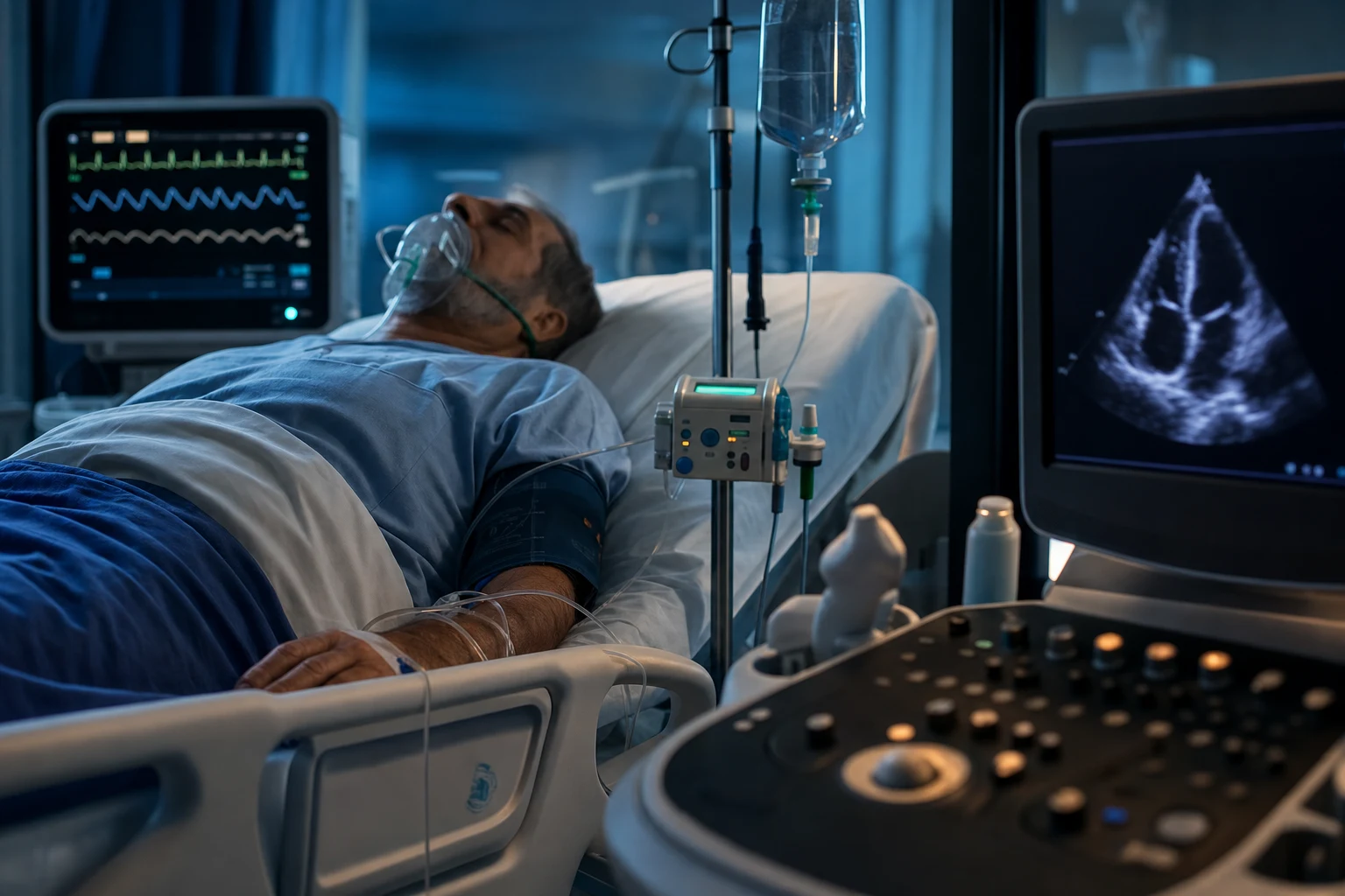
SCAI classification — the five stages

The SCAI (Society for Cardiovascular Angiography and Interventions) classification stratifies the cardiogenic shock into five stages — A through E — moving from the at-risk patient to the extremis. The stages parallel the temporal and the severity progression of the shock; each stage carries a progressively higher mortality (A under 5 per cent through E over 80 per cent). The classification guides the escalation of the monitoring and the therapy — the higher the stage, the more aggressive and the more invasive the support.[10]
The stages:[10]
- Stage A — At risk. The patient with the risk factors but NOT yet in the shock (the large anterior STEMI, the known HFrEF with the acute decompensation). The haemodynamics normal; the perfusion normal. The mortality low.
- Stage B — Beginning shock. The hypoperfusion beginning; the hypotension may respond to the fluid or to the low-dose vasopressor. The SBP above 90, the lactate near-normal. The clinical vigilance high.
- Stage C — Classic shock. The hypoperfusion now overt — the SBP under 90 (or the MAP under 65), the lactate above 2 mmol/L, the oliguria, the cold peripheries. The inotrope and the vasopressor required to maintain the perfusion.
- Stage D — Deteriorating. The shock worsening DESPITE the inotrope and the vasopressor — the escalating doses, the failing perfusion. The indication for the prompt mechanical circulatory support.
- Stage E — Extremis. The circulatory collapse — the cardiac arrest, the ongoing CPR, the refractory VF, the crash. The ECMO / the eCPR the consideration.
The practical use of the SCAI classification — the early recognition at the stage A-B (before the overt shock of the stage C), so that the revascularisation and the haemodynamic support can prevent the progression. A patient admitted with the large anterior STEMI is the stage A — the high vigilance, the early inotrope at the first sign of the hypoperfusion, the early PCI. The escalation to the stage D-E is the failure of the early recognition.[1][10]
The SCAI stages of cardiogenic shock (click each stage)
Classic shock
Overt hypoperfusion — SBP under 90 (MAP under 65), lactate above 2 mmol/L, oliguria, cool peripheries, altered mentation. Requires inotrope ± vasopressor to maintain perfusion. Revascularisation if ACS. This is the classical exam picture.
The haemodynamic profile — Forrester subsets and the catheter findings
The cardiogenic shock has a stereotyped haemodynamic profile. The cardiac pump fails → the cardiac output falls, the ventricular filling pressures rise, the systemic vascular resistance rises (the compensatory vasoconstriction), and the mixed venous (and the central venous) oxygen saturation falls (the tissues extract more oxygen from the sluggish flow). The profile, summarised:[1][2]
- Cardiac index — low (under 2.2 L/min/m², often under 1.8 in the established shock).
- Pulmonary capillary wedge pressure (PCWP) — high (above 15 mmHg, often 20-30 with the pulmonary oedema).
- Systemic vascular resistance (SVR) — high (the compensatory vasoconstriction; the SVR rises to defend the BP).
- Mixed venous oxygen saturation (SvO₂) — low (under 65 per cent, often under 50 in the severe shock; the tissues extract maximally from the slow flow).
- Central venous pressure (CVP) — elevated in the right-heart failure, the biventricular failure, or the tamponade. [1]
The Forrester haemodynamic subsets (the classic 1976 framework, still examined) combine the wedge pressure (the "warm" vs the "wet") with the cardiac index (the "warm" vs the "cold") into four quadrants. The cardiogenic shock occupies the subset IV — the high wedge (wet) AND the low cardiac index (cold): the "cold and wet" profile. The management implication: the patient needs the inotrope (to raise the cardiac index) and the diuresis / the unloading (to lower the wedge), not the further fluid.[14][1]
Subset I — warm and dry
CI normal, PCWP normal
- Cardiac index above 2.2 L/min/m², wedge under 18 mmHg
- No hypoperfusion, no pulmonary congestion
- Benign prognosis; no specific haemodynamic intervention
- Continue surveillance and treat the underlying infarct
Subset II — warm and wet
CI normal, PCWP high
- Cardiac index maintained, but wedge elevated (pulmonary oedema)
- Pump output preserved at the cost of high filling pressure
- Treat with diuresis and vasodilators (nitrate) to lower the wedge
- Forerunner of pump failure if untreated
Subset III — cold and dry
CI low, PCWP normal
- Low cardiac index with normal wedge — hypovolaemia with low contractility
- Clinically the hypoperfusion without the oedema
- Cautious fluid challenge to find the optimal preload (Starling)
- Differentiate from the true hypovolaemic shock (history, echo)
Subset IV — cold and wet (the shock)
CI low, PCWP high
- Cardiac index under 2.2, wedge above 18 — the cardiogenic shock
- Both hypoperfusion AND pulmonary congestion
- Inotrope (dobutamine / milrinone) + diuresis + vasodilator (if BP allows)
- Mechanical support if refractory; revascularise if ACS
- Mortality highest of the four subsets
Cardiogenic shock
Pump failure
- Cardiac index LOW (under 2.2)
- PCWP HIGH (above 18)
- SVR HIGH (compensatory vasoconstriction)
- SvO₂ LOW (under 65%)
- Cool, clammy peripheries; pulmonary oedema common
Septic shock
Distributive / vasodilatory
- Cardiac index HIGH (initially, with the low SVR)
- PCWP LOW or normal (until fluid-resuscitated)
- SVR LOW (the pathological vasodilation)
- SvO₂ HIGH (over 70%, often above 80% — impaired extraction)
- Warm peripheries early; the so-called warm shock
Hypovolaemic shock
Volume loss
- Cardiac index LOW
- PCWP LOW
- SVR HIGH (compensatory)
- SvO₂ LOW
- Cool peripheries; clear lung fields; responds to fluid
Obstructive shock (PE / tamponade)
Mechanical obstruction
- Cardiac index LOW
- PCWP HIGH in tamponade / RV infarct; LOW in massive PE
- SVR HIGH (compensatory)
- SvO₂ LOW
- Distinguish with echo: tamponade, clot-in-transit, RV strain
The pathophysiology — the vicious cycle
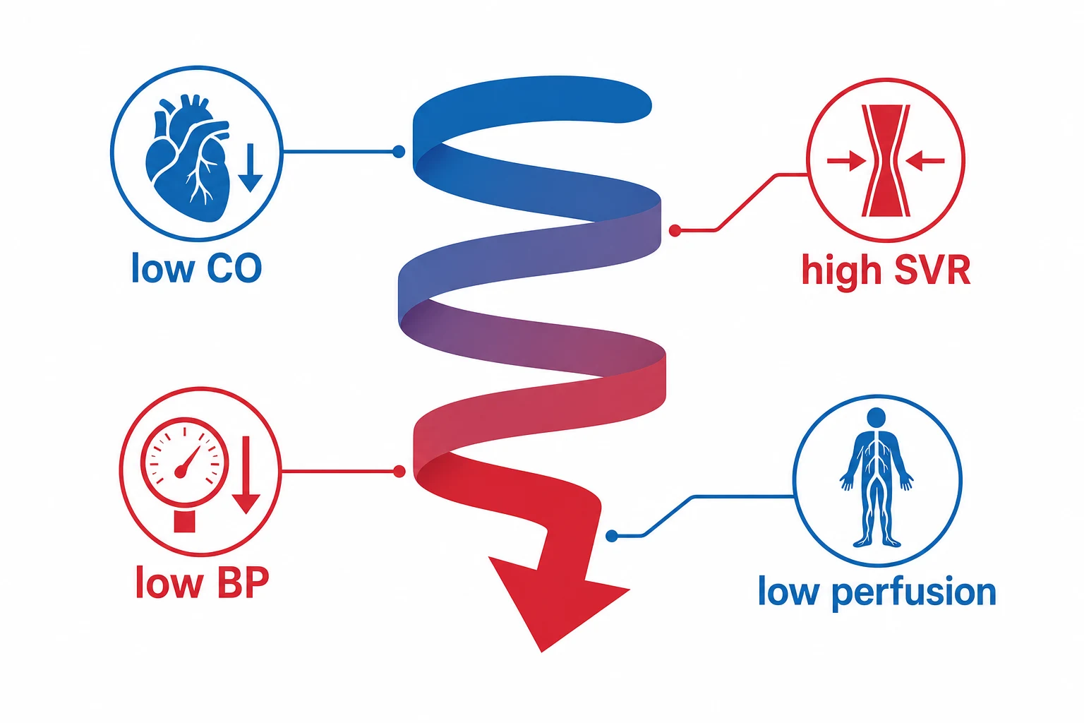
The heart fails → the CO falls → the BP falls → the compensatory the sympathetic the drive (the tachycardia, the vasoconstriction) → the SVR rises → the afterload increases → the failing the ventricle cannot the pump against the high afterload → the stroke volume falls further → the CO falls further → the worsens.[1][1]
The cycle also involves the compensatory tachycardia (which increases the myocardial oxygen demand while reducing the diastolic filling time) and the renin-angiotensin-aldosterone system (the sodium and water retention → the preload overload → the pulmonary oedema).[1]
The systemic inflammatory response is the second amplifier. The cardiogenic shock is no longer seen as a purely haemodynamic problem — it triggers a systemic inflammatory response syndrome (SIRS) indistinguishable from the sepsis (the nitric oxide release, the cytokine cascade, the inducible NOS, the reperfusion injury). The inflammatory vasoplegia supervenes on the original compensatory vasoconstriction, producing a mixed shock phenotype (the cardiogenic PLUS the distributive). This is why some patients with the established cardiogenic shock have a LOWER-than-expected SVR and require the vasopressor in addition to the inotrope. The inflammation also drives the reperfusion injury (after the PCI), the microcirculatory failure, and the multi-organ dysfunction that dominates the late course.[1][10]
The causes
- The acute coronary syndrome (the commonest — 80 per cent of the cardiogenic shock). The large the infarct (the anterior, the multivessel) or the mechanical complication (the VSD, the papillary muscle rupture, the free wall rupture).[1][1]
- The decompensated heart failure (the chronic HFrEF with the acute decompensation).[1]
- The myocarditis (the fulminant — the young, the viral).[1]
- The arrhythmia (the VT, the AF with the rapid ventricular response, the complete heart block).[1]
- The valvular (the acute severe MR, the acute severe AR, the prosthetic valve thrombosis).[1]
- The drug toxicity (the beta-blocker, the CCB, the TCA — the negative inotropy).[1]
- The post-cardiotomy (the after the cardiac surgery).[1]
The ACS-related causes fall into three groups: the primary LV pump failure (the large infarct, the multivessel disease — the commonest), the mechanical complication (the ventricular septal rupture at 3-5 days, the papillary muscle rupture with the severe MR, the free wall rupture with the tamponade — the days 1-5), and the right ventricular infarct (the inferior MI with the RV involvement — the high right-sided filling pressures, the clear lung fields). Each has a distinct management: the pump failure → the revascularisation + the inotrope; the mechanical complication → the urgent surgical repair with the MCS as the bridge; the RV infarct → the fluid loading (the RV is preload-dependent) + the inotrope + the sinus rhythm maintained (the AV synchrony critical for the RV filling) + the AV sequential pacing if the block.[1][1]
LV pump failure
The commonest (large MI)
- Large anterior or multivessel infarct
- Reduced LVEF on echo, global or anterior hypokinesis
- Pulmonary oedema, high wedge, low cardiac index
- Treat with revascularisation + inotrope + MCS if refractory
Mechanical complication
Days 1-5 post-MI
- Ventricular septal rupture: new pansystolic murmur, thrill, biventricular failure
- Papillary muscle rupture: acute severe MR, pulmonary oedema (often posteromedial papillary, single supply from PDA)
- Free wall rupture: sudden tamponade, PEA arrest, electromechanical dissociation
- Echo diagnostic; urgent surgical repair with MCS as the bridge
RV infarct
Inferior MI with RV involvement
- Inferior STEMI with right-sided leads (V4R ST elevation)
- Clear lung fields with high JVP — the "dry lungs, full neck veins" picture
- Hypotension worse with nitrates / diuretics (preload-dependent)
- Treat with FLUID loading, inotrope, maintain AV synchrony, AVR pacing if block
The clinical
The signs of the poor perfusion:[1][1]
- The cool, the clammy skin (the peripheral vasoconstriction).
- The oliguria (the renal hypoperfusion).
- The altered mental state (the cerebral hypoperfusion).
- The hypotension (the SBP below 90 or the MAP below 65).
- The pulmonary oedema (the LV failure) OR the clear lung fields (the RV failure — the isolated right).
- The echo: the reduced LV function (the low EF, the low CO), the RV dilatation (if the RV infarct), the mechanical complication (the VSD, the MR).[1]
The bedside echocardiogram is the single most important diagnostic test in the suspected cardiogenic shock. It answers the four critical questions within minutes: (1) Is the LV function reduced? (the global hypokinesis = the pump failure; the regional = the ACS); (2) Is there a mechanical complication? (the colour Doppler for the MR, the VSD; the effusion for the tamponade / the free wall rupture); (3) Is the RV involved? (the RV dilatation, the McConnell sign, the D-shape of the LV in the RV infarct or the massive PE); (4) Is the preload adequate or excessive? (the IVC size and the collapsibility, the dilated fixed IVC of the high right-sided pressures). The echo differentiates the cardiogenic from the obstructive (the tamponade, the tension pneumothorax, the PE) and from the distributive shock (the hyperdynamic LV of the sepsis — a KEY discriminator).[1][2]
The lactate is the marker of the anaerobic metabolism and the cornerstone of the resuscitation monitoring. A lactate above 2 mmol/L with the hypoperfusion signs defines the shock; the lactate clearance (the fall by at least 10 per cent per hour) is the marker of the adequate resuscitation. A rising or the static lactate signals the ongoing hypoperfusion despite the apparent haemodynamic stability.[2]
The management
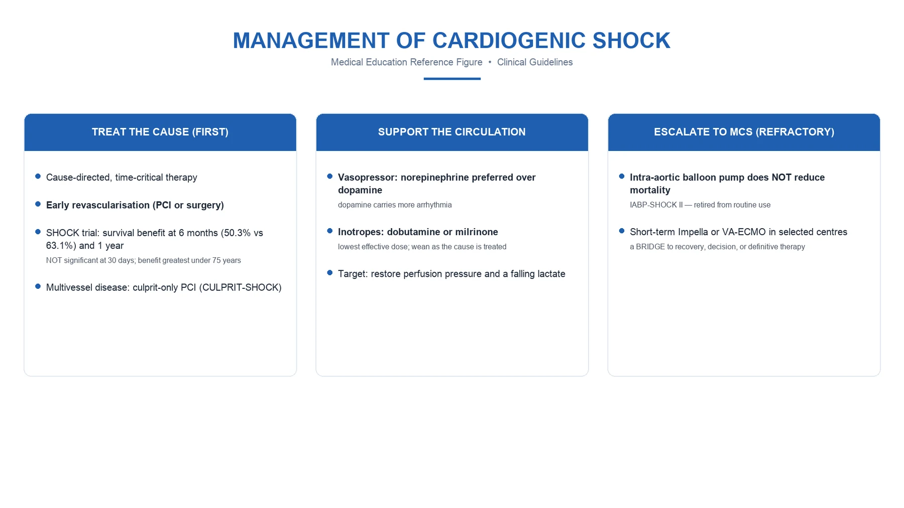
The management of cardiogenic shock — the ordered sequence
1. Recognise and classify (SCAI stage A-E)
Diagnose early — before the overt stage C shock. Hypoperfusion signs (oliguria, confusion, cold peripheries, lactate above 2) with hypotension (SBP under 90 or MAP under 65). Perform bedside echo immediately to confirm the cardiac cause, identify LV vs RV failure, and exclude mechanical complications and mimics (tamponade, PE, tension pneumothorax). Assign a SCAI stage to guide escalation.
2. Secure the airway and oxygenation
High-flow oxygen; non-invasive ventilation for the pulmonary oedema (CPAP/BiPAP reduces preload and afterload, improves oxygenation, may avoid intubation). Intubate early if the work of breathing is excessive, the consciousness is reduced, or the hypoxaemia is refractory — but anticipate haemodynamic collapse on induction (use a cardiostable induction: ketamine or etomidate, low-dose, vasopressor ready).
3. Treat the cause (revascularise the ACS immediately)
If ACS, emergency coronary angiography and PCI (or CABG). CULPRIT-SHOCK: culprit-lesion-only PCI at the index procedure, NOT multivessel PCI (lower 30-day mortality and renal failure). For the mechanical complication — urgent surgical repair with MCS as the bridge. For the arrhythmia — cardioversion or pacing. For the drug toxicity — the specific antidote.
4. Optimise the haemodynamics (inotrope ± vasopressor)
Add an inotrope for the low cardiac output (dobutamine first-line; milrinone if tolerated; levosimendan as an alternative — SURVIVE showed no survival benefit over dobutamine). Add a vasopressor if the MAP remains under 65 despite the inotrope — noradrenaline preferred (SOAP II: less arrhythmia than dopamine). Target MAP at least 65 (or 80 if known vascular disease or TBI). Aim for an arterial line, a central line, and ideally a pulmonary artery catheter or PiCCO for the guidance.
5. Escalate to mechanical circulatory support for the refractory (stage D-E)
If the perfusion remains inadequate despite the escalating pharmacological support, escalate to MCS. Impella (DanGer-SHOCK: reduced all-cause mortality in infarct-related shock) or VA-ECMO (ECLS-SHOCK and EURO-SHOCK were neutral overall — select carefully). IABP has NO routine role (IABP-SHOCK II). The MCS is a BRIDGE — to recovery, to a decision, to a durable LVAD, or to transplant.
6. Supportive care and the search for the destination
Correct the electrolytes (K 4-4.5, Mg above 0.8), the acidosis (pH above 7.2 for the inotrope effectiveness — consider bicarbonate only in extremis), the glycaemia (6-10). DVT prophylaxis (LMWH, caution with the anticoagulation). Continuous renal replacement therapy if the AKI or the fluid overload. Identify the destination — recovery, durable LVAD, transplant, or the withdrawal of care if the irreversible. Reassess every 1-2 hours with the lactate, the urine output, the mental state.
1. The identify and treat the cause
- The ACS → the urgent revascularisation (the PCI, the CABG). The SHOCK trial — the early revascularisation reduces the long-term mortality (even though the 30-day mortality was not significantly different).[1]
- The arrhythmia → the cardioversion / the pacing.
- The valvular → the surgical repair / the replacement.
- The drug toxicity → the specific antidote (the calcium + the glucagon + the high-dose insulin for the CCB / the BB; the bicarbonate for the TCA).[1]
The SHOCK trial (Hochman, NEJM 1999) established the paradigm: in the AMI cardiogenic shock, the early revascularisation (the PCI or the CABG within 24 hours) reduced the 6-month and the 1-year mortality compared with the initial medical stabilisation (the 1-year mortality 53 per cent vs 63 per cent), even though the 30-day mortality was not significantly different. The 6-year follow-up (Hochman, JAMA 2006) confirmed the durable benefit. The lesson — the early revascularisation is the standard, and the long-term survival is the endpoint that matters, not the 30-day.[3][4]
The CULPRIT-SHOCK trial (Thiele, NEJM 2017) refined the revascularisation strategy: in the multivessel-disease AMI cardiogenic shock, the culprit-lesion-only PCI at the index procedure was superior to the immediate multivessel PCI — the 30-day mortality 17.5 per cent vs 43 per cent, and the less renal-replacement therapy. The lesson — in the AMI shock, fix ONLY the culprit lesion acutely; stage the non-culprit lesions once the patient stabilises. This is now the guideline standard.[7]
2. The pharmacological support
The inotrope (to increase the contractility):[1][2]
- Dobutamine (the beta-1 — the increased contractility and the HR; the also the beta-2 vasodilation → the reduced afterload; but the tachyarrhythmia, the increased myocardial oxygen demand).[1]
- Milrinone (the PDE-3 inhibitor — the increased cAMP; the inotropy AND the vasodilation; the long half-life; the caution in the renal failure).[1]
- Levosimendan (the calcium sensitiser — the inotropy without the increased intracellular calcium; the less the arrhythmia; the vasodilation; the expensive).[2]
- Adrenaline (the potent but the high the arrhythmia, the increased the lactate, the increased the myocardial oxygen demand).[1]
The vasopressor (to maintain the perfusion pressure):[1][2]
- Noradrenaline (the alpha-1 — the vasoconstriction; the preferred over the dopamine per the SOAP II).[1]
- The target the MAP 65 (or above 70-80 if the TBI / the known the vascular disease).[2]
The SOAP II trial (De Backer, NEJM 2010) compared the dopamine with the noradrenaline in the circulatory shock (the cardiogenic, the septic, the hypovolaemic). The result — the no difference in the overall mortality, BUT the dopamine had the more arrhythmia (the atrial fibrillation especially) and the more frequent rate of discontinuation for the adverse events. The cardiogenic subgroup showed a trend toward the higher mortality with the dopamine. The lesson — the noradrenaline is the preferred first-line vasopressor in the cardiogenic shock; the dopamine is avoided (the arrhythmia, the worse outcome).[8]
The SURVIVE trial (Mebazaa, JAMA 2007) compared the levosimendan with the dobutamine in the acute decompensated heart failure. The result — the no difference in the short-term or the long-term mortality, though the levosimendan showed a transient benefit in the subgroup with the prior beta-blocker use. The lesson — the levosimendan is NOT superior to the dobutamine; the dobutamine remains the first-line inotrope for the cost-effectiveness and the familiarity, with the levosimendan as the alternative (especially in the beta-blocked patient, where the beta-agonist dobutamine is less effective).[9]
Dobutamine
Beta-1 agonist
- Beta-1: ↑ contractility, ↑ HR; mild beta-2 vasodilation (↓ afterload)
- Onset 1-2 min, short half-life (2 min) — easy to titrate
- Side effects: tachyarrhythmia, ↑ myocardial O₂ demand, myocardial ischaemia
- First-line inotrope in cardiogenic shock; dose 2-20 mcg/kg/min
- Less effective in the beta-blocked patient
Milrinone
PDE-3 inhibitor
- PDE-3 inhibition → ↑ cAMP; inotropy PLUS pulmonary and systemic vasodilation
- Long half-life (2-3 h) — slow to titrate; accumulates in renal failure
- Less arrhythmia than dobutamine; preferred if RV failure / pulmonary hypertension
- Loading 25-75 mcg/kg, infusion 0.125-0.75 mcg/kg/min
- Avoid if severe renal impairment without dose reduction
Levosimendan
Calcium sensitiser
- Sensitises troponin C to calcium — inotropy WITHOUT ↑ intracellular Ca²⁺
- Less arrhythmia; vasodilation (opens K_ATP channels); expensive
- Active metabolite has a long half-life (75+ h) — prolonged effect
- SURVIVE: no survival benefit over dobutamine; useful in beta-blocked patients
- Dose: 0.05-0.2 mcg/kg/min infusion (no loading in shock — hypotension)
Adrenaline
Non-selective catecholamine
- Beta-1, beta-2, alpha-1: potent inotropy + vasoconstriction at higher doses
- Reserve for the profound shock or the arrest; effective when dobutamine fails
- Side effects: marked arrhythmia, ↑ lactate (anaerobic glucose metabolism), ↑ O₂ demand
- May worsen the splanchnic perfusion and cause a transient lactic acidosis
- Tachyphylaxis; de-escalate to dobutamine + noradrenaline as soon as possible
Noradrenaline
Alpha-1 > beta-1
- Alpha-1 vasoconstriction restores the MAP; mild beta-1 supports contractility
- First-line vasopressor in cardiogenic shock (SOAP II)
- Less arrhythmia than dopamine or adrenaline
- Combine with dobutamine/milrinone for the low-output + hypotension
- Dose 0.05-1.0 mcg/kg/min; titrate to MAP at least 65
Dopamine
Dose-dependent effects
- Low dose: renal/mesenteric (D1) — historically for renal protection, NO proven benefit
- Mid dose: beta-1 (inotropy); high dose: alpha-1 (vasoconstriction)
- SOAP II: more arrhythmia (especially atrial fibrillation) than noradrenaline
- Trend toward higher mortality in the cardiogenic subgroup
- Avoid as a first-line vasopressor; reserve for bradycardia-induced shock
Vasopressin
V1 receptor agonist
- Catecholamine-sparing; useful in the vasodilatory / late-phase cardiogenic shock
- Fixed dose 0.01-0.04 U/min; no tachyarrhythmia
- Catecholamine-resistant shock (the septic overlap); the SCAI stage D-E
- Add to noradrenaline rather than as the sole vasopressor
3. The mechanical circulatory support (MCS)
For the refractory (the pharmacological failure):[1][2]
- The IABP (the intra-aortic balloon pump) — the diastolic augmentation (the balloon inflates in the diastole, increasing the coronary perfusion; deflates in the systole, reducing the afterload). The IABP-SHOCK II trial — the no the mortality the benefit in the ACS the cardiogenic the shock (the routine the IABP not the recommended).[1]
- The Impella (the axial-flow catheter across the aortic valve — the continuous the LV unloading; the 2.5 to the 5.0 L/min).[1]
- The VA-ECMO (the venoarterial ECMO — the full the cardiopulmonary the bypass; the 3-5 L/min; the bridge to the recovery / the transplant / the decision).[1]
- The TandemHeart (the LA-to-femoral artery the bypass).[2]
The choice of the MCS depends on the centre, the expertise, the patient, and the cause.[1]
The three landmark device trials of the recent era have reshaped the MCS landscape:[5][11][12]
- The IABP-SHOCK II (Thiele, NEJM 2012, 600 patients) — the routine IABP in the AMI cardiogenic shock showed no mortality benefit at 30 days, 6 months, 12 months, or the long-term follow-up. The routine IABP is NOT recommended; the IABP is reserved for the mechanical complication (the VSR, the MR — the bridge to surgery) and for the centre without the more advanced MCS.[5][6]
- The DanGer-SHOCK (Møller, NEJM 2024, 360 patients) — the Impella CP vs the standard care (including IABP) in the AMI cardiogenic shock. The result — the reduced all-cause mortality at 180 days (66.7 per cent vs 80.0 per cent in the per-protocol analysis). This is the first positive MCS mortality trial in the infarct-related shock — but the caveat: the benefit was confined to the per-protocol population; the trial excluded the cardiac arrest and the moderate-severe AR; the bleeding and the haemolysis were more common with the Impella. The cautious adoption.[12]
- The ECLS-SHOCK (Akin, NEJM 2023, 420 patients) — the VA-ECMO vs the standard care in the infarct-related shock. The result — the no difference in the 30-day all-cause mortality (47.8 per cent vs 49.0 per cent), with the more bleeding and the more vascular complications in the ECMO group. The EURO-SHOCK (Møller-Helgestad, EuroIntervention 2023) similarly showed no mortality benefit of the VA-ECMO. The lesson — the VA-ECMO is NOT the routine first-line MCS in the AMI shock; it is reserved for the SCAI stage D-E, the profound shock, the refractory cardiac arrest (the eCPR), the bridge to the durable support.[11][13]
IABP
Counterpulsation
- Inflates in diastole (↑ coronary perfusion), deflates in systole (↓ afterload)
- Augments cardiac output by only ~0.5-1.0 L/min — modest support
- IABP-SHOCK II (NEJM 2012): NO mortality benefit in AMI shock — routine use not recommended
- Reserved for the mechanical complication (bridge to surgery) and centres without advanced MCS
- Contraindications: significant AR, aortic dissection, severe PVD
Impella
Microaxial flow pump
- Axial-flow catheter across the aortic valve; continuous LV unloading 2.5-5.0 L/min
- Direct LV unloading (reduces wall stress, O₂ demand) — unlike IABP/ECMO
- DanGer-SHOCK (NEJM 2024): reduced 180-day mortality (per-protocol) in infarct-related shock
- Complications: limb ischaemia, bleeding, haemolysis, device malposition
- Contraindications: significant AR, LV thrombus, severe PVD, mechanical aortic valve
VA-ECMO
Full cardiopulmonary bypass
- Venoarterial: drains RA, returns to femoral artery; 3-5 L/min; supports both ventricles + oxygenation
- Most powerful support — for the SCAI stage D-E, refractory arrest (eCPR), biventricular failure
- ECLS-SHOCK and EURO-SHOCK (2023): NO routine mortality benefit in AMI shock
- Does NOT unload the LV — may worsen the afterload and the pulmonary oedema; vent (Impella/IABP) if LV distends
- Complications: bleeding, thrombosis, infection, limb ischaemia, differential hypoxia (Harlequin / north-south)
TandemHeart
LA-to-femoral artery
- Transseptal cannula drains the LA, returns to the femoral artery; 3-5 L/min
- Bypasses the LV entirely — effective LV unloading
- Technically demanding (transseptal puncture); specialist centres
- Complications: tamponade (perforation), femoral vascular injury, ARF
- Largely superseded by the Impella and the VA-ECMO
4. The supportive
- The oxygen / the ventilation (the non-invasive or the invasive; the lung-protective).[1]
- The renal replacement therapy (if the AKI / the fluid overload).[1]
- The correction of the electrolyte (the K, the Mg) + the acidosis (the pH above 7.2 for the inotrope the effectiveness).[1]
- The glycaemic control (the 6-10).[1]
- The DVT prophylaxis (the LMWH — the caution with the anticoagulation).[1]
The acidosis correction deserves the emphasis. The myocardium, the catecholamine receptors, and the vascular smooth muscle all dysfunction in the acidosis — the inotropes and the vasopressors become ineffective below the pH 7.2. The acidosis is BOTH the consequence of the shock (the hypoperfusion → the lactate) AND the amplifier (the depressed contractility, the receptor desensitisation). The correction: the adequate perfusion (the inotrope, the MCS) is the primary; the sodium bicarbonate is reserved for the pH under 7.1-7.2 with the refractory haemodynamic instability (the bicarbonate generates the CO₂ which worsens the intracellular acidosis if the ventilation is inadequate — so it is given only with the secured airway and the adequate ventilation).[1]
The landmark trials
SHOCK — early revascularisation in AMI cardiogenic shock (Hochman, NEJM 1999)
Multicentre RCT; 302 patients with AMI and cardiogenic shock
Population: Left ventricular failure within 36 h of AMI, SBP under 90 for 30 min
Key finding
No significant difference in 30-day mortality (46.7% revascularisation vs 56.0% medical, p=0.11). BUT 6-month mortality significantly lower (50.3% vs 63.1%, p=0.027); 1-year mortality 53% vs 63% (p=0.04). The 6-year follow-up (JAMA 2006) confirmed the durable survival benefit. The greatest benefit was in patients under 75 years.
Practice change
In AMI cardiogenic shock, early revascularisation (PCI or CABG) within 24 h improves LONG-TERM survival, even though 30-day mortality is not significantly different. The 30-day figure is misleading — the dying patients take time to declare themselves. Early revascularisation is the standard.
IABP-SHOCK II — routine IABP in AMI cardiogenic shock (Thiele, NEJM 2012)
Multicentre open-label RCT; 600 patients with AMI and cardiogenic shock planned for early revascularisation
Population: AMI with cardiogenic shock (SBP under 90, signs of hypoperfusion), planned for early PCI
Key finding
No significant difference in 30-day mortality (39.7% IABP vs 41.3% control, p=0.69). No difference at 6 months, 12 months, or 6-year follow-up. No difference in any subgroup (age, sex, diabetes, infarct location).
Practice change
Routine IABP in AMI cardiogenic shock provides NO mortality benefit. IABP is NOT routinely recommended. Reserve the IABP for the mechanical complication (VSR, papillary muscle rupture — as a bridge to surgery) and for centres without the more advanced MCS.
CULPRIT-SHOCK — culprit-only vs multivessel PCI in AMI shock (Thiele, NEJM 2017)
Multicentre RCT; 1075 patients with multivessel-disease AMI and cardiogenic shock
Population: AMI with cardiogenic shock and multivessel coronary disease (stenosis above 70% in a non-culprit vessel)
Key finding
Culprit-only was SUPERIOR: composite 45.9% vs 55.4% (relative risk 0.83, p=0.01). Mortality 17.5% vs 43.3% at 30 days (in the original report); less renal replacement therapy. Benefit sustained at 1 year.
Practice change
In AMI cardiogenic shock with multivessel disease, fix ONLY the culprit lesion at the index procedure. Multivessel PCI (with its longer procedure, more contrast, more ischaemic time) is harmful. Stage the non-culprit lesions once the patient stabilises. This is now the guideline standard.
SOAP II — dopamine vs noradrenaline in shock (De Backer, NEJM 2010)
Multicentre randomised trial; 1679 patients with circulatory shock
Population: Adults with circulatory shock (cardiogenic, septic, or hypovolaemic) requiring a vasopressor
Key finding
No significant difference in 28-day mortality overall (52.5% dopamine vs 48.5% noradrenaline, p=0.10). BUT dopamine had SIGNIFICANTLY MORE ARRHYTHMIA (24.1% vs 12.4%, p<0.001), especially atrial fibrillation, and more events leading to discontinuation. In the cardiogenic shock subgroup, there was a non-significant trend toward higher mortality with dopamine (p=0.11).
Practice change
Noradrenaline is the preferred first-line vasopressor in shock — it has fewer arrhythmias than dopamine, and a trend toward better outcomes in cardiogenic shock. Dopamine should not be the first-line vasopressor; reserve it for the patient with concurrent bradycardia (the so-called inotrope-chronotrope scenario).
SURVIVE — levosimendan vs dobutamine in acute heart failure (Mebazaa, JAMA 2007)
Multicentre double-blind RCT; 1327 patients with acute decompensated heart failure requiring an inotrope
Population: Adults with acute decompensated heart failure, low cardiac output, requiring inotropic support
Key finding
No significant difference in 180-day mortality (26% levosimendan vs 28% dobutamine, hazard ratio 0.91, p=0.45). No difference at 31 days or 1 year. A post-hoc subgroup of patients on prior beta-blockers showed a transient benefit with levosimendan at 31 days.
Practice change
Levosimendan is NOT superior to dobutamine in acute heart failure / low-output state. Dobutamine remains the first-line inotrope for the cost-effectiveness and familiarity. Levosimendan is a reasonable alternative, especially in the beta-blocked patient (where the beta-agonist dobutamine is less effective).
DanGer-SHOCK — Impella vs standard care in infarct-related shock (Møller, NEJM 2024)
Multicentre randomised trial; 360 patients with infarct-related cardiogenic shock (per-protocol analysis 355)
Population: AMI with cardiogenic shock (SBP under 100, lactate above 2, signs of hypoperfusion), no out-of-hospital cardiac arrest, no moderate-severe AR
Key finding
In the per-protocol analysis, Impella REDUCED 180-day mortality (66.7% vs 80.0%, relative risk 0.83, p=0.04). The intention-to-treat analysis did NOT reach significance (p=0.06). More bleeding and more haemolysis with the Impella. The benefit was confined to the strictly selected per-protocol population.
Practice change
The first positive MCS mortality trial in infarct-related shock — but the benefit is confined to a highly selected population (no arrest, no significant AR). Cautious adoption: consider the Impella CP in the SCAI stage C-D AMI shock patient who fits the trial criteria. The bleeding and haemolysis must be weighed.
ECLS-SHOCK — VA-ECMO in infarct-related shock (Akin, NEJM 2023)
Multicentre randomised trial; 420 patients with infarct-related cardiogenic shock
Population: AMI with cardiogenic shock (SBP under 90 for 30 min, or inotrope/vasopressor required, lactate above 2)
Key finding
No significant difference in 30-day mortality (47.8% VA-ECMO vs 49.0% control, relative risk 0.98, p=0.95). More bleeding (23.4% vs 9.6%) and more vascular complications with the VA-ECMO. The EURO-SHOCK trial (EuroIntervention 2023, n=406) showed a similar null result.
Practice change
Routine early VA-ECMO in AMI cardiogenic shock does NOT reduce mortality and adds bleeding / vascular harm. VA-ECMO is NOT a routine first-line MCS — reserve for the SCAI stage D-E (deteriorating / extremis), the refractory cardiac arrest (eCPR), and as a bridge to the durable LVAD or the transplant.
Prognosis
The mortality 40-60 per cent (the higher in the older, the comorbid, the delayed). The early revascularisation (the ACS), the appropriate pharmacological support, and the timely MCS (the refractory) reduce the mortality. The bridging to the transplant or the durable LVAD for the irreversible.[1][2][1]
The SCAI stage is the most powerful prognostic marker — the mortality rises steeply from the stage A (under 5 per cent) through the stage E (over 80 per cent). The other adverse markers: the lactate (the higher and the slower to clear, the worse), the renal failure (the creatinine rise and the need for the RRT), the age above 75, the out-of-hospital cardiac arrest, the anterior infarct, the multivessel disease, the delay to the revascularisation, and the biventricular failure. The SHOCK trial the only mortality-reducing intervention is the early revascularisation — every other therapy (the inotrope, the vasopressor, the MCS) is the supportive bridge, not the disease-modifier.[3][10]
Red flags
Clinical pearls
Exam practice
SAQ — AMI cardiogenic shock: classification, revascularisation, and MCS
12 minutes · 10 marks
A 64-year-old man is admitted with an anterior STEMI. He undergoes a primary PCI (the LAD stented), but 4 hours later he is cold and clammy. HR 118, BP 78/50 (MAP 59), SpO₂ 92% on 10 L O₂. JVP elevated. Bilateral crackles to mid-zones. Urine output 15 mL in the first hour. Lactate 4.8 mmol/L. Bedside echo: severely reduced LV function (LVEF ~25%), no mechanical complication, RV normal.
SAQ — The RV infarct and the differential of the clear-lung-fields shock
10 minutes · 8 marks
A 58-year-old woman presents with an inferior STEMI (V4R shows ST elevation). She is hypotensive (BP 82/58), HR 48 (sinus with the Wenckebach), JVP to the earlobes, clear lung fields, cool peripheries. Lactate 3.6 mmol/L. Bedside echo: RV dilatation with the reduced free-wall function, LV preserved.
References
- [1]Muller G, et al. ICU management of cardiogenic shock before mechanical support Curr Opin Crit Care, 2024.PMID 38872375
- [2]Terkelsen CJ, et al. Hemodynamic management of cardiogenic shock in the intensive care unit J Heart Lung Transplant, 2024.PMID 38518863
- [3]Hochman JS, Sleeper LA, Webb JG, et al. Early revascularization in acute myocardial infarction complicated by cardiogenic shock. SHOCK Investigators. Should We Emergently Revascularize Occluded Coronaries for Cardiogenic Shock N Engl J Med, 1999.PMID 10460813
- [4]Hochman JS, Sleeper LA, Webb JG, et al. Early revascularization and long-term survival in cardiogenic shock complicating acute myocardial infarction JAMA, 2006.PMID 16757723
- [5]Thiele H, Zeymer U, Neumann FJ, et al. Intraaortic balloon support for myocardial infarction with cardiogenic shock N Engl J Med, 2012.PMID 22920912
- [6]Thiele H, Zeymer U, Neumann FJ, et al. Intraaortic balloon support for cardiogenic shock N Engl J Med, 2013.PMID 23281982
- [7]Thiele H, Akin I, Sandri M, et al. PCI Strategies in Patients with Acute Myocardial Infarction and Cardiogenic Shock N Engl J Med, 2017.PMID 29083953
- [8]De Backer D, Biston P, Devriendt J, et al. Comparison of dopamine and norepinephrine in the treatment of shock N Engl J Med, 2010.PMID 20200382
- [9]Mebazaa A, Nieminen MS, Packer M, et al. Levosimendan vs dobutamine for patients with acute decompensated heart failure: the SURVIVE Randomized Trial JAMA, 2007.PMID 17473298
- [10]Baran DA, Grines CL, Bailey S, et al. SCAI clinical expert consensus statement on the classification of cardiogenic shock: This document was endorsed by the American College of Cardiology (ACC), the American Heart Association (AHA), the Society of Critical Care Medicine (SCCM), and the Society of Thoracic Surgeons (STS) in April 2019 Catheter Cardiovasc Interv, 2019.PMID 31104355
- [11]Akin M, Pauschinger M, Opitz F, et al. Extracorporeal Life Support in Infarct-Related Cardiogenic Shock N Engl J Med, 2023.PMID 37634145
- [12]Møller JE, Holmvang L, Papo D, et al. Microaxial Flow Pump or Standard Care in Infarct-Related Cardiogenic Shock N Engl J Med, 2024.PMID 38587239
- [13]Møller-Helgestad OK, Hyldebrandt JA, Banke A, et al. Venoarterial extracorporeal membrane oxygenation or standard care in patients with cardiogenic shock complicating acute myocardial infarction: the multicentre, randomised EURO SHOCK trial EuroIntervention, 2023.PMID 37334659
- [14]Forrester JS, Diamond G, Chatterjee K, Swan HJ Medical therapy of acute myocardial infarction by application of hemodynamic subsets (second of two parts) N Engl J Med, 1976.PMID 790194