ICU · Resuscitation & shock
Endpoints of Resuscitation — Lactate, ScvO2, Mottling, Urine, Capillary Refill
Also known as Resuscitation endpoints · Adequacy of resuscitation · Lactate clearance · ScvO2 · Central venous oxygen saturation · Mottling score · Capillary refill time · ANDROMEDA-SHOCK · Goal-directed therapy · Pcv-aCO2 gap · Venous-to-arterial CO2 gap · Base deficit · De-resuscitation · Fluid overload · Sublingual microcirculation · NIRS StO2 · ROSE fluid framework
Resuscitation endpoints span three tiers used together rather than in isolation: macro-haemodynamic endpoints such as MAP and urine output, metabolic endpoints such as lactate clearance and ScvO2, and peripheral perfusion endpoints such as capillary refill time, mottling score, and skin temperature.
On this page & tools
Your progress
Saved locally on this device.
Target exams
Overview & definition
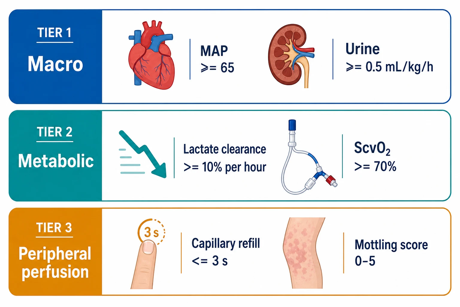
The question "have I resuscitated enough?" has no single answer. Resuscitation endpoints span three tiers, used together rather than in isolation:[1]
- Macro-haemodynamic — mean arterial pressure (MAP), urine output.
- Metabolic — lactate (and lactate clearance), central or mixed venous oxygen saturation (ScvO2 / SvO2).
- Peripheral perfusion — capillary refill time (CRT), mottling score, skin temperature.
The field has moved from Rivers' rigid protocolized early goal-directed therapy (EGDT, driven by ScvO2) toward individualised perfusion-targeted resuscitation. ProCESS showed protocolized EGDT is not superior to usual care, and ANDROMEDA-SHOCK showed peripheral-perfusion (capillary-refill) targeting is at least as good as lactate targeting.[1][3][5]

Tier 1 — macro-haemodynamic endpoints
- MAP at least 65 mmHg — the traditional perfusion-pressure target. Adequate for most; aim higher (75-85) in chronic hypertension, and lower is acceptable in young fit patients. MAP alone does not confirm adequate flow or perfusion.[1]
- Urine output at least 0.5 mL/kg/h — a classic marker of renal perfusion; aim for at least 1 mL/kg/h in shock. It lags behind correction of perfusion and is confounded by diuretics, AKI and glycosuria.[1]
- CVP — a static filling pressure, not a reliable endpoint (it does not predict fluid responsiveness and does not correlate with blood volume). Do not chase a CVP target.[1]
Tier 2 — metabolic endpoints
Lactate and lactate clearance
Hyperlactataemia reflects tissue hypoperfusion (anaerobic metabolism), catecholamine-driven glycolysis, impaired hepatic clearance, or beta-2 agonists. A rising or persistently high lactate signals ongoing inadequacy.[1]
- Lactate clearance — aim for a fall of at least 10 per cent per hour (or normalisation within 24-48 h).[1]
- Jansen (AJRCCM 2010) — lactate-guided therapy (lower the lactate over 2 hours, with fluids or transfusion if it fails to fall) reduced hospital mortality in the subgroup with an initial lactate above 3.0 mmol/L, supporting lactate clearance as a target.[2]
Caveat: lactate is not always hypoperfusion. Beta-2 agonists (salbutamol), malignancy, thiamine deficiency, metformin, and hepatic failure all raise lactate without hypoperfusion — interpret it in context.[1]
ScvO2 / SvO2
ScvO2 (central venous, from the SVC) and SvO2 (mixed venous, from the pulmonary artery) reflect the balance between oxygen delivery and consumption; a low value implies inadequate delivery or increased extraction.[1]
- Rivers EGDT (NEJM 2001) — targeting ScvO2 at least 70 per cent (alongside CVP and MAP) reduced in-hospital mortality from 46.5 to 30.5 per cent in severe sepsis and septic shock, establishing ScvO2 as a resuscitation target.[1]
- ProCESS (NEJM 2014) — protocolized EGDT (the Rivers bundle) was not superior to usual care (mortality around 18-21 per cent in all arms). Modern usual care (early antibiotics, routine lactate, more ICU support) has eroded the benefit of a rigid ScvO2-driven protocol.[5]
- ScvO2 is normally around 70 per cent; in sepsis it may be high (impaired oxygen extraction), so a normal ScvO2 does not exclude tissue hypoxia.[1]
Tier 3 — peripheral perfusion endpoints
Capillary refill time (CRT)
CRT is measured by pressing on the distal phalanx (or the knee) for 5-10 seconds and timing return of colour. Normal is at most 3 seconds. Prolonged CRT reflects peripheral vasoconstriction and hypoperfusion.[3]
- ANDROMEDA-SHOCK (JAMA 2019) — in 424 patients with septic shock, a strategy targeting peripheral perfusion (CRT at most 3 s) was compared with lactate-guided resuscitation. CRT targeting produced lower 28-day mortality (34.9 vs 43.4 per cent; the predefined significance threshold was not met, but the trend strongly favoured CRT) with less organ dysfunction and fewer interventions. Peripheral-perfusion targeting is feasible and at least as good as, possibly better than, lactate targeting.[3]
Mottling score
Mottling is patchy purplish skin discolouration from heterogeneous hypoperfused skin areas, typically around the knee. The Ait-Oufella mottling score (0-5) grades its extent:[4]
- 0 — no mottling
- 1 — small, confined to the centre of the knee
- 2 — moderate, not beyond the knee cap
- 3 — extending to the top of the kneecap
- 4 — extending to the middle of the thigh
- 5 — extending beyond the middle of the thigh
Ait-Oufella (Intensive Care Medicine 2011) showed the score strongly and independently predicts 14-day and 28-day mortality in septic shock, and that a fall in mottling over time tracks recovery. It is a simple, bedside, repeatable perfusion endpoint.[4]
Skin temperature
A cool, mottled periphery with a warm core signals vasoconstriction and ongoing shock; warming of the extremities parallels restoration of perfusion.[1]
Static vs dynamic, and responsiveness
Macro and metabolic endpoints say whether to resuscitate; dynamic tests (passive leg raise, SVV/PPV, echocardiography) say whether the patient will respond to fluid before you give it. Resuscitate to a perfusion target, but give fluid only if the patient is fluid-responsive (see the fluid-responsiveness topic).[1]
The integrated approach
Combine the tiers:[1]
- Restore a perfusion pressure (MAP at least 65) and organ flow (urine, lactate clearance).
- Confirm peripheral perfusion normalises (CRT at most 3 s, mottling resolves).
- Give fluid only if responsive; otherwise use vasopressors or inotropes.
- Reassess frequently — endpoints trend together as the patient recovers.
Tier 4 — microcirculatory and cellular endpoints (the missing link)
Macro-haemodynamics (MAP, urine) and global metabolic markers (lactate, ScvO2) describe the convective delivery of oxygen to the capillary bed, but the cell only "sees" the diffusive unloading of oxygen at the tissue. In sepsis, distributive shunting, microthrombi, endothelial glycocalyx shedding, and mitochondrial dysfunction create a dissociation between restored global flow and ongoing tissue hypoxia — the phenomenon Ince and others have called "loss of haemodynamic coherence". Several bedside markers attempt to bridge this gap.[1]
Venous-to-arterial CO2 gap (Pcv-aCO2 gap, ΔPCO2)
The Fick principle applied to CO2: CO2 production divided by cardiac output equals the arterio-venous CO2 difference. A widening Pcv-aCO2 gap (≥6 mmHg / >0.8 kPa) at a "normal" MAP, lactate and ScvO2 signals that cardiac output is still too low for the metabolic demand — CO2 is not being washed out of the tissues. It is the most accessible bedside marker of occult low flow.[1]
- Measured from paired arterial and central venous gas (the venous sample is from the SVC, paired within minutes of the arterial sample).
- A normal gap (<6 mmHg) alongside normal ScvO2 and clearing lactate is the strongest combined evidence that the macro-circulation is matched to demand.
- Caveat: gap narrows with high flow states (sepsis, hyperdynamic circulation) and widens with anaerobic CO2 generation, so it integrates flow and anaerobic metabolism. A normal gap with a high lactate suggests microcirculatory shunting rather than low cardiac output — resuscitation should target the microcirculation, not more fluid.
Base deficit / standard bicarbonate
Base deficit (the negative of base excess) quantifies the amount of base required to titrate whole blood to pH 7.40 at PCO2 40 mmHg. A base deficit worse than −5 mmol/L, or a persistently negative base excess, indicates ongoing metabolic acidosis from anaerobic metabolism (lactate, ketones, renal failure) or hyperchloraemia from excessive saline.[1]
- Trends more quickly than lactate (it responds within minutes to restored flow).
- Caveat: saline resuscitation itself causes a hyperchloraemic metabolic acidosis that worsens base deficit without any tissue hypoperfusion. A widening base deficit after 3–4 L of 0.9% saline may be the saline, not the patient. Use balanced crystalloid and interpret base deficit alongside lactate and the strong ion difference.
ScvO2 deep dive — when high is bad
A high ScvO2 (>80%) is not reassurance — it can indicate impaired oxygen extraction (cytopathic dysoxia, mitochondrial dysfunction, microcirculatory shunting) in late sepsis, where oxygen is delivered to the capillary bed but the cell cannot use it. A falling ScvO2 in a resuscitating patient is reassuring (extraction normalising); a rising ScvO2 with worsening lactate is ominous. This is the central paradox that drove the move away from isolated ScvO2 targeting toward peripheral-perfusion (CRT, mottling) and microcirculatory endpoints.[1]
Tier 5 — bedside imaging endpoints (POCUS)
Point-of-care ultrasound has supplanted static pressure targets (CVP) as the bedside tool to assess cardiac and volume status in real time. POCUS does not replace the perfusion tiers above; it answers "what is the heart doing and is it empty or full" before the next fluid bolus.[1]
Inferior vena cava assessment
- IVC diameter and collapsibility. A small (<1.5 cm), vigorously collapsing (>50% with sniff) IVC suggests a volume-responsive state; a fixed, plethoric (>2.5 cm) IVC with <10% collapse suggests high right-sided pressures and that more fluid will not help.
- Caveats: IVC assessment is unreliable in the spontaneously breathing patient with high respiratory effort (negative intrathoracic pressure distorts the IVC); in the intubated patient, a passive-leg-raise or fluid challenge with a corresponding change in IVC is more informative than a single static measurement. A plethoric IVC in cardiogenic shock is a contraindication to further fluid. IVC correlates poorly with absolute volume status — use it only to predict responsiveness in the right clinical context. [1]
Focused cardiac ultrasound (FoCUS / FATE)
- Assess the four basic windows (parasternal long and short, apical 4-chamber, subcostal). Answer four questions: (1) Is the LV small and hyperdynamic (hypovolaemia)? (2) Is the LV dilated and poorly contracting (cardiogenic shock)? (3) Is the RV dilated with septal shift (PE, RV failure)? (4) Is there a pericardial effusion with chamber collapse (tamponade)?
- Hyperdynamic small LV ("kissing walls") is the classic echo finding of fluid-responsive hypovolaemic or distributive shock — confirms that a fluid challenge is appropriate.
- A dilated, poorly contracting LV redirects therapy from fluid to inotrope; a dilated RV with septal bowing suggests PE or RV infarct and prompts consideration of thrombolysis or ino-pulmonary vasodilators rather than fluid.
- E/e' and LVOT VTI give a quantitative estimate of filling pressure and stroke volume — LVOT VTI change ≥10–15% with a passive leg raise predicts fluid responsiveness better than any static pressure. [1]
Lung ultrasound as a resuscitation endpoint
B-lines (comet-tail artefacts) on lung ultrasound indicate increased extravascular lung water. The appearance of new, diffuse B-lines during resuscitation is an early warning of fluid-induced pulmonary oedema and a signal to stop giving fluid — earlier than the chest X-ray and earlier than a falling SpO2 from pulmonary oedema. A normal A-line pattern with a hyperdynamic LV confirms ongoing fluid responsiveness; conversion from A- to B-lines during a fluid challenge is a stop signal.[1]
Tier 6 — emerging endpoints (research-to-bedside)
Sublingual microcirculation (SDF / IDF imaging)
Hand-held sidestream dark-field (SDF) and incident dark-field (IDF) imaging devices visualise the sublingual microcirculation directly at the bedside. In sepsis, the characteristic findings are: a reduced proportion of perfused small vessels (PPV), increased heterogeneity of flow (some capillaries fast-flow, neighbouring capillaries stalled), and a shift of flow from the capillaries to larger non-gas-exchanging venules.[1]
- De Backer and colleagues showed in landmark work that microcirculatory dysfunction in sepsis is independent of macro-haemodynamic restoration — patients whose MAP, lactate and ScvO2 normalise can still have a profoundly abnormal sublingual microcirculation, and this predicts mortality.
- The microcirculation recovers more slowly than the macro-circulation (hours to days), and therapies that improve macro-flow (fluids, vasopressors) do not necessarily restore micro-flow — nitroglycerin, dobutamine and terlipressin have all been studied as microcirculatory rescue agents.
- Limitation: operator-dependent, semi-quantitative scoring (De Backer density, Perfused Vessel Density, Microvascular Flow Index); not yet a routine clinical endpoint but a Fellowship-examinable concept and increasingly used in research and tertiary centres.
Near-infrared spectroscopy (NIRS / StO2)
NIRS measures tissue oxygen saturation (StO2) non-invasively through the thenar eminence (skeletal muscle) or cerebral cortex. StO2 reflects the local balance of oxygen delivery and consumption at the tissue level — conceptually a "tissue ScvO2".[1]
- A vessel-occlusion test (VOT) — inflate a cuff on the upper arm to suprasystolic for 3 minutes, then release — and measure the rate of StO2 recovery (deoxygenation and reoxygenation slopes). A slow StO2 recovery slope after cuff release reflects impaired microvascular reactivity and predicts multi-organ failure and mortality in sepsis and trauma.
- Limitations: wide inter-patient variability, machine-dependent, confounded by skin pigmentation and oedema; not yet a standard resuscitation target but an emerging bedside monitor in specialist centres.
Tissue capnometry and gastric tonometry
Gastric and sublingual PCO2 measure local tissue CO2; the gap between regional and arterial PCO2 (e.g. sublingual-to-arterial PCO2 gap) widens in low-flow states and reflects tissue hypoperfusion earlier than global lactate. Largely supplanted by sublingual microcirculatory imaging in research but remains a Fellowship-concept-level endpoint.[1]
When to stop resuscitating — fluid overload harm and de-resuscitation
This is the modern frontier of resuscitation science: the harm of over-resuscitation is now recognised to be as great as the harm of under-resuscitation. Liberal fluid strategies cause tissue oedema (pulmonary, cardiac, renal, gut, brain), abdominal compartment syndrome, impaired wound healing, and prolonged mechanical ventilation. A landmark conceptual framework (Malbrain, Vincent and others) divides fluid therapy into four phases: ROSE — Resuscitation, Optimisation, Stabilisation, and Elimination (de-resuscitation).[1]
Harms of positive fluid balance
- Pulmonary: pulmonary oedema, ARDS, prolonged mechanical ventilation. The FACTT trial (sub-study of ARDSNet) showed that a conservative fluid strategy (~negative fluid balance) improved lung function and shortened ventilation without increasing non-pulmonary organ failure.
- Cardiac: myocardial oedema reduces compliance; pericardial constraint; raised right-sided pressures worsen tricuspid regurgitation and renal venous congestion.
- Renal: renal interstitial oedema and venous congestion (high CVP is the strongest haemodynamic predictor of AKI in heart failure and sepsis — Mullens). Intra-abdominal hypertension from gut oedema compounds this.
- Abdominal: gut wall oedema, bacterial translocation, intra-abdominal hypertension progressing to abdominal compartment syndrome (sustained IAP >20 mmHg with new organ failure — a surgical emergency).
- Coagulation: dilutional coagulopathy; hyperchloraemic acidosis (from 0.9% saline) impairs coagulation and renal perfusion.
- Mortality: a positive cumulative fluid balance on day 3 of septic shock is an independent predictor of mortality (Sakr and others; the MENDS2 and CLASSIC trials targeted conservative fluid strategies for this reason). [1]
The CLASSIC trial — fluid restriction in septic shock
CLASSIC trial — restrictive vs liberal IV fluid in septic shock (Møller, Intensive Care Medicine 2022; NEJM 2022)
Document type
Multicentre randomised controlled trial — Denmark/Sweden/UK/Norway/Belgium
Population
1554 adults with septic shock after initial resuscitation
Intervention
Restrictive (lower cumulative fluid target, more vasopressors earlier) vs standard liberal fluid strategy
Result
No significant difference in 90-day mortality (primary outcome). Restrictive group received ~1.2 L less fluid by day 5. Secondary analyses suggested less severe AKI and fewer patients needing RRT in the restrictive group.
Clinical bottom line
A restrictive fluid strategy after initial resuscitation is safe and may reduce AKI. Supports a shift to early vasopressors and less cumulative fluid after the first few hours. The mortality benefit was not statistically significant but the safety of less fluid was clearly established.
De-resuscitation — the Elimination phase
Once the patient is no longer in shock (MAP adequate, lactate clearing, perfusion restored) and has a positive cumulative fluid balance, the priority inverts: remove the excess fluid. This is de-resuscitation. Tools include:[1]
- Diuretics — furosemide (bolus or infusion) titrated to a net negative fluid balance of 0.5–1 L/day in the stable patient. Use a urinary sodium/chloride to guide (a chloride >100 mmol/L suggests there is chloride to lose with diuretic; a low chloride suggests diuretic resistance).
- Hypertonic saline + loop diuretic in hyponatraemic, volume-overloaded patients.
- Albumin to maintain oncotic pressure during diuresis in hypoalbuminaemic patients.
- Renal replacement therapy with net ultrafiltration for the patient in established oliguric AKI who cannot be diuresed.
- Stop the fluid — minimise maintenance fluids, drug diluents, KVO flushes; account for every millilitre. Many stable ICU patients on day 3 are still receiving 2–3 L/day of maintenance fluid that is silently contributing to a positive balance.
The integrated resuscitation and de-resuscitation algorithm
- RECOGNISE shock — lactate >2 mmol/L, MAP <65, CRT >3 s, mottling, oliguria, altered mentation. Identify the type (hypovolaemic, distributive, cardiogenic, obstructive) with POCUS.
- RESTORE perfusion pressure — early IV fluid 250–500 mL boluses of balanced crystalloid ONLY if fluid-responsive (passive leg raise, SVV/PPV, IVC). Up to 30 mL/kg in the first 3 hours of septic shock (Surviving Sepsis); stop earlier if not responsive. Add norepinephrine early to defend MAP ≥65 — do not chase MAP with fluid in the non-responder.
- CONFIRM macro-flow and metabolic endpoints trend together — MAP ≥65, urine >0.5 mL/kg/h, lactate clearance ≥10%/h, base deficit improving, Pcv-aCO2 gap <6 mmHg.
- CONFIRM peripheral perfusion normalises — CRT ≤3 s, mottling score falling, warm peripheries. This is the ANDROMEDA-SHOCK endpoint.
- STOP resuscitating when perfusion targets are met — do not chase a number (ScvO2, CVP, lactate to zero). Check lung ultrasound for B-lines; if present, the patient has pulmonary oedema — stop fluid now.
- SWITCH to de-resuscitation — once stable for 12–24 h and still in positive fluid balance, begin furosemide to achieve net negative balance 0.5–1 L/day. Monitor for hypovolaemia (rising lactate, falling MAP, oliguria — restart careful resuscitation if these appear). Target return to dry weight within 3–5 days.
- REASSESS daily — daily weights, fluid balance, lung ultrasound, IVC, B-lines. Persistent oedema or a static positive balance predicts prolonged ventilation and AKI.
Comparison of resuscitation endpoints — a Fellowship viva framework
Macro vs metabolic vs peripheral vs microcirculatory endpoints
| Tier | Endpoint | Target | Pros | Cons / pitfalls |
|---|---|---|---|---|
| Macro | MAP | ≥65 mmHg (75–85 in chronic HTN) | Universal, easy, cheap | Confirms pressure not flow or perfusion; normal MAP in distributive shock does not exclude tissue hypoxia |
| Macro | Heart rate | Trending down to <90 | Simple | Confounded by pain, fever, agitation, beta-agonists; tachycardia lags recovery |
| Macro | Urine output | >0.5 mL/kg/h (≥1 in shock) | Renal perfusion surrogate | Lags recovery by hours; confounded by diuretics, AKI, glycosuria, pre-renal resolution |
| Macro | CVP | (do not target) | Ubiquitous | Static, does not predict responsiveness or volume; abandoned as an endpoint |
| Metabolic | Lactate clearance | ≥10% per hour | Tracks hypoperfusion; prognostic | Confounded by β2-agonists, malignancy, hepatic failure, metformin, thiamine |
| Metabolic | ScvO2 | ≥70% | Rivers EGDT target | Normal/high in sepsis does not exclude tissue hypoxia; needs central line |
| Metabolic | Base deficit | Normalise (better than −5) | Faster than lactate | Confounded by saline-induced hyperchloraemic acidosis |
| Metabolic | Pcv-aCO2 gap | <6 mmHg | Detects occult low flow | Needs paired gases; widened gap also in anaerobic CO2 generation |
| Peripheral | Capillary refill | ≤3 s | Cheap, bedside, validated (ANDROMEDA) | Operator-dependent; confounded by ambient temperature, skin pigmentation |
| Peripheral | Mottling score | Falling towards 0 | Strong mortality predictor (Ait-Oufella) | Confounded by skin type; cannot assess in dark skin or bandaged limbs |
| Imaging | IVC | Collapse >50% (responsive) | Bedside, repeatable | Unreliable in spontaneously breathing; correlates poorly with volume |
| Imaging | FoCUS / echo | Hyperdynamic small LV (responsive) | Differentiates shock type; LVOT VTI dynamic | Operator skill-dependent |
| Imaging | Lung US (B-lines) | A-line pattern maintained | Detects fluid overload early | Confounded by pre-existing lung disease |
| Micro | SDF/IDF sublingual | PPV normalising | Direct visualisation of the target organ | Specialist, semi-quantitative |
| Micro | NIRS StO2 / VOT | Recovery slope normalising | Non-invasive; tissue-level | High variability; machine-dependent |
Fluid-responsive vs fluid-non-responsive shock — how the workup changes therapy
| Test / finding | Interpretation | Next therapy |
|---|---|---|
| PLR raises LVOT VTI ≥10–15% | Fluid-responsive | Give 250–500 mL balanced crystalloid bolus, reassess |
| IVC collapse >50% with sniff + small hyperdynamic LV | Likely fluid-responsive | Trial of fluid |
| Pcv-aCO2 gap ≥6 mmHg | Cardiac output too low for demand | If responsive — fluid; if not — inotrope (dobutamine/milrinone) |
| IVC plethoric + dilated LV with poor EF | Cardiogenic, not responsive | Inotrope; stop fluid; consider MCS |
| Dilated RV + septal shift | Obstructive (PE/RV failure) | Treat cause (thrombolysis); cautious fluid; inotrope; avoid high PEEP |
| New diffuse B-lines during bolus | Pulmonary oedema from fluid | Stop fluid immediately; diurese; PEEP; review strategy |
The three "EGDT trials" — Rivers, ProCESS, ARISE, ProMISe (theTriumvirate)
| Trial | Year | Setting | Result | Practice change |
|---|---|---|---|---|
| Rivers (EGDT) | 2001, NEJM | Single centre, Detroit | EGDT (ScvO2 ≥70%, CVP 8–12, MAP ≥65, Hct 30%, dobutamine if low ScvO2) reduced in-hospital mortality 46.5% → 30.5% | Established protocolised early goal-directed therapy as the standard for a decade |
| ProCESS | 2014, NEJM | 31 US centres | Protocolised EGDT not superior to usual care (mortality ~18–21% all arms) | Eroded the EGDT protocol; usual care had improved enough to match it |
| ARISE | 2014, NEJM | 51 Australasian centres | EGDT not superior to usual care (mortality ~15–19%) | Confirmed ProCESS in a different healthcare system |
| ProMISe | 2015, NEJM | 56 UK centres | EGDT not superior and cost more (mortality ~19–29%) | Completed the trio; EGDT protocol abandoned in its original form |
| Net message | Modern usual care (early antibiotics, lactate, routine ICU) gives the Rivers benefit without the rigid protocol and ScvO2 catheter | Resuscitate to perfusion (MAP, lactate, CRT, mottling), not to a fixed ScvO2/CVP bundle |
SAQ — Endpoints of resuscitation in septic shock: the integrated approach
10 minutes · 10 marks
A 55-year-old man in septic shock from a urinary focus has received 30 mL/kg of crystalloid and is now on noradrenaline at 0.3 mcg/kg/min. His MAP is 70 mmHg, lactate 3.2 mmol/L (was 5.1), capillary refill time 6 s, with mottling around the knees, and his central venous saturation is 65%. The registrar asks whether he is adequately resuscitated.
SAQ — Lactate as a resuscitation target: interpretation and clearance
10 minutes · 10 marks
A 70-year-old woman in septic shock has a lactate of 6.5 mmol/L on ICU admission, falling to 4.0 mmol/L at 2 hours and 2.8 mmol/L at 4 hours after 30 mL/kg of crystalloid. The team asks what the lactate tells them and whether they should keep giving fluid.
Clinical pearls
Key trials and evidence
ANDROMEDA-SHOCK — CRT vs lactate targeting in septic shock (Hernández, JAMA 2019; PMID 30772908)
Document type
Multicentre randomised controlled trial — Argentina/Chile/Ecuador/Colombia/Paraguay/Peru
Population
424 adults with septic shock within 4 h of vasopressor initiation
Intervention
Peripheral-perfusion targeting (CRT ≤3 s) vs lactate-guided resuscitation for 8 h
Result
28-day mortality 34.9% (CRT) vs 43.4% (lactate); predefined Bayesian significance threshold not met by frequentist criteria but posterior probability of benefit >99% on reanalysis. CRT group received fewer fluids and vasopressors.
Clinical bottom line
Capillary-refill targeting is feasible, safe, and at least as good as — probably better than — lactate targeting, with fewer interventions. Re-established peripheral perfusion as a first-tier resuscitation endpoint.
Rivers — early goal-directed therapy (NEJM 2001; PMID 11794169)
Document type
Single-centre randomised controlled trial — Detroit
Population
263 patients with severe sepsis/septic shock and ScvO2 <70% or lactate ≥4
Intervention
Protocolised EGDT (ScvO2 ≥70%, CVP 8–12, MAP 65–90, Hct ≥30%, dobutamine if low ScvO2) vs usual care for first 6 h
Result
In-hospital mortality 30.5% (EGDT) vs 46.5% (usual); established protocolised resuscitation and ScvO2 as a resuscitation target.
Clinical bottom line
The defining trial of protocolised early resuscitation. Subsequently moderated by ProCESS/ARISE/ProMISe — modern usual care achieves the benefit without the rigid protocol.
ProCESS — protocolised vs usual care in septic shock (NEJM 2014; PMID 24635773)
Document type
Multicentre randomised controlled trial — 31 US academic centres
Population
1341 adults with septic shock
Intervention
Protocolised EGDT (Rivers bundle) vs protocolised standard therapy vs usual care
Result
60-day and 90-day mortality similar across all arms (~18–21%). No benefit of the EGDT protocol over modern usual care.
Clinical bottom line
The trial that ended the era of mandatory ScvO2-driven EGDT. Modern usual care (early antibiotics, lactate, ICU support) matches protocolised care without the cost and invasiveness of a central ScvO2 line.
ARISE — EGDT vs usual care in Australasia (NEJM 2014)
Document type
Multicentre randomised controlled trial — 51 Australasian centres
Population
1600 adults with early septic shock
Intervention
EGDT (Rivers) vs usual care
Result
90-day mortality 18.6% (EGDT) vs 18.8% (usual); no difference in any secondary outcome.
Clinical bottom line
Confirmed ProCESS in a different healthcare system. EGDT is not superior to modern usual care.
ProMISe — EGDT vs usual care in the UK (NEJM 2015; Mouncey)
Document type
Multicentre randomised controlled trial — 56 UK NHS hospitals
Population
1260 adults with early septic shock
Intervention
EGDT (Rivers) vs usual care
Result
90-day mortality 19.3% (EGDT) vs 18.8% (usual); EGDT cost more and did not improve quality of life.
Clinical bottom line
Completed the ProCESS/ARISE/ProMISe trio. EGDT in its original Rivers form is abandoned; the components that survive are early antibiotics, lactate, and perfusion-targeted (not ScvO2-targeted) resuscitation.
Jansen — early lactate-guided therapy (AJRCCM 2010; PMID 20463176)
Document type
Multicentre open-label randomised controlled trial — Netherlands
Population
363 ICU patients with lactate ≥3 mmol/L
Intervention
Lactate-guided resuscitation (aim ≥20% reduction per 2 h for 8 h) vs no lactate guidance
Result
Reduced hospital mortality when adjusted for APACHE (post hoc). Established the ≥10–20% per hour lactate clearance benchmark.
Clinical bottom line
The source of the '≥10% lactate clearance per hour' target cited in every sepsis guideline. Lactate clearance is a validated metabolic endpoint; interpret in context (β2-agonists, hepatic failure, malignancy).
Ait-Oufella — mottling score predicts mortality in septic shock (Intensive Care Medicine 2011; PMID 21373821)
Document type
Prospective observational cohort — single French centre
Population
79 patients with septic shock
Intervention
Mottling score 0–5 assessed at inclusion
Result
Mottling score independently predicted 14-day and 28-day mortality; a score of 0–2 had much lower mortality than 3–5. A fall in mottling over 6 h tracked recovery.
Clinical bottom line
Validated the 0–5 mottling score as a simple, bedside, repeatable perfusion endpoint that independently predicts mortality. Pair it with CRT (ANDROMEDA-SHOCK).
SEPSISPAM — high vs low MAP target in septic shock (Asfar, NEJM 2014)
Document type
Multicentre randomised controlled trial — France
Population
776 adults with septic shock
Intervention
MAP target 80–85 (high) vs 65–70 (low) for 5 days
Result
No difference in 28-day mortality (primary). Less need for RRT in the high-MAP group overall, and in the subgroup with chronic hypertension.
Clinical bottom line
MAP 65 is adequate for most; consider targeting 80–85 in chronic hypertensives to protect the kidney. Higher MAP does not reduce overall mortality and increases arrhythmia risk.
CLASSIC — restrictive vs standard IV fluid in septic shock (Møller, Intensive Care Medicine 2022 / NEJM 2022)
Document type
Multicentre randomised controlled trial — Scandinavia/UK/Belgium
Population
1554 adults with septic shock after initial resuscitation
Intervention
Restrictive fluid strategy (less cumulative fluid, earlier vasopressors) vs standard care
Result
No significant difference in 90-day mortality (primary). Restrictive group received ~1.2 L less fluid by day 5 with fewer severe AKI episodes and less RRT use in secondary analyses.
Clinical bottom line
A restrictive fluid strategy after initial resuscitation is safe and may protect the kidney. Supports early vasopressors and the de-resuscitation paradigm.
FACTT / ARDSNet Fluid and Catheter Treatment Trial (Wiedemann, Chest 2008; NEJM 2006)
Document type
Randomised controlled trial — NHLBI ARDS Network
Population
1000 patients with ALI/ARDS
Intervention
Conservative fluid strategy (targetled to lower central venous pressure) vs liberal fluid for 7 days
Result
No mortality difference but the conservative group had more ventilator-free days, more ICU-free days, and improved oxygenation without increasing non-pulmonary organ failure.
Clinical bottom line
The evidence for de-resuscitation in critically ill patients: a conservative/negative fluid balance in established critical illness improves lung function and shortens ventilation.
Surviving Sepsis Campaign 2021 (Evans, Intensive Care Medicine / Critical Care Medicine 2021)
Document type
International consensus guideline — SCCM/ESICM
Scope
Management of sepsis and septic shock
Key recommendations on endpoints
Suggest MAP ≥65 (strong); suggest at least 30 mL/kg crystalloid in first 3 h (weak); suggest lactate as a guide to resuscitation (weak); suggest dynamic measures to assess fluid responsiveness (best practice); suggest norepinephrine as first-line vasopressor (strong)
Clinical bottom line
The current international reference. Endorses perfusion-targeted resuscitation, dynamic fluid responsiveness, and lactate clearance; explicitly de-emphasises CVP.
Prognosis
Failure to clear lactate, persistent mottling, and a prolonged CRT all independently predict mortality in shock. ANDROMEDA-SHOCK validated peripheral-perfusion targeting; Ait-Oufella validated the mottling score; ProCESS redirected practice away from rigid ScvO2-driven protocols.[1][3][4][5]
[1]Red flags
References
- [1]Rivers E, Nguyen B, Havstad S, et al. Early goal-directed therapy in the treatment of severe sepsis and septic shock N Engl J Med, 2001.PMID 11794169
- [2]Jansen TC, van Bommel J, Schoonderbeek FJ, et al. Early lactate-guided therapy in intensive care unit patients: a multicenter, open-label, randomized controlled trial Am J Respir Crit Care Med, 2010.PMID 20463176
- [3]Hernandez G, Ospina-Tascon GA, Damiani LP, et al.; ANDROMEDA-SHOCK investigators. Effect of a Resuscitation Strategy Targeting Peripheral Perfusion Status vs Serum Lactate Levels on 28-Day Mortality Among Patients With Septic Shock: The ANDROMEDA-SHOCK Randomized Clinical Trial JAMA, 2019.PMID 30772908
- [4]Ait-Oufella H, Lemoinne S, Boelle PY, et al. Mottling score predicts survival in septic shock Intensive Care Med, 2011.PMID 21373821
- [5]The ProCESS Investigators. Yealy DM, Kellum JA, Huang DT, et al. A randomized trial of protocol-based care for early septic shock N Engl J Med, 2014.PMID 24635773