ICU · Resuscitation & shock
Hypovolaemic & Haemorrhagic Shock
Also known as Haemorrhagic shock · Hypovolaemic shock · Damage control resuscitation · Massive transfusion protocol · Damage control surgery · Trauma-induced coagulopathy · Lethal triad · Permissive hypotension · ATLS shock classification · Fluid responsiveness
Hypovolaemic and haemorrhagic shock — blood loss reduces preload and cardiac output. The four ATLS classes guide recognition: class I under 15 per cent (compensated), class II 15-30 per cent, class III 30-40 per cent, class IV above 40 per cent. Hypotension is a LATE sign — the body compensates with tachycardia, then vasoconstriction, then a narrowing pulse pressure, before blood pressure finally falls. Modern management is damage control resuscitation: permissive hypotension (SBP 80-90) until bleeding is controlled (except TBI — needs SBP above 110), 1:1:1 massive transfusion, damage control surgery, and TXA within 3 hours. Resuscitation endpoints are lactate clearance, urine output above 0.5 mL/kg/hr, and normal mental state. Trauma-induced coagulopathy and the lethal triad (acidosis, hypothermia, coagulopathy) drive mortality and must be prevented.
On this page & tools
Your progress
Saved locally on this device.
Target exams
Overview & definition
Hypovolaemic / haemorrhagic shock is blood loss reducing venous return (preload) and cardiac output. It is the leading cause of preventable trauma death. Modern management is damage control resuscitation: permissive hypotension until bleeding is controlled, 1:1:1 massive transfusion, damage control surgery, and TXA.[1][1]
Hypovolaemic shock is the archetype of the low-preload / high-SVR / low-CO shock phenotype. The central problem is loss of circulating volume, not pump failure (cardiogenic) or vasodilation (distributive). Everything else follows from that single defect: reduced venous return reduces stroke volume (Frank-Starling), the sympathetic nervous system compensates, and a series of clinical signs unfold in a predictable, examinable order. Recognition of the compensated phase — before blood pressure falls — is the single most important skill, because by the time a trauma patient is hypotensive they have lost at least 30% of their blood volume and the mortality curve has steepened.[1]
The four ATLS classes of haemorrhagic shock
The ATLS classification stratifies haemorrhagic shock by percentage blood volume lost (adult blood volume ≈ 70 mL/kg, so a 70 kg adult has ~5 L). Each class has a recognisable clinical signature. The examinable point — repeated below because it is the single most tested concept — is that blood pressure is maintained (compensated) until class III, so a normal blood pressure does NOT exclude significant blood loss.[1][1]
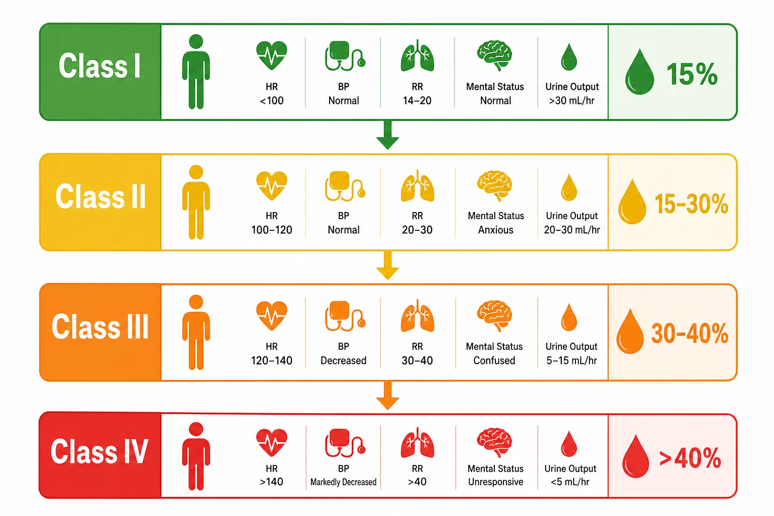
| Class | Blood loss | HR | BP | CNS | Urine |
|---|---|---|---|---|---|
| I | Under 15% | Under 100 | Normal | Normal | Normal |
| II | 15-30% | 100-120 | Normal | Anxious | 20-30 mL/h |
| III | 30-40% | 120-140 | Decreased | Confused | 5-15 mL/h |
| IV | Above 40% | Above 140 | Markedly decreased | Lethargic | Negligible |
Class I — under 15% blood loss (compensated, minimal)
Loss of up to 750 mL in a 70 kg adult. The body tolerates this without any measurable physiological compromise — this is the volume given in a standard blood donation. Heart rate is under 100, blood pressure normal, pulse pressure normal, mental state normal, urine output normal (above 30 mL/h, or above 0.5 mL/kg/hr). Capillary refill may be slightly prolonged. Management is to identify and stop the source of bleeding; fluid resuscitation is rarely required beyond what is needed to maintain the circulating volume, and the patient can usually be managed with crystalloid if needed. The trap is assuming the well-looking trauma patient is fine — class I can silently progress to class IV if an occult source (intra-abdominal, retroperitoneal, pelvic) is missed.[1]
Class II — 15-30% blood loss (compensated, mild)
Loss of 750-1500 mL. This is where compensation becomes clinically visible but blood pressure is still maintained. Heart rate rises to 100-120 (early tachycardia is the first detectable sign). Pulse pressure narrows because diastolic pressure rises from catecholamine-driven vasoconstriction while systolic is preserved — a narrowed pulse pressure is a more sensitive marker of significant volume loss than the absolute systolic pressure. The patient is anxious/hostile (mild CNS hypoperfusion), urine output falls to 20-30 mL/h. Management: give 1-2 L warmed balanced crystalloid bolus and reassess; identify and control bleeding.[1][1]
Class III — 30-40% blood loss (decompensated, moderate)
Loss of 1500-2000 mL. This is the decompensation threshold — the point at which sympathetic compensation is exhausted and blood pressure finally falls. Most patients are hypotensive by this stage (typically 30% of patients are hypotensive at 30% loss; virtually all by 40%). Heart rate 120-140, pulse pressure markedly narrowed then systolic falls, the patient is confused, oliguric (5-15 mL/h). Class III shock is a surgical / MTP emergency — this is the patient who needs blood, not crystalloid, and immediate bleeding control. Delays here are lethal.[1]
Class IV — above 40% blood loss (decompensated, severe)
Loss of more than 2000 mL (above 2 L). The patient is lethargic or obtunded, markedly hypotensive, heart rate above 140 (or paradoxically bradycardic in the pre-arrest agonal phase), negligible urine output, cold and mottled peripheries. This is imminent cardiac arrest from exsanguination — activate the massive transfusion protocol immediately, take to theatre / angioembolisation without delay. Mortality is high; survival depends on the speed of haemorrhage control and prevention of the lethal triad.[1][1]
Pathophysiology
Blood loss reduces venous return (preload) and stroke volume (Frank-Starling). Compensatory sympathetic mechanisms (tachycardia, vasoconstriction) maintain blood pressure initially (classes I-II). Decompensation (classes III-IV) occurs once loss exceeds about 30 per cent — BP falls, perfusion is compromised, and shock ensues.[1][1]
Trauma-induced coagulopathy (TIC) is an endogenous coagulopathy of severe trauma (tissue-factor release, protein-C activation, hyperfibrinolysis), compounded by resuscitation (dilution, acidosis, hypothermia).[2][8]
The compensation cascade — the order of signs
The body defends blood pressure through a stereotyped, ordered sequence of sympathetic and neuroendocrine responses. Understanding the ORDER is the key to recognising compensated shock:[1][1]
- Tachycardia (earliest sign) — reduced venous return reduces stroke volume; baroreceptors sense the fall and drive sympathetic output. Heart rate rises to maintain cardiac output (CO = HR × SV). This appears before any blood pressure change.
- Vasoconstriction (increased SVR) — catecholamines constrict arterioles and venules, raising systemic vascular resistance and venous tone (the venous side mobilises the unstressed volume into the circulation). This is why diastolic pressure rises early.
- Narrowed pulse pressure — because diastolic rises (vasoconstriction) while systolic is relatively preserved, the pulse pressure (systolic − diastolic) narrows. A narrowed pulse pressure is a sensitive marker of significant volume loss that precedes the fall in systolic pressure.
- Hypotension (LATE sign) — once loss exceeds ~30%, compensation is exhausted; systolic pressure falls. By now the patient is in class III-IV shock. A normal systolic pressure does not mean the patient is well-resuscitated.
- End-organ hypoperfusion — oliguria, altered mental state, cold peripheries, raised lactate. These are the markers of tissue-level shock regardless of the blood pressure number. [1]
The ordered compensation cascade in hypovolaemic shock (recognise the EARLY signs)
1. Tachycardia (FIRST detectable sign)
Baroreceptors sense falling preload → sympathetic outflow → heart rate rises to maintain cardiac output (CO = HR × SV). Appears before any blood pressure change. CAVEAT: tachycardia is non-specific (pain, anxiety, drugs), and a few patients (elite athletes, beta-blocked, elderly) may NOT mount a tachycardia — its absence does not exclude shock.
2. Vasoconstriction (rising SVR, rising diastolic)
Catecholamines constrict arterioles and the venous capacitance bed, raising systemic vascular resistance and mobilising the unstressed venous volume into the circulation. Diastolic blood pressure RISES early as a direct mechanical consequence of arteriolar constriction.
3. Narrowed pulse pressure (sensitive marker)
Systolic is relatively preserved (stroke volume maintained by tachycardia) while diastolic rises (vasoconstriction), so pulse pressure (systolic − diastolic) NARROWS. A narrowed pulse pressure is one of the most sensitive bedside markers of significant volume loss and precedes the fall in systolic pressure by a wide margin.
4. Hypotension (LATE — decompensation)
Once loss exceeds ~30% of blood volume, sympathetic compensation is exhausted and systolic pressure falls. The patient is now in class III-IV shock. A normal systolic pressure therefore does NOT exclude life-threatening haemorrhage — about 30% of volume can be lost before any fall in blood pressure.
5. End-organ hypoperfusion (the real target)
Oliguria (urine output under 0.5 mL/kg/hr), altered mental state (confusion, agitation, then lethargy), cold/mottled peripheries, prolonged capillary refill, raised serum lactate. These reflect tissue-level oxygen debt and are the TRUE indicators of shock — resuscitate to their correction, not to a blood pressure number.
Compensated shock (class I-II)
Under 30% blood loss
- Blood pressure MAINTAINED by sympathetic drive
- Tachycardia and narrowed pulse pressure are the early tells
- Patient anxious/mildly confused; oliguria beginning
- Lactate may already be rising (anaerobic metabolism)
- Treatable with crystalloid + source control if caught here
Decompensated shock (class III-IV)
Above 30% blood loss
- Blood pressure has FALLEN — compensation exhausted
- Marked tachycardia, confused/lethargic, oliguric/anuric
- Cold mottled peripheries; high lactate; base deficit
- Blood (MTP), not crystalloid, is the resuscitation fluid
- Immediate surgical/angiographic bleeding control required
The lethal triad
Three interlocking processes worsen coagulopathy and mortality:[2][1]
- Acidosis (pH below 7.2 → reduced coagulation-factor enzyme activity, impaired fibrin polymerisation).[2]
- Hypothermia (core below 35 → slowed coagulation cascade, impaired platelet function).[2]
- Coagulopathy (dilutional plus TIC, plus consumption).[2]
Each worsens the others in a vicious cycle; prevention is central.[2] Once the lethal triad is fully established (pH under 7.2, temperature under 35°C, INR above 1.5), mortality approaches 100%. The triad is far easier to prevent than to reverse — the entire damage-control philosophy is built on this premise.[7]
Fluid resuscitation — the initial approach and the response paradigm
The initial fluid strategy depends on whether the bleeding is controlled or uncontrolled. This distinction is the foundation of modern trauma resuscitation and was established by the landmark Bickell (1994) trial.[3]
Controlled haemorrhagic shock (non-trauma, or bleeding already stopped)
Give a 1-2 L warmed balanced crystalloid bolus (Hartmann / compound sodium lactate, or Plasma-Lyte — NOT normal saline, which causes hyperchloraemic acidosis that worsens coagulopathy), then reassess the response:[1][1]
- Rapid response — vital signs normalise and stay normal: the patient had a volume deficit and bleeding has stopped. Continue maintenance, investigate the cause.
- Transient response — vital signs improve then deteriorate again as the bolus wears off: this indicates ongoing haemorrhage. This is the trigger to activate the massive transfusion protocol (MTP), switch to blood-based resuscitation, and achieve definitive bleeding control (surgery / endoscopy / angioembolisation).
- No response / minimal response — severe ongoing loss or a different shock phenotype (cardiogenic from myocardial contusion/tamponade, distributive from neurogenic shock in spinal injury). Reassess the diagnosis with point-of-care ultrasound (FAST, RUSH/E-FAST); the "non-responder" to crystalloid in trauma is bleeding until proven otherwise. [1]
Uncontrolled haemorrhagic shock (active trauma bleeding)
In the actively bleeding trauma patient, aggressive crystalloid is harmful — it dilutes clotting factors, dislodges clots ("pop the clot"), worsens acidosis, and causes a lethal triad. The strategy is damage control resuscitation (below): permissive hypotension, minimise crystalloid, resuscitate with blood in 1:1:1 ratio, achieve bleeding control.[3][9]
The fluid-response algorithm — assess after the 1-2 L crystalloid bolus
Rapid and sustained response
Vital signs normalise and remain normal after the bolus. Implies a finite volume deficit with bleeding already controlled. Continue maintenance fluid, investigate and definitively treat the source. No blood products required unless haemoglobin is low.
Transient response (bleeding is ONGOING)
Vital signs improve with the bolus then drift back down as it redistributes/metabolises. This is the classical sign of CONTINUING haemorrhage. Activate the massive transfusion protocol, switch from crystalloid to blood-based resuscitation (1:1:1), and secure definitive bleeding control (surgery / angioembolisation / endoscopy). Do not keep chasing with crystalloid.
No or minimal response
Failure to respond to an adequate crystalloid bolus means either (a) massive ongoing loss exceeding the rate of infusion, or (b) a different shock phenotype — cardiogenic (myocardial contusion, tamponade, tension pneumothorax), obstructive, or distributive (neurogenic from spinal cord injury). Perform point-of-care ultrasound (FAST / E-FAST / RUSH) to re-evaluate; in trauma, assume ongoing haemorrhage until proven otherwise.
Crystalloid-led resuscitation
Traditional / controlled bleeding
- 1-2 L warmed balanced crystalloid bolus, then reassess
- Appropriate for NON-haemorrhagic hypovolaemia (dehydration, DKA, sepsis)
- Appropriate for trauma ONLY once bleeding is controlled
- Harms in active bleeding: dilution, clot disruption, acidosis, lethal triad
- Normal saline causes hyperchloraemic acidosis — use Hartmann / Plasma-Lyte
Blood-led (DCR) resuscitation
Active uncontrolled haemorrhage
- Minimise crystalloid; resuscitate with blood products
- 1:1:1 RBC:FFP:platelets (PROPPR), TXA within 3h, empiric calcium
- Permissive hypotension SBP 80-90 until bleeding controlled (except TBI)
- Goal-directed via ROTEM/TEG once bleeding slows
- Definitive bleeding control (surgery / angio / endoscopy) is the real treatment
Damage control resuscitation (DCR)
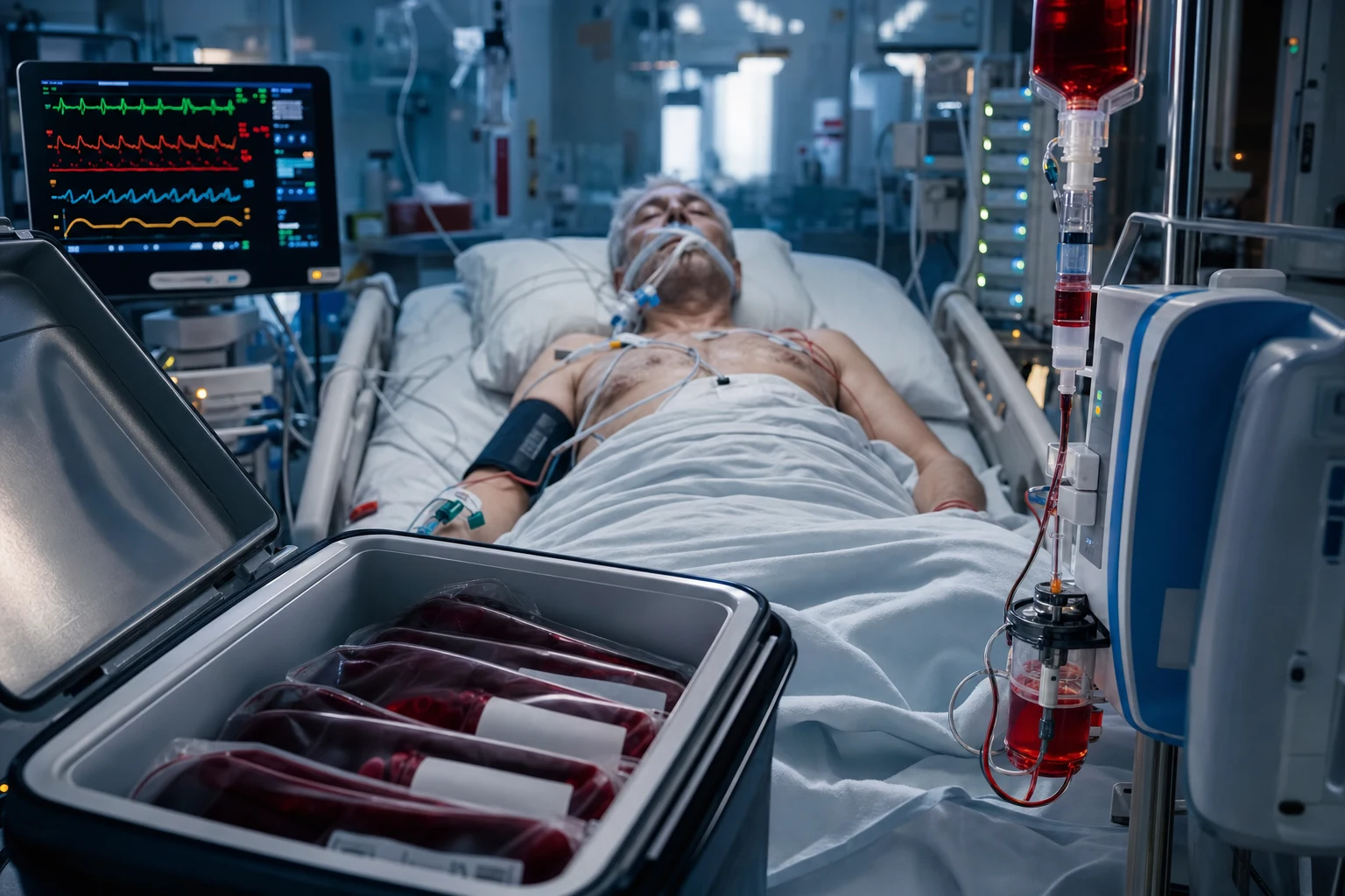
Damage control resuscitation is the integrated package that replaced the old "give two litres of saline and run to theatre" approach. Its rationale: aggressive crystalloid and normotension in the actively bleeding patient deepen the lethal triad and worsen mortality. DCR instead buys time and physiology until the bleed is definitively controlled.[2][9]
1. Permissive hypotension.[1][2][10]
- Target MAP 50-65 (or SBP 80-90) until bleeding is controlled. Avoid normotension, which dislodges clots and dilutes clotting factors.[1]
- Restrict fluids; allow permissive hypotension to minimise clot disruption.[2]
- Do NOT use permissive hypotension in TBI — the brain-injured patient needs a CPP (MAP minus ICP) above 60, so maintain normotension (SBP above 110).[1][7]
2. 1:1:1 massive transfusion.[1][4]
- RBC : FFP : platelets 1 : 1 : 1 (the PROPPR trial showed equivalent overall mortality but earlier haemostasis and fewer deaths from exsanguination at 24h).[4]
- Give cryoprecipitate / fibrinogen to keep fibrinogen above 2 g/L.[7]
- TXA 1 g IV within 3 hours of injury (CRASH-2 — reduces mortality; after 3 hours there is no benefit and possible harm).[5][6]
3. Damage control surgery.[2]
- Rapid control of bleeding (packing, ligation, balloon tamponade, temporary vascular shunt).[2]
- Not definitive repair — stabilise first, then return to theatre (typically 24-48 h) for definitive surgery once stable.[2]
4. Warming and correction of acidosis.[2][1]
- Warmed IV fluids, forced-air warming, and body-cavity lavage as needed.[2]
- Empiric calcium (citrate in stored blood chelates Ca²⁺) — CaCl₂ 10 mmol per ~4 units blood products.[7]
Permissive hypotension in depth
Permissive hypotension means deliberately tolerating a sub-normal blood pressure in the actively bleeding patient, on the principle that restoring full pressure before haemostasis mechanically disrupts clots and accelerates bleeding. The evidence base is Bickell (1994): in penetrating torso trauma, delaying fluid resuscitation until operative control of bleeding improved survival (70% vs 62%) compared with immediate aggressive fluids.[3] Dutton (2002) showed that titrating to a lower target SBP (~70 mmHg) versus conventional (~100 mmHg) during active haemorrhage was safe and did not increase mortality.[10]
Permissive hypotension (active bleeding)
SBP 80-90 mmHg until control
- Deliberate, temporary sub-normal BP until haemorrhage control
- Rationale: normotension "pops the clot", dilutes factors, accelerates bleeding
- Target: SBP 80-90 mmHg (MAP ~50-60) — consciousness and peripheral perfusion
- Give BLOOD, not crystalloid, to hold this floor
- Once bleeding controlled, restore normotension
Normotensive resuscitation (bleeding controlled, or TBI)
Conventional targets
- Restore normal blood pressure once haemostasis achieved
- MANDATORY in TBI — need SBP above 110 (MAP above 80) for CPP above 60
- Mandatory in spinal cord injury with cord hypoperfusion
- Use in non-trauma bleeding (variceal, AAA, obstetric) — extrapolation unproven
- Elderly and comorbid tolerate deep hypotension poorly — apply judgement
Tranexamic acid (TXA) — CRASH-2 and the 3-hour rule
Tranexamic acid is an anti-fibrinolytic (lysine analogue blocking plasminogen activation) that counteracts the hyperfibrinolytic component of trauma-induced coagulopathy. The dose is 1 g IV over 10 minutes, then 1 g over 8 hours.[5]
CRASH-2 (2010, 20,211 patients) showed TXA reduced all-cause mortality (14.5% vs 16.0%) and bleeding death, with no increase in thromboembolism.[5] The crucial refinement came from the time-window analysis (2011): benefit is greatest within 1 hour of injury, present within 3 hours, but TXA increases mortality when given more than 3 hours after injury.[6]
Bickell — immediate vs delayed fluid resuscitation (NEJM 1994)
Single-centre RCT; 598 hypotensive patients with penetrating torso trauma
Population: Adults with penetrating torso injury and SBP under 90 mmHg
Key finding
Delayed resuscitation improved survival (70% vs 62%, p=0.04) and reduced length of stay. Postoperative complications were also lower in the delayed group.
Practice change
In penetrating torso trauma with shock, aggressive pre-operative crystalloid worsens outcome. Delay resuscitation until bleeding is controlled — the foundation of permissive hypotension and DCR.
CRASH-2 (Lancet 2010) and the 2011 time-window analysis
Multicentre placebo-controlled RCT; 20,211 trauma patients with or at risk of major bleeding
Population: Adult trauma patients with significant haemorrhage, 274 hospitals across 40 countries
Key finding
TXA reduced all-cause mortality (14.5% vs 16.0%, p=0.0035) and bleeding death (4.9% vs 5.7%). The 2011 time-window analysis showed benefit greatest within 1h, present within 3h, but TXA INCREASED bleeding death when given more than 3 hours after injury (4.4% vs 3.1%).
Practice change
The 3-hour rule: give TXA 1 g IV ASAP (ideally within 1h, pre-hospital if possible). Never give TXA more than 3 hours after injury.
PROPPR (Holcomb, JAMA 2015)
Multicentre RCT; 680 severely injured adults predicted to need massive transfusion
Population: Trauma patients at 12 Level I trauma centres in North America
Key finding
No significant difference in 24h mortality (12.7% vs 17.0%, p=0.09) or 30-day mortality. BUT 1:1:1 achieved earlier haemostasis and fewer exsanguination deaths at 24h, with no increase in ARDS or multi-organ failure.
Practice change
1:1:1 is safe and reasonable empirically during active massive bleeding — it improves early haemostasis. No overall mortality advantage, so switch to viscoelastic-guided therapy as soon as possible.
Resuscitation endpoints — what to target
The goal of resuscitation is restoration of tissue oxygen delivery and the clearance of oxygen debt, not a particular blood pressure number. Resuscitating to a "normal" blood pressure alone is inadequate — a patient can be normotensive and still in shock at the tissue level (ongoing anaerobic metabolism, rising lactate). The validated endpoints are:[1][1]
- Lactate clearance — serum lactate reflects the magnitude of anaerobic metabolism (oxygen debt). The target is lactate returning to normal (under 2 mmol/L), ideally with a fall of at least 10% per hour (or 50% in the first 6 hours, analogous to the Jansen lactate-clearance concept). A rising or static lactate signals ongoing hypoperfusion. Lactate is the single best global marker of adequacy of resuscitation.
- Urine output above 0.5 mL/kg/hr — renal perfusion is a sensitive, real-time, bedside index of adequate cardiac output and tissue perfusion. Oliguria (under 0.5 mL/kg/hr) signals under-resuscitation. A catheter and an hourly urine measure are mandatory in any shocked patient.
- Normal mental state — the brain is the most perfusion-sensitive organ; a clear sensorium indicates adequate cerebral perfusion (and by inference adequate cardiac output). Confusion, agitation, or lethargy = ongoing shock.
- Base deficit / bicarbonate — a base deficit worse than −5 mmol/L (or worsening trend) reflects metabolic acidosis from hypoperfusion; it should normalise with adequate resuscitation.
- Central venous oxygen saturation (ScvO₂) — above 70% indicates adequate oxygen delivery relative to extraction (the principle from early goal-directed therapy).
- Capillary refill time under 2 seconds and warm, well-perfused peripheries — a return of the vasoconstricted, mottled periphery to normal indicates SVR normalising as shock resolves.
- Resolution of metabolic acidosis (pH above 7.35, normal strong ion difference). [1]
Global perfusion endpoints
The primary targets
- Lactate clearance — under 2 mmol/L, fall of at least 10% per hour
- Urine output above 0.5 mL/kg/hr (renal perfusion)
- Normal mental state (cerebral perfusion)
- Base deficit normalising (better than −5 mmol/L)
- ScvO₂ above 70%
Haemodynamic / perfusion signs
The supporting signs
- Blood pressure restored to baseline (after bleeding controlled)
- Heart rate falling back toward normal
- Pulse pressure widening (SVR normalising)
- Warm peripheries, capillary refill under 2 seconds
- Resolution of metabolic acidosis on blood gas
What endpoints do NOT prove
Common traps
- A normal blood pressure alone does NOT exclude ongoing shock
- Central venous pressure (CVP) is a poor predictor of fluid responsiveness
- Normalisation of heart rate can lag or be masked (beta-blockers, athletes)
- Haemoglobin concentration can be misleading early (haemodilution is delayed)
- Persistent lactate despite normal BP = ongoing hypoperfusion — keep going
Prognosis
Mortality depends on the speed of bleeding control and prevention of the lethal triad. Damage control resuscitation reduces mortality compared with traditional aggressive fluid resuscitation.[1][2][1] Trauma-induced coagulopathy on arrival multiplies mortality by 3-4 times; the lethal triad, once established, carries a mortality approaching 100%. Early recognition (class II, before hypotension), prompt MTP activation, and rapid definitive bleeding control are the modifiable determinants of survival.[8]
The lethal triad of massive haemorrhage (click each)
INR above 1.5
Trauma-induced coagulopathy (endogenous) plus dilutional coagulopathy (iatrogenic) plus consumption. INR above 1.5 on arrival defines TIC. The three elements reinforce one another; once fully established, mortality approaches 100%.
Special populations and pitfalls
The elderly patient
Comorbidity + beta-blockers
- Less physiological reserve; tolerate hypotension poorly
- May not mount tachycardia (beta-blockers, conduction disease) — HR is unreliable
- Comorbid coronary disease means permissive hypotension risks myocardial ischaemia
- Apply permissive hypotension cautiously; a higher BP target may be appropriate
The pregnant patient
Changed physiology
- Baseline tachycardia and increased blood volume mask loss (up to 35% before signs)
- Uterine compression of IVC in supine position worsens preload — nurse left lateral tilt
- Resuscitate the MOTHER — maternal survival is the best thing for the fetus
- Permissive hypotension less studied; conventional targets often preferred
The paediatric patient
Exceptional compensatory reserve
- Children compensate extremely well — can maintain BP until 45-50% volume loss
- A falling blood pressure in a child is a RED FLAG indicating decompensation
- Use weight-based fluid boluses: 20 mL/kg crystalloid, repeat, then blood
- Bradycardia and hypotension in a bleeding child is pre-arrest
The athlete / beta-blocked patient
Masked tachycardia
- Resting bradycardia means the usual tachycardic response is blunted
- Beta-blockade prevents the HR rise even in severe shock
- Rely on pulse pressure, lactate, urine output, and mental state instead
- Have a low threshold to investigate for occult bleeding
Differential diagnosis of shock that is NOT responding
A trauma patient who fails to respond to adequate resuscitation may have an alternative or additional cause of shock — mechanical, cardiogenic, or obstructive. Point-of-care ultrasound (E-FAST / RUSH) is the key diagnostic tool:[1]
- Tension pneumothorax — hypotension, distended neck veins, unilateral absent breath sounds, tracheal deviation. Needle/finger thoracostomy immediately.
- Cardiac tamponade — Beck's triad (hypotension, muffled heart sounds, raised JVP), pulsus paradoxus. Pericardiocentesis / thoracotomy.
- Myocardial contusion / cardiogenic shock — blunt cardiac injury can cause pump failure; echocardiography shows reduced contractility.
- Neurogenic shock (spinal cord injury) — hypotension with bradycardia (unlike the tachycardia of hypovolaemia), warm peripheries (loss of sympathetic tone).
- Missed source of bleeding — pelvic fracture, retroperitoneal haematoma, intrathoracic, long-bone fracture (each femur fracture can hold 1-2 units). Re-survey the patient. [1]
Monitoring the resuscitating patient
Monitoring the haemorrhagic-shock patient — from arrival to stabilisation
1. Primary survey and adjuncts (A-E + E-FAST)
Airway with cervical spine control; Breathing (oxygen, exclude tension pneumothorax); Circulation (two large-bore cannulae, blood samples including crossmatch, ROTEM/TEG, venous gas + lactate); Disability (GCS, pupils — TBI changes the BP target); Exposure. Perform E-FAST / RUSH to identify free fluid, tamponade, pneumothorax.
2. Arterial line + continuous haemodynamics
Insert an arterial line early for beat-to-beat blood pressure (essential for titrating permissive hypotension) and arterial blood gas access. Monitor heart rate, pulse pressure, and mean arterial pressure continuously. A falling diastolic or widening pulse pressure signals re-bleeding.
3. Hourly urine output + serial lactate
Urinary catheter for hourly urine output (target above 0.5 mL/kg/hr). Serial lactate every 1-2 hours until normalising (target fall of at least 10% per hour, under 2 mmol/L). Serial blood gas for pH, base deficit, haemoglobin.
4. Coagulation and viscoelastic monitoring
During active MTP, draw ROTEM/TEG, fibrinogen, platelets, and ionised calcium every 30-60 min. Drive component therapy from the viscoelastic trace (CT → FFP; A10/α-angle → fibrinogen; MA/MCF → platelets; ML/LY30 → TXA). Keep fibrinogen above 2 g/L and ionised Ca²⁺ above 1.0 mmol/L.
5. Temperature and perfusion
Continuous core temperature — keep above 36°C with forced-air warming and warmed fluids. Monitor capillary refill, peripheral temperature, and mental state as end-organ perfusion markers. Re-survey frequently for re-bleeding or missed injuries.
Red flags
Exam practice
SAQ — Haemorrhagic shock classification and resuscitation strategy
12 minutes · 10 marks
A 32-year-old man is brought to the emergency department 50 minutes after a motorcycle crash. He is anxious and confused. HR 128, BP 96/64 (pulse pressure 32), SpO₂ 96% on 15 L O₂. Urine output 15 mL in the first hour. Lactate 5.2 mmol/L. FAST is positive in the abdomen.
SAQ — Why hypotension is a late sign, and the non-responder
10 minutes · 8 marks
A 24-year-old woman is brought in after a fall. She is pale and clammy. HR 115, BP 110/80 (pulse pressure 30), capillary refill 4 seconds, lactate 3.8 mmol/L. She is given 2 L of warmed Hartmann solution. Her BP rises to 120/85 briefly, then over 20 minutes falls back to 100/78.
Clinical pearls
References
- [1]Boutros A, et al. Resuscitation for Hypovolemic Shock Surg Clin North Am, 2017.PMID 29132511
- [2]Duchesne JC, et al. Damage control resuscitation Blood Rev, 2015.PMID 25631636
- [3]Bickell WH, Wall MJ Jr, Pepe PE, et al. The use and clinical importance of a substrate-specific electrode for rapid determination of blood lactate concentrations JAMA, 1994.PMID 7966896
- [4]Holcomb JB, Tilley BC, Baraniuk S, et al. (PROPPR Study Group) Transfusion of plasma, platelets, and red blood cells in a 1:1:1 vs a 1:1:2 ratio and mortality in patients with severe trauma: the PROPPR randomized clinical trial JAMA, 2015.PMID 25647203
- [5]CRASH-2 trial collaborators Effects of tranexamic acid on death, vascular occlusive events, and blood transfusion in trauma patients with significant haemorrhage (CRASH-2): a randomised, placebo-controlled trial Lancet, 2010.PMID 20554319
- [6]CRASH-2 collaborators The importance of early treatment with tranexamic acid in bleeding trauma patients: an exploratory analysis of the CRASH-2 randomised controlled trial Lancet, 2011.PMID 21439633
- [7]Spahn DR, Bouillon B, Cerny V, et al. The European guideline on management of major bleeding and coagulopathy following trauma: fifth edition Crit Care, 2019.PMID 30917843
- [8]Brohi K, Singh J, Heron M, Coats T Acute coagulopathy of trauma: hypoperfusion induces systemic anticoagulation and hyperfibrinolysis J Trauma, 2008.PMID 18469643
- [9]Cannon JW, Khan MA, Raja AS, et al. Damage control resuscitation in patients with severe traumatic hemorrhage: A practice management guideline from the Eastern Association for the Surgery of Trauma J Trauma Acute Care Surg, 2017.PMID 28225743
- [10]Dutton RP, Mackenzie CF, Scalea TM Molecular Mechanisms in Root Nodule Development J Plant Growth Regul, 2000.PMID 11038225