ICU · Resuscitation & shock
Massive Transfusion, Blood Products & Tranexamic Acid
Also known as Massive transfusion · Massive haemorrhage protocol · Blood products in resuscitation · Tranexamic acid · TXA · PROPPR trial · CRASH-2 · TRALI · TACO · Damage control resuscitation · Permissive hypotension · Haemostatic resuscitation · Damage control surgery · Viscoelastic testing · TEG · ROTEM · WOMAN trial · HALT-IT · Lethal triad
Massive transfusion in resuscitation turns on two evidence-based questions: the component ratio (PROPPR — 1:1:1 vs 1:1:2 RBC:plasma:platelets) and tranexamic acid (CRASH-2 — mortality benefit, but only within 3 hours of injury; WOMAN in postpartum haemorrhage, HALT-IT negative in GI bleed, TICH-2 in intracerebral haemorrhage). Damage control resuscitation (permissive hypotension, haemostatic 1:1:1 resuscitation, damage control surgery), viscoelastic (TEG/ROTEM)-guided component therapy, fibrinogen replacement above 1.5-2.0 g/L, calcium replacement (citrate chelation), the lethal triad of hypothermia-acidosis-coagulopathy, and awareness of transfusion reactions (TRALI, TACO, acute haemolysis) are central.
On this page & tools
Your progress
Saved locally on this device.
Target exams
Overview & definition
Massive transfusion supports the bleeding patient while bleeding is controlled. It turns on two evidence-based questions: the component ratio (PROPPR — a 1:1:1 ratio of RBC:plasma:platelets) and tranexamic acid (CRASH-2 — a mortality benefit, but only if given within 3 hours). The keys to a good outcome are early activation of the massive haemorrhage protocol (MHP), ratio-based then viscoelastic-guided component therapy, calcium replacement (citrate chelation), and vigilance for transfusion reactions.[1][3][1]
Modern trauma resuscitation is organised around damage control resuscitation (DCR) — three integrated pillars: (1) permissive hypotension until bleeding is controlled, (2) haemostatic resuscitation with blood products in a balanced 1:1:1 ratio and minimal crystalloid, and (3) damage control surgery — definitive haemorrhage control takes priority over definitive organ repair. DCR exists to break the vicious circle of the lethal triad (hypothermia, acidosis, coagulopathy), which is the physiology that kills the bleeding patient long before their surgical injuries do.[9][1]
Definitions & the massive haemorrhage protocol
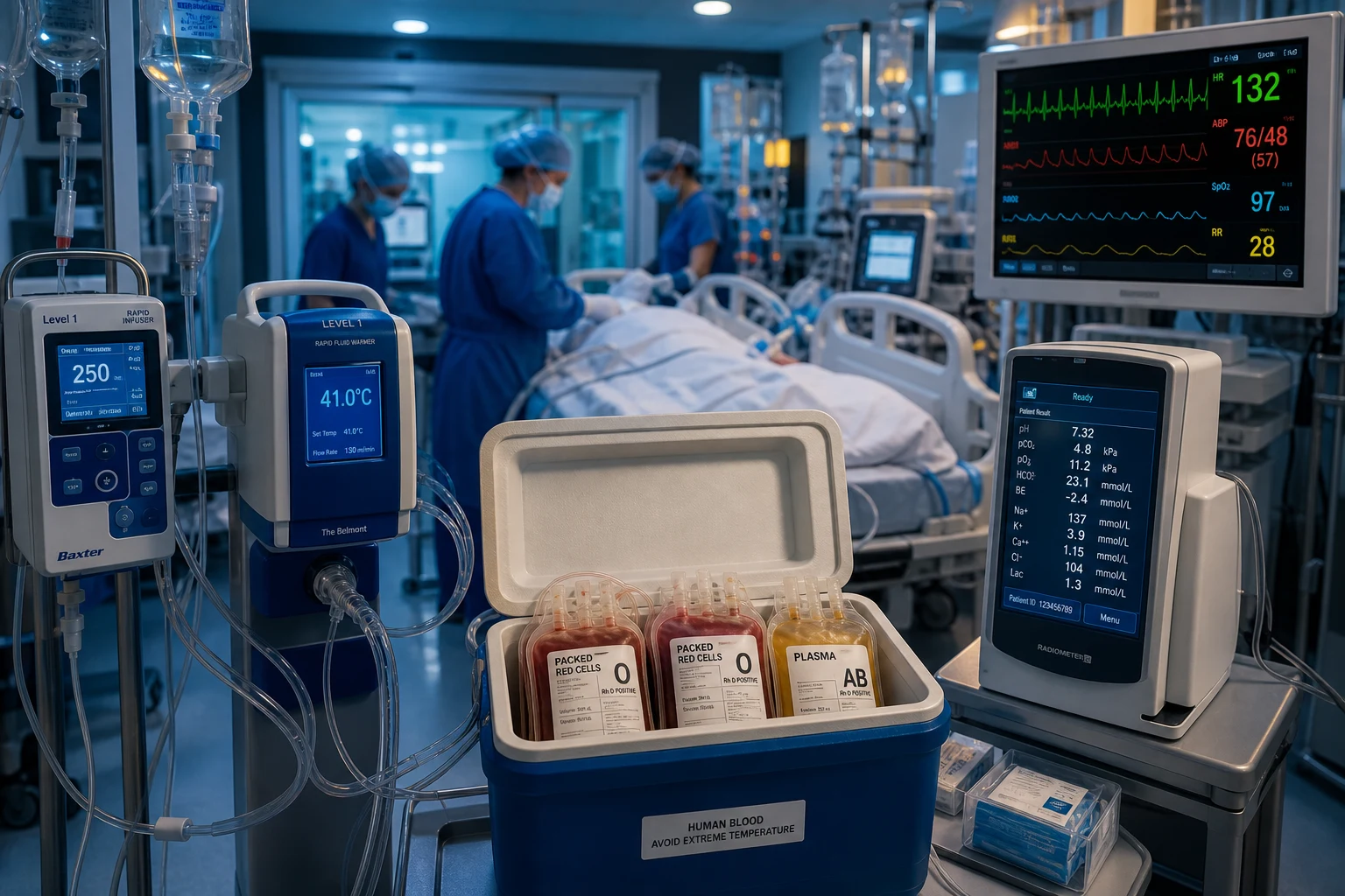
Massive transfusion is commonly defined as the transfusion of 10 or more units of red cells within 24 hours, or 4 or more units within 1 hour with ongoing bleeding, or replacement of one blood volume within 24 hours.[1] In practice these thresholds are often exceeded only after the patient has already been sick for some time — which is why the operational trigger is the massive haemorrhage protocol, not the unit count. The clinically useful concept is major haemorrhage: blood loss that is uncontrolled, ongoing, or producing shock — activate the protocol on physiology, not on a tally.[9]
Definitions of massive transfusion — exam and operational
| Definition | Threshold | Source / use | Practical note |
|---|---|---|---|
| 10 units RBC / 24 h | Cumulative red cells | ASA / AABB (most widely taught) | Easy to remember; met late in the illness |
| 4 units RBC / 1 h | Rapid transfusion with ongoing bleeding | Many MHP activation criteria | The trigger most aligned with the physiology |
| 1 blood volume / 24 h | ~10 units RBC in a 70 kg adult (~70 mL/kg) | Approximation; better in children | Loss of one entire blood volume |
| >50% blood volume / 3 h | ~3.5 L / 3 h | Common alternative | For very rapid bleeds |
| Massive haemorrhage protocol (MHP) operational | Any uncontrolled + ongoing blood loss with shock | AABB / hospital policies | Activate on physiology, not on a count — do not wait |
The massive haemorrhage protocol (MHP) is a pre-defined, team-activated pathway that delivers blood products rapidly. The principles:[3][1][10]
- Activate early — do not wait for the patient to meet the definition. A single phone call releases a pre-packed box of RBC, plasma and platelets in a fixed ratio.
- Ratio-based initially, then switch to viscoelastic-guided (TEG/ROTEM) component therapy once bleeding slows and a trace is available.
- Communicate — a dedicated team leader, a timekeeper, and a scribe; closed-loop communication with the blood bank.
- Two large-bore cannulae, a rapid infuser with a warming unit, and a blood-gas analyser at the bedside.
- Sample for laboratory on activation — full blood count, coagulation, fibrinogen, group-and-save / crossmatch, ionised calcium, potassium, lactate, venous gas (for pH and base excess).
- Pre-warmed, pressurised, calcium-replaced, group-confirmed. The four operational words.
A systematic review and meta-analysis of MTP implementation in trauma showed that introduction of a formal massive transfusion protocol is associated with improved survival (roughly a one-third reduction in mortality).[10] The benefit is plausibly driven by earlier balanced-ratio transfusion and reduced crystalloid use.
Massive haemorrhage protocol — activation to delivery
RECOGNISE and ACTIVATE
Uncontrolled + ongoing blood loss with shock (or predicted to need >=4 units). One phone call to blood bank activates the protocol. Do NOT wait for laboratory confirmation. Notify anaesthetics, surgery, haematology, and senior medical officer simultaneously.
RELEASE pack 1 — uncrossmatched O-negative + 1:1:1 components
Two units O-negative (O-positive acceptable in males / post-menopausal females if O-negative scarce) delivered within minutes, followed by a pre-packed box of 6 units RBC + 4 units FFP + 1 adult dose platelets (~1:1:1). Some protocols add cryoprecipitate (10 units) up-front when fibrinogen is expected to be low.
SECURE the haemorrhage — call the surgeon / endoscopist / interventional radiologist NOW
Bleeding control is definitive treatment; transfusion only buys time. Activate the operating theatre, endoscopy, or interventional radiology in parallel with — not after — pack release. Damage control surgery for trauma; endoscopic / IR haemostasis for GI bleed; surgical / IR control for obstetric and ruptured aneurysm.
GIVE tranexamic acid 1 g IV bolus then 1 g over 8 h
ONLY if within 3 h of injury / haemorrhage onset (CRASH-2, WOMAN). After 3 h the benefit is lost and harm may follow. Avoid in upper GI bleed (HALT-IT showed harm — venous thromboembolism without mortality benefit).
WARM, calcium-replace, avoid crystalloid
Forced-air warming to >36 degC; monitor ionised calcium and replace (calcium chloride 1 g via central line, or calcium gluconate peripherally). Minimise crystalloid — it dilutes clotting factors, causes acidosis (hyperchloraemia), and worsens the lethal triad.
REASSESS with viscoelastic testing (TEG/ROTEM) and laboratory
Once bleeding slows, switch from ratio-based to goal-directed component therapy. Use a low fibrinogen trace / Clauss fibrinogen to drive cryoprecipitate or fibrinogen concentrate; a prolonged clotting time to drive FFP or PCC; a low maximum amplitude to drive platelets. Re-check Hb, INR, platelets, fibrinogen, ionised Ca, K+, and venous pH every 30-60 min.
STAND DOWN when haemostasis is achieved
Confirm surgical / endoscopic control, normalising lactate, stable haemoglobin, and a normalising viscoelastic trace. Notify blood bank. Debrief the team. Review for transfusion reactions (TRALI/TACO especially within 6 h of stand-down).
What is in a typical 'box' — adult pack composition
| Component | Amount per box | Volume | Target |
|---|---|---|---|
| Packed red cells (PRBC) | 4-6 units | ~250-300 mL/unit | Hb 70-90 g/L (higher in TBI / ischaemia) |
| Fresh frozen plasma (FFP) | 4 units | ~200-250 mL/unit | INR <1.5 |
| Platelets | 1 adult dose (1 apheresis unit or ~6 pooled) | ~200-300 mL | Platelets >50 (or >100 in active bleed / TBI) |
| Cryoprecipitate | 10 units (adult dose) | ~150 mL total | Fibrinogen >1.5-2.0 g/L |
| Calcium | Titrate — CaCl2 1 g central / Ca-gluconate peripherally | 10 mL | Ionised Ca >1.0 mmol/L |
Damage control resuscitation (DCR)
Damage control resuscitation integrates three principles delivered from the moment of injury through definitive haemorrhage control:[9][1]
The three pillars of damage control resuscitation
| Pillar | Principle | Practical execution | Evidence |
|---|---|---|---|
| 1. Permissive hypotension | Keep the SBP modest (typically 80-90 mmHg, or MAP 65) until haemorrhage is controlled — a normal BP dislodges clots and dilutes clotting factors via increased bleeding | Titrate fluids / vasopressors to SBP 80-90 (or conscious-level) until bleeding controlled; then restore normotension | Avoid in TBI, ischaemic heart disease, elderly — these need perfusion pressure |
| 2. Haemostatic resuscitation | Replace lost blood with blood in a balanced ratio (1:1:1 PRBC:FFP:platelets), minimise crystalloid, give plasma early | PROPPR 1:1:1; prehospital plasma (PAMPer); early fibrinogen, calcium, TXA | PROPPR (1:1:1); PAMPer (prehospital plasma mortality benefit) |
| 3. Damage control surgery | Definitive haemorrhage control first; definitive organ repair later | Rapid control of bleeding + contamination ("the operation of least invasive surgery"); pack, close, rewarm, correct coagulopathy in ICU; return to theatre at 24-48 h for definitive repair | Standard of care since 1990s |
Permissive hypotension
The aim is to maintain enough perfusion to keep the brain and heart alive without raising the pressure enough to blow off clots. A typical target is SBP 80-90 mmHg (or MAP ~65 mmHg) until haemorrhage control is achieved, then restore normotension. Clinical signs of adequate perfusion (mentation, urine output, lactate clearance) guide the target.[9]
Permissive hypotension — who is in, who is out
| Permissive hypotension IS appropriate | Permissive hypotension is NOT appropriate (avoid) |
|---|---|
| Penetrating torso trauma with ongoing haemorrhage | Traumatic brain injury (TBI) — hypotension doubles mortality; SBP must stay >110 mmHg |
| Short transport time to definitive haemorrhage control | Ischaemic heart disease / known coronary disease |
| Young, previously fit patient | Elderly — limited cardiovascular reserve; titrate to higher SBP |
| Suspected active arterial bleeding being surgically controlled | Spinal cord injury — risk of secondary ischaemic injury |
| Prolonged transport / delayed haemorrhage control — cumulative hypoperfusion harms | |
| Aortic stenosis / critical aortic disease — depend on preload and pressure |
Caveat: the classic trials of permissive hypotension (e.g., Bickell 1994, in penetrating trauma) excluded head injury. The principle is time-limited — prolonged permissive hypotension trades short-term clot preservation for cumulative organ hypoperfusion (renal, mesenteric, cardiac). Once bleeding is controlled, restore normotension and normalise lactate. [1]
Damage control surgery
Damage control surgery is the surgical expression of the same philosophy: stop the bleeding, stop the contamination, get out. The patient is too sick for a definitive operation. The phases:[1][9]
Damage control surgery — the staged approach
Phase 1 — Damage control laparotomy / thoracotomy (operation 1)
Rapid control of haemorrhage (ligation, shunting, packing) and contamination (resect / staple off perforated bowel without anastomosis). Target <60-90 min on table. Temporary closure (e.g., vacuum dressing, towel-clip) — NOT primary fascial closure.
Phase 2 — ICU resuscitation (the "lethal triad" phase)
Active rewarming (>36 degC), correct acidosis (improve perfusion; bicarbonate only for pH <7.1 with intractable shock), correct coagulopathy (FFP, cryoprecipitate, platelets, calcium guided by TEG/ROTEM), and support perfusion. This is where the patient "finishes" the resuscitation the operating theatre could not.
Phase 3 — Definitive repair (operation 2, at 24-48 h)
Once warm, non-acidotic, and coagulopathy corrected, return to theatre: remove packs, restore bowel continuity (anastomosis or stoma), close the abdomen definitively. Sometimes further operations are needed (operation 3, 4...).
The ratio question (1:1:1) — PROPPR
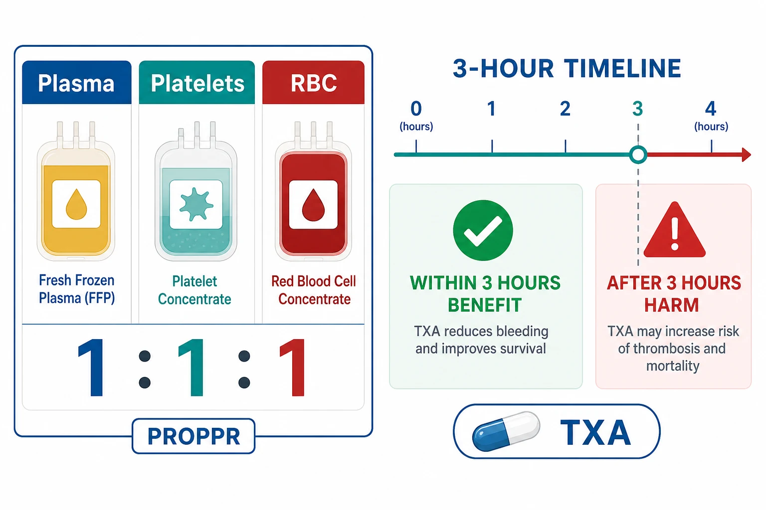
The PROPPR trial (Holcomb, JAMA 2015) randomised 680 patients with severe trauma and active bleeding to a 1:1:1 versus 1:1:2 ratio of plasma:platelets:RBC.[3]
- The 1:1:1 group had lower 24-hour mortality (12.7 vs 17.0 per cent) and fewer deaths from exsanguination.[3]
- 30-day mortality was not significantly different (22.4 vs 26.1 per cent).[3]
- No increase in complications (no more multi-organ failure, ARDS, sepsis, or thromboembolism) with the higher plasma/platelet ratio.[3]
The rationale is the endotheliopathy of trauma: severe shock disrupts the endothelial glycocalyx and consumes clotting factors, so early plasma restores both volume and the damaged endothelium — hence the move away from crystalloid and RBC-only resuscitation toward a balanced ratio.[1][9]
Prehospital plasma (PAMPer, Sperry, NEJM 2018) extended this principle to the field. In trauma patients at risk of haemorrhagic shock transported by air, two units of thawed plasma given before hospital arrival reduced 30-day mortality (23 vs 33 per cent; mortality ratio 0.65). The signal supports the concept that early plasma is itself a resuscitative drug — it repairs the endothelial glycocalyx and reduces the shock-driven coagulopathy before the patient reaches hospital.[12]
PROPPR — 1:1:1 vs 1:1:2 (plasma:platelets:RBC) — the headline numbers
| Outcome | 1:1:1 group | 1:1:2 group | Significance |
|---|---|---|---|
| 24-hour mortality | 12.7% | 17.0% | Significantly lower with 1:1:1 |
| Death from exsanguination (24 h) | 9.2% | 14.6% | Significantly fewer with 1:1:1 |
| 30-day mortality | 22.4% | 26.1% | NOT significant (P=0.26) |
| ARDS / MOF / sepsis / VTE | No increase | — | 1:1:1 not more harmful |
| Median PRBC in 24 h | 9 units | 9 units | Same RBC use |
| Median FFP in 24 h | 7 units | 5 units | More plasma in 1:1:1 |
| Time to haemostasis | Shorter | Longer | Favoured 1:1:1 |
How to interpret the 24-hour vs 30-day divergence: PROPPR's primary endpoint was 24-hour (and 30-day) mortality. The 1:1:1 group had fewer early deaths from exsanguination — the most plausibly treatment-related outcome. The 30-day curve converged because patients saved at 24 hours could still die later of their injuries (head injury, sepsis, MOF). The trial is generally read as supporting 1:1:1 as the initial ratio, because (a) it reduces early exsanguination, the one outcome most directly influenced by the transfusion strategy, and (b) it does so without adding harm. The European and AABB guidelines endorse a 1:1:1 (or plasma:RBC at least 1:2) initial target.[3][9]
Tranexamic acid (TXA)
Mechanism. TXA is a synthetic lysine analogue that reversibly blocks the lysine-binding site of plasminogen, preventing its conversion to plasmin. Plasmin normally degrades fibrin, so TXA is antifibrinolytic — it stabilises the clot.[1]
In more detail: plasminogen binds fibrin through lysine-binding kringle domains; tissue plasminogen activator (tPA) then cleaves plasminogen to plasmin, which digests the fibrin mesh. TXA occupies the lysine-binding site, so plasminogen cannot dock onto fibrin and is not converted to plasmin. The net effect is clot preservation. TXA is ~6-10 times more potent than the older aminocaproic acid, is renally excreted (reduce dose in renal impairment), and has a plasma half-life of ~2 hours.[1][6]
Dosing (the CRASH-2 / WOMAN regimen): 1 g IV bolus over 10 minutes, then 1 g infused over 8 hours. In children: 15 mg/kg bolus (max 1 g) then 2 mg/kg/h infusion. In obstetric (WOMAN) and most surgical contexts the same bolus + infusion is used. The dose is renally cleared — reduce in significant renal impairment.[1][5][1]
CRASH-2 (Lancet 2010) randomised over 20,000 trauma patients to TXA (a 1 g IV bolus, then 1 g over 8 hours) versus placebo, within 8 hours of injury.[1]
- TXA reduced all-cause mortality (14.5 vs 16.0 per cent; relative risk 0.91) and death due to bleeding (4.9 vs 5.7 per cent), with no increase in vascular occlusive events.[1]
The 3-hour window (the CRASH-2 exploratory analysis, Lancet 2011): the benefit is time-critical. Treatment within 3 hours of injury reduced death from bleeding; treatment after 3 hours increased the risk of bleeding death.[2] Give TXA early — ideally at the scene or on arrival.
A meta-analysis of individual patient data from >40,000 bleeding patients (Gayet-Ageron, Lancet 2018) confirmed the time-dependence across trauma, postpartum, and surgical bleeding: the risk of bleeding death doubled for every 15-minute delay in antifibrinolytic administration.[6]
WOMAN (Lancet 2017) extended the case for TXA beyond trauma to postpartum haemorrhage: in over 20,000 women with PPH, TXA reduced death from bleeding (1.5 vs 1.9 per cent) with no increase in thromboembolic events or maternal complications — and no excess deaths in the subgroup treated after 3 hours (the obstetric mechanism is different and the harm signal seen in trauma did not reproduce).[5]
CRASH-3 (Lancet 2019) examined TXA in traumatic brain injury. It supports TXA in mild-to-moderate TBI given early, where it reduces death; the benefit in severe TBI is uncertain, and the risk-benefit is unfavourable once injury is many hours old.[4]
TXA in non-traumatic bleeding — when it works and when it does NOT
TXA across the bleeding syndromes — what the trials show
| Trial / context | Population | Regimen | Result | Take-home |
|---|---|---|---|---|
| CRASH-2 (2010)[1] | Trauma with significant haemorrhage | 1 g bolus + 1 g/8 h, within 8 h | Reduced all-cause and bleeding death; only within 3 h | Give early in trauma; avoid after 3 h |
| CRASH-2 timing (2011)[2] | IPD re-analysis | — | Within 3 h: benefit; after 3 h: harm | The 3-hour rule |
| WOMAN (2017)[5] | Postpartum haemorrhage | 1 g bolus + 1 g/8 h | Reduced death from bleeding; no harm after 3 h; no excess VTE | TXA is standard in PPH |
| CRASH-3 (2019)[4] | Traumatic brain injury (mild-moderate) | 1 g bolus + 1 g/8 h | Reduced death in mild-moderate TBI given early; uncertain in severe | Give early in TBI; do not give late |
| TICH-2 (2018)[8] | Spontaneous intracerebral haemorrhage | 1 g bolus + 1 g/8 h | No benefit on functional outcome or mortality; smaller haematoma growth | Not routinely recommended in ICH |
| HALT-IT (2020)[7] | Acute upper GI bleeding | 1 g bolus + 3 g/24 h | No mortality benefit; increased venous thromboembolism and seizures | Do NOT give TXA in upper GI bleed |
Why HALT-IT was negative (exam reasoning): upper GI bleeding is rarely driven by primary hyperfibrinolysis — it is a mechanical / arterial problem (peptic ulcer, varix). TXA cannot clot a spurting artery. The patients are also older, often cirrhotic (with rebalanced but fragile haemostasis), and the high-dose 24-h regimen delivered more drug than the trauma regimen, accounting for the excess VTE and seizures. The lesson: TXA is not a universal haemostat — it works where fibrinolysis is pathological, and harms where it is not.[7][1]
TXA — when to give, when to withhold
GIVE in trauma with significant haemorrhage (within 3 h)
CRASH-2: 1 g IV bolus over 10 min, then 1 g over 8 h. The single most evidence-supported pharmacological haemostat in trauma. Give at the scene if possible. Time-zero is the time of injury, not arrival.
GIVE in postpartum haemorrhage (within 3 h)
WOMAN: same regimen. No excess thromboembolism. Standard of care; give as soon as PPH is diagnosed. Obstetric bleeding is treated with TXA + uterotonics + balloon tamponade + interventional radiology / surgery.
GIVE in mild-moderate TBI (early)
CRASH-3 supports early TXA in mild-to-moderate TBI (GCS 9-15). The benefit in severe TBI is uncertain. Do NOT give late (>3 h) in TBI.
CONSIDER in elective major surgery with high bleeding risk
Cardiac, orthopaedic (joint replacement), and spinal surgery — reduces surgical blood loss and transfusion. Surgical dosing is context-specific (often weight-based).
WITHHOLD after 3 h in trauma (CRASH-2 / WOMAN subgroup)
After 3 h, TXA may increase bleeding death in trauma. The mechanism is uncertain (late fibrinolysis shutdown? rebound hypercoagulability?). The 3-h rule is the exam point.
WITHHOLD in upper GI bleeding (HALT-IT)
No mortality benefit and excess VTE + seizures. GI bleeding is not primarily fibrinolytic. Use endoscopic haemostasis, PPI, and (for varices) octreotide + antibiotics + banding.
WITHHOLD (or use cautiously) with active thromboembolism, acquired colour-vision disturbance, subarachnoid haemorrhage (seizure risk)
Contraindications: active intravascular thrombosis, history of seizures (TXA lowers seizure threshold, especially with high dose), inherited colour blindness (cannot report visual adverse effects). Reduce dose in renal failure (renally excreted).
Blood components, targets & calcium
- Red cells — maintain an adequate oxygen-carrying capacity. In active bleeding, transfuse to a haemoglobin of around 70-90 g/L (higher in TBI or ischaemic heart disease).[1]
- Fresh frozen plasma (FFP) — corrects multiple factor deficiency; target an INR below 1.5.[1]
- Platelets — target a count above 50 (above 100 in active bleeding or TBI).[1]
- Cryoprecipitate / fibrinogen concentrate — fibrinogen falls early in major haemorrhage. Maintain the Clauss fibrinogen above 1.5-2.0 g/L with cryoprecipitate (fibrinogen concentrate is faster and pathogen-reduced where available).[1]
- Calcium — stored blood contains citrate, which chelates calcium and causes ionised hypocalcaemia (negative inotropy, hypotension, coagulopathy). Monitor ionised calcium and replace it (calcium chloride via a central line, or calcium gluconate peripherally).[1]
- Prothrombin complex concentrate (PCC) — for rapid reversal of warfarin or factor deficiency, faster and smaller-volume than FFP.[1]
Blood components — what each does, target, and pitfalls
| Component | Volume (adult) | Target | Onset / half-life | Key pitfalls |
|---|---|---|---|---|
| PRBC | ~250-300 mL/unit | Hb 70-90 g/L | RBC survival ~30-60 d | Stored cells: low 2,3-DPG (left-shifted O2 curve early), high K+ (hyperkalaemia in rapid transfusion), citrate load |
| FFP | ~200-250 mL/unit | INR <1.5; factors restored | Clotting factors: hours-days | Large volume; needs thawing (~20-30 min); TRALI risk (use male / never-pregnant-female donor plasma to reduce) |
| Platelets | ~200-300 mL (1 adult dose) | Platelets >50 (>100 if bleeding/TBI) | Short — recheck in 4-6 h | Stored at room temp (bacterial growth risk); ABO-mismatch acceptable if life-threatening |
| Cryoprecipitate | ~150 mL (10-unit pool) | Fibrinogen >1.5-2.0 g/L | Fibrinogen half-life ~3-5 d | Contains factor VIII, XIII, vWF, fibronectin + fibrinogen; needs thawing |
| Fibrinogen concentrate | 4-6 g typical dose | Fibrinogen >1.5-2.0 g/L | Fast — reconstituted in minutes | Pathogen-reduced; faster and smaller-volume than cryoprecipitate; preferred in many European centres |
| PCC (3- or 4-factor) | 25-50 IU/kg | Rapid warfarin reversal; INR <1.5 | Minutes | Thrombosis risk; only reverses warfarin (does NOT replace all factors); carry-on heparin if needed |
Calcium and citrate toxicity — the silent killer of massive transfusion
Each unit of stored blood contains citrate-phosphate-dextrose (CPD) anticoagulant. Citrate chelates ionised calcium (and magnesium). At the rapid transfusion rates of an MHP (>1 unit every 5 minutes, or a rapid infuser at full flow), the liver cannot metabolise citrate fast enough, and ionised hypocalcaemia develops. This is worse in hypothermia, acidosis, and hepatic dysfunction (all common in shock).[1]
Effects of ionised hypocalcaemia:
- Negative inotropy and vasodilation — refractory hypotension that does not respond to catecholamines.
- Coagulopathy — calcium is a cofactor (factor IV) in the coagulation cascade; hypocalcaemia functionally impairs clot formation despite adequate factor levels.
- Prolonged QT, arrhythmias, reduced responsiveness to adrenaline/vasopressin. [1]
Management: monitor ionised calcium (not total calcium, which is misleading in this setting). Replace empirically during rapid transfusion — calcium chloride 1 g IV via a central line (preferred, more bioavailable Ca2+) or calcium gluconate 1-2 g IV peripherally (safe but provides less ionised calcium per mmol). A practical rule: 1 g calcium chloride for every 4 units of blood products during rapid transfusion, titrated to ionised calcium.[1][9]
Viscoelastic testing (TEG / ROTEM)
Viscoelastic testing (TEG / ROTEM) guides goal-directed therapy once bleeding slows: a low fibrinogen trace drives cryoprecipitate; a prolonged clotting time drives FFP or PCC; a low maximum amplitude drives platelets.[1][11]
Viscoelastic tests are whole-blood point-of-care assays that measure the dynamics of clot formation from a fresh sample — initiation, propagation, stabilisation, and lysis. Unlike conventional coagulation tests (PT/INR, aPTT, fibrinogen, platelet count), which are performed on platelet-poor plasma and take 30-60 minutes, TEG/ROTEM report in 10-30 minutes and capture the interaction of cells and plasma — closer to in-vivo haemostasis. They are the recommended tool for guiding component therapy in major haemorrhage (European guideline 2019).[9][11]
TEG vs ROTEM — the two platforms
| Feature | TEG (Haemonetics) | ROTEM (Tem International) |
|---|---|---|
| Mechanism | Cup oscillates; pin suspended in blood detects resistance | Pin rotates; cup fixed; optical/electromagnetic detection |
| Sample | Whole blood (citrated or native) | Whole blood (citrated, recalcified) |
| Key parameters | R (reaction time), K, alpha angle, MA (maximum amplitude), LY30 (lysis at 30 min) | CT (clotting time), CFT, alpha angle, MCF (maximum clot firmness), ML (maximum lysis) |
| What each reflects | R/CT = clotting factors; alpha angle = fibrinogen/clot kinetics; MA/MCF = platelet + fibrin clot strength; LY30/ML = fibrinolysis | Same map |
| Activators / assays | Kaolin, rapid-TEG, functional fibrinogen, platelet mapping | INTEM (intrinsic), EXTEM (extrinsic), FIBTEM (fibrinogen), APTEM (aprotinin — lysis) |
| Time to actionable result | 10-30 min | 10-30 min |
| POCT availability | Common in cardiac surgery / trauma | Common in trauma / liver transplant / cardiac |
Goal-directed component therapy by viscoelastic trace (EXTEM/FIBTEM/INTEM)
Fibrinogen low? (FIBTEM MCF <10 mm, or Clauss fibrinogen <1.5 g/L)
Give CRYOPRECIPITATE 10 units OR fibrinogen concentrate 4-6 g. Fibrinogen is the FIRST factor to fall in major haemorrhage and the one most predictive of ongoing bleeding. Re-check after the dose.
Clotting factors low / prolonged CT? (EXTEM CT >80 s, or INR >1.5)
Give FRESH FROZEN PLASMA 4 units (15 mL/kg). For rapid reversal of warfarin, give PROTHROMBIN COMPLEX CONCENTRATE (PCC) 25-50 IU/kg instead — faster and smaller volume.
Platelet contribution low? (EXTEM vs FIBTEM differential low; platelet count <50)
Give 1 adult dose PLATELETS. Target >50 (or >100 in active bleeding / TBI / antiplatelet therapy).
Hyperfibrinolysis on trace? (LY30 >7.5% / ML >15%, or APTEM corrects EXTEM)
Give TRANEXAMIC ACID 1 g IV bolus (then 1 g/8 h if within 3 h window). Viscoelastic evidence of hyperfibrinolysis is the strongest indication for antifibrinolytics.
Trace normal but still bleeding?
Re-examine for SURGICAL bleeding (a slipped ligature, uncontrolled vessel). A normal viscoelastic trace in an actively bleeding patient is a surgical problem until proven otherwise — call the surgeon.
The lethal triad — hypothermia, acidosis, coagulopathy
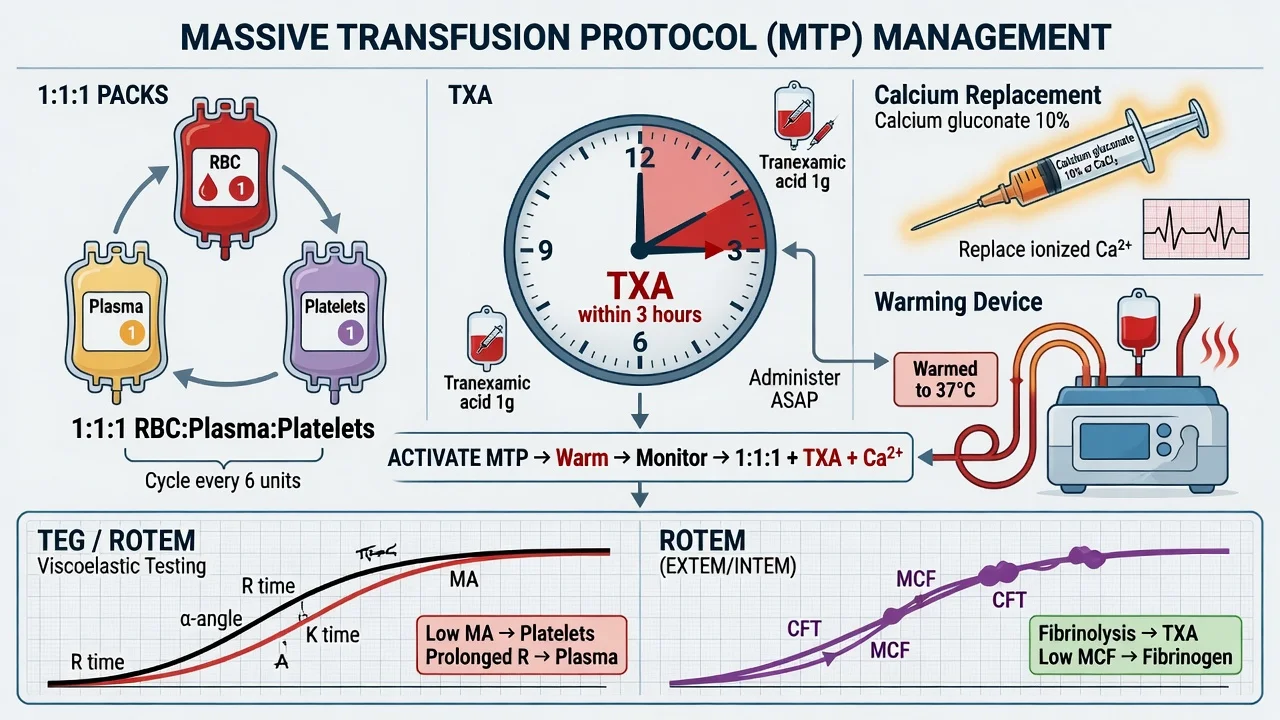
The lethal triad is the metabolic death spiral of uncontrolled major haemorrhage. Each element worsens the others, and the spiral is rapidly fatal if not broken:[1][9]
The lethal triad — mechanism and how each element drives the others
| Element | Mechanism in major haemorrhage | How it worsens the triad |
|---|---|---|
| Hypothermia | Heat loss (exposure, cold fluids, open cavity), impaired thermogenesis (shock) | Hypothermia <34 degC impairs clotting factor enzyme activity (each 1 degC drop ~10% slower), activates fibrinolysis, and reduces platelet function |
| Acidosis | Tissue hypoperfusion -> anaerobic glycolysis -> lactic acidosis; aggravated by hyperchloraemic saline | pH <7.2 reduces clotting factor activity by >50%, impairs fibrin polymerisation, and blunts catecholamine responsiveness -> more hypotension -> more acidosis |
| Coagulopathy | Consumption, dilution (crystalloid/colloid), hypothermia, acidosis, hypocalcaemia, anticoagulants | Coagulopathy -> more bleeding -> worse dilution and hypoperfusion -> worse hypothermia and acidosis |
Breaking the lethal triad — concurrent corrective actions
WARM aggressively — temperature is the most easily corrected and most quickly fatal element
Forced-air warmer, warmed IV fluids (all fluids through a fluid warmer during MHP), warmed inspired gases, raise the ambient temperature. Target core temperature >36 degC. Hypothermia is independent predictor of mortality in trauma.
CORRECT ACIDOSIS by restoring perfusion, NOT by giving bicarbonate
Treat the cause (haemorrhage control, restore circulating volume with blood products, improve cardiac output). Avoid 0.9% saline (hyperchloraemic acidosis) — use balanced crystalloid if any crystalloid. Sodium bicarbonate is reserved for pH <7.1-7.2 with refractory shock or hyperkalaemia.
CORRECT COAGULOPATHY with blood products and haemostatic agents
1:1:1 ratio (PROPPR), fibrinogen >1.5-2.0 g/L, platelets >50, INR <1.5, TXA within 3 h, calcium replacement, PCC for warfarin. Guide with TEG/ROTEM.
CONTROL THE BLEEDING — the only definitive correction
The triad cannot be fully reversed while bleeding continues. Surgical / endoscopic / interventional-radiology haemorrhage control is the definitive intervention; the steps above buy time to achieve it.
Acidosis severity and clotting function
| Arterial pH | Coagulation factor activity | Clinical implication |
|---|---|---|
| 7.40 (normal) | 100% | Normal |
| 7.30 | ~70% | Mild impairment |
| 7.20 | ~50% | Significant impairment — coagulopathy contributes to bleeding |
| <7.10 | <30% | Severe coagulopathy; also catecholamine-resistant shock |
Exam point: the lethal triad is why normothermia, permissive hypotension (time-limited), blood-product-first resuscitation, and damage control surgery form a coherent strategy — each is designed to prevent or break one arm of the spiral. Crystalloid resuscitation worsens all three arms (dilution, hypothermia, acidosis) and is the historical practice that DCR explicitly replaced.[9][1]
Transfusion reactions & complications
- Acute haemolytic reaction — ABO incompatibility from a clerical error; fever, hypotension, flank pain, haemoglobinuria, DIC. Stop the transfusion immediately, support the circulation, maintain urine output, and send blood and unit samples for repeat typing.[1]
- TRALI (transfusion-related acute lung injury) — the leading cause of transfusion-related death. Donor anti-leucocyte antibodies (or lipid mediators) damage the pulmonary endothelium, causing non-cardiogenic pulmonary oedema with bilateral infiltrates and hypoxaemia within 6 hours. Treat supportively — oxygen and ventilation; diuretics do not help (it is not volume overload).[1]
- TACO (transfusion-associated circulatory overload) — true volume overload; hypertension, raised JVP, pulmonary oedema responsive to diuretics. Distinguish from TRALI by the volume responsiveness.[1]
- Febrile non-haemolytic / allergic — mild; paracetamol or antihistamine; stop and reassess if severe (anaphylaxis in IgA-deficient patients).[1]
- Transfusion-transmitted infection — now very rare with screening.[1]
- Electrolyte and temperature — hyperkalaemia (potassium leaches from stored red cells, dangerous in rapid transfusion), hypocalcaemia (citrate), and hypothermia — the same lethal triad risks as massive resuscitation.[1]
TRALI vs TACO — the exam-defining distinction
| Feature | TRALI | TACO |
|---|---|---|
| Mechanism | Donor anti-leucocyte antibodies (classically multiparous female donors) / lipid mediators damage pulmonary endothelium | True circulatory volume overload |
| Onset | Within 6 h of transfusion (usually within 1-2 h) | Within 6 h (often during or immediately after) |
| Pulmonary oedema | Non-cardiogenic — bilateral infiltrates, low/normal cardiac pressures | Cardiogenic — bilateral infiltrates, raised cardiac filling pressures |
| JVP / BP | Normal / low (often hypotensive) | Raised JVP, hypertension |
| Echocardiogram | Normal LV function | Often impaired / dilated LV |
| BNP | Normal | Raised |
| Fever | Often present | Usually absent |
| Response to diuretics | Minimal (NOT volume overload) | Good (diureses) |
| Management | Supportive — oxygen, lung-protective ventilation; NO diuretics; report to blood bank; avoid further plasma from implicated donor | Diurese (furosemide); sit upright; oxygen; reduce transfusion rate; consider NIV |
| Mortality | High (leading cause of transfusion death) | Lower but significant, especially elderly / cardiac |
| Recurrence | Low if donor excluded | Higher if cardiac / renal dysfunction persists |
Other transfusion reactions — recognition and management
| Reaction | Mechanism | Onset | Features | Management |
|---|---|---|---|---|
| Acute haemolytic | ABO incompatibility (clerical error) — donor RBC destroyed by recipient anti-A/anti-B | Minutes to hours | Fever, flank pain, hypotension, haemoglobinuria, DIC, renal failure | STOP transfusion immediately; support circulation; maintain urine output (fluids/mannitol/diuretics); repeat typing; report to blood bank |
| Febrile non-haemolytic (FNHTR) | Cytokines in stored product; recipient anti-leucocyte antibodies | During / within 1-2 h | Fever, rigors, mild | Slow/stop; paracetamol; leucodepleted products prevent recurrence |
| Mild allergic (urticaria) | Donor plasma protein (often IgA in IgA-deficient recipient) | During | Urticaria, pruritus | Slow; antihistamine; usually safe to resume |
| Anaphylactic | Anti-IgA in IgA-deficient recipient; or other plasma protein | Minutes | Bronchospasm, hypotension, angioedema | STOP; adrenaline (IM), fluids, intubation if needed; IgA-deficient products thereafter |
| Delayed haemolytic | Anamnestic response to prior allosensitisation (e.g., Kidd, Duffy) | Days (3-14) | Falling Hb, mild jaundice, positive direct antiglobulin test | Usually no acute intervention; identify antibody for future compatibility |
| Transfusion-associated graft-vs-host | Viable donor T-cells engraft in immunocompromised recipient | 1-2 weeks | Fever, rash, diarrhoea, pancytopenia, liver dysfunction; often fatal | Irradiated (not just leucodepleted) products for immunocompromised, Hodgkin, stem-cell transplant, purine-analogue therapy |
| Post-transfusion purpura | Anti-HPA antibodies destroy recipient platelets | 5-10 days | Severe thrombocytopenia, bleeding | IV immunoglobulin; plasma exchange |
| Bacterial sepsis (platelets) | Room-temperature storage permits bacterial growth | During / shortly after | High fever, rigors, septic shock | STOP; cultures (patient + unit); broad-spectrum antibiotics; supportive |
Metabolic complications specific to massive / rapid transfusion
| Complication | Mechanism | Clinical effect | Prevention / management |
|---|---|---|---|
| Hypocalcaemia | Citrate chelation | Hypotension, prolonged QT, coagulopathy, catecholamine resistance | Monitor ionised Ca; replace CaCl2 (central) or Ca-gluconate (peripheral) during rapid transfusion |
| Hyperkalaemia | K+ leaches from stored RBC; rises with storage age | Arrhythmia, cardiac arrest (especially in rapid transfusion and renal failure) | Use fresh (<7-10 d) RBC for massive transfusion if possible; washed / additive-reduced RBC; monitor K+; treat hyperkalaemia |
| Hypothermia | Cold stored product infused rapidly | Worsens coagulopathy, arrhythmia | Fluid warmer for all products during MHP; forced-air patient warming |
| Acidosis (early) | Stored RBC pH ~6.5-7.0 from citric acid / lactic acid | Compounds shock acidosis | Correct perfusion; usually self-corrects as citrate metabolised to bicarbonate |
| Alkalosis (later) | Citrate -> bicarbonate metabolism | Shifts Hb-O2 curve left; hypokalaemia | Usually mild and self-limiting |
Prognosis
Outcome depends on the speed of bleeding control, an early balanced ratio (PROPPR), early TXA (CRASH-2, within 3 hours), and avoidance of the lethal triad and transfusion reactions. Late TXA (after 3 hours) and delayed protocol activation worsen mortality.[1][2][3][1]
Determinants of outcome in massive transfusion
| Factor | Effect on outcome | Evidence |
|---|---|---|
| Time to haemorrhage control | The dominant determinant | Observational; DCS standard |
| Early TXA (within 3 h) | Reduced bleeding and all-cause death | CRASH-2, WOMAN, IPD meta-analysis |
| Late TXA (after 3 h) | Increased bleeding death (trauma) | CRASH-2 timing |
| Balanced 1:1:1 ratio | Fewer early deaths from exsanguination | PROPPR |
| Prehospital plasma | Reduced mortality at risk of shock | PAMPer |
| MHP / formal protocol | Reduced mortality | MHP systematic review |
| Lethal triad present | Each element independently predicts mortality; combined spiral is fatal | Trauma registries |
| Calcium <0.9 mmol/L (ionised) on admission | Strongly predicts mortality | Observational trauma data |
| Hypofibrinogenaemia on admission | Strong predictor of massive bleeding and death | Trauma coagulopathy studies |
Trial cards — the evidence base
CRASH-2 2010 — Tranexamic Acid in Trauma Haemorrhage
Multinational randomised, placebo-controlled trial, 20,211 trauma patients with significant haemorrhage (PMID 20554319)
Population: Adult trauma patients with significant bleeding, or systolic BP <90 / HR >110, within 8 h of injury
Key finding
All-cause death 14.5% TXA vs 16.0% placebo (RR 0.91; 95% CI 0.85-0.97; P=0.0035). Bleeding death 4.9% vs 5.7% (RR 0.85). NO increase in vascular occlusive events (fatal/non-fatal MI, stroke, PE, DVT).
Practice change
TXA reduces mortality in bleeding trauma patients with no thromboembolic cost. The landmark trial that made TXA standard in trauma. The cheap, transportable, globally applicable haemostatic.
CRASH-2 exploratory analysis 2011 — The 3-hour window
Pre-specified exploratory analysis of CRASH-2 by time to treatment (PMID 21439633)
Population: Subgroups stratified by time from injury to TXA administration
Key finding
Treatment within 3 h: reduced bleeding death (RR ~0.72). Treatment AFTER 3 h: INCREASED bleeding death (RR ~1.44 in the >3 h group). No clear mechanism; possibly late fibrinolysis shutdown or rebound.
Practice change
The 3-hour rule. TXA in trauma must be given within 3 h of injury — ideally at the scene. Beyond 3 h the harm exceeds the benefit. This is the most exam-tested TXA fact.
PROPPR 2015 — Plasma:Platelets:RBC Ratio in Severe Trauma
Multicentre randomised trial, 680 patients with severe trauma and active bleeding (PMID 25647203)
Population: Adult trauma patients predicted to need massive transfusion (AIS >=3, SBP <90 or HR >120 within 1 h, and activator of MTP)
Key finding
24-h mortality 12.7% (1:1:1) vs 17.0% (1:1:2); P=0.03. Death from exsanguination 9.2% vs 14.6%. 30-day mortality NOT significantly different (22.4% vs 26.1%; P=0.26). No difference in complications.
Practice change
1:1:1 is the initial ratio of choice — fewer early exsanguination deaths, no added harm. The modern basis for ratio-based resuscitation.
CRASH-3 2019 — Tranexamic Acid in Traumatic Brain Injury
International randomised, placebo-controlled trial, ~12,700 TBI patients (PMID 31623894)
Population: Adults with traumatic brain injury, within 3 h of injury (primary analysis), GCS 3-15
Key finding
In mild-to-moderate TBI (GCS 9-15), early TXA reduced head-injury-related death. In severe TBI (GCS 3-8), benefit uncertain and the risk-benefit is unfavourable when given late. No excess vascular occlusive events overall.
Practice change
Early TXA for mild-to-moderate TBI is supported; do NOT give late in TBI. The benefit in severe TBI is unproven — avoid over-extrapolation from CRASH-2.
WOMAN 2017 — Tranexamic Acid in Postpartum Haemorrhage
International randomised, double-blind, placebo-controlled trial, 20,060 women with PPH (PMID 28456509)
Population: Women with clinical diagnosis of postpartum haemorrhage after vaginal or caesarean delivery
Key finding
Death from bleeding 1.5% TXA vs 1.9% placebo (RR 0.81; P=0.045). No increase in thromboembolic events, maternal complications, or deaths in women treated after 3 h. Hysterectomy reduced.
Practice change
TXA is standard of care in PPH. Notably, unlike trauma, the harm signal after 3 h did NOT reproduce — the obstetric mechanism of fibrinolysis differs. Give TXA in all clinically significant PPH.
HALT-IT 2020 — Tranexamic Acid in Acute Upper GI Bleeding
International randomised, double-blind, placebo-controlled trial, 12,009 patients (PMID 32563378)
Population: Adults with acute upper or lower GI bleeding
Key finding
No difference in death from bleeding (3.7% vs 3.8%). INCREASED venous thromboembolism (0.8% vs 0.4%) and seizures (0.6% vs 0.1%) with TXA. No benefit in any subgroup.
Practice change
Do NOT give TXA in acute upper GI bleeding. Unlike trauma/PPH, GI bleeding is not primarily fibrinolytic, and the high-dose regimen caused harm. The negative counterpart to CRASH-2.
TICH-2 2018 — Tranexamic Acid in Intracerebral Haemorrhage
International randomised, placebo-controlled trial, 2,325 patients (PMID 29778325)
Population: Adults with acute spontaneous intracerebral haemorrhage within 8 h
Key finding
No significant difference in functional outcome or mortality. Slightly less haematoma growth with TXA but no clinical benefit. No excess thromboembolic events.
Practice change
TXA is NOT routinely recommended in spontaneous intracerebral haemorrhage. The lesson: a benefit in trauma bleeding does not extrapolate to non-fibrinolytic bleeding.
PAMPer 2018 — Prehospital Plasma in Trauma
Multicentre randomised trial, 501 trauma patients transported by air medical services (PMID 30044935)
Population: Adult trauma patients at risk for haemorrhagic shock (hypotension, tachycardia, or penetrating injury)
Key finding
30-day mortality 23.2% (plasma) vs 33.0% (standard); OR 0.65 (95% CI 0.43-0.97; P=0.03). Benefit greatest in severely injured (ISS >=25).
Practice change
Extends haemostatic resuscitation to the prehospital phase. Early plasma (before hospital) reduces mortality in trauma at risk of haemorrhagic shock — supports the endotheliopathy-of-trauma rationale for early plasma.
Rossaint 2019 — European Trauma Bleeding Guideline (5th edition)
Multidisciplinary clinical practice guideline, GRADE methodology (PMID 30917843)
Population: Adult trauma patients with major bleeding / coagulopathy
Key finding
Recommends: early MHP activation; 1:1:1 or plasma:RBC at least 1:2; target fibrinogen >1.5 g/L with fibrinogen concentrate / cryoprecipitate; TXA within 3 h; permissive hypotension (SBP 80-90) until haemorrhage controlled (NOT in TBI); viscoelastic-guided component therapy; calcium monitoring; damage control surgery.
Practice change
The definitive European reference for major trauma bleeding. Maps directly onto the exam answers.
SAQ — Damage control resuscitation in major trauma
10 minutes · 10 marks
A 28-year-old man arrives by helicopter after a high-speed motorbike crash. He has a severe pelvic fracture and an open femur fracture, BP 70/40, HR 135, lactate 7. The massive haemorrhage protocol is activated. Outline the principles of damage control resuscitation and the role of viscoelastic testing.
SAQ — Obstetric massive haemorrhage and the role of fibrinogen
10 minutes · 10 marks
A 35-year-old woman undergoes emergency caesarean section for placenta accreta and develops massive post-partum haemorrhage. She has received 6 units of red cells, 4 units of FFP, and 1 pool of platelets. Her fibrinogen is 1.8 g/L and she continues to bleed. Discuss the fibrinogen target and the role of TXA in obstetric haemorrhage.
Clinical pearls
[1]Red flags
References
- [1]CRASH-2 trial collaborators, Shakur H, Roberts I, et al. Effects of tranexamic acid on death, vascular occlusive events, and blood transfusion in trauma patients with significant haemorrhage (CRASH-2): a randomised, placebo-controlled trial Lancet, 2010.PMID 20554319
- [2]CRASH-2 collaborators The importance of early treatment with tranexamic acid in bleeding trauma patients: an exploratory analysis of the CRASH-2 randomised controlled trial Lancet, 2011.PMID 21439633
- [3]Holcomb JB, Tilley BC, Baraniuk S, et al.; PROPPR Study Group. Transfusion of plasma, platelets, and red blood cells in a 1:1:1 vs a 1:1:2 ratio and mortality in patients with severe trauma: the PROPPR randomized clinical trial JAMA, 2015.PMID 25647203
- [4]CRASH-3 trial collaborators. Effects of tranexamic acid on death, disability, vascular occlusive events and other morbidities in patients with acute traumatic brain injury (CRASH-3): a randomised, placebo-controlled trial Lancet, 2019.PMID 31623894
- [5]WOMAN Trial Collaborators. Effect of early tranexamic acid administration on mortality, hysterectomy, and other morbidities in women with post-partum haemorrhage (WOMAN): an international, randomised, double-blind, placebo-controlled trial Lancet, 2017.PMID 28456509
- [6]Gayet-Ageron A, Prieto-Merino D, Ker K, et al.; ANTISEPTIC Trial Collaboration. Effect of treatment delay on the effectiveness and safety of antifibrinolytics in acute severe haemorrhage: a meta-analysis of individual patient-level data from 40 138 bleeding patients Lancet, 2018.PMID 29126600
- [7]HALT-IT Trial Collaborators. Effects of a high-dose 24-h infusion of tranexamic acid on death and thromboembolic events in patients with acute gastrointestinal bleeding (HALT-IT): an international randomised, double-blind, placebo-controlled trial Lancet, 2020.PMID 32563378
- [8]Sprigg N, Robson K, Bath P, et al.; TICH-2 Investigators. Tranexamic acid for hyperacute primary IntraCerebral Haemorrhage (TICH-2): an international randomised, placebo-controlled, phase 3 superiority trial Lancet, 2018.PMID 29778325
- [9]Rossaint R, Bouillon B, Cerny V, et al. The European guideline on management of major bleeding and coagulopathy following trauma: fifth edition Crit Care, 2019.PMID 30917843
- [10]Nascimento B, Calder S, Rizoli S, et al. The effect of massive transfusion protocol implementation on the survival of trauma patients: a systematic review and meta-analysis Blood Transfus, 2020.PMID 32955420
- [11]Schöchl H, Nienaber U, Hofer G, et al. Effect of coagulation factor concentrate administration on ROTEM® parameters in major trauma Scand J Trauma Resusc Emerg Med, 2015.PMID 26514413
- [12]Sperry JL, Guyette FX, Brown JB, et al.; PAMPer Study Group. Prehospital Plasma during Air Medical Transport in Trauma Patients at Risk for Hemorrhagic Shock N Engl J Med, 2018.PMID 30044935