ICU · Resuscitation & shock
Neurogenic Shock (Spinal Cord Injury)
Also known as Neurogenic shock · Spinal shock · Spinal cord injury shock · Distributive shock spinal · MAP target spinal cord
Neurogenic shock — the loss of the sympathetic tone from the spinal cord injury (above T6). The vasodilation (the warm, the dry, the flushed — the loss of the alpha) + the bradycardia (the loss of the cardiac sympathetic — the unopposed vagal). The MAP target 85-90 (the spinal cord perfusion). The noradrenaline (the alpha-1 — the vasoconstriction) + the atropine / the isoprenaline (the bradycardia). The distinguish from the hypovolaemic (the warm + the dry vs the cool + the clammy).
On this page & tools
Your progress
Saved locally on this device.
Target exams
Overview & definition
Neurogenic shock — the loss of the sympathetic tone from the spinal cord injury (above T6). The unopposed parasympathetic → the vasodilation (the warm, the dry, the flushed skin — the loss of the alpha) + the bradycardia (the unopposed vagal — the loss of the cardiac sympathetic T1-T4). The MAP target 85-90 mmHg (the spinal cord perfusion). The distinct from the hypovolaemic shock (the warm vs the cool).[1][1]
The critical unifying concept. Neurogenic shock is a distributive shock — the same family as the septic, the anaphylactic, and the vasoplegic post-cardiac-surgery. All share the loss of the vascular tone (the low SVR). But neurogenic shock is the ONLY distributive shock (indeed the ONLY shock of ANY type) that causes bradycardia rather than tachycardia. Every other shock — the hypovolaemic, the cardiogenic, the septic, the anaphylactic, the obstructive — drives a compensatory tachycardia. The neurogenic shock does NOT, because the sympathetic cardiac accelerator fibres (T1-T4) are severed along with the vasoconstrictor fibres. The unopposed vagal wins. Bradycardia + hypotension + warm dry skin in a trauma patient = neurogenic shock until proven otherwise.[1][4]
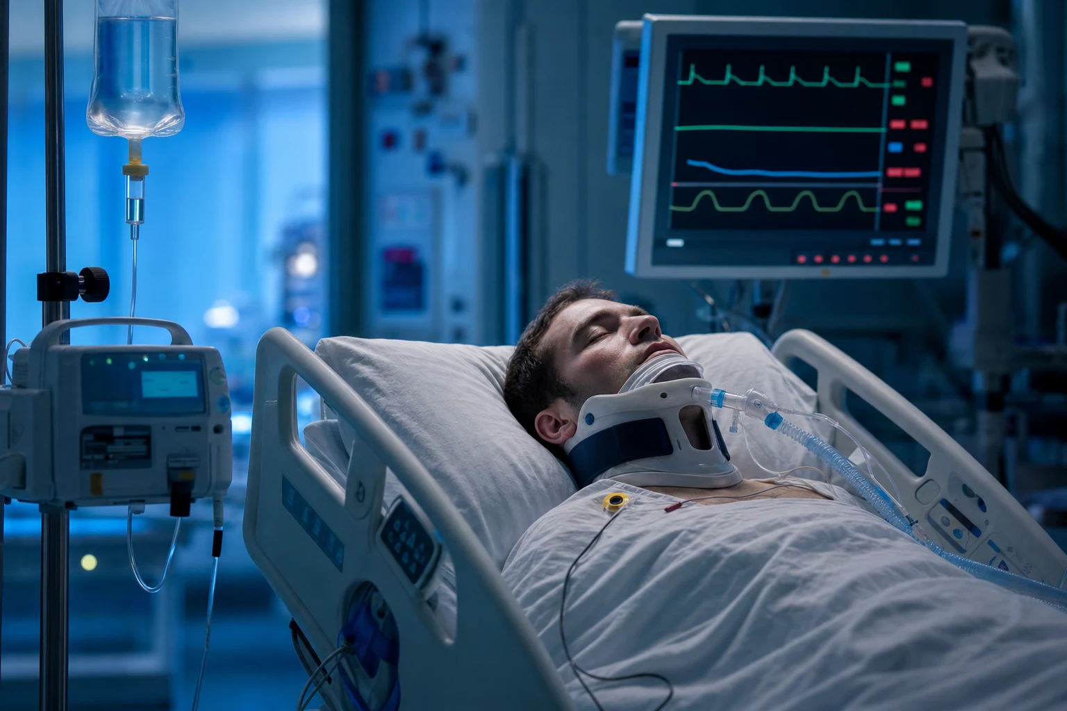
The pathophysiology
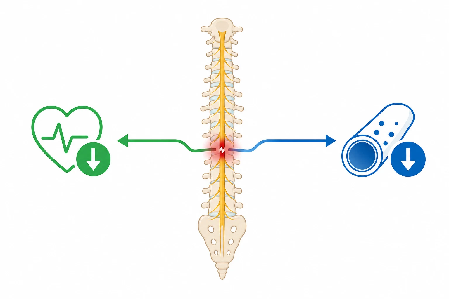
The spinal cord injury (above T6) disrupts the descending sympathetic pathways from the brainstem to the sympathetic chain. The result:[1][1]
- The vasodilation — the loss of the sympathetic alpha-1 vasoconstrictor tone to the peripheral vasculature → the massive vasodilation → the reduced SVR → the hypotension.[1]
- The bradycardia — the loss of the cardiac sympathetic outflow (T1-T4) → the unopposed vagal (parasympathetic) tone → the bradycardia. The distinct from the hypovolaemic shock (which causes the compensatory tachycardia).[1]
- The temperature dysregulation — the loss of the sympathetic vasoconstriction → the poikilothermia (the body assumes the ambient temperature).[1]
The detailed pathophysiology — the three lost loops
The sympathetic nervous system is the body's "fight or flight" accelerator. It is controlled from the brainstem (the rostral ventrolateral medulla, the RVLM) and projects DOWN the spinal cord in the descening bulbospinal pathways to synapse on the intermediolateral (IML) cell column of the spinal cord grey matter — from T1 to L2. From the IML, the pre-ganglionic neurons exit via the ventral roots to the sympathetic chain, then the post-ganglionic fibres reach every target organ: the blood vessels (alpha-1 vasoconstriction), the heart (beta-1 chronotropy + inotropy), the bronchi (beta-2 bronchodilation), the sweat glands, the adrenals.[4][1]
A spinal cord injury above the level of T6 (and especially the cervical cord, above T1) severs ALL of these descending fibres below the lesion. The sympathetic chain below the lesion is now cut off from central command — it fires on its own intrinsic tone (minimal), not on brainstem-directed drive. Three specific consequences:[4]
-
The loss of the vasomotor tone (the alpha-1). The splanchnic and peripheral vasculature below the lesion lose their tonic sympathetic vasoconstriction → the massive arteriolar and venous vasodilation → the pooling of blood in the capacitance vessels (the splanchnic bed, the lower-limb veins) → the reduced venous return → the reduced preload → the reduced cardiac output → the hypotension. The SVR falls dramatically. This is mechanically identical to the vasodilation of the anaphylaxis or the sepsis — it is a DISTRIBUTIVE shock — but the trigger is the severed sympathetic rather than the mediator release.[1][1]
-
The loss of the cardiac accelerator tone (the beta-1, T1-T4). The cardiac sympathetic fibres arise from T1 to T4 (sometimes T5). A cervical cord injury severs these. The heart is now under the UNOPPOSED influence of the vagus nerve (the parasympathetic, the CN X — which travels outside the spinal cord, via the vagus, and is therefore SPARED). The result: the bradycardia. This is the single most distinguishing feature of the neurogenic shock — and the one that catches out the junior who expects a tachycardia in every shock. No other form of shock produces a bradycardia as part of its primary pathophysiology. (The hypovolaemic, the cardiogenic, the septic, the anaphylactic — all tachycardic.)[4][1]
-
The loss of the thermoregulatory tone. The skin vasculature below the lesion cannot vasoconstrict to conserve heat → the body assumes the temperature of the environment (poikilothermia). The patient may be hypothermic in a cool ED, or febrile in a warm resus bay — neither reflects a true core-temperature derangement. The sweating below the lesion is also abolished (the loss of the sympathetic sudomotor fibres) → the skin is DRY below the lesion (but may sweat above, as the lesion-sparing fibres above fire excessively to compensate).[1][1]
Why above T6 — the level matters
The higher the cord lesion, the more of the sympathetic outflow is lost, and the worse the neurogenic shock:[2][4]
- Cervical (C1-C8) lesions — the loss of the ENTIRE sympathetic outflow (T1-L2) + the cardiac accelerator fibres (T1-T4). The most severe neurogenic shock. ALSO the loss of the phrenic nerve (C3-C5) → the diaphragmatic paralysis → the respiratory failure requiring mechanical ventilation. The combination of the profound vasodilation + the bradycardia + the respiratory failure makes the high cervical SCI one of the most fragile ICU patients.[4]
- Upper thoracic (T1-T5) lesions — the loss of the cardiac accelerator fibres + the upper-body vasomotor tone. The neurogenic shock is significant (the bradycardia + the hypotension) but the diaphragm is spared (the C3-C5).[2]
- Lower thoracic (T6-T12) lesions — the cardiac accelerator fibres (T1-T4) are SPARED (the lesion is BELOW them) → NO bradycardia. Some vasodilation in the splanchnic/lower-limb bed (mild hypotension) but the cardiac sympathetic is intact → the compensatory tachycardia is preserved. Neurogenic shock is RARE below T6 — the hypotension in a T6-L2 injury should prompt an aggressive search for the OTHER cause (the blood loss).[2][3]
- Lumbar (below L1) lesions — NO neurogenic shock (the IML column is the T1-L2 — the lesion is at or below it).[1]
The timing and the natural history
The neurogenic shock develops within 30 minutes to hours of the injury (the time for the residual catecholamines already released to be washed out, and for the loss of the tonic sympathetic to become haemodynamically apparent). It peaks at 24-72 hours and may persist for 1-3 weeks (the average 7-10 days, sometimes up to 4-6 weeks in the complete high cervical lesion) before the intrinsic spinal sympathetic tone partially recovers. The longer the neurogenic shock, the worse the prognosis (the more complete the cord injury).[3][4]
The bradycardia may be profound. Heart rates of 30-40 bpm are not uncommon in the high cervical lesion. The symptomatic bradycardia (the hypotension refractory to the vasopressor alone, the syncope, the AV block, the asystole) is a real risk in the first 1-2 weeks — the atropine and/or the isoprenaline and/or the chronotrope is part of the toolkit, not an afterthought.[4][5]
The autonomic dysreflexia — the LATE counterpart (NOT in the acute phase)
For completeness and to avoid exam confusion: the autonomic dysreflexia is a DIFFERENT phenomenon that occurs in the REHABILITATION phase (weeks to months after the injury), in patients with lesions above T6. A noxious stimulus below the lesion (a full bladder, a faecal impaction, an ingrown toenail) triggers a massive reflex sympathetic discharge below the lesion (unopposed, because the descending inhibitory pathways are severed) → the life-threatening hypertension. The baroreceptors sense the hypertension → trigger the vagal (bradycardia) and the vasodilation ABOVE the lesion (the flushed, the sweating, the pounding headache above the lesion; the cold, the pale below). This is NOT neurogenic shock (it is hypertension, not hypotension; it is weeks later, not the acute phase) — but the examiners love to contrast the two, so know the difference.[1][1]
The clinical — the distinguishing features
| Feature | Neurogenic | Hypovolaemic |
|---|---|---|
| Heart rate | Bradycardia | Tachycardia |
| Skin | Warm, dry, flushed | Cool, clammy, pale |
| Temperature | Poikilothermia | Normal (or cool from exposure) |
| JVP | Normal or low | Low |
| Urine output | Low (from hypotension) | Low (from hypovolaemia) |
| Sensory level | Present (the cord) | Absent |
Key: the neurogenic shock has the BRADYCARDIA + the WARM skin (the loss of the sympathetic), while the hypovolaemic has the tachycardia + the cool skin (the compensatory sympathetic).[1]
The timing. The neurogenic shock develops within 30 minutes to hours of the injury. The spinal shock (a different concept — the transient loss of the spinal reflexes below the lesion, lasting days to weeks) is distinct from the neurogenic shock (the haemodynamic).[1]
Neurogenic shock vs ALL other shock types — the heart rate is the differentiator
| Feature | Neurogenic | Hypovolaemic | Cardiogenic | Septic | Anaphylactic | Obstructive |
|---|---|---|---|---|---|---|
| Heart rate | BRADYCARDIA (unique) | Tachycardia | Tachycardia | Tachycardia | Tachycardia | Tachycardia (or relative brady) |
| Skin | Warm, dry, flushed | Cool, clammy, pale | Cool, clammy, pale | Warm, flushed (early) | Warm, flushed + urticaria | Cool, clammy |
| SVR | LOW (vasodilation) | HIGH (compensatory) | HIGH (compensatory) | LOW (vasodilation) | LOW (vasodilation) | HIGH (compensatory) |
| Cardiac output | Low (relative — low preload) | Low | LOW (the pump fails) | HIGH (early — hyperdynamic) | Low (relative) | Low |
| JVP / preload | Normal or low | LOW | HIGH (the failing pump backs up) | Normal or low | Low (venous pooling) | HIGH (tamponade / tension PTX) |
| Mechanism | Loss of sympathetic tone (SCI) | Volume loss | Pump failure | Vasodilation + capillary leak | Mast-cell mediator release | Mechanical obstruction to flow |
| First-line agent | Noradrenaline + atropine | Fluid / blood | Inotrope (dobutamine) | Fluid + noradrenaline | IM adrenaline | Relieve obstruction |
The full clinical picture
The classic triad in the trauma patient with a cervical or upper-thoracic SCI: (1) hypotension, (2) bradycardia, (3) warm dry flushed skin. All three must be present for the textbook diagnosis — but in the real trauma patient, the co-existent blood loss (the hypovolaemia) may mask the warm dry skin (replacing it with the cool clammy pale) or the bradycardia (replacing it with the tachycardia from the hypovolaemic reflex). The mixed picture (the neurogenic + the hypovolaemic) is the rule, not the exception, in the polytrauma patient — and the failure to recognise BOTH is a leading cause of the preventable death.[2][6]
- The blood pressure. The systolic commonly 70-90 mmHg in the untreated high cervical lesion. The diastolic falls disproportionately (the loss of the diastolic vascular tone) → the WIDE pulse pressure may be masked by the profound vasodilation → the narrow pulse pressure is more typical once the bradycardia reduces the stroke volume variability. The orthostatic hypotension is extreme — sit the patient up at your peril (the venous pooling → the catastrophic preload drop).[4]
- The heart rate. The bradycardia (30-50 bpm) is the rule in the cervical lesion. May be the sinus bradycardia, the junctional escape, the AV block (first, second — Mobitz I or II, or complete), or the asystolic pauses (the vagally-mediated). The symptomatic bradycardia is the indication for the chronotrope (the atropine, the isoprenaline, the dopamine, or the adrenaline infusion).[4][5]
- The skin. Below the lesion: the warm, the dry, the flushed (the loss of the sympathetic vasoconstriction + the sudomotor). Above the lesion: the cool, the pale, the sweaty (the compensatory sympathetic SURGE in the intact fibres above — the body's futile attempt to raise the BP). The LINE OF DEMARCATION at the sensory level is a clinical giveaway — examine for it.[1]
- **The abdomen.**The distended, the hypoactive-bowel, the areflexic (the paralytic ileus from the loss of the splanchnic sympathetic tone → the unopposed parasympathetic → the atony). May mimic or conceal an intra-abdominal catastrophe — the FAST scan / the CT is mandatory.[6]
- The respiratory. The high cervical lesion → the diaphragmatic paralysis (the C3-C5 phrenic) → the shallow, the rapid, the paradoxical breathing (the accessory-muscle-only, the abdominal paradox). The lower cervical lesion → the intercostal paralysis (the T1-T11) → the preserved diaphragm but the poor cough and the secretion retention. Anticipate the respiratory failure and the early intubation.[1]
- The neurological. The flaccid paralysis + the absent reflexes below the lesion (the spinal shock, the acute phase — NOT the spasticity that develops weeks later). The sensory level (the pinprick, the light touch) on the exam localises the lesion. The bulbocavernosus reflex (the S2-S4 — the contraction of the anal sphincter on the tug of the glans penis or the Foley catheter pull) — when ABSENT, confirms the spinal shock (the acute phase); when it RETURNS, marks the end of the spinal shock (but NOT necessarily the end of the neurogenic shock). The ASIA Impairment Scale (A-E) quantifies the completeness of the injury and the prognosis.[1][5]
The diagnosis — the rule-out, not the rule-in
The diagnostic principle. Neurogenic shock is a diagnosis of inclusion (the SCI is present) AND exclusion (the other causes of the hypotension are ruled out). The temptation — especially in the ED with a known cervical SCI and the classic triad — is to STOP looking once the neurogenic is "found." This is the error. The neurogenic shock does NOT exclude the co-existent blood loss — and the missed intra-abdominal bleed is the killable mistake.[2][6]
The stepwise diagnostic approach
-
Recognise the trauma + the SCI. Any patient with a mechanism compatible with the SCI (the high-speed MVC, the diving injury, the fall from height, the direct blow to the neck/back) AND any neurological sign (the weakness, the sensory level, the priapism — pathognomonic in the acute male SCI) is at risk. Immobilise (the cervical collar, the spinal board), then examine.[6]
-
Document the haemodynamics. The bradycardia + the hypotension + the warm dry skin = the neurogenic triad. But the tachycardia in a known SCI does NOT exclude the neurogenic — it may indicate the CO-EXISTENT hypovolaemia (the blood loss) on top of the neurogenic, OR a lower lesion (below T6) where the cardiac accelerator is spared. The hypotension in a SCI requires the EXPLANATION, not the assumption.[2][6]
-
RULE OUT THE HYPOVOLAEMIA — the single most important step. The hypotension in the trauma patient is the hypovolaemic UNTIL PROVEN OTHERWISE, even when the SCI is known. Actively search for the bleeding:[6]
- The physical exam — the chest (the flail segment, the penetrating wound, the subcutaneous emphysema → the intrathoracic), the abdomen (the distension, the tenderness, the seatbelt sign → the intra-abdominal), the pelvis (the instability, the bruising → the pelvic fracture), the long bones (the deformity, the swelling → the femur, the tibia), the external (the lacerations, the scalp — the scalp can hold 1-2 L).
- The FAST scan (Focused Assessment with Sonography in Trauma) — the bedside, the repeatable, the rapid. The Morrison's pouch, the splenorenal recess, the pouch of Douglas, the pericardium. The positive FAST = the intra-abdominal blood → the laparotomy. The negative FAST does NOT exclude the retroperitoneal or the hollow-viscus injury → the CT if stable.
- The CT trauma series — the head, the cervical spine, the chest, the abdomen/pelvis (with IV contrast) — the definitive imaging once the patient is stable enough for the trip. The chest CT for the aortic injury (the widened mediastinum, the traumatic aortic transection — a killer in the high-speed deceleration). The abdomino-pelvic CT for the solid-organ laceration, the hollow-viscus injury, the retroperitoneal haematoma.
- The bloods — the FBC (the Hb may be normal in the EARLY acute bleed — the haemodilution takes hours; the serial Hb is more useful), the cross-match, the lactate (the rising lactate = the ongoing tissue hypoperfusion from the blood loss), the venous/arterial blood gas (the base deficit — a sensitive marker of the occult hypovolaemia).[6]
-
Establish the diagnosis of the neurogenic shock — ONLY after the hypovolaemia is excluded (or treated). The combination of (a) the known cervical/upper-thoracic SCI, (b) the hypotension unexplained by the blood loss, (c) the bradycardia, (d) the warm dry flushed skin below the lesion, (e) the documented sensory level, AND (f) the negative FAST / the CT excluding the bleed → the neurogenic shock is the working diagnosis. Begin the neurogenic-specific management (the MAP 85-90, the noradrenaline, the atropine).[1][1]
-
The serial reassessment. The mixed shock (the neurogenic + the hypovolaemic) is common. Even after the neurogenic is "diagnosed," the patient may develop the delayed bleed (the splenic laceration that was initially haemodynamically stable, the pelvic fracture with the ongoing venous ooze). The serial lactate, the serial Hb, the serial FAST, the serial clinical exam — the vigilance continues.[6]
The differential of the bradycardia + hypotension (beyond neurogenic)
Not every bradycardia + hypotension in a trauma patient is the neurogenic. Consider:[6][1]
- The hypovolaemic shock with the late bradycardia — the profoundly exsanguinated patient (the >40% blood-volume loss) may develop a bradycardia as a pre-terminal event (the vagally-mediated "the heart gives up"). This is NOT neurogenic shock — it is the agonal bradycardia of the extreme hypovolaemia. The skin is COOL and CLAMMY (not warm dry). The treatment is the BLOOD, not the atropine.[1]
- The obstructive shock — the tension pneumothorax, the massive haemothorax, the cardiac tamponade. The obstructive shock may present with a relative bradycardia (or a tachycardia). The distinguishing: the JVP is HIGH (distended — in the tension pneumothorax and the tamponade; LOW in the massive haemothorax), the muffled heart sounds (the tamponade), the tracheal deviation and the unilateral absent breath sounds (the tension pneumothorax). The FAST / the bedside ultrasound is the rapid differentiator.[1]
- The beta-blocker or the calcium-channel-blocker overdose — the iatrogenic/drug bradycardia + hypotension. The history (the patient's medication list, the empty pill bottles). The treatment is the glucagon (the beta-blocker) and the calcium / the high-dose insulin (the calcium-channel-blocker).[1]
- The vagal / the vasovagal — the brief, the self-limited, the situational (the painful procedure, the sight of blood). Resolves with the recumbency and the leg raise. Not sustained.[1]
- The high spinal / the epidural anaesthesia — the iatrogenic (the epidural that has risen too high, blocking the cardiac accelerator fibres T1-T4). The same physiology as the neurogenic — the sympathectomy + the unopposed vagal. The treatment: the vasopressor (the noradrenaline / the metaraminol / the adrenaline) + the chronotrope. The distinction from the trauma neurogenic is the context (the anaesthetic, not the injury).[1]
Bradycardia + hypotension in the trauma patient — the differential
| Cause | Skin | JVP | Distinguishing features | Treatment |
|---|---|---|---|---|
| Neurogenic shock | Warm, dry, flushed | Normal or low | The cervical/upper-thoracic SCI + the sensory level + the poikilothermia. The ONLY shock with the bradycardia as primary. | Noradrenaline + atropine + MAP 85-90 |
| Hypovolaemic (late, pre-terminal) | Cool, clammy, pale | LOW | The >40% blood-volume loss — the agonal vagal bradycardia. The skin is COOL (not warm). The lactate is high. | BLOOD — transfuse, control the bleeding |
| Tension pneumothorax | Cool, clammy | HIGH (distended) | The unilateral absent breath sounds + the tracheal deviation + the hyperresonance. The FAST/US: the collapsed lung, the shifted mediastinum. | Needle decompression → chest tube |
| Cardiac tamponade | Cool, clammy | HIGH (distended) | The Beck triad (the muffled heart sounds + the hypotension + the JVD). The pulsus paradoxus. The FAST/US: the pericardial fluid. | Pericardiocentesis → thoracotomy |
| Cardiogenic shock | Cool, clammy | HIGH (distended) | The MI history / the ECG changes. The bilateral crackles. The FAST/US: the poor LV function. | Inotrope + revascularisation |
| Beta-blocker / CCB overdose | Variable | Variable | The drug history. The ECG: the bradycardia, the AV block. The hyperkalaemia (CCB). | Glucagon (BB) / calcium + HIET (CCB) |
| High spinal / epidural | Warm, dry (below block) | Variable | The anaesthetic context. The sympathectomy + the unopposed vagal — the SAME physiology as the neurogenic. | Vasopressor + chronotrope |
The management
1. The MAP target 85-90 mmHg.[1]
- The higher the target (vs the 65 for the general ICU) because the injured spinal cord is extremely vulnerable to the hypoperfusion — the secondary injury (the ischaemia worsens the cord damage). The MAP 85-90 for at least 7 days (the evidence from the systematic review — the improved the neurological outcome).[1]
- Noradrenaline (the alpha-1 — the vasoconstriction; the preferred — the restores the SVR).[1]
3. The bradycardia.[1]
- Atropine 0.5 mg IV (the vagal block — the symptomatic bradycardia).[1]
4. The volume.[1]
- The cautious the fluid (the neurogenic shock is NOT the hypovolaemic — the excess the fluid → the pulmonary oedema; but the vasodilation → the relative the hypovolaemia → the initial the 500 mL the crystalloid the challenge, then the vasopressor).[1]
5. The rule out the co-existent.[1][1]
- The trauma patient may have BOTH the neurogenic AND the hypovolaemic (the blood loss) — the distinguish is the key (the warm + the dry + the bradycardia vs the cool + the clammy + the tachycardia). The FAST scan / the CT to exclude the intra-abdominal / the intrathoracic the bleeding. The NOT the assume the neurogenic alone.[1]
The vasopressor choice — the receptor rationale
This is the highest-yield exam point in neurogenic shock. The vasopressor choice is NOT arbitrary — it is dictated by the receptor pharmacology and the specific pathophysiology.[1][7]
The neurogenic shock has TWO deficits: (1) the lost vascular tone (the alpha-1) AND (2) the lost cardiac accelerator tone (the beta-1, T1-T4). The ideal vasopressor addresses BOTH.[7]
Vasopressor choice in neurogenic shock — the receptor analysis
| Agent | Receptors | Effect on BP | Effect on HR | Role in neurogenic shock |
|---|---|---|---|---|
| Noradrenaline | alpha-1 (strong) + beta-1 (modest) + beta-2 (minimal) | Strong vasoconstriction → restores SVR | Modest chronotropy (beta-1) — restores the cardiac sympathetic drive the injury abolished | FIRST-LINE / PREFERRED. Addresses BOTH deficits — the vasoconstriction AND the modest chronotropy. The AHA/ASA and most guidelines prefer it. Titrate 0.05-0.5 mcg/kg/min to MAP 85-90.[7] |
| Adrenaline | alpha-1 + beta-1 (strong) + beta-2 (strong) | Vasoconstriction + inotropy | Strong chronotropy + inotropy | Second-line / alternative. When noradrenaline inadequate or unavailable. More chronotropy + arrhythmogenicity. Useful when the profound bradycardia coexists with the hypotension and the atropine fails. Dose 0.05-0.5 mcg/kg/min.[7] |
| Dopamine | D1 (low dose) + beta-1 (moderate) + alpha-1 (high dose) | Moderate vasoconstriction + inotropy | Strong chronotropy | Historical favourite, now disfavoured. More arrhythmogenic than noradrenaline (the SOAP II trial — more arrhythmias in the cardiogenic shock). BUT still has a role for the bradycardia (the chronotropy) — some units use it as the first-line when the bradycardia is the dominant problem. Dose 5-20 mcg/kg/min.[1] |
| Phenylephrine | alpha-1 ONLY (pure) | Vasoconstriction | REFLEX BRADYCARDIA (the baroreceptor reflex — the raised BP → the vagal response) | AVOID. A pure alpha-1 agent — it raises the BP but WORSENS the bradycardia (the exact deficit the neurogenic patient already has). The baroreceptor reflex (the intact vagus senses the raised BP → the vagally-mediated bradycardia) compounds the already-present unopposed-vagal bradycardia. May be used transiently if nothing else available, but switch to noradrenaline ASAP.[1][7] |
| Metaraminol | alpha-1 (predominant) + indirect beta (minimal) | Vasoconstriction | Reflex bradycardia (like phenylephrine) | AVOID for the same reason as phenylephrine. A mixed agent but predominantly alpha — the same baroreceptor-reflex bradycardia risk. Not preferred. |
| Vasopressin | V1 (vasopressin receptor) | Vasoconstriction (catecholamine-independent) | Minimal effect on HR (no reflex bradycardia) | Adjunct / refractory. Useful in the catecholamine-resistant vasodilation. Does not cause the reflex bradycardia (acts on the V1, not the baroreflex arc). Dose 0.01-0.04 U/min. Not first-line (limited evidence in neurogenic shock specifically).[1] |
The bottom line on the vasopressor. Noradrenaline is the first-line — it restores the SVR AND provides a modest beta-1 chronotropy to counter the bradycardia. The pure alpha-1 agents (phenylephrine, metaraminol) are AVOIDED because they worsen the bradycardia via the baroreceptor reflex. If the bradycardia is the dominant refractory problem, escalate to the adrenaline or the dopamine (the beta-1 chronotropy) rather than pushing the noradrenaline higher.[7]
The bradycardia management — the chronotrope ladder
The symptomatic bradycardia (the hypotension refractory to the noradrenaline, the syncope, the AV block, the asystolic pauses) requires the chronotrope IN ADDITION to the vasopressor. The ladder:[4][5]
The neurogenic shock bradycardia management — the chronotrope ladder
- ATROPINE 0.5-1 mg IV (repeat up to 3 mg total) — the vagal block. The first-line for the symptomatic bradycardia. Blocks the muscarinic receptor → the unopposed vagal is overcome → the SA node fires faster. Effective for the sinus bradycardia and the high-grade AV block of the vagal origin. May be repeated or given as an infusion. CAUTION: the atropine alone does NOT address the hypotension (the vasodilation) — combine with the vasopressor. Also: the atropine may be INEFFECTIVE in the denervated heart (the transplant patient analogy) if the bradycardia is from the loss of the sympathetic rather than the excess of the vagal — but in the neurogenic, the vagal is the dominant remaining tone, so the atropine usually works.[5]
- ISOPRENALINE (isoproterenol) infusion 2-10 mcg/min — the pure beta-1 + beta-2 agonist. The chronotropy (beta-1) + the vasodilation (beta-2 — yes, it vasodilates, which may worsen the hypotension — so always combine with the alpha vasopressor). Useful when the atropine fails. The beta-2 vasodilation is the downside — titrate carefully.
- ADRENALINE infusion 0.05-0.5 mcg/kg/min — the alpha-1 + beta-1 + beta-2. The chronotropy + the inotropy + the vasoconstriction (at higher doses). The most complete single agent when both the bradycardia and the hypotension are refractory. Escalate from the noradrenaline if the bradycardia dominates.
- DOPAMINE infusion 5-20 mcg/kg/min — the beta-1 chronotropy (moderate doses) + the alpha-1 (high doses). The chronotropy makes it useful for the bradycardia. More arrhythmogenic (the SOAP II lesson) but still has a role. Some units prefer it as the first-line when the bradycardia is the presenting feature.
- EXTERNAL / TRANSVENOUS PACING — the rescue for the refractory symptomatic bradycardia (the complete heart block, the asystolic pauses) unresponsive to the pharmacology. The transcutaneous pacing first (the quick, the available, the painful — sedate), then the transvenous (the more reliable, the longer-term). The permanent pacemaker is rarely needed (the bradycardia is usually transient, resolving as the cord oedema settles over days-weeks).[4]
The full integrated management — the first hours
The neurogenic shock integrated management — the resuscitation bay to the ICU
- RECOGNISE THE RISK — any trauma with a mechanism for the SCI (the high-speed MVC, the fall, the diving, the direct blow) → immobilise the cervical spine (the collar, the blocks, the tape), the log-roll for the transfer. Assume the SCI until excluded. The primary survey (ABCDE) — the Airway (the C-spine control), the Breathing, the Circulation (the haemorrhage control FIRST), the Disability (the GCS, the pupils, the sensory level), the Exposure (the full skin exam — look for the line of demarcation).
- SECURE THE AIRWAY AND THE BREATHING — the high cervical lesion (C3-C5) → the diaphragmatic paralysis → the early intubation (the anticipatory, not the reactive). The lower cervical / upper thoracic → the intercostal paralysis → the poor cough, the secretion retention → the early NIV or the intubation if the secretion clearance fails. RSI with the cardiovascular-stable induction (the ketamine or the etomidate — AVOID the propofol / the thiopentone which crash the already-fragile BP).[1]
- CONTROL THE HAEMORRHAGE (THE CRITICAL STEP) — the external bleeding (the direct pressure, the tourniquet for the limb), the pelvic binder (the pelvic fracture), the chest tube (the massive haemothorax), the FAST (the intra-abdominal blood → the laparotomy). DO NOT assume the hypotension is neurogenic until the bleeding is excluded. The lactate, the serial Hb, the base deficit — the markers of the occult hypovolaemia.[6]
- ESTABLISH THE MONITORING — the ECG (the bradycardia, the AV block), the arterial line (the beat-to-beat BP for the vasopressor titration — the MAP target 85-90), the central line (the noradrenaline infusion, the CVP — though the CVP is unreliable in the vasodilated state), the urine output (the Foley — the target >0.5 mL/kg/h as the perfusion marker), the serial lactate.
- START THE VASOPRESSOR — THE NORADRENALINE — the first-line. Start early (do not wait for the fluid to fail — the neurogenic shock is the vasodilatory, the fluid alone will not fix the SVR). Titrate to the MAP 85-90. Dose 0.05-0.5 mcg/kg/min (some patients need high doses — the profound vasodilation). Add the vasopressin if catecholamine-resistant.[1][7]
- TREAT THE BRADYCARDIA — the atropine 0.5-1 mg IV for the symptomatic bradycardia. Escalate to the isoprenaline / the adrenaline / the dopamine if refractory. The pacing for the refractory AV block / asystole.[5]
- THE CAUTIOUS FLUID — the 500 mL crystalloid challenge (the balanced — the Hartmann's / the Plasma-Lyte — AVOID the normal saline in the large volume — the hyperchloraemic acidosis). Assess the response (the MAP rise = the volume-responsive; the no rise = the vasodilatory, push the vasopressor). AVOID the fluid overload — the neurogenic shock is NOT the hypovolaemic, and the excess fluid → the pulmonary oedema (especially in the patient with the co-existent cardiac or the lung injury).[1]
- THE SPINAL CORD PERFUSION — MAINTAIN THE MAP 85-90 FOR 7 DAYS — the single most evidence-supported intervention. The MAP 85-90 mmHg for at least 7 days (the systematic review — the improved neurological outcome). The protocol: the noradrenaline (the first-line), the adrenaline / the dopamine (the second-line for the refractory or the bradycardia-dominant), the vasopressin (the adjunct). The arterial line for the titration. The sedation holiday (if ventilated) to assess the neurology daily.[1][5]
- THE DEFINITIVE SPINAL FIXATION — the neurosurgical / the ortho-spinal referral. The early decompression (within 24 h) for the incomplete lesion with the cord compression (the STASCIS and the AOSpine 2017 evidence — the earlier the decompression, the better the neurological outcome in the incomplete cervical SCI). The timing is the neurosurgical decision but the ICU team maintains the MAP 85-90 throughout (before, during, and after the surgery).[5]
- THE SERIAL ASSESSMENT AND THE COMPLICATION PREVENTION — the daily ASIA exam (the serial neurological assessment for the deterioration — the expanding haematoma, the inadequate perfusion), the DVT prophylaxis (the high risk — the LMWH once the bleeding excluded, the mechanical if not), the stress-ulcer prophylaxis (the PPI), the pressure-area care (the high risk — the immobile, the vasodilated, the insensate skin), the early enteral nutrition, the bowel and bladder regime (the neurogenic bowel and bladder are the norm).[1][1]
Prognosis
The higher the cord level (the cervical — the C3-C5 → the diaphragm loss + the neurogenic) and the more the complete the injury (the ASIA A), the worse. The MAP 85-90 for 7 days → the improved the neurological outcome (the one retrospective and the systematic review evidence).[1][1]
The prognostic factors
- The level. The higher the lesion, the worse. The cervical (especially C1-C4) carries the highest mortality (the respiratory failure + the neurogenic shock + the autonomic instability). The upper thoracic (T1-T5) is significant. The lower thoracic and lumbar are less haemodynamically severe (the cardiac accelerator fibres spared below T6).[2][3]
- The completeness (the ASIA scale). The ASIA A (complete — no motor or sensory below the lesion) → the worst prognosis. The ASIA B-D (incomplete) → the better, and the potential for the recovery with the early decompression + the MAP 85-90. The ASIA E (normal) → the minor injury.[5]
- The duration of the neurogenic shock. The longer the neurogenic shock persists (the bradycardia + the hypotension refractory to the weaning), the more complete the injury and the worse the prognosis. The resolution (the ability to wean the vasopressor, the return of the intrinsic heart rate) over 1-2 weeks is a favourable sign.[3][4]
- The MAP 85-90 maintained for 7 days → the improved the neurological outcome (the systematic review evidence — the retrospective data, not RCT, but the consistent direction). The secondary ischaemic injury (the hypoperfusion of the already-injured cord) is the PREVENTABLE component of the cord damage — and the MAP 85-90 is the intervention that prevents it.[1][5]
- The co-existent injuries. The traumatic brain injury (the dual diagnosis — the TBI + the SCI — the worst, as the TBI needs the CPP 60+ which needs the MAP 85-90+ for the SCI, so the targets align). The polytrauma (the blood loss, the chest injury) worsens the mortality independent of the cord.[6]
- The age and the comorbidities — the elderly, the cardiac disease, the anticoagulation (the intracord haematoma) — the worse.[3]
The evidence — the trials and the guidelines
The evidence for the MAP 85-90 for 7 days
The evidence base for the MAP 85-90 target is NOT from the RCT — it is from the retrospective case series and the systematic reviews. The original work was the Levi et al. (1993) — the retrospective before-and-after at the Miami Project — which showed the improved neurological outcome with the MAP >85 maintained for 7 days. The Vale et al. (1997) confirmed. The systematic reviews (the Blok 2011 — the Cite id 1 in this topic) collated the evidence and found the consistent signal of the benefit, though the quality is the retrospective (the level 3 evidence, not the level 1).[1][1]
The counterpoint. The Casha et al. (2016, the Cite id 7 in this topic) — a retrospective study of 144 patients — found NO benefit of the MAP augmentation to 85 over the standard resuscitation, and raised the concern of the increased complications (the fluid overload, the ARDS, the cardiac). The debate continues — but the CONSENSUS (the AANS/CNS guidelines, the AHA/ASA stroke guidelines, the ASCON/Spine guidelines) is to TARGET the MAP 85-90 for 7 days, while AVOIDING the fluid overload (use the vasopressor, not the fluid, to maintain the MAP).[1]
Levi 1993 — MAP augmentation in acute spinal cord injury
Source
Retrospective before-and-after, Miami Project (Journal of Neurosurgery)
Intervention
MAP >85 mmHg maintained for 7 days (vasopressors + fluid) vs historical controls (standard resuscitation)
Population
Acute cervical SCI (n=103)
Outcome
Improved ASIA motor score at discharge; trend to improved Frankel grade. The signal consistent with the cord-perfusion benefit.
Limitation
Retrospective, historical controls, single centre, era effect (the improved neurosurgical care over time). NOT an RCT.
Impact
Established the MAP >85 for 7 days as the standard — adopted by the AANS/CNS, the AHA/ASA, and most ICU protocols worldwide.
Vale 1997 — Combined medical and surgical treatment after acute spinal cord injury
Source
Retrospective case series, Stanford (Neurosurgery)
Intervention
MAP >85 mmHg for 7 days + early surgical decompression
Population
Acute SCI, cervical and thoracic (n=77)
Outcome
Improved neurological outcome in the incomplete lesions (ASIA B-D → ASIA C-D or better). The complete lesions (ASIA A) less responsive.
Limitation
Retrospective, no control group, combined with the surgery (cannot separate the MAP effect from the surgical effect).
Blok 2011 — Systematic review of intensive cardiopulmonary management after SCI (PMID 20030558)
Source
Systematic review, Journal of Neurotrauma
Question
Does the MAP augmentation (85-90) + the early surgery + the steroid improve the SCI outcome?
Findings
MAP 85-90 for 7 days: the consistent signal of the improved neurological outcome (level 3 evidence). The early surgery (within 24h): the STASCIS-level benefit in the incomplete cervical. The methylprednisolone (NASCIS): the controversial, the declining use.
Conclusion
The MAP 85-90 for 7 days is RECOMMENDED (moderate evidence). The early surgery for the incomplete lesion with the cord compression is RECOMMENDED. The steroid is OPTIONAL/controversial.
Casha 2016 — Futility of perfusion pressure augmentation in acute SCI (PMID 26963989)
Source
Retrospective cohort, single centre, Journal of Neurotrauma
Intervention
MAP augmentation to 85 vs standard resuscitation
Population
Acute SCI (n=144)
Outcome
NO significant improvement in the neurological outcome with the MAP augmentation. The trend to more complications (the fluid overload, the ARDS).
Limitation
Retrospective, single centre, the selection bias (the sicker patients may have been the augmented group). The counter-evidence to the Levi/Vale — the debate is unresolved.
Impact
Tempered the enthusiasm for the aggressive MAP augmentation. The consensus remains to TARGET 85-90 but to use the vasopressor (not the fluid) and to monitor for the complications.
STASCIS 2012 — Surgical Timing in Acute Spinal Cord Injury Study
Source
Multicentre prospective cohort, 6 North American centres (PLOS One)
Intervention
Early decompression (within 24 h) vs late (>24 h) in the cervical SCI
Population
Acute cervical SCI, n=313
Outcome
Early surgery: 19.8% improvement of >=2 ASIA grades vs late 8.8% (p=0.01). The early decompression improved the neurological outcome in the cervical SCI.
Impact
Established the EARLY DECOMPRESSION (within 24h) as the standard for the incomplete cervical SCI with the cord compression. The AOSpine 2017 guideline confirmed.
The high-yield exam questions — the examiner's favourites
"What is the mechanism of the bradycardia in the neurogenic shock?"
The loss of the cardiac sympathetic accelerator fibres (T1-T4) → the unopposed vagal (parasympathetic) tone → the bradycardia. The vagus is spared because it travels OUTSIDE the spinal cord (the CN X — the brainstem exits via the jugular foramen, not the cord). The cervical cord lesion severs the T1-T4 sympathetic but leaves the vagus intact → the unopposed vagal wins → the bradycardia. This is the ONLY shock state that causes a bradycardia — every other shock (the hypovolaemic, the cardiogenic, the septic, the anaphylactic, the obstructive) drives a compensatory tachycardia.[1][4]
"Why is the noradrenaline the preferred vasopressor, and why is the phenylephrine avoided?"
The noradrenaline is the alpha-1 (the vasoconstriction — the restores the SVR) + the modest beta-1 (the chronotropy — the counteracts the bradycardia). It addresses BOTH deficits of the neurogenic shock. The phenylephrine is the PURE alpha-1 — it raises the BP but triggers the baroreceptor reflex (the intact vagus senses the raised BP → the vagally-mediated bradycardia) → the WORSENING of the already-present bradycardia. The metaraminol has the same problem (predominantly alpha). The lesson: in the neurogenic shock, use the agent with the beta-1 component (the noradrenaline, the adrenaline, the dopamine), not the pure alpha (the phenylephrine, the metaraminol).[1][7]
"What is the MAP target and for how long?"
The MAP 85-90 mmHg for at least 7 days. The higher target (vs the 65 for the general ICU) because the injured spinal cord is extremely vulnerable to the secondary ischaemic injury — the hypoperfusion worsens the cord damage beyond the primary mechanical injury. The 7 days is the empiric duration from the systematic reviews (the Levi, the Vale, the Blok) — the period during which the cord oedema and the autonomic instability are maximal. Maintain with the vasopressor (the noradrenaline first-line), NOT the fluid (the neurogenic shock is NOT the hypovolaemic — the fluid overload → the pulmonary oedema).[1][5]
"How do you distinguish the neurogenic shock from the hypovolaemic shock in the trauma patient?"
The neurogenic: the bradycardia + the warm + the dry + the flushed skin below the lesion + the poikilothermia + the documented sensory level. The hypovolaemic: the tachycardia + the cool + the clammy + the pale + the narrow pulse pressure + the LOW JVP. The bradycardia is the single most distinguishing feature. BUT — the mixed picture (the neurogenic + the hypovolaemic) is common: the co-existent blood loss may convert the bradycardia to the tachycardia (the hypovolaemic reflex overriding the unopposed vagal) and the warm skin to the cool (the hypovolaemic vasoconstriction overriding the neurogenic vasodilation). The hypotension in a trauma patient with a SCI is the hypovolaemic UNTIL PROVEN OTHERWISE — the FAST scan, the CT, the serial lactate, the serial Hb. Do NOT assume the neurogenic alone.[2][6]
"What is the difference between the neurogenic shock and the spinal shock?"
The neurogenic shock is the HAEMODYNAMIC phenomenon — the hypotension + the bradycardia + the vasodilation, from the loss of the sympathetic tone. It develops within minutes-hours, peaks at 24-72h, and resolves over 1-3 weeks. The spinal shock is the NEUROLOGICAL phenomenon — the transient loss of the spinal reflexes below the lesion (the flaccid paralysis, the areflexia, the loss of the bulbocavernosus reflex), lasting days to weeks, ending when the bulbocavernosus reflex returns. The two are distinct concepts that share the word "shock" — the examiners love to test the distinction. The neurogenic shock is the BP/HR; the spinal shock is the reflexes.[1][1]
"What is the role of the methylprednisolone (the NASCIS protocol)?"
The NASCIS II (1990) and NASCIS III (1997) suggested a benefit of the high-dose methylprednisolone (the 30 mg/kg bolus then the infusion) IF given within 8 hours of the injury. BUT — the evidence is controversial (the post-hoc subgroup analysis, the marginal benefit, the significant complications — the infection, the GI bleed, the hyperglycaemia, the delayed wound healing). The CURRENT consensus (the AANS/CNS 2013, the ASCON) is that the methylprednisolone is OPTIONAL — not the standard of care, not contraindicated, a treatment option to be discussed with the neurosurgical team. Many units have ABANDONED it. The ICU team should know the protocol (if the neurosurgeon prescribes it) but should NOT initiate it unilaterally.[5]
SAQ — Neurogenic shock after spinal cord injury
10 minutes · 10 marks
A 25-year-old man is admitted after a diving accident with a C5 spinal cord injury. He is hypotensive (BP 80/50, MAP 60) with a heart rate of 45, warm dry peripheries, and a flaccid paralysis below the neck. The team asks whether this is hypovolaemic or neurogenic shock, and how to manage it.
SAQ — Trauma patient with possible spinal injury: airway and ventilation
10 minutes · 10 marks
A 40-year-old man is brought in by ambulance after a high-speed motor vehicle collision. He is unconscious with stridor, an suspected C-spine injury, and a Glasgow Coma Scale of 7. The team plans to intubate. Outline the airway management strategy.
Red flags
Clinical pearls — the high-yield points
The summary — the one-minute mental model
The neurogenic shock in one sentence: the cervical or the upper-thoracic SCI severs the sympathetic outflow → the vasodilation (the low SVR, the warm dry skin) + the unopposed vagal (the bradycardia) → the hypotension that is the ONLY shock with the bradycardia; the management is the noradrenaline (the alpha + the beta-1 — restores BOTH the BP and the HR), the atropine (the bradycardia), the MAP 85-90 for 7 days (the cord perfusion), the cautious fluid (the distributive, not the hypovolaemic), and the RULE-OUT of the co-existent blood loss (the FAST / the CT — the missed bleed kills).[1][1]
References
- [1]Blok KT, et al. A systematic review of intensive cardiopulmonary management after spinal cord injury J Neurotrauma, 2011.PMID 20030558
- [2]Guly HR, et al. Proteomic analysis of residual proteins in blades and petioles of fallen leaves of Brassica napus Plant Biol (Stuttg), 2015.PMID 25294336
- [3]Saadeh WS, et al. Zika Virus Disease: Case Report and Review of Literature Pediatr Emerg Care, 2016.PMID 27749669
- [4]Lehmann KG, et al. Psychomotor retardation is linked to frontal alpha asymmetry in major depression J Affect Disord, 2015.PMID 26363266
- [5]Hadley MN, et al. High-yield resveratrol production in engineered Escherichia coli Appl Environ Microbiol, 2011.PMID 21441338
- [6]Velmahos GC, et al. Urinary concentrations of organophosphorus insecticide metabolites in Japanese workers Chemosphere, 2012.PMID 22455950
- [7]Bouchard JA, et al. Posterior scleral reinforcement on progressive high myopic young patients Optom Vis Sci, 2014.PMID 24509544
- [8]Inoue T, et al. Ibuprofen May Not Increase Bleeding Risk in Plastic Surgery: A Systematic Review and Meta-Analysis Plast Reconstr Surg, 2018.PMID 28938369