ICU · Resuscitation & shock
Obstructive Shock — Tamponade, Tension Pneumothorax, Massive PE
Also known as Obstructive shock · Cardiac tamponade · Tension pneumothorax · Massive pulmonary embolism · Beck's triad · Pericardiocentesis · Needle thoracostomy
Obstructive shock — the mechanical obstruction to the cardiac output. The three causes (the tamponade, the tension pneumothorax, the massive PE). The common feature: the impaired the ventricular the filling (the reduced the preload) → the reduced the CO → the shock. The immediate the recognition + the decompression. The Beck's triad (the tamponade), the tracheal the deviation (the tension), the right the heart the strain (the PE).
On this page & tools
Your progress
Saved locally on this device.
Target exams
Overview & definition
Obstructive shock — the mechanical obstruction to the ventricular filling or the outflow, producing the reduced cardiac output despite a normal pump function. The three causes: the cardiac tamponade, the tension pneumothorax, and the massive pulmonary embolism. The common feature: the impaired the venous return (the preload) → the reduced the stroke volume → the shock. The immediate recognition + the decompression is life-saving.[1][1]

The three causes
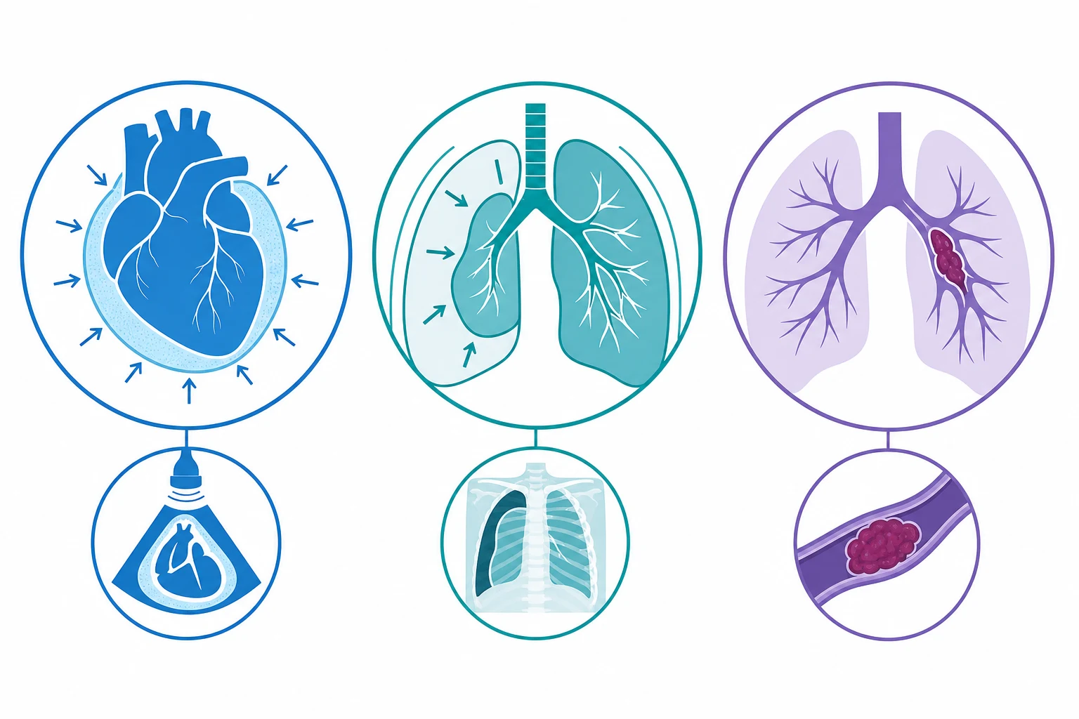
1. Cardiac tamponade
The pericardial fluid under the pressure compresses the heart, preventing the ventricular filling.[1][1]
Beck's triad (the classic — for the acute tamponade):
- The hypotension.
- The distended the neck the veins (the elevated the JVP / the CVP).
- The muffled the heart the sounds.[1]
The clinical. The pulsus paradoxus (the SBP drops above 10 mmHg on the inspiration — the exaggerated the normal). The tachycardia. The tachypnoea. The PEA arrest (the common the arrest the rhythm).[1]
The echo. The pericardial effusion. The RA collapse (the early diastole). The RV collapse (the late diastole). The plethoric the IVC (the dilated, the non-collapsing). The swinging the heart.[1][1]
The management. The immediate pericardiocentesis (the echo-guided; the subxiphoid). The fluid the challenge (the volume the loading — the buys the time by increasing the filling the pressure). The NOT the diuretics / the vasodilators (the worsen the filling). The surgical the window (the recurrent).[1]
2. Tension pneumothorax
The air accumulates in the pleural space under the pressure, compressing the mediastinum and the great vessels.[1][1]
The clinical. The tracheal deviation (the away from — the late). The hypoxaemia. The hypotension. The tachycardia. The hyperresonance + the absent breath sounds (the affected side). The distended neck veins. The time-the-critical.[1]
The management. The immediate needle thoracostomy (the 14G the cannula in the 2nd ICS the mid-clavicular OR the 5th ICS the mid-axillary — the decompress; the rush of the air). The then the formal the chest the drain (the Seldinger the or the surgical). The NOT the wait for the CXR (the clinical the diagnosis).[1]
3. Massive pulmonary embolism
The large embolus obstructs the pulmonary circulation, increasing the RV afterload, causing the RV failure and the reduced LV filling.[1][1]
The clinical. The sudden the dyspnoea, the hypoxaemia, the hypotension, the tachycardia. The risk factors (the DVT, the post-op, the malignancy, the immobility). The right-the-heart the strain (the echo — the RV the dilatation, the McConnell the sign, the tricuspid the regurgitation).[1]
The echo. The RV dilatation. The McConnell sign (the RV the apical the sparing — the hypokinetic the RV the free the wall with the hyperkinetic the apex). The D-shaped the septum (the paradoxic the septal the motion — the RV the overload). The plethoric the IVC.[1]
The management. The immediate the thrombolysis (the alteplase — if the massive with the hypotension; the 50 mg the IV the bolus or the 100 mg the infusion). The NOT the wait for the CT (the echo the diagnostic). The anticoagulation (the heparin). The surgical the embolectomy / the catheter the embolectomy (if the thrombolysis the contraindicated / the failed).[1]
The differentiation
The common the feature: the elevated the JVP / the CVP (the venous the congestion — the blood cannot the enter the heart) + the hypotension (the reduced the CO). The differentiation by the clinical + the echo + the CXR.[1]
| Feature | Tamponade | Tension PTX | Massive PE |
|---|---|---|---|
| JVP | Elevated | Elevated | Elevated |
| Breath sounds | Normal | Unilateral absent | Normal (may be diffuse wheeze) |
| CXR | Enlarged heart | Unilateral hyperlucency | May be normal |
| Echo | Effusion, RA/RV collapse | Absent lung sliding, compressed heart | RV dilatation, McConnell sign |
| Intervention | Pericardiocentesis | Needle thoracostomy | Thrombolysis |
Prognosis
The mortality the depends on the rapidity of the decompression. The delay → the PEA arrest → the death. The immediate the decompression the life-the-saving.[1][1]
SAQ — Massive pulmonary embolism: diagnosis and thrombolysis
10 minutes · 10 marks
A 60-year-old woman, day 7 post elective hip replacement, collapses on the ward with sudden dyspnoea. She is hypotensive (BP 75/40), tachycardic (HR 130), SpO2 86% on high-flow oxygen, with distended neck veins. A bedside echocardiogram shows a dilated right ventricle with septal bowing into the LV. The team asks for the diagnosis and immediate management.
SAQ — Tension pneumothorax: recognition and emergency decompression
10 minutes · 10 marks
A 25-year-old man is brought into the resuscitation bay after a stab wound to the right chest. He is in severe respiratory distress, hypotensive (BP 75/45), tachycardic (HR 130), with absent breath sounds on the right and tracheal deviation to the left. The team prepares for action.
Red flags
Pathophysiology — the unified mechanism
Obstructive shock is the one shock state where the pump is normal but the plumbing is blocked. All three causes converge on the same endpoint — impaired ventricular filling → reduced preload → reduced stroke volume → reduced cardiac output → shock — but each blocks a different point of the circuit.[1][1]
flowchart TD
    A[Tamponade<br/>pericardial fluid/clot] --> E[Impaired ventricular FILLING]
    B[Tension PTX<br/>intrathoracic pressure] --> E
    C[Massive PE<br/>pulmonary vascular obstruction] --> E
    E --> F[Reduced PRELOAD]
    F --> G[Reduced stroke volume]
    G --> H[Reduced cardiac output]
    H --> I[HYPOPERFUSION = shock]
    H --> J[Compensatory tachycardia<br/>+ vasoconstriction]
    J --> K[Elevated JVP / CVP<br/>the venous return is blocked]
The signature haemodynamic fingerprint that unifies the three is the combination the examiners drill: elevated filling pressures (raised JVP/CVP) WITH hypotension. In hypovolaemic shock the JVP is flat; in obstructive shock it is distended — the blood cannot get INTO or THROUGH the heart, so it piles up on the venous side. This single observation at the bedside re-routes the whole differential.[1]
The three causes — where each blocks the circuit
| Cause | Site of obstruction | Primary haemodynamic lesion | Distinguishing finding |
|---|---|---|---|
| Cardiac tamponade | Pericardial space (fluid/clot around the heart) | Compresses the heart in diastole → cannot fill | Raised & EQUALISED diastolic pressures; RA/RV collapse on echo; pulsus paradoxus |
| Tension pneumothorax | Pleural space + mediastinum (air under pressure) | Compresses the great veins + mediastinal shift → impaired venous return + increases PVR | Unilateral absent breath sounds, hyperresonance, tracheal deviation (late) |
| Massive PE | Pulmonary arterial tree (embolus) | Acute RV afterload ↑ → RV failure → reduced LV preload | RV strain on echo (McConnell sign), S1Q3T3, deep venous thrombosis |
The pericardial pressure-volume curve (tamponade)
The pericardium is a stiff, nearly inextensible fibrous sac. The critical concept is the pericardial pressure-volume relationship: it is flat at low volumes (a little extra fluid barely raises pressure) then turns near-vertical once reserve capacity is exhausted. Fluid accumulating slowly (weeks) lets the pericardium creep and stretch — up to 2 L can collect without tamponade. Fluid accumulating fast (hours — trauma, aortic dissection into the pericardium, post-surgical bleed) hits the vertical part of the curve immediately, and as little as 150–200 mL can be lethal.[5][8]
Once intrapericardial pressure exceeds intracardiac filling pressure, the chambers invert during diastole: the right atrium collapses first (early diastole, when RA pressure is lowest), then the right ventricle (late diastole). Stroke volume falls, the heart rate rises to compensate, and the patient mounts exaggerated inspiratory effort — producing pulsus paradoxus (an exaggerated version of the normal <10 mmHg inspiratory drop in systolic BP).[8]
The intrathoracic pressure effect (tension pneumothorax)
A one-way valve lets air into the pleural space on inspiration but blocks its exit. Intrapleural pressure climbs above atmospheric, collapsing the ipsilateral lung AND compressing everything in the mediastinum. The dominant haemodynamic lesion is compression of the thin-walled great veins (SVC/IVC) and the right heart — venous return is physically squeezed off, so preload collapses even though the JVP is distended. A secondary injury is mediastinal shift kinking pulmonary vessels and acutely raising pulmonary vascular resistance, loading the RV. Tracheal deviation is a LATE, pre-terminal sign — by the time you see it, the patient is minutes from arrest.[1]
The ventricular interdependence effect (massive PE)
A large embolus suddenly occludes the pulmonary vascular bed → pulmonary artery pressure spikes → the thin-walled RV, built for low pressure, dilates and fails. The failing, dilated RV bulges into the LV through the interventricular septum (D-shaped septum / paradoxic septal motion) — LV preload and compliance fall, LV stroke volume falls, and cardiac output collapses. This is acute cor pulmonale. The elevated CVP here reflects RV failure, not volume overload: giving more fluid to an already-failing, dilated RV can worsen septal shift and actually drop cardiac output.[2][4]
Distinguishing clinical features at the bedside
Because the three causes share the elevated-JVP-plus-hypotension fingerprint, the discriminating signs are what the bedside exam and the POCUS add. Memorise the syndrome clusters rather than isolated signs.[1][1]
Bedside differentiation — the syndrome clusters
| Feature | Tension pneumothorax | Cardiac tamponade | Massive PE |
|---|---|---|---|
| Insidious vs sudden | Usually sudden (trauma, positive-pressure ventilation, line insertion) | Variable — slow (malignancy) or sudden (trauma, dissection, post-op) | Sudden (dyspnoea, syncope, collapse) |
| Breath sounds | UNILATERAL absent | Normal (muffled heart sounds) | Normal; may be diffuse |
| Percussion | Unilateral HYPERRESONANCE | Normal | Normal |
| Trachea | Deviated AWAY (late sign) | Central | Central |
| Neck veins | Distended (may be absent if hypovolaemic) | Distended | Distended |
| Heart sounds | Normal/distant | MUFFLED | Loud P2, tricuspid regurgitation murmur |
| Pulsus paradoxus | May be present | CLASSIC (>10 mmHg) | May be present |
| Chest/mediastinum | Hyperlucency, depressed hemidiaphragm, mediastinal shift | Enlarged cardiac silhouette (globular) | May be normal; oligoaemia (Westermark), Hampton's hump |
| ECG | Usually normal (small voltage if huge) | Low voltage ± ELECTRICAL ALTERNANS | S1Q3T3, sinus tachycardia, T-wave inversion V1-V4, right axis |
| POCUS/echo | Absent lung sliding, absent B-lines, stratosphere sign | Effusion + RA/RV collapse + plethoric IVC | RV dilatation, McConnell sign, D-shaped septum, TR |
| Definitive act | Needle thoracostomy → chest tube | Pericardiocentesis | Thrombolysis ± catheter/surgical embolectomy |
The three classic exam triads
- Beck's triad (acute tamponade, from trauma): hypotension + muffled heart sounds + distended neck veins. Classical on paper, rare (<20%) in practice — most modern tamponade is subacute and presents with vague dyspnoea and a raised JVP.[8]
- Tension pneumothorax cluster: tracheal deviation + absent breath sounds + hyperresonance on the affected side, with hypoxaemia and hypotension. Tracheal deviation is a late pre-terminal sign — do not wait for it.[1]
- Massive PE cluster: sudden dyspnoea + hypoxaemia + hypotension with risk factors and RV strain on echo. The ECG S1Q3T3 is memorable but neither sensitive nor specific.[2]
ECG findings
ECG in the three causes of obstructive shock
| Cause | ECG finding | Mechanism / significance |
|---|---|---|
| Tamponade | Low QRS voltage (<5 mm limb leads) | Fluid conducts electricity away from the chest wall (short-circuits the signal) |
| Tamponade | Electrical alternans (beat-to-beat varying QRS amplitude/axis) | The heart SWINGS freely in a large effusion, changing its electrical axis each beat — pathognomonic for large effusion / pre-tamponade |
| Tamponade | PR depression / diffuse ST elevation | If due to pericarditis (the underlying cause) |
| Tension PTX | Usually normal | May show small voltages if the pneumothorax is enormous; right-axis deviation with a left-sided PTX from heart displacement |
| Massive PE | Sinus tachycardia (the commonest) | Non-specific |
| Massive PE | S1Q3T3 (deep S in I, Q in III, inverted T in III) | Acute RV strain/dilatation; classic but seen in only ~10-50%; absence does NOT exclude PE |
| Massive PE | Right axis deviation, RBBB (incomplete/complete) | RV strain/conduction delay |
| Massive PE | T-wave inversion V1–V4 (and inferior leads) | RV strain; a negative T in V1 plus T inversion in V1-V4 has high specificity for central PE |
| Massive PE | S1Q3T3 + right axis + RBBB + TWI = Sgarbossa-McGinn-Wellens PE pattern | The full "right heart strain" picture |
Pearl: S1Q3T3 was described by McGinn & White in 1935 — it is a teaching favourite because it is so memorable, but in modern practice sinus tachycardia plus a suggestive story and RV strain on echo is far more common. A normal ECG never excludes massive PE.[4]
Echocardiography and POCUS — the decisive test
Bedside ultrasound (FOCUS — Focused Cardiac Ultrasound) is the single most useful investigation in suspected obstructive shock: it confirms the cause within seconds at the bedside and is the bridge to definitive decompression. Every arrested or peri-arrest patient should have a cardiac POCUS.[5][7]
POCUS findings by cause
| View | Tamponade | Tension PTX | Massive PE |
|---|---|---|---|
| Subcostal / IVC | Plethoric IVC (>2.1 cm, <50% collapse) | IVC variably distended | Plethoric IVC |
| Parasternal long / short | Effusion, RV collapse (late diastole) | ABSENT lung sliding on affected side (M-mode: barcode / stratosphere sign) | RV dilatation, D-shaped (flattened) septum |
| Apical 4-chamber | Effusion, RA collapse (early diastole), swinging heart | May see compressed, displaced heart | RV/LV ratio >1 (RV bigger than LV), McConnell sign |
| Lung windows | — | NO lung sliding, NO B-lines on the affected side (sliding + B-lines on the normal side) | Bilateral lung sliding; may show bilateral B-lines if infarction/oedema |
The McConnell sign (massive PE)
McConnell sign = akinesia of the RV free wall with SPARED, hyperkinetic RV apex — gives a distinctive flickering-apex appearance. It is highly suggestive of acute PE (sensitivity ~77%, specificity ~94% in the original description) because chronic RV overload hypertrophies the apex, whereas acute PE spares it. Its absence does not exclude PE, and it can appear in other acute RV stress states, but in the right context it is a strong pointer toward reperfusion.[2][4]
The FALLS protocol (Fluid Administration Limited by Lung Sonography)
The BLUE / FALLS protocols use lung and cardiac POCUS to separate shock phenotypes. In the FALLS approach you administer fluid while watching the lung for B-lines: if the JVP is high and B-lines appear with little fluid, think cardiogenic (or tamponade); if the patient remains flat with a huge IVC and a clear chest, think obstructive — then cardiac POCUS sorts tamponade from PE from tension PTX.[1]
Cardiac tamponade — expanded management
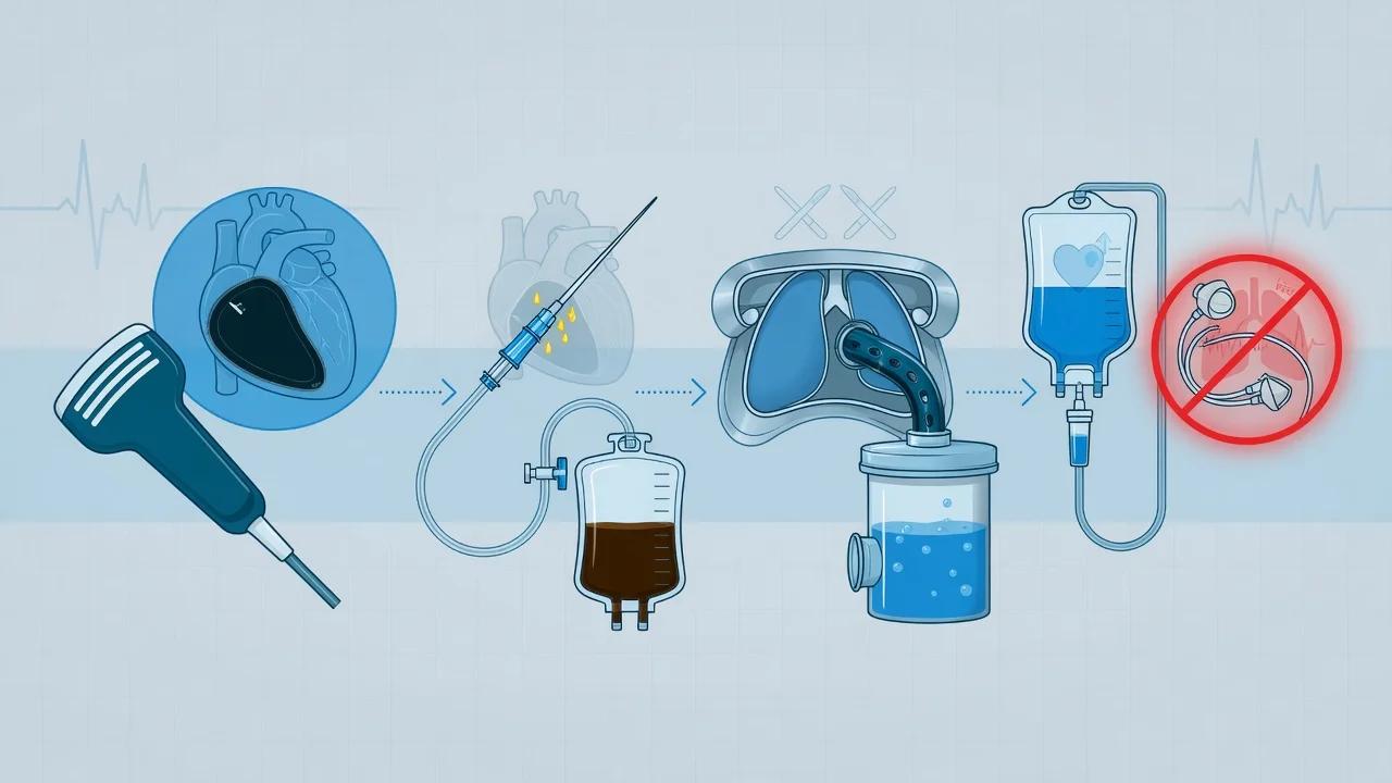
Tamponade management — pericardiocentesis is life-saving
- RECOGNISE — hypotension + tachycardia + distended neck veins + pulsus paradoxus + POCUS showing effusion with RA/RV collapse = tamponade → call for help, prepare for urgent echo-guided pericardiocentesis
- GIVE A FLUID BOLUS (250–500 mL crystalloid) — raises venous return to temporarily push MORE blood past the pericardial constraint (buys time while you set up). Give small boluses and reassess — a huge bolus will not overcome a fixed pericardial constraint and may cause pulmonary oedema if there is a mixed picture
- AVOID POSITIVE-PRESSURE VENTILATION unless the pericardium can be drained the instant the patient is intubated — PPV raises intrathoracic pressure, drops venous return, and can precipitate cardiac arrest in tamponade. If intubation is unavoidable: lowest PEEP, lowest pressures, drain IMMEDIATELY after induction
- AVOID DIURETICS AND VASODILATORS — the patient is preload-dependent; nitrates, opiates (relative), and diuretics all lower preload and worsen output
- START A VASOPRESSOR (noradrenaline) as a temporising bridge if profoundly hypotensive before drainage — definitive treatment is still pericardiocentesis
- PERFORM ECHO-GUIDED PERICARDIOCENTESIS (Seldinger, pigtail drain). Approach: subxiphoid (1–2 cm below the left xiphocostal angle, aim at the left shoulder, 15–30° to skin) or apical (5th ICS mid-clavicular — closest to a large apical effusion). Confirm needle position with agitated saline (bubbles appear in the pericardial space, not in the RV). Aspirate slowly — rapid removal of >500 mL risks acute RV dilatation ("cardiac decompression syndrome")
- LEAVE A DRAIN (pigtail) for continued drainage and to detect reaccumulation; flush q8h; remove when output <50 mL/24 h AND no reaccumulation on echo
- SEND FLUID for cytology (malignancy — send ≥50 mL), culture ± AFB/TB-PCR, cell count, glucose, protein, LDH, triglycerides (chylopericardium), ADA (TB)
- TREAT THE CAUSE — malignancy (pericardial window ± sclerotherapy); uraemia (dialysis); TB (anti-TB therapy + steroids); infection (antibiotics); autoimmune (steroids/immunosuppression); aortic dissection with haemopericardium → SURGERY, not pericardiocentesis alone
Tension pneumothorax — needle thoracostomy technique
Tension pneumothorax — immediate decompression
- CLINICAL DIAGNOSIS — DO NOT WAIT FOR A CXR. Unilateral absent breath sounds + hyperresonance + hypoxaemia/hypotension (± tracheal deviation, distended neck veins) on the affected side = decompress NOW. A CXR is for confirmation AFTER decompression
- CHOOSE THE SITE:
- 2nd intercostal space, mid-clavicular line (classic anterior approach) — fast, memorable, but the 14G cannula may be too short in a muscular or large-breasted patient (the chest wall here can be >5 cm)
- 5th intercostal space, mid-axillary line (lateral/anterior axillary approach) — increasingly preferred (longer needle-to-pleura distance is more consistent with the 5 cm+ cannula length; preferred in ATLS 10th edition and by many trauma services); aligns with the chest-tube site
- NEEDLE THORACOSTOMY: 14G (or larger-bore) cannula, insert JUST ABOVE the upper border of the rib (avoid the neurovascular bundle running below each rib). A rush of air + improvement in BP/saturation confirms success. In an arrested patient, bilateral needle decompression is part of the reversible-causes protocol
- CONVERT TO A FORMAL CHEST TUBE — needle decompression is only a bridge. Insert an intercostal drain (Seldinger for small, or blunt-dissection 28–32Fr for trauma/haemothorax) at the 5th ICS mid-axillary line, connect to underwater seal
- REASSESS — if no improvement after decompression, suspect: wrong side, kinked/blocked cannula, cannula too short (consider a longer/firmer device), or a different diagnosis (massive PE, tamponade)
- POST-DECOMPRESSION CXR — confirm lung re-expansion and tube position; check for re-expansion pulmonary oedema after rapid drainage of a large pneumothorax
Needle decompression — 2nd ICS mid-clavicular vs 5th ICS mid-axillary
| Feature | 2nd ICS mid-clavicular | 5th ICS mid-axillary |
|---|---|---|
| Ease of access in supine trauma patient | Good | Good (lateral approach) |
| Chest-wall thickness at this site | Often >4–5 cm (especially males, large BMI) — 14G cannula may not reach the pleura | Generally thinner, more reliable penetration |
| Landmark ease | Easy (sternal angle → 2nd rib → 2nd ICS) | Easy (anterior axillary line, 5th ICS) |
| Risk of injury | Internal mammary artery, great vessels | Long thoracic nerve, lung, intercostal vessels |
| Current guideline preference | Traditional; still taught | Increasingly preferred (ATLS 10th, many trauma services) |
| Conversion to chest tube | Separate site | Same site as chest tube — logistically easier |
Massive PE — reperfusion strategy
Massive PE — risk stratification and reperfusion
- STRATIFY THE RISK — High-risk (massive): sustained hypotension (SBP <90 mmHg for ≥15 min) or shock → needs immediate reperfusion. Intermediate-risk (submassive): normotensive but RV dysfunction + positive troponin. Low-risk: neither.[4]
- GIVE OXYGEN, SUPPORT THE CIRCULATION — high-flow O2 for hypoxaemia; cautious fluids (250 mL boluses — the failing RV does not tolerate large volume); noradrenaline to restore coronary perfusion pressure to the ischaemic RV; consider dobutamine/milrinone for RV inotropy
- START ANTICOAGULATION — therapeutic LMWH or unfractionated heparin (UFH preferred if reperfusion is planned, as it is short-acting and reversible)
- HIGH-RISK PE → IMMEDIATE REPERFUSION:
- Systemic thrombolysis (first line): alteplase 100 mg over 2 h (or 50 mg IV bolus in peri-arrest). Halt heparin during the infusion. Absolute contraindications (haemorrhagic stroke, active bleeding, recent neurosurgery/intracranial trauma) → go to catheter/surgical embolectomy
- Surgical embolectomy: if thrombolysis contraindicated or failed, or in centres with immediate capability; also preferred for paradoxical embolus (clot-in-transit across a PFO) or if a mechanical valve is present
- Percutaneous catheter-directed therapy: ultrasound-assisted thrombolysis (EKOS) or catheter embolectomy — lower systemic lytic dose, useful for intermediate-high risk or contraindication to full-dose lysis
- INTERMEDIATE-RISK PE: anticoagulate; monitor closely (ward with telemetry) — reserve thrombolysis for clinical deterioration (the PEITHO trial showed reduced decompensation but at the cost of major bleeding, including intracranial haemorrhage).[3]
- INVESTIGATE THE SOURCE — lower-limb doppler; consider IVC filter only if anticoagulation is contraindicated or recurrent embolism despite therapeutic anticoagulation (NOT routine)
- TRANSITION TO LONG-TERM ANTICOAGULATION — DOAC or warfarin once stable; at least 3 months for provoked PE, lifelong for unprovoked/recurrent
Reperfusion options for high-risk (massive) PE
| Option | Indication | Pro | Con |
|---|---|---|---|
| Systemic thrombolysis (alteplase 100 mg/2 h) | First line for high-risk PE with shock; also for suspected PE arrest | Fast, widely available, no theatre needed | Major bleeding (~6–10%); intracranial haemorrhage (~2%); absolute contraindications |
| Surgical embolectomy | Thrombolysis contraindicated/failed; clot-in-transit across PFO; large free-floating RA/RV thrombus | Definitive clot removal; no systemic lytic | Needs cardiothoracic theatre/cpB; not universally available |
| Catheter-directed thrombolysis / embolectomy (EKOS, AngioJet, FlowTriever) | Intermediate-high risk; high-risk with high bleeding risk; centres with IR capability | Lower lytic dose → less bleeding; rapid haemodynamic improvement | Needs interventional radiology; not universally available |
| VA-ECMO | Refractory collapse / arrest as bridge to embolectomy | Supports circulation; oxygenation; buys time | High bleeding/complication rate; specialist centre |
Key trials and evidence
Kucher 2006 — Massive pulmonary embolism in ICOPER (PMID 16432055)
Source
Circulation — retrospective analysis of the International Cooperative PE Registry (2,392 patients; 108 with massive PE defined as SBP <90 mmHg)
Headline result
90-day mortality in massive PE was 52.4% vs 14.7% in non-massive PE
Practice gap
68% of massive-PE patients did NOT receive thrombolysis, surgical or catheter embolectomy
On thrombolysis
Thrombolysis did not significantly reduce 90-day mortality (HR 0.79, 95% CI 0.44–1.43) — a registry limitation (selection bias), NOT evidence that lysis is ineffective
Clinical bottom line
Massive PE is highly lethal; advanced reperfusion is underused. Observational data, so treat as descriptive — it quantifies the stakes, it does not define treatment
PEITHO 2014 — Meyer (PMID 24716681)
Source
NEJM — RCT of tenecteplase vs placebo in 1,006 normotensive patients with intermediate-risk PE (RV dysfunction + positive troponin)
Primary outcome
Tenecteplase reduced death or haemodynamic decompensation by 56% (2.6% vs 5.6%)
Safety
Significantly MORE major bleeding (11.5% vs 2.4%) and MORE intracranial haemorrhage (2.4% vs 0.2%); two fatal ICH in the elderly
Clinical bottom line
In submassive PE, routine thrombolysis does NOT improve survival and increases bleeding. Reserve lysis for clinical deterioration (falling BP, worsening hypoxia). It does NOT apply to massive PE with shock — those still get full-dose lysis.
ESC 2019 / 2020 PE Guidelines — Konstantinides (PMID 31473594)
Source
European Heart Journal — multidisciplinary ESC/ERS task force guideline
Risk stratification
High-risk (massive: shock/hypotension) → immediate reperfusion. Intermediate (submassive: RV strain ± troponin) → anticoagulate + monitor. Low-risk → anticoagulate, consider early discharge
Reperfusion
Systemic thrombolysis first line for high-risk; surgical/catheter embolectomy if lysis contraindicated or failed; catheter-directed therapy for selected intermediate-high risk
Anticoagulation
DOACs preferred for most; UFH if reperfusion planned
Clinical bottom line
The current European standard for PE — drives the 'lyse the shocked patient, anticoagulate the rest' rule and the move to catheter-directed therapy
ESC 2015 Pericardial Disease Guidelines — Adler (PMID 28855243) and Spodick NEJM 2003 (PMID 12672859)
Source
European Heart Journal guideline + NEJM clinical review on tamponade
Diagnosis
Echo is gold standard: RA collapse in early diastole (most sensitive), RV collapse in late diastole (most specific), IVC plethora, respiratory variation in mitral (>25%) and tricuspid (>40%) inflow
Management
Echo-guided pericardiocentesis (Seldinger, pigtail drain); fluid bolus temporises; AVOID positive-pressure ventilation, diuretics, vasodilators; surgical window for recurrent effusion
Clinical bottom line
Tamponade is a clinical + echo diagnosis; pericardiocentesis is life-saving; treat the underlying cause to prevent reaccumulation
Special scenarios
Obstructive shock in specific contexts
| Scenario | Special feature | Management twist |
|---|---|---|
| Mechanically ventilated patient | Tension PTX can develop rapidly under positive pressure; tamponade worsens with PPV | High index of suspicion; bilateral needle decompression in the deteriorating ventilated patient; minimise PPV/PEEP in tamponade |
| Iatrogenic (post-CVC, post-PCI, post-paceport) | Sudden collapse during/after a procedure | POCUS immediately; pericardiocentesis for haemopericardium; tamponade from CVC erosion, PCI perforation, pacer wire |
| Trauma | Tension PTX and tamponade coexist (blunt cardiac injury, haemopericardium) | ATLS primary survey; needle thoracostomy + chest tube; ED thoracotomy if pulseless with tamponade |
| Malignancy | Most common cause of tamponade; recurrent | Pericardiocentesis + pericardial window ± sclerotherapy; treat underlying cancer |
| Pregnancy | PE risk 4–5× higher; aortocaval compression changes haemodynamics | Left lateral tilt; weight-adjusted LMWH; systemic lysis if high-risk PE (alteplase does not cross placenta significantly) |
| Post-cardiac surgery | Tamponade is ORGANISED CLOT; chest-tube output stops | SURGICAL re-exploration — pericardiocentesis will fail |
| Aortic dissection (Type A) | Haemopericardium = surgical emergency | BP control (beta-blocker first); theatre for ascending aorta repair; pericardiocentesis only as bridge |
Prognosis
Outcome by cause and speed of decompression
| Cause | Outcome determinant | Mortality |
|---|---|---|
| Tension pneumothorax | Speed of decompression | Near 100% untreated; <10% with immediate needle + chest tube |
| Cardiac tamponade | Speed of drainage + underlying cause | Malignancy: median survival 2–4 months (cancer-driven); viral/idiopathic: excellent; post-surgical: 20–30% if delayed |
| Massive PE | Initial haemodynamics + reperfusion | Massive PE 90-day mortality ~52% (ICOPER); rises sharply with arrest |
| Aortic dissection + tamponade | Surgery | Very high without emergency repair |
References
- [1]Levy B, et al. Obstructive Shock, from Diagnosis to Treatment Rev Cardiovasc Med, 2022.PMID 39076909
- [2]Kucher N, Rossi E, De Rosa M, Goldhaber SZ Massive pulmonary embolism Circulation, 2006.PMID 16432055
- [3]Meyer G, Vicaut E, Danays T, et al. Fibrinolysis for patients with intermediate-risk pulmonary embolism N Engl J Med, 2014.PMID 24716681
- [4]Konstantinides SV, Meyer G, Becattini C, et al. 2019 ESC Guidelines for the diagnosis and management of acute pulmonary embolism developed in collaboration with the European Respiratory Society (ERS): The Task Force for the diagnosis and management of acute pulmonary embolism of the European Society of Cardiology (ESC) Eur Respir J, 2019.PMID 31473594
- [5]Adler Y, Charron P, Imazio M, et al. Update on angiotensin II: new endocrine connections between the brain, adrenal glands and the cardiovascular system Endocr Connect, 2017.PMID 28855243
- [6]Imazio M, Brucato A, Spodick DH, Adler Y Injury risks of EMS responders: evidence from the National Fire Fighter Near-Miss Reporting System BMJ Open, 2015.PMID 26068510
- [7]Tsang TS, Enriquez-Sarano M, Freeman WK, et al. Intra-articular injection composed of steroid, iohexol and local anaesthetic: is it stable? Br J Radiol, 2009.PMID 19001468
- [8]Spodick DH Influenza vaccination and reduction in hospitalizations for cardiac disease and stroke among the elderly N Engl J Med, 2003.PMID 12672859