ICU · Resuscitation
The Principles of Resuscitation
Also known as Resuscitation · Fluid resuscitation · Fluid responsiveness · Oxygen delivery · Goals of resuscitation · Early goal-directed therapy · Massive transfusion
Resuscitation is the restoration of oxygen delivery to the tissues in the patient in shock, and it is the first act of intensive care. This topic builds the examiner's framework on five ideas. First, the goal — the global oxygen delivery and the perfusion, and the principle that resuscitation targets the perfusion, not a single pressure. Second, fluid — when to give it (only the fluid-responsive patient benefits), how to judge responsiveness (the dynamic indices, the passive leg raise), and the choice of fluid (balanced crystalloids over saline; colloids offer no outcome advantage). Third, blood — the restrictive transfusion threshold and the damage-control ratio in massive haemorrhage. Fourth, the endpoints of resuscitation — the lactate clearance and the peripheral perfusion (the capillary refill), not the central venous pressure. Fifth, the evidence — the EGDT era and its refinement by ProCESS and ARISE, the ANDROMEDA-SHOCK capillary-refill strategy, and the shift from protocolised targets to individualised, perfusion-directed resuscitation.
On this page & tools
Your progress
Saved locally on this device.
5 MCQs with explanations
Target exams
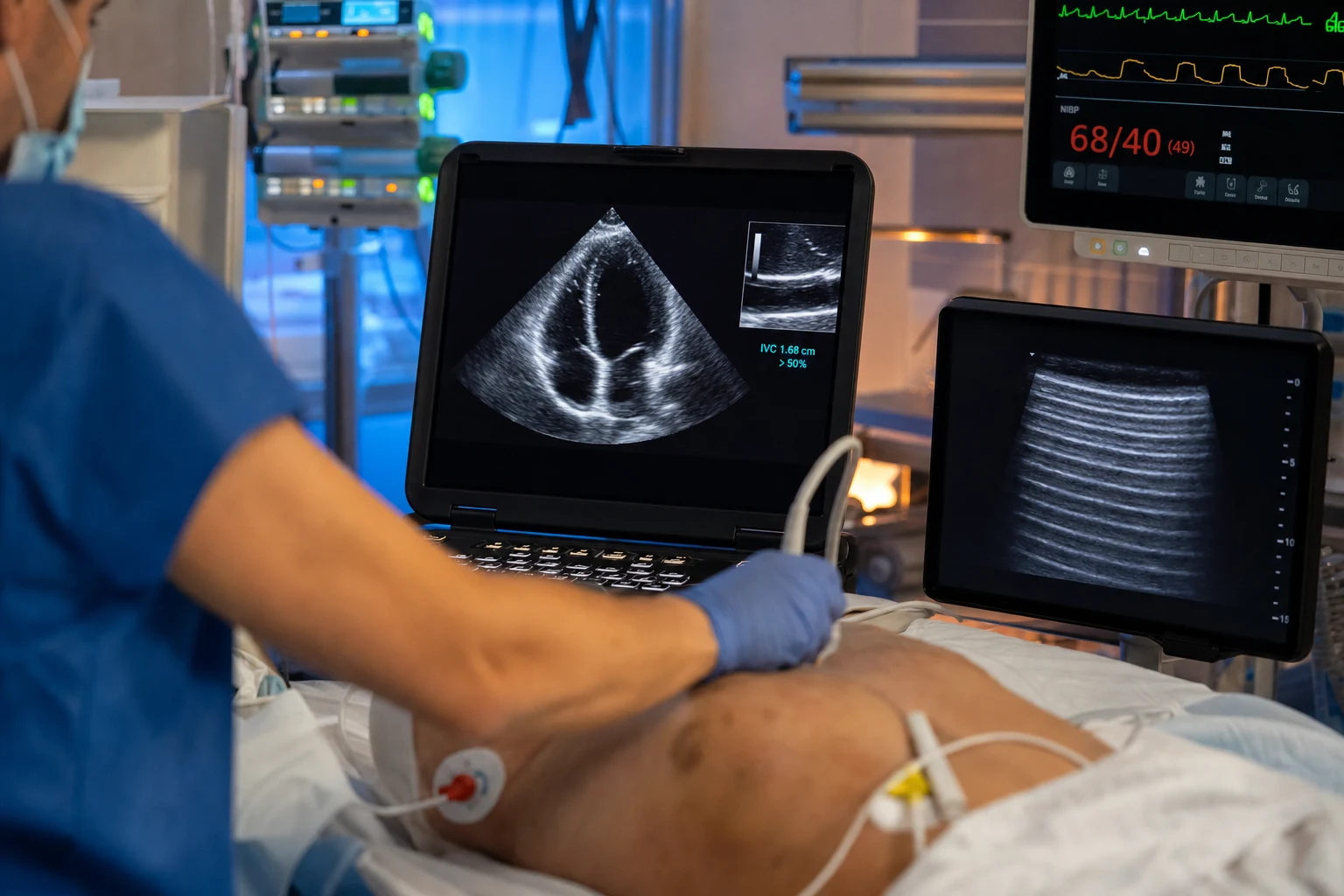
Overview & definition
Resuscitation is the restoration of oxygen delivery to the tissues in the patient whose perfusion is failing — the patient in shock. It is the first act of intensive care, and its goal is straightforward to state and difficult to execute: to deliver enough oxygen to meet the tissues' demand, and to do so before the hypoxia of under-resuscitation gives way to the oedema, the congestion and the abdominal compartment of over-resuscitation.[1][1]
The framework rests on five ideas, each of which the evidence of the last two decades has refined. The goal is the global oxygen delivery and the perfusion, not a single pressure. Fluid helps only the patient who is fluid-responsive, and that responsiveness is judged by the dynamic indices, not the central venous pressure. Blood is given to a restrictive threshold, and in massive haemorrhage to a damage-control ratio. The endpoints of resuscitation are the lactate clearance and the peripheral perfusion. And the evidence has moved from the protocolised targets of early goal-directed therapy to an individualised, perfusion-directed approach.[1][2]
The goal: oxygen delivery and perfusion
Global oxygen delivery (DO2) is the product of the cardiac output and the arterial oxygen content (CaO2): DO2 = CO × CaO2, where CaO2 is governed by the haemoglobin and the saturation. Delivery fails when the cardiac output falls (hypovolaemia, pump failure, obstructive shock) or when the content falls (anaemia, hypoxaemia), and the resuscitation is the correction of whichever has failed — fluid and vasopressors for the output, blood and oxygen for the content.[1]
The goal of resuscitation is to restore the perfusion — the delivery of oxygen that meets demand — and the markers of adequate perfusion are a clearing lactate, a normalising capillary refill, a warming periphery and a recovering urine output. A mean arterial pressure is a means to that end, not the end itself; the pressure target is the lowest that restores perfusion, because a higher pressure achieved with more fluid and more vasopressor carries its own harm.[1]
The perfusion checklist — the four targets
The resuscitationist defends four targets in the first hours, each a window onto the adequacy of perfusion rather than an end in itself, and each titrated to the individual. A single target in isolation misleads — the pressure may be adequate while the periphery is cold and the lactate is rising — so the four are examined together at the bedside.[1][19]
- A mean arterial pressure of at least 65 mmHg — the perfusion pressure that, in most patients, restores autoregulation in the cerebral, coronary and splanchnic beds. SEPSISPAM showed that targeting 65 mmHg was as safe as targeting 85 mmHg in the unselected patient with septic shock, and the chronically hypertensive patient may need a higher target to clear their mentation and their urine, while the young and the previously normotensive may perfuse at a lower one.[17]
- A lactate clearance of at least 10 per cent — lactate is the metabolic signature of anaerobic under-perfusion, and its clearance over the first hours of resuscitation is both a measure of adequacy and a prognostic sign. A clearance of 10 per cent or more over two hours (a fall of roughly 20 per cent over the first two hours) is associated with improved survival, and a lactate that fails to clear warns that the resuscitation is inadequate or the cause uncontrolled.[24][25]
- A urine output above 0.5 mL/kg/h — the recovering urine output is the kidney's witness that the renal perfusion has been restored; oliguria persists in the under-resuscitated and warns of the incipient acute kidney injury that complicates shock.
- A central venous oxygen saturation (ScvO2) of 70 per cent or above — the saturation of blood returning to the right heart reflects the balance between the oxygen delivered and the oxygen consumed; a low ScvO2 (below 70 per cent) signals that the tissues are extracting harder than the delivery is providing, and was the cornerstone of the original EGDT protocol, though it has been superseded by the peripheral-perfusion markers as the routine target.[1]
The four resuscitation targets — what each measures, and its pitfalls
| Target | Threshold | What it measures | Pitfall / caveat |
|---|---|---|---|
| Mean arterial pressure | ≥ 65 mmHg | The perfusion pressure to the vital beds | The chronically hypertensive may need a higher target; the young may perfuse lower. A "normal" MAP does not exclude under-perfusion (cold periphery, rising lactate). SEPSISPAM found no benefit to targeting 85 vs 65 mmHg.[17] |
| Lactate clearance | ≥ 10% over 2 h (≈20% over the first 2 h) | The resolution of anaerobic metabolism | Lactate rises in beta-agonist use, malignancy, hepatic failure and mitochondrial dysfunction (type-B hyperlactataemia) — a non-clearing lactate is not always under-perfusion. The absolute value matters less than the trend.[24] |
| Urine output | > 0.5 mL/kg/h | Renal cortical perfusion | Confounded by diuretics, osmotic agents (mannitol, contrast), pre-existing CKD, and the AKI of established shock (the kidney that cannot make urine regardless of perfusion). A passing furosemide-induced diuresis is not perfusion. |
| ScvO2 | ≥ 70% | The balance of delivery and consumption | A normal ScvO2 in septic shock (the "shunting" or mitochondrial-failure patient) can coexist with tissue hypoxia; a low ScvO2 is specific, a normal one not wholly reassuring. Replaced by the capillary refill as the routine target.[1][10] |
Fluid: give it only to the responsive, and judge responsiveness dynamically
Fluid increases the stroke volume only in the patient whose ventricles are operating on the ascending limb of the Frank-Starling curve — the fluid-responsive patient — and roughly half of critically ill patients are fluid-responsive at any moment. Giving fluid to a non-responsive patient does not increase the cardiac output; it only raises the venous pressure, causes oedema, and contributes to the harm of a positive fluid balance (which is independently associated with mortality).[4]
The central venous pressure does not predict fluid responsiveness — it is a static, pressure-based measure that bears no reliable relationship to the ventricular response to a fluid bolus, as the systematic review of Marik established. The fluid decision is made with the dynamic indices, which challenge the circulation and measure the response:[4][5]
- The pulse pressure variation (ΔPP) and the stroke volume variation — in the mechanically ventilated patient, the cyclical changes in intrathoracic pressure swing the stroke volume, and a ΔPP above about 13 per cent predicts fluid responsiveness with a high sensitivity and specificity.[5]
- The passive leg raise (PLR) — a reversible, endogenous fluid challenge that transfers venous blood from the legs to the heart; a rise in cardiac output (measured by an echocardiographic or arterial-flow surrogate) of about 10 per cent predicts responsiveness, and it works in the spontaneous-breathing and the arrhythmic patient where the ΔPP fails.[6]
- A fluid challenge — a small, rapidly given bolus (e.g. 250 mL of colloid over a minute) with a real-time measure of the response, distinguishing the responder from the non-responder without committing to a large volume.
- The inferior vena cava (IVC) variability — a point-of-care echocardiographic measure of the IVC diameter's respiratory swing. In the ventilated patient a distensibility above 18 per cent and in the spontaneously breathing patient a collapsibility above 50 per cent suggest a volume-responsive right heart, and it is the bedside index of choice where the arterial line is absent or the ΔPP is unreliable.
- The end-expiratory occlusion test (EEXPO) — a 15-second pause at end-expiration removes the intrathoracic pressure that, in the ventilated patient, has been impeding venous return; a rise in cardiac output (or arterial pulse pressure) of 5 per cent predicts fluid responsiveness, and it works in the arrhythmic patient and the patient with right-heart failure where the ΔPP fails.[21]
- The mini-fluid challenge — a tiny (100 mL) bolus given rapidly over one minute, with a real-time flow measure (aortic VTI or pulse-pressure surrogate); a rise in flow of 10 per cent or more distinguishes the responder, and — unlike a 500 mL bolus — it commits no fluid to the non-responder, the very patient the test is meant to protect.[22]
The dynamic indices of fluid responsiveness — what works where
| Index | Threshold | Needs | Ventilated? | Spontaneously breathing? | Arrhythmia? | Pitfall |
|---|---|---|---|---|---|---|
| Pulse pressure variation (ΔPP) / stroke volume variation | > 13% | Arterial line (or PiCCO/Vigileo) | Yes | No (unreliable) | No | Fails with low tidal volume (<8 mL/kg), open chest, low lung compliance, right-heart failure, spontaneous breathing efforts.[5] |
| Passive leg raise (PLR) | > 10% rise in CO/SV | Real-time CO/SV measure (echo VTI, oesophageal Doppler, calibrated arterial waveform) | Either | Yes | Yes | Must start from semi-recumbent (45°) → supine + 45° legs; the effect lasts ~60–90 s; abdominal compression gives a false positive. The gold standard when spontaneous effort or arrhythmia rules out ΔPP.[6] |
| End-expiratory occlusion (EEXPO) | > 5% rise in CO/PP | Arterial line + real-time flow | Yes | No | Yes | Requires 15 s apnoeic pause (tolerated only in the deeply sedated/paralysed); less familiar but powerful where ΔPP fails.[21] |
| IVC variability | Distensibility > 18% (ventilated) / collapsibility > 50% (spontaneous) | Point-of-care ultrasound | Either | Yes | Yes | Operator-dependent; subxiphoid view only; misleading in the intubated with high PEEP, right-heart failure, or abdominal hypertension. A coarse screen, not a precise measure. |
| Mini-fluid challenge | > 10% rise in CO after 100 mL | Real-time flow measure | Either | Yes | Yes | Must be given fast (over 1 min) and measured immediately; a slow infusion blurs the signal. Best coupled with the PLR as a confirmatory test.[22] |
Fluid: the choice — balanced crystalloids, and no advantage to colloids
The choice of fluid is governed by two trials. SMART (NEJM 2018) showed that balanced crystalloids (lactated Ringer's or Plasma-Lyte) modestly but significantly reduced the composite of death, new renal-replacement therapy and persistent renal dysfunction compared with saline, and they avoid the hyperchloraemic acidosis of large-volume saline — so balanced crystalloids are preferred for crystalloid resuscitation.[7]
Colloids offer no outcome advantage over crystalloids. The CRISTAL trial (JAMA 2013) found no mortality difference between colloid- and crystalloid-based resuscitation in hypovolaemic shock; albumin and saline were equivalent in the SAFE trial; and the starch colloids are harmful (renal injury). The cheaper, safer crystalloid — preferably balanced — is the default.[8][1]
The crystalloid-vs-colloid evidence — SAFE, CHEST, 6S and ALBIOS
The crystalloid-versus-colloid question was settled by a quartet of large trials, and the answer is the same in each: the colloid buys no outcome advantage and, in the case of the starches, buys real harm.[11][18]
- The SAFE trial (NEJM 2004, n ≈ 7000) — the definitive albumin-vs-saline comparison: 4% albumin and saline gave equivalent 28-day mortality in a heterogeneous ICU population, deflating two decades of theoretical enthusiasm for colloid. A pre-specified sub-group analysis hinted at harm from albumin in traumatic brain injury, and albumin is now reserved for the specific indication (the cirrhotic with spontaneous bacterial peritonitis, the large-volume paracentesis, the hypo-oncotic patient) rather than as a routine resuscitation fluid.[11]
- The CHEST trial (NEJM 2012, n ≈ 7000) — hydroxyethyl starch (HES 130/0.4) versus saline in a general ICU population: no mortality difference, but a significant increase in renal-replacement therapy. The starch was no better, and it injured the kidney.[12]
- The 6S trial (NEJM 2012, n ≈ 800) — HES 130/0.42 versus Ringer's acetate in severe sepsis: the starch increased mortality and the need for renal-replacement therapy. In the septic patient the starch is not neutral — it kills and it destroys kidneys. This, with CHEST, ended the HES era.[13]
- The ALBIOS trial (NEJM 2014, n ≈ 1800) — 20% albumin to maintain a serum albumin of 30 g/L versus crystalloid in severe sepsis and septic shock: no mortality difference. Even targeted albumin in sepsis confers no outcome benefit.[18]
SAFE — Saline versus Albumin Fluid Evaluation (PMID 15163774)
Document type
Multicentre randomised controlled trial — New England Journal of Medicine
Population
6997 critically ill adults requiring fluid resuscitation across 16 ICUs (Australia and New Zealand)
Intervention
4% albumin versus normal saline for all fluid resuscitation
Headline finding
28-day mortality identical (20.9% albumin vs 21.1% saline). No difference in single-organ failure, days in ICU, days of ventilation, or renal-replacement therapy. Sub-group: albumin trended toward harm in traumatic brain injury.
Clinical bottom line
The definitive refutation of albumin as a routine resuscitation fluid. Albumin and saline are equivalent for outcomes; reserve albumin for its specific indications (cirrhosis with SBP, large-volume paracentesis, severe hypoalbuminaemia), not as a default.
CHEST — Crystalloid versus Hydroxyethyl Starch Trial (PMID 23075127)
Document type
Multicentre randomised controlled trial — New England Journal of Medicine
Population
7000 critically ill adults requiring fluid resuscitation
Intervention
6% hydroxyethyl starch (HES 130/0.4) versus normal saline
Headline finding
No difference in 90-day mortality (18% vs 17%). However, HES significantly increased the rate of renal-replacement therapy (7.0% vs 5.8%) and was associated with more rash and pruritus.
Clinical bottom line
HES offers no benefit and confers a real renal-injury signal. Combined with 6S, this ended the use of HES in critical care. Do not reach for a starch to resuscitate.
6S — Scandinavian Starch for Severe Sepsis (PMID 22738085)
Document type
Multicentre randomised controlled trial — New England Journal of Medicine
Population
804 adults with severe sepsis across 26 Scandinavian ICUs
Intervention
6% hydroxyethyl starch (HES 130/0.42) versus Ringer's acetate
Headline finding
HES INCREASED 90-day mortality (51% vs 43%, RR 1.17) and the need for renal-replacement therapy (22% vs 16%, RR 1.35). The starch was actively harmful in the septic patient.
Clinical bottom line
The decisive evidence that HES is dangerous in sepsis — not merely neutral. The regulatory consequence (EMA restriction, FDA warning) followed. Balanced crystalloids are the resuscitation fluid in septic shock.
Vasopressors: defending the perfusion pressure
When the fluid has restored the preload and the perfusion still fails on a low mean arterial pressure — the distributive shock of sepsis, the vasodilation of anaesthesia and anaphylaxis — the vasopressor defends the perfusion pressure. The choice of agent is governed by the receptor pharmacology and by three large trials, and the synthesis is a clear hierarchy.[19]
Noradrenaline (norepinephrine) is the first-line vasopressor in shock. It is a pure alpha-agonist with modest beta-1 activity — it raises the vascular tone, restores the mean arterial pressure, and the modest inotropy preserves (rather than sacrifices) the cardiac output. It is titrated to a mean pressure of 65 mmHg (or the lowest that perfuses the individual), and it is the agent of first choice in the Surviving Sepsis Campaign guideline.[19]
Dopamine is not the equivalent. SOAP II (NEJM 2010) compared dopamine with noradrenaline in 1679 patients with shock: there was no mortality difference overall, but dopamine caused more arrhythmia (mostly atrial fibrillation) and, in the pre-specified cardiogenic-shock sub-group, a higher mortality. Dopamine is reserved for the select indications (the bradycardic shock, the temporary chronotropy) and is otherwise avoided — the arrhythmia burden is the harm.[14]
Vasopressin is the adjunct, not the first agent. It is a pure V1-receptor agonist that raises the vascular tone by a different pathway than the catecholamines, it is relatively deficient in late septic shock, and at a fixed low dose (0.03 U/min) it "spares" the noradrenaline. VASST (NEJM 2008) found no mortality difference overall between vasopressin and noradrenaline, but a signal of benefit in the less-severe septic shock; VANISH (JAMA 2016) found that early vasopressin did not reduce the kidney-failure-free days compared with noradrenaline. The synthesis: vasopressin is added as a second agent when the noradrenaline dose creeps up (often above 0.25–0.5 µg/kg/min), to spare catecholamine dose, and it is not the agent you reach for first.[15][16]
Adrenaline (epinephrine) is the third-line agent and the choice in refractory shock. It is a potent alpha- and beta-agonist — it restores the pressure and drives the inotropy — but it raises the lactate (a beta-2 glycolytic effect that confounds the lactate endpoint), it drives tachyarrhythmia, and at high dose it injures the splanchnic bed. It is added to noradrenaline and vasopressin in the refractory case, or used in anaphylaxis (where it is first-line) and the beta-blocker-overdose bradycardic shock. SEPSISPAM confirmed that the target pressure (65 vs 85 mmHg) does not change the outcome, so escalating the agent to chase a higher pressure is the wrong reflex; the reflex is to defend a perfusing pressure and examine the perfusion, not to escalate to a number.[17]
The vasopressors — receptor profile, role, and the trial that defines it
| Agent | Receptor profile | First-line role | Key trial | Caveat |
|---|---|---|---|---|
| Noradrenaline (norepinephrine) | α1 >> β1 (no β2) | First-line in septic, vasodilatory and most shock | SOAP II (vs dopamine) — equivalent mortality, fewer arrhythmias[14] | May worsen splanchnic and digital perfusion at high dose; extravasation causes necrosis (central line preferred). The SSC 2021 first-line agent.[19] |
| Vasopressin | V1 pure (no adrenergic) | Adjunct (added when noradrenaline > 0.25–0.5 µg/kg/min) | VASST, VANISH — no mortality benefit; catecholamine-sparing[15][16] | Fixed low dose 0.03 U/min (do NOT titrate — it is not a titratable drug); causes mesenteric and digital ischaemia; do NOT use first-line. |
| Adrenaline (epinephrine) | α1 + β1 + β2 | Third-line / refractory; first-line in anaphylaxis, beta-blocker bradycardia | SOAP II sub-group (worse than noradrenaline in cardiogenic) | Raises lactate (β2 glycolysis — confounds the lactate endpoint); tachyarrhythmia; splanchnic vasoconstriction. Escalate to it, do not start with it. |
| Dopamine | DA + β1 + α1 (dose-dependent) | Avoid as a vasopressor (select: bradycardic shock, temporary chronotropy) | SOAP II — more arrhythmia, worse cardiogenic sub-group[14] | The arrhythmia burden (atrial fibrillation) is the harm. Largely retired from the vasopressor role. |
| Metaraminol / phenylephrine | α1 pure | Temporary vasopressor (periprocedural, no central line) | — | Pure alpha: raises pressure at the cost of stroke volume and cardiac output; a bridge, not a resuscitation agent. |
| Angiotensin II | AT1 | Investigational / refractory vasodilatory shock (ATHOS-3) | — | Reserve for the truly refractory vasoplegia; expensive, thrombosis risk. |
SOAP II — dopamine versus norepinephrine in shock (PMID 20200382)
Document type
Multicentre randomised controlled trial — New England Journal of Medicine
Population
1679 adults with any form of shock (cardiogenic, septic, hypovolaemic) across 8 centres
Intervention
Dopamine versus norepinephrine as the first-line vasopressor
Headline finding
No difference in 28-day mortality (52.5% vs 48.5%). Dopamine had MORE arrhythmic events (24.1% vs 12.4%). A pre-specified sub-group showed higher mortality with dopamine in cardiogenic shock.
Clinical bottom line
Norepinephrine is the first-line vasopressor in shock — equivalent mortality with far fewer arrhythmias. Dopamine is retired from the routine vasopressor role.
SEPSISPAM — high versus low blood-pressure target in septic shock (PMID 24635770)
Document type
Multicentre randomised controlled trial — New England Journal of Medicine
Population
776 adults with septic shock already on vasopressors
Intervention
Mean arterial pressure target of 65 versus 85 mmHg
Headline finding
No difference in 28-day or 90-day mortality. In the pre-specified chronically hypertensive sub-group, the higher target reduced the need for renal-replacement therapy.
Clinical bottom line
A MAP of 65 mmHg is the default target. Individualise upward only for the chronic hypertensive. Do not escalate vasopressors to chase a higher number unless the perfusion demands it.
VANISH — early vasopressin versus norepinephrine in septic shock (PMID 27483065)
Document type
Multicentre factorial randomised controlled trial — JAMA
Population
409 adults with septic shock
Intervention
Early vasopressin (titrated 0–0.06 U/min) versus norepinephrine; with hydrocortisone or placebo in the factorial
Headline finding
No difference in kidney-failure-free days (the primary outcome). No mortality difference. Vasopressin did not prevent acute kidney injury in septic shock.
Clinical bottom line
Vasopressin is an adjunct, not a first-line agent. Use it to spare catecholamine dose once the norepinephrine requirement rises; do not expect it to protect the kidney.
Blood: the restrictive threshold and the damage-control ratio
Blood is a resuscitation fluid, and two principles govern its use. The restrictive transfusion threshold was established by the TRICC trial (NEJM 1999): a transfusion trigger of 70 g/L (a haemoglobin of 70, targeting 70 to 90) was as safe as a liberal threshold (100 g/L) in critically ill patients, and conserves a scarce resource. The exception is the patient with active ischaemia (an acute coronary syndrome), in whom a higher threshold is reasonable.[9]
In massive haemorrhage, the principle is damage-control resuscitation: the early, ratio-based delivery of blood products to prevent the lethal triad of acidosis, hypothermia and coagulopathy. A plasma-to-red-cell-to-platelet ratio approaching 1:1:1 is the standard (from the PROPPR trial's signal of less early death from haemorrhage, balanced against a small risk of more transfusion), with tranexamic acid early in trauma (the CRASH-2 data), permissive hypotension until the bleeding is controlled, and the avoidance of the clear-fluid dilution that worsens coagulopathy.[1][1]
Endpoints: the lactate and the peripheral perfusion
The endpoint of resuscitation is the restoration of perfusion, and the two best-validated surrogates are the lactate clearance and the peripheral perfusion.[10][1]
The lactate reflects the anaerobic metabolism of under-perfusion, and its clearance over the first hours of resuscitation is a marker of the adequacy of resuscitation and a prognostic sign — a failure to clear portends a worse outcome. The capillary refill time is a bedside surrogate of peripheral perfusion, and the ANDROMEDA-SHOCK trial (JAMA 2019) showed that a resuscitation strategy targeting the capillary refill was, if anything, favourable to one targeting the lactate — re-centring the bedside physical examination (a warm, well-perfused periphery with a brisk capillary refill) as a resuscitation endpoint.[10]
The lactate clearance — the metabolic endpoint
The lactate is the metabolic witness of anaerobic under-perfusion, and its clearance over the first hours of resuscitation is both a measure of adequacy and a powerful prognostic sign. The seminal work of Nguyen showed that a lactate clearance of 10 per cent or more over the first two hours was independently associated with improved survival in severe sepsis and septic shock, and a clearance of roughly 20 per cent over the first two hours is a common target. A lactate that fails to clear — that plateaus or rises despite the resuscitation — warns that the resuscitation is inadequate, the source uncontrolled, or the perfusion failing on a hidden cause (ongoing haemorrhage, mesenteric ischaemia, cardiogenic failure).[24][25]
The lactate has its pitfalls, and the examiner expects them named. A non-clearing lactate is not always under-perfusion: beta-agonist therapy (salbutamol, adrenaline) drives glycolysis and raises the lactate; hepatic failure slows its clearance; malignancy and mitochondrial dysfunction (sepsis-induced cytopathic dysoxia) generate a "type-B" hyperlactataemia independent of perfusion. The lesson: treat the trend, the perfusion and the patient together, not the number in isolation.[1]
The peripheral perfusion — the bedside endpoint
The peripheral examination is the cheapest and, since ANDROMEDA-SHOCK, among the most powerful of the resuscitation endpoints. The capillary refill time — firm pressure on the distal phalanx of a finger for 10 seconds, release, and time the return of colour; a refill above 3 seconds is abnormal — and the knee mottling score (0 to 5, by the area of mottling around the knee) are the two bedside measures. Ait-Oufella showed that the mottling score independently predicted mortality in septic shock, and ANDROMEDA-SHOCK showed that a capillary-refill-targeted strategy was favourable to a lactate-targeted one — re-centring the bedside physical examination as a resuscitation target.[10][23]
ANDROMEDA-SHOCK — capillary-refill versus lactate-targeted resuscitation (PMID 30772908)
Document type
Multicentre randomised controlled trial — JAMA
Population
424 adults with septic shock in the first 6 hours of resuscitation
Intervention
A resuscitation strategy targeting peripheral perfusion (capillary refill time ≤ 3 s) versus one targeting serum lactate clearance (≥ 20% per 2 h)
Headline finding
The capillary-refill arm had LOWER 28-day mortality (34.9% vs 43.4%, with a Bayesian re-analysis showing a >99% posterior probability of benefit). It achieved the perfusion with LESS fluid and fewer vasopressors. The trial was stopped early for futility in the lactate arm on an intention-to-treat analysis, but the Bayesian re-analysis and the consistent direction of effect re-centred the peripheral examination.
Clinical bottom line
A perfusion-targeted resuscitation (the capillary refill at the bedside) is at least as good as a lactate-targeted one, achieved with less fluid — a powerful endorsement of the physical examination and of the individualised, perfusion-directed approach.
The evidence: from EGDT to individualised, perfusion-directed resuscitation
The modern era began with early goal-directed therapy (EGDT) — the Rivers trial (NEJM 2001), in which a protocol of central-venous-oxygenation-guided resuscitation in the first six hours of severe sepsis reduced mortality, and which reshaped practice around the "first six hours."[1]
The protocol was then tested head-to-head and refined by ProCESS (NEJM 2014) and ARISE (NEJM 2014): both found that the formal EGDT protocol (with a central line, an ScvO2 target, and protocolised blood and inotrope) was no better than usual care in their contemporary settings — a tribute both to how thoroughly the "first six hours" principle had been absorbed into routine practice and to the diminishing return of the protocolised targets. The synthesis is that the early recognition and the early antibiotics, fluid and vasopressors are what matter, and the rigid protocol is not.[2][3]
The current direction is individualised, perfusion-directed resuscitation — the perfusion examined at the bedside (the capillary refill, the lactate, the mottling), the fluid given only to the responsive, the pressure targeted to the lowest that perfuses, and the resuscitation de-escalated as the perfusion recovers. The protocol has given way to the principle.[2][10]
The trio that buried protocolised EGDT — ProCESS, ARISE and ProMISe
Three contemporaneous multicentre trials — ProCESS (United States, NEJM 2014), ARISE (Australasia, NEJM 2014) and ProMISe (United Kingdom, NEJM 2015) — independently randomised over 4000 patients with early septic shock to the formal Rivers-era EGDT protocol (central venous catheter, ScvO2 target ≥ 70%, protocolised blood and inotrope) versus usual care. All three found no mortality benefit from the protocol — and, importantly, the EGDT arm used more central lines, more blood, more inotropes and more ICU days, at greater cost. The synthesis is that the principle embedded in the protocol (recognise early, give antibiotics, restore perfusion, watch the lactate) is what saves lives; the rigid protocol itself is obsolete.[2][3][20]
The three EGDT re-test trials — what each showed
| Trial | Setting | n | EGDT mortality | Usual care mortality | Bottom line |
|---|---|---|---|---|---|
| ProCESS (NEJM 2014)[2] | USA, 31 centres | 1341 | 21.0% (6 h) | 18.2% (usual care), 18.9% (protocolised standard) | EGDT no better; more central lines and inotropes in EGDT arm |
| ARISE (NEJM 2014)[3] | ANZ + HK, 51 centres | 1600 | 18.6% | 18.8% | EGDT no better; more central lines, blood, inotropes |
| ProMISe (NEJM 2015)[20] | UK, 56 hospitals | 1260 | 29.5% | 29.2% | EGDT no better; higher costs, no quality-of-life benefit |
What endures from EGDT — the "first six hours"
What survives the trio is the principle of the early bundle, not the protocolised targets. The mandate to recognise sepsis in the first hours, draw the cultures, give the broad-spectrum antibiotics early, restore the perfusion with fluid to the responsive, and watch the lactate and the capillary refill — this is the durable legacy. Seymour showed, in a New York state-wide cohort, that completion of the 3-hour sepsis bundle within 3 hours was associated with lower mortality, and that each hour of delay in antibiotics after the onset of hypotension added measurable mortality. The early recognition and the early antibiotic are what matter; the central line and the ScvO2 target are not.[26]
ProMISe — Protocolised Management in Sepsis (PMID 26597979)
Document type
Multicentre randomised controlled trial — New England Journal of Medicine
Population
1260 adults with early septic shock across 56 UK NHS hospitals
Intervention
Rivers-era EGDT protocol versus usual resuscitation care
Headline finding
No difference in 90-day mortality (29.5% vs 29.2%). EGDT used significantly more central venous catheters, more blood transfusions, more inotropes, and cost more — for no benefit.
Clinical bottom line
The third and final nail in the protocolised-EGDT coffin. The early-bundle principle endures; the ScvO2-guided central-line protocol does not.
Seymour — time to treatment and mortality in mandated sepsis care (PMID 28528569)
Document type
State-wide cohort study (New York) — New England Journal of Medicine
Population
49,331 adults with sepsis and septic shock across 149 hospitals, following state-mandated 3-hour bundle reporting
Key finding
Completion of the 3-hour bundle within 3 hours was associated with lower mortality (OR 0.85). The longest delays — in antibiotics after the onset of septic shock — were each associated with an additional ~0.4% absolute mortality per hour.
Clinical bottom line
The quantitative justification for the early bundle: give the antibiotic, draw the cultures, give the fluid, measure the lactate — early. This is the durable legacy of the EGDT era.
Management: the integrated plan
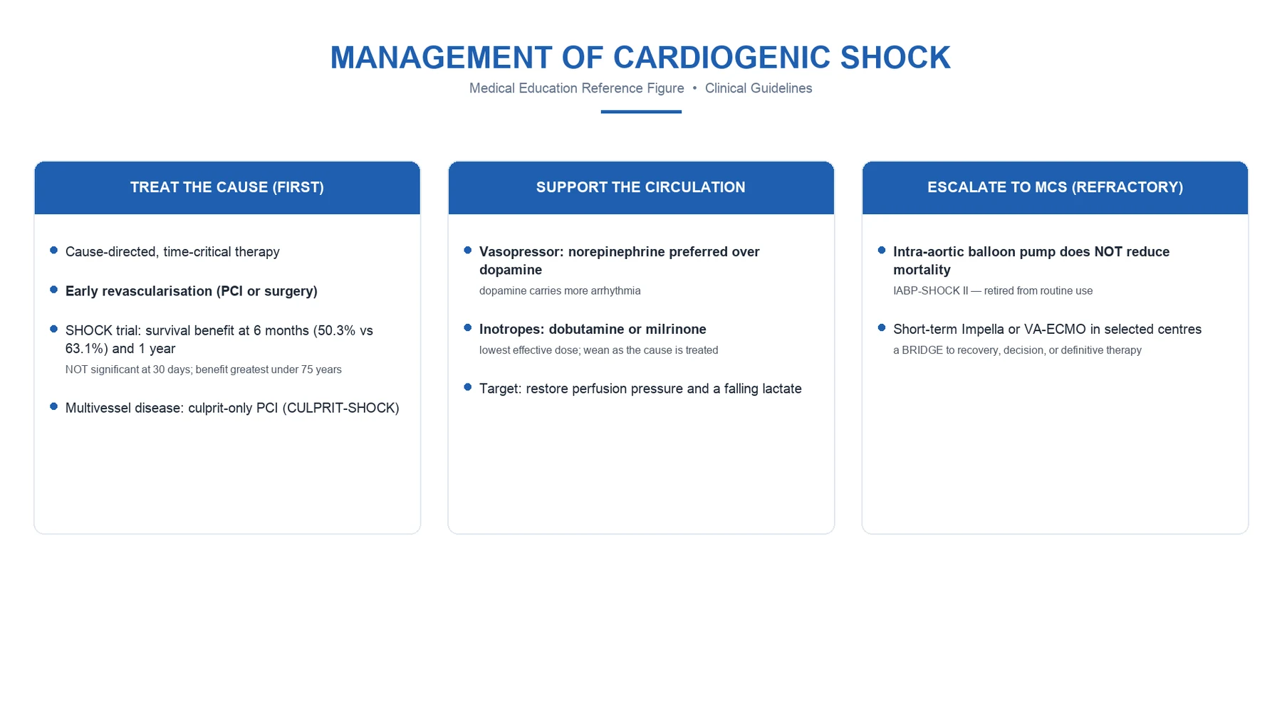
The resuscitation of the patient in shock proceeds in a fixed sequence, individualised to the cause.[2][1]
- Recognise the shock — the hypoperfusion (a raised lactate, a prolonged capillary refill, oliguria, altered mentation) and identify the type (hypovolaemic, distributive, cardiogenic, obstructive) by the examination and a focused echocardiogram.
- Restore the delivery — fluid to the responsive patient (test it first), blood to the anaemic, oxygen to the hypoxaemic, and a vasopressor to defend the mean arterial pressure in the vasodilatory. In massive haemorrhage, the damage-control ratio and tranexamic acid.
- Target the perfusion, not a pressure — the lowest mean pressure that restores a clearing lactate, a brisk capillary refill, a warming periphery and a recovering urine output.
- De-escalate as the perfusion recovers — stop the fluid, wean the vasopressor, and avoid the positive balance that causes the oedema, the congestion and the compartment syndromes of over-resuscitation. [1]
The first six hours of resuscitation in septic shock — the bedside sequence
- RECOGNISE THE SHOCK (minutes 0–15) — the hypoperfusion (raised lactate > 2 mmol/L, capillary refill > 3 s, oliguria, altered mentation, mottling) and the vasodilation (warm periphery, wide pulse pressure) or the hypovolaemia (cold, narrow). Send the cultures, draw the lactate, the venous gas, the full blood count and the renal and liver panels. Establish two large-bore cannulae and full monitoring (ECG, SpO2, NIBP, ideally an arterial line).[19][26]
- RESTORE THE DELIVERY (minutes 15–60) — give the broad-spectrum antibiotic WITHIN THE FIRST HOUR, and source-control early. Test the fluid responsiveness (PLR or ΔPP), and give a balanced-crystalloid bolus (250–500 mL over 15 min) ONLY to the responsive patient, reassessing after each bolus. Begin noradrenaline early if the MAP is below 65 mmHg and not correcting — do not wait for "enough fluid" to start the vasopressor in the vasodilated patient.[7][14][19]
- TARGET THE PERFUSION (minutes 60–360) — examine the perfusion as a panel every 30–60 minutes: the lactate (aim for ≥10% clearance per 2 h), the capillary refill (aim ≤3 s), the mottling (aim improving), the urine output (aim >0.5 mL/kg/h). Continue fluid only if the patient remains responsive AND the perfusion is still failing. Titrate noradrenaline to the lowest MAP that perfuses (65 mmHg default; higher only for the chronic hypertensive whose perfusion fails at 65).[10][17][24]
- ADD THE ADJUNCTS AS NEEDED (minutes 60–360) — add vasopressin 0.03 U/min when the noradrenaline dose rises above 0.25–0.5 µg/kg/min (catecholamine-sparing); transfuse to a haemoglobin of 70 g/L (higher only for active ischaemia); consider hydrocortisone 200 mg/day if the vasopressor requirement continues to escalate. Reserve the central venous ScvO2 for the patient whose perfusion is failing without explanation.[9][15][16]
- DE-ESCALATE (hours 6–24) — once the lactate is clearing, the capillary refill is brisk, the mottling has resolved and the urine is recovering, STOP the fluid and begin to wean the vasopressor. Aim for a net-zero or negative fluid balance in the days that follow — the positive balance of over-resuscitation is itself a predictor of mortality. The skilled resuscitator knows the moment to stop.[2][1]
Monitoring resuscitation at the bedside
Monitoring divides into the perfusion, the responsiveness and the cause.[10][1]
- The perfusion — the lactate (and its clearance), the capillary refill time, the mottling, the urine output, and the conscious state; the bedside examination is re-centred by the ANDROMEDA-SHOCK data.
- The responsiveness — the dynamic indices (the ΔPP, the stroke volume variation) in the ventilated patient, and the passive leg raise (with a real-time flow measure) in any patient; the central venous pressure does not predict it.
- The cardiac output and the function — a focused echocardiogram (the stroke volume, the ventricular function, the filling) and, in the complex case, an advanced haemodynamic monitor.
- The harm of over-resuscitation — the fluid balance, the venous pressure, the oxygenation (for pulmonary oedema) and the abdominal pressure. [1]
Prognosis and the harm of both under- and over-resuscitation
The prognosis is the prognosis of the cause, but two iatrogenic errors shape the outcome: under-resuscitation, which prolongs the hypoperfusion and the organ injury; and over-resuscitation, whose positive fluid balance is independently associated with mortality and which causes the pulmonary oedema, the intra-abdominal hypertension and the wound failure that complicate recovery. The skilled resuscitator gives the fluid to the responsive patient, achieves the perfusion, and then stops.[2][1]
SAQ — Fluid responsiveness and the choice of fluid in septic shock
10 minutes · 10 marks
A 60-year-old woman with septic shock from a urinary source has received 1.5 L of Hartmann`s solution. Her MAP is 60, lactate 3.5, and her central venous pressure is 12 mmHg. The registrar wants to give another bolus `because the CVP is low`. Outline your approach to the next fluid decision.
SAQ — The harm of over-resuscitation and the perils of a positive fluid balance
10 minutes · 10 marks
A 70-year-old man with septic shock has received 6 L of crystalloid over 8 hours. He is now anuric, with a PaO2/FiO2 of 150 on FiO2 0.6, a CVP of 18 mmHg, and a soft abdomen with a bladder pressure of 25 mmHg. Discuss the iatrogenic complications of his fluid balance and the de-resuscitation strategy.
Clinical pearls
Red flags
References
- [1]Rivers E, Nguyen B, Havstad S, et al.; Early Goal-Directed Therapy Collaborative Group. Early goal-directed therapy in the treatment of severe sepsis and septic shock N Engl J Med, 2001.PMID 11794169
- [2]The ProCESS Investigators; Yealy DM, Kellum JA, Huang DT, et al. A randomized trial of protocol-based care for early septic shock N Engl J Med, 2014.PMID 24635773
- [3]The ARISE Investigators and ANZICS Clinical Trials Network; Peake SL, Delaney A, Bailey M, et al. Goal-directed resuscitation for patients with early septic shock N Engl J Med, 2014.PMID 25272316
- [4]Marik PE, Baram M, Vahid B. Does central venous pressure predict fluid responsiveness? A systematic review of the literature and the tale of seven mares Chest, 2008.PMID 18628220
- [5]Michard F, Boussat S, Chemla D, et al. Relation between respiratory changes in arterial pulse pressure and fluid responsiveness in septic patients with acute circulatory failure Am J Respir Crit Care Med, 2000.PMID 10903232
- [6]Monnet X, Rienzo M, Osman D, et al. Passive leg raising predicts fluid responsiveness in the critically ill Crit Care Med, 2006.PMID 16540963
- [7]Semler MW, Self WH, Wanderer JP, et al.; SMART Investigators. Balanced Crystalloids versus Saline in Critically Ill Adults N Engl J Med, 2018.PMID 29485925
- [8]Annane D, Siami S, Jaber S, et al.; CRISTAL Investigators. Effects of fluid resuscitation with colloids vs crystalloids on mortality in critically ill patients presenting with hypovolemic shock: the CRISTAL randomized trial JAMA, 2013.PMID 24108515
- [9]Hébert PC, Wells G, Blajchman MA, et al.; Transfusion Requirements in Critical Care Investigators. A multicenter, randomized, controlled clinical trial of transfusion requirements in critical care. Transfusion Requirements in Critical Care Investigators, Canadian Critical Care Trials Group N Engl J Med, 1999.PMID 9971864
- [10]Hernández G, Ospina-Tascón GA, Damiani LP, et al.; ANDROMEDA-SHOCK Investigators. Effect of a Resuscitation Strategy Targeting Peripheral Perfusion Status vs Serum Lactate Levels on 28-Day Mortality Among Patients With Septic Shock: The ANDROMEDA-SHOCK Randomized Clinical Trial JAMA, 2019.PMID 30772908
- [11]Finfer S, Bellomo R, Boyce N, French J, Myburgh J, Norton R; SAFE Study Investigators. A comparison of albumin and saline for fluid resuscitation in the intensive care unit N Engl J Med, 2004.PMID 15163774
- [12]Myburgh JA, Finfer S, Bellomo R, et al.; CHEST Investigators; Australian and New Zealand Intensive Care Society Clinical Trials Group. Hydroxyethyl starch or saline for fluid resuscitation in intensive care N Engl J Med, 2012.PMID 23075127
- [13]Perner A, Haase N, Guttormsen AB, et al.; 6S Trial Group; Scandinavian Critical Care Trials Group. Hydroxyethyl starch 130/0.42 versus Ringer's acetate in severe sepsis N Engl J Med, 2012.PMID 22738085
- [14]De Backer D, Biston P, Devriendt J, et al.; SOAP II Investigators. Comparison of dopamine and norepinephrine in the treatment of shock N Engl J Med, 2010.PMID 20200382
- [15]Russell JA, Walley KR, Singer J, et al.; VASST Investigators. Vasopressin versus norepinephrine infusion in patients with septic shock N Engl J Med, 2008.PMID 18305265
- [16]Gordon AC, Mason AJ, Thirunavukkarasu N, et al.; VANISH Investigators. Effect of Early Vasopressin vs Norepinephrine on Kidney Failure in Patients With Septic Shock: The VANISH Randomized Clinical Trial JAMA, 2016.PMID 27483065
- [17]Asfar P, Meziani F, Hamel JF, et al.; SEPSISPAM Investigators. High versus low blood-pressure target in patients with septic shock N Engl J Med, 2014.PMID 24635770
- [18]Caironi P, Tognoni G, Masson S, et al.; ALBIOS Study Investigators. Albumin replacement in patients with severe sepsis or septic shock N Engl J Med, 2014.PMID 24635772
- [19]Evans L, Rhodes A, Alhazzani W, et al. Executive Summary: Surviving Sepsis Campaign: International Guidelines for the Management of Sepsis and Septic Shock 2021 Crit Care Med, 2021.PMID 34643578
- [20]Mouncey PR, Osborn TM, Power GS, et al.; ProMISe Trial Investigators. Protocolised Management In Sepsis (ProMISe): a multicentre randomised controlled trial of the clinical effectiveness and cost-effectiveness of early, goal-directed, protocolised resuscitation for emerging septic shock Health Technol Assess, 2015.PMID 26597979
- [21]Monnet X, Osman D, Ridel C, Lamia B, Jakob M, Teboul JL. Predicting volume responsiveness by using the end-expiratory occlusion in mechanically ventilated intensive care unit patients Crit Care Med, 2009.PMID 19237902
- [22]Muller L, Toumi M, Bousquet PJ, et al.; AZUReA Group. An increase in aortic blood flow after an infusion of 100 ml colloid over 1 minute can predict fluid responsiveness: the mini-fluid challenge study Anesthesiology, 2011.PMID 21792056
- [23]Ait-Oufella H, Lemoinne S, Boelle PY, et al. Mottling score predicts survival in septic shock Intensive Care Med, 2011.PMID 21373821
- [24]Nguyen HB, Rivers EP, Knoblich BP, et al. Early lactate clearance is associated with improved outcome in severe sepsis and septic shock Crit Care Med, 2004.PMID 15286537
- [25]Jansen TC, van Bommel J, Woodward R, Mulder PG, Bakker J. The prognostic value of blood lactate levels relative to that of vital signs in the pre-hospital setting: a pilot study Crit Care, 2008.PMID 19091118
- [26]Seymour CW, Gesten F, Prescott HC, et al. Time to Treatment and Mortality during Mandated Emergency Care for Sepsis N Engl J Med, 2017.PMID 28528569