ICU · Resuscitation
Classification and management of shock
Also known as Types of shock · Shock classification · Cardiogenic, hypovolaemic, distributive, obstructive shock
Shock is inadequate tissue perfusion resulting in cellular hypoxia and organ dysfunction. Four types: (1) Hypovolaemic (fluid/blood loss — trauma, GI bleed, burns, dehydration). (2) Cardiogenic (pump failure — MI, myocarditis, cardiomyopathy). (3) Distributive (vasodilation — sepsis 1, anaphylaxis, neurogenic, adrenal). (4) Obstructive (mechanical obstruction — PE, tamponade, tension pneumothorax). Diagnosis: clinical (hypotension, tachycardia, cool/warm extremities, oliguria, altered mental status) + haemodynamic parameters (CO, SVR, CVP, SvO2). Treatment: treat underlying cause + supportive (fluids, vasopressors, inotropes, oxygen). Key: identify the TYPE of shock to guide therapy.
On this page & tools
Your progress
Saved locally on this device.
Target exams
Red flags
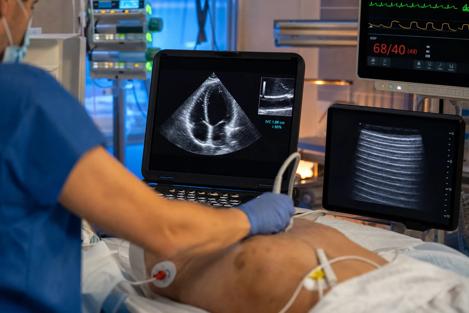
Definition and why a BP number is not enough
Shock is the acute, generalised circulatory failure that delivers insufficient oxygen and substrates to tissues, tipping cells from aerobic to anaerobic metabolism and, if uncorrected, into irreversible organ damage and death.[1]
Three reasons a blood pressure reading alone is a poor surrogate: [1]
- BP is a derived variable.
MAP = CO × SVR. CO can fall markedly while SVR rises to compensate — so MAP is preserved even as tissue perfusion collapses. The patient in compensated shock has a "normal" BP and dying cells. - MAP is a late, decompensated sign. By the time hypotension appears, ~30–40% of circulating volume may be lost and the sympathetic/RAAS/ADH reserve is exhausted. The window of reversibility is narrowing.
- Perfusion is regional, not global. The body triages: blood is shunted to brain and heart at the expense of splanchnic, renal, muscle and skin beds. A normal central BP can coexist with profound gut and renal ischaemia (lactate rising, urine falling).[2]
The practical consequence: define shock by evidence of inadequate tissue perfusion (cool/clammy or warm flushed skin, mottling, prolonged capillary refill, oliguria, altered mentation, rising lactate) — not by a systolic number.[1]
The 4 types
Hypovolaemic
Fluid/blood loss
- Causes: haemorrhage (trauma, GI bleed, rupture), dehydration (vomiting, diarrhoea, burns, DKA)
- Haemodynamics: LOW CVP, LOW CO, HIGH SVR (compensatory vasoconstriction)
- Clinical: cool, clammy, pale, oliguria, narrow pulse pressure
- Treatment: fluids (crystalloid) ± blood (if haemorrhage). Restore intravascular volume.
Cardiogenic
Pump failure
- Causes: acute MI (#1), myocarditis, cardiomyopathy, arrhythmia, valvular failure, drug toxicity
- Haemodynamics: HIGH CVP (back-up from failing pump), LOW CO, HIGH SVR
- Clinical: cool, clammy, pulmonary oedema (bilateral crackles), gallop rhythm, raised JVP
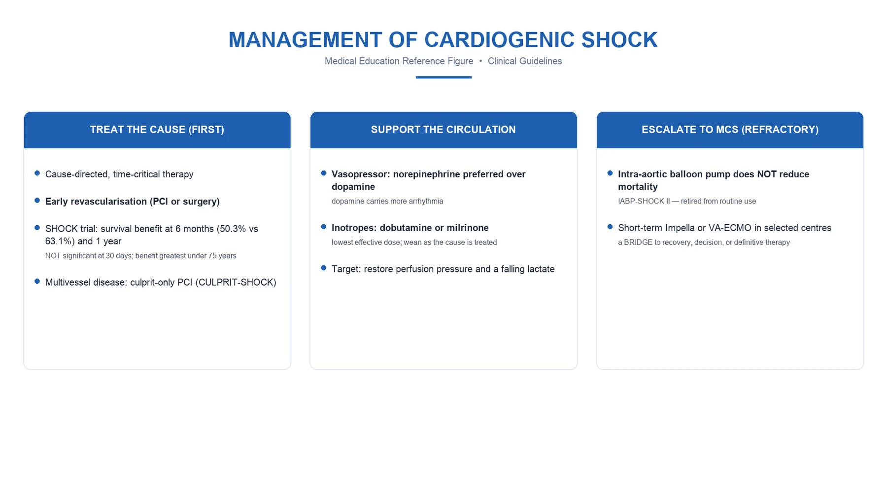
- Treatment: inotropes (dobutamine, milrinone), vasopressors (noradrenaline for BP), mechanical support (IABP, Impella, VA-ECMO), treat cause (PCI for MI)
Distributive
Vasodilation
- Causes: sepsis (#1), anaphylaxis, neurogenic (spinal cord injury), adrenal insufficiency
- Haemodynamics: LOW CVP, LOW SVR (massive vasodilation), normal or HIGH CO (initially — high output state)
- Clinical: WARM, flushed, bounding pulse, wide pulse pressure (early). Late: cold (decompensated).
- Treatment: vasopressors (noradrenaline — restore SVR), fluids (restore volume — vasodilated capacitance vessels), treat cause (antibiotics for sepsis, adrenaline for anaphylaxis)
Obstructive
Mechanical obstruction
- Causes: massive PE (#1), cardiac tamponade, tension pneumothorax
- Haemodynamics: LOW CO (obstructed flow), variable CVP/SVR
- Clinical: distended neck veins, muffled heart sounds (tamponade), unilateral breath sounds (tension PTX), evidence of DVT (PE)
- Treatment: RELIEVE OBSTRUCTION (thrombolysis/Embolectomy for PE, pericardiocentesis for tamponade, needle decompression for tension PTX)
Haemodynamic profiles by shock type
The haemodynamic signature of each shock type is one of the most frequently examined concepts in CICM/FFICM/EDIC. Use it at the bedside (with a pulmonary artery catheter or transpulmonary thermodilution — PiCCO) to confirm the type and titrate therapy.[2]
Haemodynamic profile of each shock type (typical patterns)
Hypovolaemic
Low preload
- CI (cardiac index): LOW (< 2.5 L/min/m²)
- SVR: HIGH (> 1500 dyn·s·cm⁻⁵) — compensatory vasoconstriction
- PCWP: LOW (< 8 mmHg) — underfilled LV
- CVP/ScvO2: LOW CVP (< 5 mmHg); LOW ScvO2 (< 65%) — high extraction
- Pulse pressure: NARROW (low stroke volume)
Cardiogenic
Pump failure
- CI: LOW (< 2.2 L/min/m²)
- SVR: HIGH (> 1500) — compensatory
- PCWP: HIGH (> 18 mmHg) — LV failure → back-up → pulmonary oedema
- CVP/ScvO2: HIGH CVP (≥ 12 mmHg); LOW ScvO2 (< 65%) — extraction maximal
- Pulse pressure: NARROW
Distributive (septic)
Vasoplegia
- CI: HIGH early (> 4.0) — hyperdynamic; may fall late as septic cardiomyopathy sets in
- SVR: LOW (< 800) — the defining lesion
- PCWP: LOW-NORMAL (5–12 mmHg) — relative hypovolaemia from capillary leak
- CVP/ScvO2: LOW CVP early; HIGH ScvO2 (> 70%, often > 80%) — impaired extraction / AV shunting / cytopathic dysoxia
- Pulse pressure: WIDE (high stroke volume + low diastolic from vasodilation)
Obstructive
Mechanical
- CI: LOW (< 2.5) — outflow obstructed or ventricular interdependence
- SVR: HIGH (compensatory) — except late in massive PE where it may collapse
- PCWP: variable — EQUALISES in tamponade (RA/RV/LV pressures all ~15–25 mmHg); low/normal in tension PTX; RA > PCWP with RV strain in PE
- CVP/ScvO2: HIGH CVP (back-pressure); LOW ScvO2
- Pulse pressure: NARROW; paradoxus > 10 mmHg in tamponade/tension PTX
Quick exam-ready reference: the 4×4 grid
| Parameter | Hypovolaemic | Cardiogenic | Distributive | Obstructive |
|---|---|---|---|---|
| CO/CI | ↓↓ | ↓↓ | ↑ (early) → ↓ (late) | ↓↓ |
| SVR | ↑↑ | ↑↑ | ↓↓ | ↑ (or normal) |
| PCWP | ↓↓ | ↑↑ | ↓ / normal | normal / ↑ (tamponade equalises) |
| SvO₂/ScvO₂ | ↓ | ↓ | ↑ (impaired extraction) | ↓ |
| CVP | ↓↓ | ↑↑ | ↓ early | ↑↑ |
Cool shock vs warm shock — the bedside decision
The single most useful bedside distinction because it dictates the first therapeutic move: fluids/inotropes for cool shock; vasopressors for warm shock.[1]
Cool shock vs warm shock
Cool shock
High SVR, low CO
- Shock types: hypovolaemic, cardiogenic, obstructive
- Extremities: cold, clammy, pale, mottled; capillary refill > 3 s
- Pulse pressure: NARROW (low stroke volume; diastolic rises from vasoconstriction)
- Diastolic BP relatively preserved; systolic falls first
- SvO2/ScvO2: LOW (tissues extracting maximally)
- First move: give FLUIDS (if hypovolaemic), INOTROPES (if cardiogenic), or RELIEVE OBSTRUCTION (if obstructive)
Warm shock
Low SVR, normal/high CO
- Shock types: distributive (septic, anaphylactic, neurogenic, adrenal)
- Extremities: warm, flushed, dry; bounding pulse; wide pulse pressure
- Pulse pressure: WIDE (low diastolic from vasodilation; high stroke volume)
- Diastolic BP falls early and dramatically (unopposed vasodilation)
- SvO2/ScvO2: HIGH (impaired extraction — the paradox of septic shock)
- First move: VASOPRESSORS (noradrenaline first-line) to restore SVR + treat cause
Pathophysiology: the shock cascade
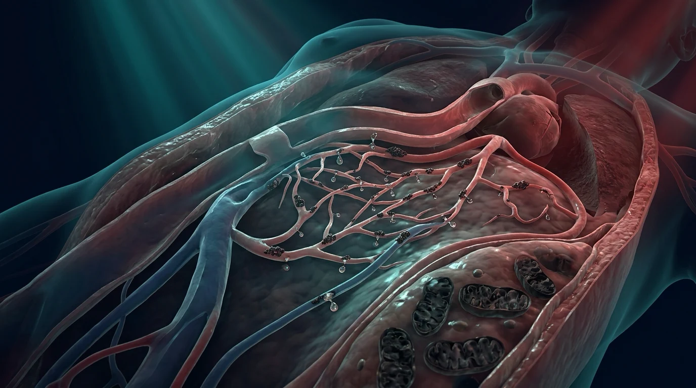
Shock is not a static state — it is a progressive cascade with three phases, each with a different prognosis and a different cellular signature.[1]
- Compensated (early) shock. Cardiac output or effective circulating volume falls. Baroreceptors in the carotid sinus and aortic arch sense the fall in stretch and fire the sympathetic nervous system. Heart rate and contractility rise; venous and arteriolar tone increase. RAAS, ADH and the adrenergic axis retain salt and water and constrict splanchnic, renal and cutaneous beds. BP is preserved; lactate may already be rising.
- Decompensated (progressive) shock. The compensatory reserve is exhausted. SVR can no longer hold MAP, hypotension appears, coronary and cerebral perfusion fall, myocardial ischaemia worsens pump failure (a vicious cycle), capillary leak worsens (further dropping preload), and the microcirculation begins to fail — heterogeneous flow, stopped capillaries, oxygen shunts past static cells.[10][13]
- Irreversible shock. Cellular ATP is depleted, mitochondrial membrane permeability rises (cytochrome c release, apoptosis), lysosomal membranes rupture, and massive cell death cascades through organ systems. Even full restoration of perfusion at this stage no longer prevents MODS and death. The cellular threshold of irreversibility is the biological substrate of "you can't resuscitate a corpse."[1]
The clinical implication: the earlier shock is recognised, the more reversible it is. Lactate, capillary refill and urine output change before BP does.[1]
Compensatory mechanisms
The body defends perfusion pressure to brain and heart through four overlapping neurohumoral axes. Knowing each axis explains both the clinical signs and the targets of the drugs used to support shock.[1]
| Mechanism | Trigger | Effect | Clinical correlate | Therapeutic lever |
|---|---|---|---|---|
| Sympathetic nervous system | Baroreceptor unloading | ↑ HR, ↑ contractility, venoconstriction (↑ preload), arteriolar constriction (↑ SVR) | Tachycardia, cold peripheries, narrowed pulse pressure, sweating | β-blockade unmasks shock; inotropes/vasopressors substitute for it |
| RAAS | Renal hypoperfusion, β1 at JG cells | Angiotensin II (potent vasoconstrictor) + aldosterone (Na⁺/water retention) | Oliguria, Na⁺ retention; ACE-inhibitors can precipitate shock | Replace volume; avoid ACEi/ARBs in shock |
| ADH (vasopressin) | Hypothalamic osmoreceptors, low pressure | Water retention (V2) + vasoconstriction (V1) | Diluted urine (low Na⁺); relative vasopressin deficiency in late septic shock | Exogenous vasopressin (VASST)[6] |
| Adrenal axis (cortisol) | Stress | Permissive effect on catecholamine vascular tone; anti-inflammatory; mobilises substrate | Relative adrenal insufficiency → refractory vasoplegia | Hydrocortisone 200 mg/day in refractory septic shock (SSC 2021)[4] |
Three further points worth knowing for vivas: [1]
- Tachycardia is not universal. Neurogenic shock (cord lesion above T6) and β-blocked, elderly, or athletic patients may be bradycardic in shock — do not be reassured by a normal heart rate.
- Venoconstriction matters more than is taught. ~70% of circulating blood is in the venous reservoir; sympathetic venoconstriction auto-transfuses ~500 mL in early shock. Loss of this is why induction of anaesthesia precipitates catastrophic hypotension in the under-resuscitated shock patient.
- Microcirculatory dysfunction is the final common pathway. Even when MAP, CO and DO₂ are restored, the microcirculation may remain deranged — heterogeneous flow, stopped capillaries, oxygen shunts past static cells — the so-called macrocirculation–microcirculation dissociation of sepsis.[10][13][14]
Oxygen delivery and consumption (DO₂/VO₂)
The single most physiologically coherent framework for understanding shock is the relationship between oxygen delivery (DO₂) and oxygen consumption (VO₂).[1]
The equations
- DO₂ (oxygen delivery) =
CO × CaO₂≈CO × 1.34 × Hb × SaO₂(+ dissolved O₂, usually negligible).- Normal DO₂ ≈ 1000 mL/min (CO 5 L/min × 200 mL O₂/L blood).
- VO₂ (oxygen consumption) =
CO × (CaO₂ − CvO₂)≈ 250 mL/min at rest.- Extraction ratio = VO₂/DO₂ ≈ 25% normally.
- SvO₂ (mixed venous saturation) reflects the balance. Normal SvO₂ ≈ 75% (ScvO₂ ≈ 70%). [1]
The critical DO₂ and supply-dependency
As DO₂ falls, healthy tissues maintain VO₂ by extracting more (SvO₂ falls) until a critical DO₂ threshold (~300 mL/min/m²) below which extraction can no longer compensate, VO₂ becomes supply-dependent, and anaerobic metabolism begins — lactate rises. This is the inflection point that defines shock.[1]
| Variable | Normal | Compensated shock | Critical shock |
|---|---|---|---|
| DO₂ | ~1000 mL/min | Falling | < 300 mL/min/m² (critical) |
| VO₂ | ~250 mL/min (independent) | Maintained by ↑ extraction | Supply-dependent ↓ |
| SvO₂ | 75% | 50–65% (high extraction) | < 50% |
| Extraction ratio | 25% | 30–50% | > 50% |
| Lactate | < 2 mmol/L | 2–4 mmol/L | > 4 mmol/L |
The septic paradox — pathological oxygen extraction
In distributive (septic) shock the picture inverts: DO₂ is normal or high (hyperdynamic state) yet lactate rises and SvO₂ is high (> 70%). This is impaired oxygen utilisation — AV shunting, mitochondrial dysfunction (cytopathic dysoxia), microcirculatory heterogeneity.[10][13] A high ScvO₂ in a shocked patient is not reassuring — it signals failure to extract.
Clinical implication: in cardiogenic/hypovolaemic shock you raise DO₂ (inotropes, fluids, transfusion). In septic shock the problem is utilisation — you must restore the microcirculation, clear the source, and accept that high ScvO₂ does not equal adequate resuscitation.[1]
Lactate as a marker of shock
Lactate is the single most useful biochemical marker in shock because it reflects the cellular switch to anaerobic metabolism — the defining biochemical lesion.[1]
Why lactate rises in shock
- Tissue hypoxia (Type A — the dominant mechanism in shock). When DO₂ falls below the critical threshold, pyruvate cannot enter the aerobic Krebs cycle and is shunted to lactate. This is true anaerobic glycolysis — the lactate is the by-product of inadequate DO₂.
- Impaired clearance. The liver (Cori cycle) normally clears lactate; hepatic hypoperfusion or dysfunction in shock slows clearance and lactate accumulates even at normal production.
- Accelerated aerobic glycolysis (Type B component). Sepsis and adrenaline-driven β2 stimulation drive aerobic glycolysis faster than pyruvate can be oxidised — lactate rises without tissue hypoxia. This is why not all hyperlactataemia in sepsis equals hypoperfusion, and why aggressive fluid for a "high lactate" alone is wrong.[1]
- Mitochondrial dysfunction (cytopathic dysoxia). In late sepsis, mitochondria cannot use oxygen even when delivered — pyruvate → lactate regardless of DO₂.[10]
Lactate as a resuscitation target
- Baseline lactate ≥ 4 mmol/L defines severe hyperlactataemia and, with hypotension, septic shock (Sepsis-3).[4]
- Lactate clearance ≥ 10% per 2 hours is the most validated target — equivalent to ScvO₂-guided therapy in the Jansen LACTATE trial.[11]
- Failure to clear lactate is the most powerful single predictor of mortality, more so than the absolute value.
- Normalised lactate at 24–48 h correlates with survival.[1]
Pitfalls of lactate interpretation
- Lactate may be normal in early haemorrhage — measure and re-measure.
- Lactate rises with seizures, shivering, β2-agonists, malignancy, metformin, thiamine deficiency — consider Type B causes.
- Lactate falls slowly even with good resuscitation (clearance half-life ~hours) — do not chase the number with fluids; reassess the whole patient.
- Capillary lactate > arterial lactate gradient reflects microcirculatory failure — a marker of severity.[13]
Clinical assessment — the five "windows" of perfusion
A structured bedside examination using five windows lets you diagnose shock, classify the type, and follow the response to therapy without waiting for bloods.[2]
| Window | What it tells you | Sign of good perfusion | Sign of poor perfusion |
|---|---|---|---|
| Skin | Peripheral vasoconstriction (sympathetic drive) | Warm, pink, capillary refill < 3 s, no mottling | Cool, clammy, pale, mottled, CRT > 3 s |
| Kidney | Splanchnic + renal perfusion | Urine output > 0.5 mL/kg/h | Oliguria, rising creatinine |
| Brain | Cerebral perfusion | Alert, oriented, calm | Agitated, confused, drowsy |
| Metabolic | Cellular oxygenation | Lactate normalising / clearing > 10%/2 h | Rising lactate, metabolic acidosis |
| Cardiac | Cardiac output, HR, pulse character | HR normalising, strong pulse, warm peripheries | Tachycardia/bradycardia, thready pulse, cold peripheries |
Remember: a single window can mislead (warm skin in early sepsis). Use all five together — concordance confirms adequacy of resuscitation; discordance (lactate still rising with "normal" MAP and urine output) demands escalation.[1]
Bedside investigations in undifferentiated shock
- ABG with lactate — acidosis (metabolic with raised lactate), base deficit (> −5 correlates with lactate and mortality), oxygenation.
- Venous gas — ScvO₂ from a central line (or SvO₂ from a PA catheter). Low (< 65%) in cool shock; paradoxically high (> 70%) in sepsis.
- ECG — STEMI/NSTEMI (cardiogenic), arrhythmia, RV strain (PE — S1Q3T3, right axis, T inversion V1–V3).
- Bedside echocardiogram (FoCUS/FATE/RUSH) — the single most useful test: hyperdynamic small LV (hypovolaemic), dilated poorly contracting LV (cardiogenic), RV dilatation with septal bowing (PE), pericardial effusion with RA/RV diastolic collapse (tamponade), absent lung sliding with swept heart (tension PTX).[2]
- Bloods — FBC (Hb for haemorrhage; WCC for sepsis), coagulation (DIC), CRP/procalcitonin, troponin, group & save/crossmatch, cultures (blood, urine, sputum, CSF) before antibiotics if possible but never delay antibiotics.[9]
- Lactate (serial) — at 0, 2, 4, 6 h to track clearance.[11]
- CXR — pneumothorax, widened mediastinum (aortic), pulmonary oedema, pneumonia source.
- POCUS — IVC collapsibility (preload), lung B-lines (pulmonary oedema), FAST for free fluid in trauma, abdominal aorta.
- Advanced monitoring (if not responding): arterial line (beat-to-beat BP, pulse pressure variation for fluid responsiveness), CVP/ScvO₂ line, transpulmonary thermodilution (PiCCO — GEDV, EVLW, PPV), rarely PA catheter.[2][8]
Resuscitation targets (end-points of resuscitation)
The goal is restored tissue oxygenation, not a number. Use a combination:[2][4]
- MAP ≥ 65 mmHg (SEPSISPAM — 65 sufficient; higher targets do not improve outcome and increase arrhythmia). Adjust higher (75–80) for chronic hypertension; accept lower (50–65) in haemorrhagic shock with uncontrolled bleeding (permissive hypotension).
- Urine output ≥ 0.5 mL/kg/h — best clinical perfusion marker.
- Lactate clearance ≥ 10% per 2 h.[11]
- ScvO₂ ≥ 70% / SvO₂ ≥ 65% (Rivers EGDT — though the ProCESS/ARISE/ProMISe trio showed no mortality benefit of protocolised ScvO₂ targeting over usual care).[3][7][15][16]
- Normalising capillary refill (< 3 s) — the ANDROMEDA-SHOCK trial showed capillary refill targeting was at least as good as lactate targeting in septic shock.
- Improving conscious level / mentation.
- Falling base deficit / improving pH.
FlowSteps: approach to undifferentiated shock
A systematic approach to the shocked patient
1. Recognise shock early (before the BP falls)
Look for the compensated signs: tachycardia, narrowed pulse pressure, cool/mottled peripheries, capillary refill > 3 s, oliguria, agitation or drowsiness, rising lactate. A normal BP does not exclude shock.
2. ABC + 100% oxygen + 2 large-bore cannulae
Airway (intubate early if GCS < 8 or tiring), Breathing (high-flow oxygen; target SpO2 ≥ 94%), Circulation (two 14–16G cannulae; send bloods including lactate, group/crossmatch, cultures).
3. Classify the shock type within 5 minutes
History + examination + bedside echo + ABG. Decide: is it COOL (hypovolaemic/cardiogenic/obstructive — needs fluids/inotropes/relief of obstruction) or WARM (distributive — needs vasopressors)? This single binary decision drives the first drug.
4. Treat the reversible cause SIMULTANEOUSLY
Do not wait for full workup. Antibiotics + source control within 1 h for sepsis (Kumar).<Cite id="9" /> IM adrenaline for anaphylaxis. Needle decompression/thoracostomy for tension PTX. Pericardiocentesis for tamponade. Activation of massive transfusion for haemorrhage. PCI for STEMI. Thrombolysis for massive PE.
5. Fluid challenge — reassess, do not blanket
250–500 mL balanced crystalloid over 15–30 min (SMART — balanced > saline).<Cite id="5" /> Reassess: did CO/MAP/lactate improve? If responsive, repeat. If not, STOP fluids — they are not fluid-responsive; more fluid causes pulmonary oedema and abdominal compartment syndrome.
6. Start vasopressors early if not fluid-responsive
Noradrenaline first-line (alpha-1 vasoconstriction + mild beta-1). Target MAP ≥ 65. Add vasopressin (VASST) <Cite id="6" /> if escalating noradrenaline; consider hydrocortisone 200 mg/day in refractory septic shock (SSC 2021).<Cite id="4" />
7. Add inotropes if low cardiac output
If ScvO2 < 70%, lactate not clearing, echo shows poor LV function, or cool peripheries persist despite adequate MAP — add dobutamine (or milrinone if pulmonary hypertension/right-heart failure).
8. Monitor with serial reassessment
Reassess all five perfusion windows hourly. Serial lactate. urine output hourly. Escalate monitoring (arterial line, CVP/ScvO2, PiCCO) if not improving. Re-image with echo.
9. Address mixed shock
Most ICU shock is mixed (e.g. septic cardiomyopathy + vasoplegia + capillary-leak hypovolaemia). Treat each component — vasopressor for the vasoplegia, inotrope for the cardiomyopathy, judicious fluid for the hypovolaemia.
10. Define and treat the endpoint
MAP ≥ 65, lactate clearing > 10%/2 h, ScvO2 ≥ 70%, urine > 0.5 mL/kg/h, warm peripheries, improving mentation. Consider de-resuscitation once stable.
FlowSteps: assessing fluid responsiveness
Only ~50% of haemodynamically unstable ICU patients are fluid-responsive. Giving fluid to the non-responsive half causes harm (pulmonary oedema, abdominal compartment syndrome, worse AKI). Assess before you bolus.[8]
Fluid responsiveness — what works and what does not
Use a DYNAMIC test, never static markers
CVP, IVC diameter, PAD pressure are poor predictors of fluid responsiveness (Cecconi/ESICM consensus).<Cite id="2" /> Use a dynamic challenge that tests the Frank-Starling curve: preload-increasing manoeuvre + real-time CO measurement.
Passive leg raise (PLR) — the best reversible test
Start semi-recumbent 45°. Tilt the whole bed to trunk-flat + legs at 45° for 60–90 s. Transfers ~300 mL venous blood. A rise in CO/SV/LVOT VTI ≥ 10% = fluid responsive. MUST measure CO in real time (echo LVOT VTI, PiCCO, arterial waveform) — a PLR without CO is useless.
Fluid bolus — the mini-fluid challenge
100–250 mL colloid or crystalloid over 1 min. Rise in CO ≥ 10–15% = responsive. More physiological than PLR but carries a small fluid load.
Pulse pressure variation / stroke volume variation (PPV/SVV)
In a fully sedated, ventilated patient with tidal volume ≥ 8 mL/kg, sinus rhythm and no right-heart failure: PPV > 12–13% or SVV > 10% predicts fluid responsiveness. Useless if spontaneously breathing, arrhythmia, low TV, open chest, or RV failure.
End-expiratory occlusion test (EEO)
15-s expiratory hold in a ventilated patient. Rise in arterial pulse pressure or CO ≥ 5% predicts responsiveness. Works in arrhythmia where PPV fails.
IVC distensibility (limited use)
Distensibility index > 18% (ventilated) or collapse > 50% with sniff (spontaneous) suggests low preload/high responsiveness. Sensitivity moderate — use as adjunct, not alone.
STOP when the patient is no longer responsive
Reassess after every bolus. The cumulative fluid balance at 72 h correlates with mortality — fluid is a drug with a toxic ceiling (CLASSIC, CLOVERS).
TrialCard: landmark evidence in shock
Rivers — Early Goal-Directed Therapy (EGDT)
Single-centre RCT: 263 patients with severe sepsis/septic shock in the ED
Population: Patients with lactate ≥ 4 mmol/L or SBP ≤ 90 after fluid
Key finding
EGDT reduced in-hospital mortality (30.5% vs 46.5%, p=0.009) and 28-day mortality (33.3% vs 49.2%). Changed practice worldwide and launched the Surviving Sepsis Campaign.
Practice change
The foundational trial of protocolised resuscitation. Single-centre, unblinded, and later not reproduced — but established the principle of lactate, ScvO2 and time-bound resuscitation.
ProCESS (Angus, NEJM 2014)
Multicentre RCT: 1341 patients with early septic shock in 31 US EDs
Population: Septic shock patients
Key finding
No mortality difference between EGDT (21%), standard-protocol (18.2%) and usual care (18.9%). No difference in 90-day mortality, 1-year mortality, or need for organ support.
Practice change
With modern usual care (early antibiotics, fluids, lactate monitoring), the invasive EGDT protocol offered no benefit over protocolised or usual care. Rivers-style ScvO2 catheters largely abandoned.
ARISE (ANZICS, NEJM 2014)
Multicentre RCT: 1600 patients with early septic shock in 51 Australasian ICUs/EDs
Population: Patients ≥ 18 years with refractory septic shock or lactate ≥ 4
Key finding
No mortality difference (18.6% EGDT vs 18.4% usual care). No difference in ICU/hospital stay, organ support, or adverse events.
Practice change
Independently confirmed ProCESS: EGDT offered no advantage over high-quality usual care. The 'EGDT trilogy' (ProCESS/ARISE/ProMISe) retired invasive early goal-directed therapy.
ProMISe (Mouncey, HTA 2015)
Multicentre RCT: 1260 patients with early septic shock in 56 UK hospitals
Population: Adults with early septic shock
Key finding
No mortality difference (29.5% EGDT vs 29.2% usual care). EGDT had higher SOFA scores and more cardiovascular support use at 90 h.
Practice change
The third of the EGDT trilogy — completed the demonstration that invasive protocolised EGDT (central venous ScvO2 catheter, mandatory transfusion/inotrope) confers no benefit over modern usual care in high-income settings.
SMART (Semler, NEJM 2018)
Pragmatic cluster-crossover RCT: 15,802 adults in 5 ICUs
Population: All critically ill adults (medical + surgical)
Key finding
Balanced crystalloids reduced mortality (10.3% vs 11.1%, p=0.02) and MAKE30 (death/dialysis/persistent renal dysfunction: 14.3% vs 15.4%, p=0.04). Benefit larger in sepsis.
Practice change
Balanced crystalloids preferred over saline for nearly all ICU resuscitation. The most important fluid trial of the decade — modest absolute benefit (~1%) across a large heterogeneous population.
VASST (Russell, NEJM 2008)
Multicentre RCT: 778 patients with septic shock on vasopressors
Population: Adults with septic shock requiring noradrenaline
Key finding
No difference in 28-day mortality (35.4% vs 39.3%, p=0.26). Pre-specified subgroup with less severe shock (5–15 μg/min norepinephrine) had lower mortality with vasopressin (26.5% vs 35.7%).
Practice change
Low-dose vasopressin (fixed 0.03 U/min) is a catecholamine-sparing adjunct, not a replacement for noradrenaline. Useful in escalating or catecholamine-resistant septic shock.
Kumar — antibiotics in septic shock (CCM 2006)
Multicentre retrospective cohort: 2154 patients with septic shock
Population: Adults with septic shock and hypotension
Key finding
Mortality rose 7.6% for every hour of delay in effective antibiotics after onset of hypotension (each hour OR ~1.119). Survival was 79.0% with antibiotics within 1 h, falling progressively to ~25% if delayed > 5 h.
Practice change
Foundational evidence for the Surviving Sepsis Campaign Hour-1 bundle: give broad-spectrum antibiotics within 1 hour of recognising septic shock. Time-to-antibiotic is the single most modifiable mortality determinant.
Jansen — LACTATE trial (AJRCCM 2010)
Multicentre open-label RCT: 348 patients with lactate ≥ 3 mmol/L
Population: ICU patients with raised lactate (not necessarily septic)
Key finding
Lactate-guided therapy reduced in-hospital mortality when adjusted for APACHE II (19.8% vs 29.7%; adjusted HR 0.61, p=0.007) and shortened ICU stay. No excess fluid use.
Practice change
Established serial lactate measurement as a resuscitation target — equivalent in effect size to ScvO2-guided therapy (Rivers) but cheaper and less invasive. Lactate clearance ≥ 10%/2 h is now the standard target.
BICAR-ICU (Jaber, Lancet 2018)
Multicentre RCT: 389 ICU patients with severe metabolic acidaemia (pH ≤ 7.20)
Population: Adults with pH 7.20 or less, PaCO2 ≤ 45, bicarbonate ≤ 20
Key finding
No significant difference in the primary outcome overall. Pre-specified subgroup with AKI (Creatinine × 2) had reduced 28-day mortality (46% vs 63%, p=0.017) and need for RRT.
Practice change
Sodium bicarbonate may be considered for severe metabolic acidaemia (pH ≤ 7.20) in patients with concomitant AKI. Not a blanket therapy — acidosis is a marker; treat the cause.
SSC 2021 (Evans, CCM 2021)
International consensus guidelines (Surviving Sepsis Campaign, joint ESICM/SCCM)
Population: Adults with sepsis or septic shock
Key finding
Recommends: measure lactate; blood cultures before antibiotics; broad-spectrum antibiotics within 1 h; 30 mL/kg crystalloid for septic shock or lactate ≥ 4; noradrenaline first-line vasopressor; add vasopressin (not first-line); consider hydrocortisone 200 mg/day if vasopressor-refractory; target MAP ≥ 65.
Practice change
The current global standard of care for septic shock — the distributive shock archetype. Defines the resuscitation targets used in the other shock types by analogy.
SAQ — Mixed shock: classifying and resuscitating the shocked patient
10 minutes · 10 marks
A 68-year-old man is admitted to ICU 4 hours after emergency laparotomy for a perforated diverticulum. He is intubated and mechanically ventilated. HR 122, BP 78/45 (MAP 56), temp 38.9°C, lactate 5.2 mmol/L, urine output 10 mL/h for the last 2 h. He is peripherally warm with a bounding pulse and a wide pulse pressure. Noradrenaline is running at 0.35 mcg/kg/min. Bedside echocardiography shows a hyperdynamic, small left ventricle with EF ~65%.
SAQ — DO2/VO2, lactate clearance and the endpoints of resuscitation
10 minutes · 10 marks
A 55-year-old woman in septic shock from a urinary source has received 30 mL/kg crystalloid, broad-spectrum antibiotics and noradrenaline at 0.4 mcg/kg/min. Her MAP is now 68 mmHg, lactate 4.8 mmol/L (was 6.5), ScvO2 82%, and she remains oliguric. The registrar asks whether she needs more fluid and an inotrope to 'improve oxygen delivery.'
Clinical pearls (core)
Additional exam-exhaustive pearls
Red flags
Quick-reference summary table
| Domain | Hypovolaemic | Cardiogenic | Distributive (septic) | Obstructive |
|---|---|---|---|---|
| Defect | Low preload | Pump failure | Vasodilation | Mechanical obstruction |
| CO | ↓↓ | ↓↓ | ↑ early → ↓ late | ↓↓ |
| SVR | ↑↑ | ↑↑ | ↓↓ | ↑ / normal |
| PCWP | ↓↓ | ↑↑ | ↓ / normal | normal / ↑ (equalised in tamponade) |
| CVP | ↓↓ | ↑↑ | ↓ early | ↑↑ |
| SvO2 | ↓ | ↓ | ↑ (impaired extraction) | ↓ |
| Skin | Cold, clammy | Cold, clammy | Warm early / cold late | Cold |
| Pulse pressure | Narrow | Narrow | Wide | Narrow; paradoxus |
| First drug | Fluids ± blood | Inotrope | Noradrenaline + fluids | RELIEVE obstruction |
| Prognosis | 20–40% mortality | 40–50% | 30–50% | Variable — PE 30–60% |
| Killer trial | CRASH-2 (TXA) | IABP-SHOCK II | SSC 2021 / ProCESS/ARISE | — |
References
- [1]Vincent JL, De Backer D. Circulatory shock N Engl J Med, 2013.PMID 24171518
- [2]Cecconi M, De Backer D, Antonelli M, et al. Consensus on circulatory shock and hemodynamic monitoring. Task force of the European Society of Intensive Care Medicine Intensive Care Med, 2014.PMID 25392034
- [3]Rivers E, Nguyen B, Havstad S, et al. Early goal-directed therapy in the treatment of severe sepsis and septic shock N Engl J Med, 2001.PMID 11794169
- [4]Evans L, Rhodes A, Alhazzani W, et al. Executive Summary: Surviving Sepsis Campaign: International Guidelines for the Management of Sepsis and Septic Shock 2021 Crit Care Med, 2021.PMID 34643578
- [5]Semler MW, Self WH, Rice TW, et al. Balanced Crystalloids versus Saline in Critically Ill Adults N Engl J Med, 2018.PMID 29768150
- [6]Russell JA, Walley KR, Singer J, et al. Vasopressin versus norepinephrine infusion in patients with septic shock N Engl J Med, 2008.PMID 18305265
- [7]Angus DC, Yealy DM, Kellum JA, et al. Protocol-based care for early septic shock N Engl J Med, 2014.PMID 25054724
- [8]Monnet X, Marik PE, Teboul JL. Prediction of fluid responsiveness: an update Ann Intensive Care, 2016.PMID 27858374
- [9]Kumar A, Roberts D, Wood KE, et al. Duration of hypotension before initiation of effective antimicrobial therapy is the critical determinant of survival in human septic shock Crit Care Med, 2006.PMID 16625125
- [10]De Backer D, Creteur J, Preiser JC, et al. Microvascular blood flow is altered in patients with sepsis Am J Respir Crit Care Med, 2002.PMID 12091178
- [11]Jansen TC, van Bommel J, Schoonderbeek FJ, et al. Early lactate-guided therapy in intensive care unit patients: a multicenter, open-label, randomized controlled trial Am J Respir Crit Care Med, 2010.PMID 20463176
- [12]Jaber S, Paugam C, Futier E, et al. Sodium bicarbonate therapy for patients with severe metabolic acidaemia in the intensive care unit (BICAR-ICU): a multicentre, open-label, randomised controlled, phase 3 trial Lancet, 2018.PMID 29910040
- [13]De Backer D, Donadello K, Sakr Y, et al. Microcirculatory alterations in patients with severe sepsis: impact of time of assessment and relationship with outcome Crit Care Med, 2013.PMID 23318492
- [14]Sakr Y, Gath V, Oishi J, et al. Characterization of buccal microvascular response in patients with septic shock Eur J Anaesthesiol, 2010.PMID 20090537
- [15]ARISE Investigators, ANZICS Clinical Trials Group, Peake SL, et al. Goal-directed resuscitation for patients with early septic shock N Engl J Med, 2014.PMID 25272316
- [16]Mouncey PR, Osborn TM, Power GS, et al. Protocolised Management In Sepsis (ProMISe): a multicentre randomised controlled trial of the clinical effectiveness and cost-effectiveness of early, goal-directed, protocolised resuscitation for emerging septic shock Health Technol Assess, 2015.PMID 26597979