ICU · Shock states
Shock States — Classification, Profiles and Management
Also known as Shock · Distributive shock · Cardiogenic shock · Hypovolaemic shock · Obstructive shock · Haemodynamic profiles · Vasopressors
Shock is the failure of oxygen delivery to meet tissue demand, and its classification into four types — distributive, cardiogenic, hypovolaemic and obstructive — is the framework that directs the resuscitation. This topic builds the examiner's framework on the pathophysiology (the oxygen-delivery equation and which determinant each shock type fails), the haemodynamic profiles (the cardiac output and the systemic vascular resistance that separate the four), the specific management of each (early revascularisation in cardiogenic shock; the vasopressor choice — norepinephrine over dopamine, the place of vasopressin; the blood-pressure target and the corticosteroid question in septic shock), and the evidence — the SHOCK, IABP-SHOCK II and CULPRIT-shock trials in cardiogenic shock, and SOAP II, VASST, SEPSISPAM and ADRENAL in distributive shock.
On this page & tools
Your progress
Saved locally on this device.
Target exams
Overview & definition
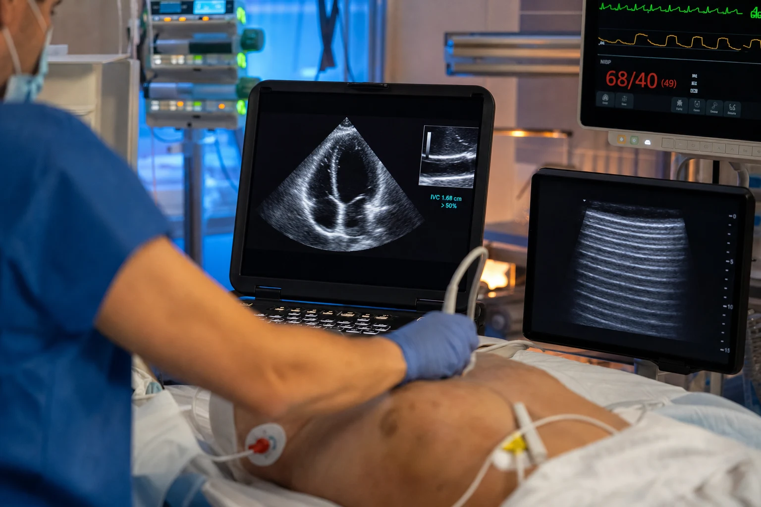
Shock is the failure of oxygen delivery to meet the metabolic demand of the tissues — a state of cellular hypoxia that, if uncorrected, progresses to multi-organ failure and death. It is recognised by the hypoperfusion (a raised lactate, a prolonged capillary refill, oliguria, an altered mentation) and the haemodynamic derangement (hypotension, tachycardia), and it is classified into four types by which determinant of delivery has failed.[1][1]
The four types — distributive, cardiogenic, hypovolaemic and obstructive — are not merely descriptive; each fails a different part of the oxygen-delivery equation, each has a characteristic haemodynamic profile, and each demands a different resuscitation. The classification is the framework that directs the treatment, and the bedside task is to identify the type (or types, for they coexist) and to correct its cause.[1]
Pathophysiology: oxygen delivery and the four failures
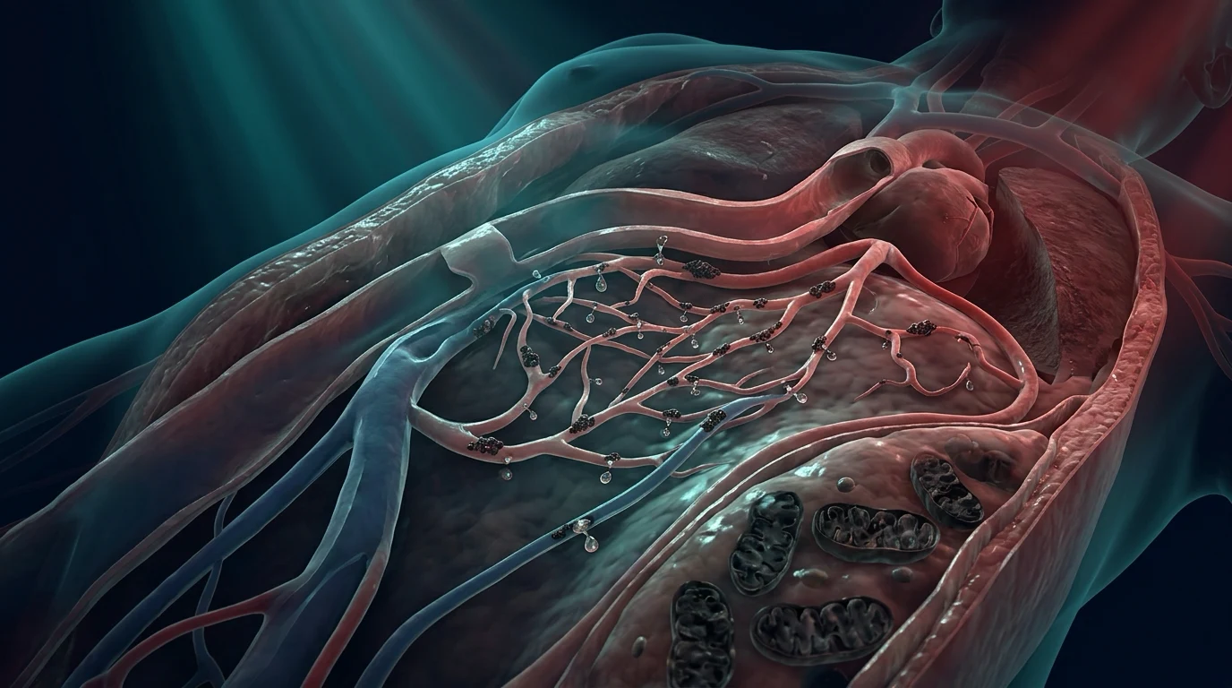
Global oxygen delivery is the product of the cardiac output and the arterial oxygen content (DO2 = CO × CaO2), and the cardiac output is governed by the heart rate, the preload, the contractility and the afterload. Each shock type fails a different determinant:[1]
- Hypovolaemic shock — a fall in the preload (haemorrhage, dehydration, third-space loss); the cardiac output falls, the peripheral resistance rises (the vasoconstriction that compensates), and the periphery is cold.
- Cardiogenic shock — a fall in the contractility (myocardial infarction, myocarditis, the decompensated failing heart); the output falls, the filling pressure rises, and the periphery is cold.
- Obstructive shock — an obstruction to the output or the filling (tension pneumothorax, massive pulmonary embolism, cardiac tamponade); the output falls mechanically, and the periphery is cold.
- Distributive shock — a fall in the afterload (the vasoplegia of sepsis, anaphylaxis, neurogenic shock); the cardiac output is high (or normal) but the resistance is low, the blood is maldistributed, and the periphery is warm. [1]
The first three (hypovolaemic, cardiogenic, obstructive) are the "cold" shocks — a low output, a high tone, a cool periphery — and distributive is the "warm" shock — a high output, a low tone, a warm periphery. The bedside examination of the periphery is the first clue to the type.[1][1]
Cellular pathophysiology: the oxygen delivery-consumption mismatch
Shock is, at its core, a mismatch between oxygen delivery (DO2) and oxygen consumption (VO2). In health, DO2 is approximately 1000 mL/min and VO2 approximately 250 mL/min, leaving a generous reserve — tissues can extract more oxygen when demand rises. As DO2 falls in shock, oxygen extraction rises to defend VO2, and the mixed venous oxygen saturation (SvO2) drops as a marker of the increasing extraction. Below a critical DO2 threshold (approximately 300 mL/min/m², or 8 mL/kg/min), extraction can no longer compensate and VO2 falls in proportion to DO2 — anaerobic metabolism begins, lactate accumulates, and the cell tips from compensated hypoxia into dysoxia.[1][19]
The biochemical consequences define the shock state: [1]
- Anaerobic glycolysis — pyruvate is shunted to lactate rather than entering the Krebs cycle, yielding 2 ATP per glucose instead of 36; the proton accumulation produces a metabolic (lactic) acidosis with an increased anion gap. A serum lactate above 2 mmol/L (in the absence of an alternative cause) is a defining marker of tissue hypoperfusion; above 4 mmol/L it is the threshold for "severe hyperlactataemia" and an independent mortality predictor.[12]
- Mitochondrial dysfunction — in septic shock specifically, the inflammatory cascade (cytokines, nitric oxide, reactive oxygen species) impairs mitochondrial oxidative phosphorylation, producing a cellular oxygen utilisation defect ("cytopathic hypoxia") — the cell cannot use the oxygen that is delivered. This explains the paradox of septic shock: high DO2 and a high SvO2 (often >70%) despite profound tissue hypoxia. It is the rationale for early recognition and the limitations of simply pushing more oxygen.[12][19]
- The microcirculatory failure — even when macro-haemodynamics are restored, the microcirculation may remain deranged (the loss of capillary density, the heterogeneity of flow between capillaries). The capillary refill time and the mottling score are the bedside windows onto the microcirculation, and they are the targets of the ANDROMEDA-SHOCK resuscitation strategy.[14]
- The lactate clearance — the rate at which the serum lactate falls is a marker of the resolution of the perfusion deficit, and a clearance of >10% per hour is associated with improved survival. A rising or static lactate despite apparent macro-haemodynamic restoration signals ongoing hypoperfusion, anaerobic metabolism, or impaired hepatic clearance.[16]
Classification: the four types and their causes
The four types of shock are distinguished by which determinant of the oxygen-delivery equation has failed, and each has a characteristic set of causes and a haemodynamic profile that directs the resuscitation.[19]
Hypovolaemic shock — a failure of the preload, the volume returning to the heart. The causes divide into:
- Haemorrhagic — external (trauma, GI bleed, surgical blood loss) or internal (haemothorax, intra-abdominal, retroperitoneal, long-bone and pelvic fracture, ruptured ectopic, ruptured aortic aneurysm). The blood volume lost is the determinant: 15% (Class I, compensated), 15–30% (Class II, tachycardia and narrowed pulse pressure), 30–40% (Class III, hypotension and confusion), >40% (Class IV, lethargy and cold clamped periphery).
- Non-haemorrhagic — dehydration (vomiting, diarrhoea, burns, polyuria of DKA or diabetes insipidus), third-space loss (pancreatitis, peritonitis, ileus, crush injury), and the profound diuresis of hyperglycaemic crises. [1]
Distributive shock — a failure of the vascular tone and the distribution of blood (and in sepsis, of cellular oxygen use). The causes are:
- Septic shock — the archetype; the vasoplegia of infection with a maldistributed, leaking circulation and a cellular oxygen-utilisation defect. Defined by Sepsis-3 as sepsis with vasopressor dependency to maintain MAP ≥65 mmHg and a serum lactate >2 mmol/L despite adequate volume resuscitation.[12][13]
- Anaphylactic shock — the explosive IgE-mediated release of histamine and tryptase causing vasoplegia, bronchospasm, and capillary leak, classically within minutes of exposure to an allergen (NMBA, antibiotic, contrast, food, venom).[1]
- Neurogenic shock — the loss of sympathetic vasomotor tone from a high spinal-cord injury (above T6), producing a warm, dry, vasodilated periphery with an inappropriately bradycardic heart rate (unwitnessed hypovolaemia would be tachycardic).[1]
- Other distributive states — the post-cardiac-arrest vasoplegia, the systemic inflammatory response of pancreatitis or burns, the adrenal crisis (cortisol deficiency unmasks vasoplegia), and the early phase of hepatic failure.
Cardiogenic shock — a failure of the pump (the contractility, less commonly a critical arrhythmia). The causes divide into:
- Ischaemic — acute myocardial infarction with >40% left-ventricular myocardium loss (the classic), mechanical complications (acute mitral regurgitation from papillary muscle rupture, ventricular septal rupture, free-wall rupture), and right-ventricular infarction.
- Non-ischaemic — decompensated chronic heart failure, fulminant myocarditis (giant-cell, viral, autoimmune), takotsubo (stress) cardiomyopathy, peripartum cardiomyopathy, drug toxicity (beta-blocker, calcium-channel-blocker, anthracycline), and the post-cardiac-arrest stunning.
- Arrhythmogenic — sustained ventricular tachycardia, fast atrial fibrillation or flutter with a failing heart, complete heart block. [1]
Obstructive shock — a mechanical obstruction to either the filling (the venous return) or the output (the ejection) of the heart. The causes are few and the corrections are specific:
- Cardiac tamponade — fluid, blood, or clot in the pericardial space compressing the right heart in diastole; corrected by pericardiocentesis or a surgical window.
- Tension pneumothorax — air under positive pressure in the pleural space compressing the mediastinum, the great vessels, and the contralateral lung; corrected by immediate needle decompression followed by an intercostal drain.
- Massive pulmonary embolism — clot obstructing the pulmonary outflow tract, acutely raising the right-ventricular afterload; corrected by systemic thrombolysis, catheter embolectomy, or surgical embolectomy.
- Other — severe pulmonary hypertension, an intra-thoracic abdominal compartment syndrome, a mediastinal tumour, a tension haemothorax, or an intra-aortic balloon pump malposition. [1]
The four types may coexist — the septic patient with a stress cardiomyopathy or an ischaemic insult; the trauma patient with a tension pneumothorax, hypovolaemia, and a pericardial tamponade. The bedside task is to identify the dominant type and the contributing types, and to correct each.[1][19]
The haemodynamic profiles: cardiac output and systemic vascular resistance
The four types are separated by their cardiac output and their systemic vascular resistance (SVR), and the profile directs the resuscitation.[1][1]
- Distributive — a high (or normal) cardiac output with a low SVR. The clue is a warm, vasodilated periphery with a shock state; the management is the vasopressor (for the SVR) and the treatment of the cause (the infection, the allergen, the cord injury).
- Cardiogenic — a low cardiac output with a high SVR. The clue is a cold, clammy periphery with a raised filling pressure; the management is the inotrope, the unloading of the ventricle, and the mechanical support, with the cause treated (the infarct revascularised).
- Hypovolaemic — a low cardiac output with a high SVR. The clue is a cold periphery with a low filling pressure; the management is the fluid (or the blood) to restore the preload.
- Obstructive — a low cardiac output with a compensatory high SVR. The clue is the specific syndrome (the tracheal deviation of the pneumothorax, the distended neck veins of the tamponade, the right-heart strain of the embolism); the management is the relief of the obstruction. [1]
The cardiac output and the SVR are inferred at the bedside from the examination, the focused echocardiogram, and (in the complex case) an advanced haemodynamic monitor; the central venous pressure is no guide to the fluid-responsiveness question that runs through them all.[9][10]
Haemodynamic profiles by shock type — cardiac output, SVR, PCWP, SvO2
| Parameter | Hypovolaemic | Cardiogenic | Obstructive | Distributive |
|---|---|---|---|---|
| Cardiac output (CO) | Low | Low | Low | High (or normal) |
| Systemic vascular resistance (SVR) | High (compensatory vasoconstriction) | High (compensatory vasoconstriction) | High (compensatory) | Low (vasoplegia) |
| Pulmonary capillary wedge pressure (PCWP) | Low (underfilled) | High (backward failure) | High (tamponade/PE) or normal | Low or normal (capillary leak; vasodilated venous capacitance) |
| Mixed venous saturation (SvO2) | Low (high extraction) | Low (high extraction) | Low (high extraction) | High (impaired extraction — cytopathic hypoxia) |
| Central venous pressure (CVP) | Low | High | High | Low or normal |
| Stroke volume | Low | Low | Low | High, normal, or late-low |
| Heart rate | Tachycardic | Tachy- or bradycardic | Tachycardic (except neurogenic) | Tachycardic |
| Periphery | Cold, clammy, mottled | Cold, clammy | Cold, often distended neck veins | Warm, vasodilated (early) |
| The bedside clue | History of loss; flat IVC; low CVP | Wet lungs, raised JVP, cold periphery; echo: poorly contracting LV | Specific syndrome (Beck's triad, tracheal deviation, RV strain) | Warm periphery with shock; fever or source; resolved after adequate fluid |
| Cardiac silhouette on echocardiography | Small, hypercontractile, kissing LV walls | Dilated, hypocontractile LV | Right-heart dilation/strain (PE, tamponade); RV collapse in tamponade | Normal or hypercontractile LV (early); late may develop septic cardiomyopathy |
| The first response | Volume (or blood) restoration | Inotrope, offload, revascularise | Relieve the obstruction | Vasopressor, fluid if responsive, treat cause |
The compensated vs decompensated shock state — the clinical spectrum
| Domain | Compensated shock | Decompensated shock |
|---|---|---|
| Mean arterial pressure | Maintained (vasoconstriction and tachycardia compensate) | Below 65 mmHg (or >40 mmHg below baseline in the chronic hypertensive) |
| Heart rate | Tachycardic (the early sign — except neurogenic and the late, bradycardic child) | Tachy- or bradycardic (bradycardia is pre-arrest) |
| Capillary refill | Prolonged (>3 s) — the earliest reliable sign | Greatly prolonged (>5 s) or instantaneous flash (vasoplegia) |
| Mottling | Often present around knees | Extensive, confluent, >50% of lower limbs — high mortality |
| Skin | Pale, cool, clammy (cold shock) or warm, flushed (early distributive) | Mottled, cold, cyanotic, clammy |
| Mental state | Anxious, agitated (early cerebral hypoperfusion) | Confused, drowsy, obtunded — a sign of cerebral hypoperfusion |
| Urine output | Reduced (<0.5 mL/kg/h) | Anuric (<0.1 mL/kg/h) — renal hypoperfusion |
| Lactate | Raised (often 2–4 mmol/L) | Markedly raised (>4 mmol/L, rising) |
| Base deficit | Mild (−2 to −5) | Severe (<−5) |
| Respiratory rate | Tachypnoeic (compensatory for metabolic acidosis, or from lung injury) | Slow, shallow, sighing (pre-arrest — fatigue) |
Clinical recognition: compensated vs decompensated, and the perfusion markers
The recognition of shock rests on three pillars: the haemodynamic derangement, the perfusion deficit, and the biochemical footprint. Hypotension alone is a late and unreliable sign — by the time the blood pressure falls, compensatory mechanisms have failed, and the patient is at the threshold of decompensation. The skilled clinician recognises the compensated shock state (the tachycardia, the cool periphery, the rising lactate, the falling urine output, the anxious or restless patient) and treats it before the blood pressure collapses.[19][1]
The perfusion markers are the four corners of the bedside examination: [1]
- The capillary refill time — firm pressure on the distal phalanx of a finger (or the sternum, for central refill) for 5 seconds; the time to return of colour. A normal CRT is <3 s; a CRT >3 s is abnormal and signals peripheral hypoperfusion. It is the single most useful bedside perfusion marker, the ANDROMEDA-SHOCK target, and a sensitive marker of resuscitation adequacy.[14]
- The mottling score — patchy cyanotic discolouration of the skin, classically around the knees (Ait-Oufella 2011 mottling score, 0–5 by area). It correlates with mortality in septic shock and is a marker of severe microcirculatory failure.[1]
- The urine output — the kidney is the most sensitive monitor of the perfusion. A urine output <0.5 mL/kg/h is oliguria; <0.1 mL/kg/h is approaching anuria. Hourly measurement is the standard in the shocked patient.
- The conscious state — confusion, agitation, drowsiness, or obtundation reflect cerebral hypoperfusion. The GCS or the four-hour AVPU is the quick bedside test; a falling conscious state is an emergency.
- The lactate — the biochemical footprint of anaerobic metabolism. A normal lactate does not exclude shock (compensated distributive shock can have a normal lactate), but a raised lactate is the single most powerful prognostic marker.[12][16]
The compensated shock state is the syndrome of tachycardia, a narrowed pulse pressure, a cool periphery, a prolonged capillary refill, oliguria, a mild metabolic acidosis, and a raised lactate, with a preserved mean arterial pressure. The patient looks unwell but is not (yet) hypotensive — and they are the patient to find, because the decompensated shock is a late and dangerous stage. The decompensated state is hypotension, a falling conscious state, anuria, a rising lactate, and multi-organ failure — the patient who is about to arrest.[19]
Distributive shock
Distributive shock — septic, anaphylactic and neurogenic — is a failure of the vascular tone that maldistributes the blood and, in sepsis, a cellular failure of oxygen use. It is the commonest shock in the ICU.[11][1]
Septic shock is the archetype: the vasoplegia of infection, with a high cardiac output, a low SVR, and a maldistributed, leaking circulation. The resuscitation is the early antibiotics and source control, the fluid to the responsive patient, and the vasopressor to defend the mean arterial pressure — and the perfusion endpoints (the lactate, the capillary refill) confirm the recovery. The early, aggressive resuscitation of the first six hours (the EGDT era) endures, even as its rigid protocol has been superseded.[11]
Anaphylactic shock is the explosive, IgE-mediated vasoplegia and bronchospasm of a severe allergic reaction, and it is an emergency treated with intramuscular adrenaline first, then the fluid and the airway. Neurogenic shock is the vasoplegia of a high spinal-cord injury (the loss of the sympathetic tone), with a warm, dry periphery and an inappropriately slow heart rate, treated with the fluid and the vasopressor.[1]
Cardiogenic shock
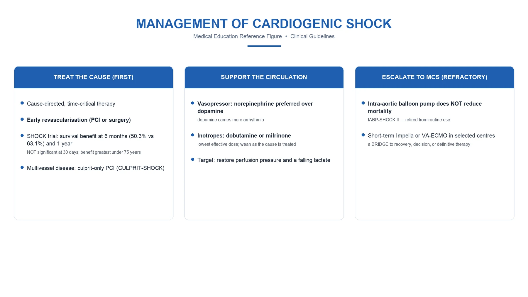
Cardiogenic shock — the failing pump — is most often the acute myocardial infarction, and its management has been reshaped by three trials.[1][2]
The SHOCK trial (NEJM 1999) established that early revascularisation (percutaneous or surgical) of the infarct-related artery reduced the long-term mortality of cardiogenic shock complicating myocardial infarction, and it is the foundation: the cardiogenic-shock patient goes to the catheter lab. The CULPRIT-shock trial (NEJM 2017) then showed that a culprit-lesion-only PCI strategy was superior to immediate multivessel PCI (less mortality and renal failure at 30 days) — so only the culprit is dilated in the acute setting, with the other lesions staged later.[2][4]
The IABP-SHOCK II trial (Lancet 2013) showed that the intra-aortic balloon pump did not reduce mortality in infarction-related cardiogenic shock, and it is no longer routine; the mechanical support of the modern era is the Impella and the veno-arterial ECMO, reserved for the refractory case as a bridge to recovery or to a durable therapy. The pharmacological platform is the inotrope (dobutamine, milrinone, adrenaline) and the vasopressor, the unloading of the ventricle, and the correction of the cause.[3][1]
Hypovolaemic shock
Hypovolaemic shock is the failure of the preload — haemorrhagic or non-haemorrhagic (dehydration, third-space loss, burns) — and it is the "cold, low-pressure" shock corrected by the restoration of the volume.[1]
In the haemorrhagic form, the management is the damage-control resuscitation — the blood products in a 1:1:1 ratio, the tranexamic acid, the permissive hypotension, and the surgical control of the bleeding. In the non-haemorrhagic form, it is the balanced crystalloid to the responsive patient — and in both, the fluid is given only to the responsive (tested by the pulse-pressure variation or the passive leg raise), because over-resuscitation is itself harmful.[9][10]
Obstructive shock
Obstructive shock is the mechanical obstruction to the output or the filling, and it is the shock most often missed — because its correction is specific and time-critical, and the resuscitation that does not relieve the obstruction is futile.[1]
- Tension pneumothorax — the tracheal deviation, the unilateral hyperresonance and absent breath sounds, the hypotension; the needle decompression, then the chest drain.
- Cardiac tamponade — the distended neck veins, the muffled heart sounds, the hypotension (Beck's triad), the pulsus paradoxus; the echocardiogram confirms, and the pericardiocentesis (or the surgical window) relieves.
- Massive pulmonary embolism — the right-heart strain, the raised right-sided pressures, the hypotension; the systemic thrombolysis (or the catheter or surgical embolectomy) for the unstable patient. [1]
The principle is that the obstructive shock is corrected by the relief of the obstruction, and the fluid and the vasopressor are the holding measures while it is achieved.[1]
Vasopressors and inotropes
The vasopressor restores the SVR; the inotrope restores the contractility; and the choice is governed by the shock type and the evidence.[5][1]
Norepinephrine is the first-line vasopressor for the distributive shock. The SOAP II trial (NEJM 2010) compared dopamine with norepinephrine and found no mortality difference overall, but more arrhythmias with dopamine (notably atrial fibrillation) — so norepinephrine is preferred, and dopamine is reserved for the exceptional indication (the bradycardic shock).[5]
Vasopressin is a second-line agent in septic shock — a pure vasoconstrictor (a V1 agonist) that complements the catecholamine. The VASST trial (NEJM 2008) found no overall mortality difference between vasopressin and norepinephrine, but vasopressin is used as a catecholamine-sparing adjunct in the high-norepinephrine patient, and it is weaned as the shock resolves.[6]
The inotropes — dobutamine (a beta-1 agonist) for the low-output cardiogenic shock, milrinone (a phosphodiesterase inhibitor) for the pulmonary-vasculature unloading, and adrenaline for the refractory — are reserved for the documented low-output shock, and they increase the myocardial oxygen demand that must be supplied.[1]
The blood-pressure target and the corticosteroid question
Two further questions arise in distributive shock: the target mean arterial pressure and the corticosteroid.[7][8]
The SEPSISPAM trial (NEJM 2014) compared a high (80 to 85 mmHg) with a low (65 to 70 mmHg) mean arterial pressure target in septic shock and found no difference in mortality — so the lower target (65 mmHg) is the standard, with the higher target reserved for the chronic hypertensive (in whom a low pressure may precipitate an acute kidney injury). The high-target group had more atrial fibrillation.[7]
The ADRENAL trial (NEJM 2018) tested hydrocortisone in septic shock and found no difference in 90-day mortality (though a faster shock reversal and fewer blood transfusions) — so hydrocortisone is not routine, but it is a reasonable adjunct in the septic shock that is refractory to adequate fluid and vasopressor. (The companion trial, APPROVe/CoVASS, was similar.)[8]
Shock indices: lactate clearance, ScvO2, base deficit, shock index
Shock is monitored not by a single number but by a panel of indices that together describe the perfusion, the oxygen debt, and the response to resuscitation. Each captures a different dimension, and the Fellowship candidate should know the normal, the threshold for concern, and the evidence behind each.[16][19]
The shock indices — what each measures, the normal, the threshold, the prognostic weight
| Index | What it measures | Normal | Threshold for concern | Prognostic weight |
|---|---|---|---|---|
| Serum lactate | Anaerobic metabolism (tissue hypoxia, mitochondrial dysfunction) | <1.5 mmol/L | >2 mmol/L (Sepsis-3 threshold); >4 mmol/L = severe hyperlactataemia | Independent mortality predictor; rising or static lactate = ongoing hypoperfusion |
| Lactate clearance | The rate at which lactate falls with resuscitation | n/a | >10% per hour (Jones 2010 target) is associated with improved survival | Each 5% increase in clearance confers ~2% absolute mortality reduction in sepsis |
| Central venous oxygen saturation (ScvO2) | Balance between O2 delivery and consumption | >70% | <70% = inadequate delivery (high extraction); >80% in sepsis can signal cytopathic hypoxia | EGDT (Rivers 2001) used ScvO2 ≥70% as a resuscitation target; later trials (PROCESS/ARISE/ProMISe) showed no benefit of ScvO2-guided protocol over usual care |
| Base deficit / base excess | The metabolic (non-respiratory) acid-base footprint | −2 to +2 mmol/L | Base deficit >5 mmol/L = significant acidosis | Base deficit tracks lactate clearance; a worsening base deficit signals worsening hypoperfusion |
| Shock index (HR/SBP) | A composite marker of haemodynamic derangement | 0.5–0.7 | >0.9 abnormal; >1.0 shock; >1.4 severe; >2.0 critical | Each 0.5-unit rise associated with increased mortality; SI is more sensitive than HR or SBP alone for early shock |
| Modified shock index (HR/MAP) | Refinement of the shock index using MAP | 0.7–1.0 | >1.3 abnormal | Better predictor than HR/SBP in some series |
| Capillary refill time | The peripheral perfusion surrogate | <3 s | >3 s abnormal; >5 s severe | Target of ANDROMEDA-SHOCK resuscitation strategy; abnormal CRT independently predicts 28-day mortality |
| Mottling score (Ait-Oufella) | Microcirculatory failure extent | 0 (none) | Score 0–5 by area around knees; >2 is severe | A score of 4–5 carries mortality >90% in septic shock |
The classical ATLS classes of haemorrhagic shock — blood loss, signs, and fluid strategy
| Class | Blood loss (% of circulating volume) | Volume (70 kg adult) | Heart rate | Blood pressure | Pulse pressure | Urine output | Mental state | Fluid strategy |
|---|---|---|---|---|---|---|---|---|
| Class I | <15% | <750 mL | <100 | Normal | Normal | >30 mL/h | Slightly anxious | Crystalloid maintenance |
| Class II | 15–30% | 750–1500 mL | 100–120 | Normal | Narrowed | 20–30 mL/h | Anxious, hostile | Crystalloid resuscitation |
| Class III | 30–40% | 1500–2000 mL | 120–140 | Fallen | Narrowed | 5–15 mL/h | Confused | Blood + crystalloid |
| Class IV | >40% | >2000 mL | >140 (or bradycardic pre-arrest) | Markedly low | Markedly narrowed | Negligible | Lethargic, obtunded | Massive transfusion protocol |
The shock index (HR/SBP) is the single most under-used bedside index in early shock. A young patient with a haemorrhage may have a systolic pressure of 120 mmHg and a heart rate of 130 — a "normal blood pressure" but a shock index of 1.08, the threshold of Class II–III shock. The narrow pulse pressure (the diastolic rising faster than the systolic) is the vasoconstrictive compensation, and it precedes the fall in systolic pressure by some minutes.[1]
Management principles: the ABCDE and the resuscitation bundle
The resuscitation of the shocked patient follows the ABCDE structure of the standard ICU/ED assessment, with the shock-specific interventions layered on. The Surviving Sepsis Campaign Hour-1 bundle (Evans 2021) is the prototypical bundle for septic shock; the principles transfer to the other shock types with their type-specific causes treated in parallel.[13][20]
ABCDE resuscitation of the shocked patient — the first 60 minutes
- A — AIRWAY AND C-SPINE — Assess and secure the airway; high-flow oxygen by mask (or nasal high-flow if appropriate); consider early intubation if the airway is threatened, the work of breathing is unsustainable, or the conscious state is falling. In the trauma patient, immobilise the cervical spine until cleared.
- B — BREATHING AND VENTILATION — Examine for tension pneumothorax (tracheal deviation, unilateral hyperresonance, absent breath sounds, hypotension — decompress immediately), flail chest, massive haemothorax, or open pneumothorax. Aim for an SpO2 ≥94% and a PaO2 ≥60 mmHg; if intubated, use lung-protective ventilation (tidal volume 6 mL/kg ideal body weight, plateau pressure <30 cmH2O).
- C — CIRCULATION WITH HAEMORRHAGE CONTROL — Apply direct pressure to external bleeding; pelvic binder for the unstable pelvic fracture; identify the shock type by the examination and the focused echocardiogram. Two large-bore cannulae (or intra-osseous access). Send bloods: group and cross-match, full blood count, coagulation, lactate, venous gas, troponin, β-HCG in women. Establish a fluid challenge ONLY if the patient is fluid-responsive (test with pulse-pressure variation, passive leg raise, or the focused echo's IVC; never by the central venous pressure). Give 250–500 mL balanced crystalloid boluses and reassess; in haemorrhagic shock, switch to blood products early (1:1:1 plasma:platelets:red cells) with tranexamic acid within 3 hours.
- C (continued) — VASOPRESSOR FOR THE VASOPLEGIC — If the MAP is <65 mmHg despite adequate volume resuscitation, start norepinephrine (first-line; titrate to MAP ≥65 mmHg); add vasopressin (fixed 0.03 U/min) as a catecholamine-sparing adjunct if the norepineephrine dose is rising. Do NOT delay vasopressors to chase a "fluid target" — early norepinephrine in septic shock with low SVR is safe and effective.
- C (continued) — INOTROPE FOR THE LOW-OUTPUT SHOCK — If the focused echocardiogram shows a hypocontractile, dilated LV with a low cardiac output and the patient remains hypoperfused despite adequate preload and MAP, add an inotrope — dobutamine 2.5–10 μg/kg/min (or adrenaline infusion as a combined inotrope/vasopressor) for the cardiogenic shock; milrinone if the right ventricle or the pulmonary vasculature is the problem. Reassess for ongoing ischaemia and the need for early revascularisation.
- C (continued) — TRANSFUSION IF ANAEMIC — If the haemoglobin is <70 g/L (or <80–90 g/L in the actively ischaemic or the chronic cardiac patient), transfuse packed red cells to restore the arterial oxygen content. Relieve the obstruction in obstructive shock: needle decompression for tension pneumothorax, pericardiocentesis for tamponade, systemic thrombolysis for massive PE.
- D — DISABILITY — Assess the GCS, the pupils, the blood glucose (treat hypoglycaemia immediately — a common co-factor in the shocked patient), and consider the causes of an altered conscious state (hypoperfusion, hypoglycaemia, hypoxia, intracranial event).
- E — EXPOSURE AND EXAMINATION — Examine the whole patient (front and back); identify the source of the shock (the abdomen for peritonitis or ruptured aneurysm, the limbs for compartment syndrome or long-bone fracture, the back for sacral pressure injury, the skin for the petechiae of meningococcaemia or the rash of anaphylaxis). Maintain normothermia (avoid hypothermia in the trauma patient — the lethal triad of acidosis, coagulopathy, and hypothermia).
- TREAT THE CAUSE IN PARALLEL — Antibiotics within one hour for septic shock (after blood cultures), source control (drainage, debridement, removal of infected line), early revascularisation for the infarct-related cardiogenic shock (SHOCK, CULPRIT-shock), surgical control of bleeding, hydrocortisone in refractory septic shock (ADRENAL), adrenaline for anaphylaxis, thrombolysis for the massive PE, pericardiocentesis for tamponade.
- REASSESS THE PERFUSION, NOT THE PRESSURE — The mean arterial pressure target (65 mmHg, higher in the chronic hypertensive) is a minimum, not an end. The target is a clearing lactate, a brisk capillary refill (<3 s), a urine output >0.5 mL/kg/h, and a normal conscious state. A patient with a MAP of 75 and a rising lactate is not resuscitated; a patient with a MAP of 65 and a clearing lactate is.[7][13][20]
Management: the type-specific approach
The management of shock is the identification of the type and the correction of its cause, with the restoration of the delivery.[1][1]
- Identify the type — by the examination, the focused echocardiogram, and the haemodynamic profile (the cardiac output and the SVR); recognise the coexistence of types (the septic patient with a cardiomyopathy, the traumatised patient with a tension pneumothorax).
- Restore the delivery — the fluid (to the responsive) for the preload, the inotrope and the unloading for the contractility, the vasopressor for the SVR, the relief of the obstruction, and the blood and the oxygen for the content.
- Treat the cause — the antibiotics and the source control, the revascularisation of the infarct, the surgical control of the bleeding, the needle for the pneumothorax, the adrenaline for the anaphylaxis.
- Target the perfusion, not a pressure — the lowest mean arterial pressure (65 mmHg in the vasoplegic, higher in the chronic hypertensive) that restores a clearing lactate and a brisk capillary refill.[7]
Monitoring shock at the bedside
Monitoring divides into the perfusion, the haemodynamics, and the cause.[9][1]
- The perfusion — the lactate (and its clearance), the capillary refill, the mottling, the urine output, the conscious state.
- The haemodynamics — the mean arterial pressure, the cardiac output and the SVR (inferred or measured), the focused echocardiogram (the ventricular function, the filling, the obstruction), and the fluid-responsiveness test before any bolus.
- The cause — the electrocardiogram and the troponin (the infarct), the chest film and the echocardiogram (the pneumothorax, the tamponade), the computed tomography (the embolism, the source).
- The harm — the fluid balance (the over-resuscitation), the arrhythmia (the dopamine, the high-pressure target), and the ischaemia (the inotrope-driven demand). [1]
Prognosis and the determinants of outcome
The prognosis of shock is the prognosis of its cause and its duration — the lactate and its clearance are the most powerful single prognostic markers, and the time to the restoration of the perfusion is the determinant of the organ failure. The mortality ranges from the low (the early-resuscitated septic shock) to the high (the refractory cardiogenic shock, which still carries a mortality in the region of 40 to 50 per cent despite the revascularisation and the mechanical support).[1][2]
SAQ — Septic shock with superimposed cardiomyopathy: a mixed shock state
10 minutes · 10 marks
A 72-year-old man with type 2 diabetes and ischaemic heart disease is admitted with community-acquired pneumonia. He is warm peripherally with a wide pulse pressure, HR 124, BP 76/40 (MAP 52), lactate 6.0 mmol/L, ScvO2 78%. After 30 mL/kg crystalloid and noradrenaline 0.5 mcg/kg/min the MAP is 64, lactate 5.2, but a focused echocardiogram shows a globally hypokinetic LV with an ejection fraction of 35% and a raised JVP. The senior registrar wants to give another fluid bolus.
SAQ — Cardiogenic shock from acute MI: revascularisation and mechanical support
10 minutes · 10 marks
A 58-year-old man presents with an anterolateral STEMI and cardiogenic shock: BP 78/50 (MAP 59), HR 110, cool peripheries, bilateral pulmonary crackles, lactate 4.5 mmol/L. Echocardiography shows an akinetic anterior wall with EF 25%, no mechanical complication. Coronary angiography demonstrates a proximal LAD occlusion with severe two-vessel disease.
Red flags
Key trials and evidence
Singer 2016 — Sepsis-3 definitions (PMID 26903338)
Document type
International consensus task force — JAMA
Scope
Replaced the SIRS-based definitions of sepsis with a new framework: sepsis = life-threatening organ dysfunction caused by a dysregulated host response to infection (SOFA ≥2); septic shock = sepsis with vasopressor-requiring hypotension to maintain MAP ≥65 mmHg AND lactate >2 mmol/L despite adequate volume resuscitation.
Key change
Dropped 'severe sepsis' as a category; introduced qSOFA (RR ≥22, altered mentation, SBP ≤100) as a bedside prompt (not diagnostic); defined septic shock by the vasopressor-and-lactate combination.
Clinical bottom line
The modern framework for sepsis and septic shock. The Fellowship candidate must be able to recite the septic shock definition: vasopressor dependency to maintain MAP ≥65 mmHg with a serum lactate >2 mmol/L despite adequate volume resuscitation.
Evans 2021 — Surviving Sepsis Campaign International Guidelines (PMID 34643578)
Document type
Joint ESICM/SCCM consensus guideline — Critical Care Medicine
Scope
The 2021 update of the SSC, with 93 recommendations across the sepsis spectrum.
Key recommendations for shock
Norepinephrine first-line vasopressor (strong); suggest vasopressin 0.03 U/min as a catecholamine-sparing adjunct (weak); suggest adding adrenaline if two vasopressors insufficient (weak); suggest hydrocortisone 200 mg/day if two vasopressors insufficient to maintain MAP (weak); MAP target 65 mmHg (strong); suggest crystalloid 30 mL/kg in first 3 hours (weak).
Clinical bottom line
The reference for the modern sepsis-shock bundle. The Hour-1 bundle: measure lactate, obtain blood cultures, administer broad-spectrum antibiotics, begin rapid 30 mL/kg crystalloid for hypotension or lactate ≥4, begin vasopressors if hypotensive during/after fluids to maintain MAP ≥65.
Seymour 2017 — Time to treatment and mortality in mandated sepsis care (PMID 28528569)
Document type
Observational cohort from New York State's mandated Rory Staunton 3-hour bundle — NEJM
Population
49,331 sepsis and septic shock patients across 149 hospitals
Key finding
Each hour of delay in completing the 3-hour bundle (antibiotics, lactate, fluids) was associated with an **odds ratio of 1.04 per hour for in-hospital mortality** (1.04, 95% CI 1.02–1.05); mortality rose linearly with each hour.
Clinical bottom line
Time is of the essence in septic shock. The Hour-1 bundle and the 'each hour costs lives' message is grounded in this dataset. Antibiotics within one hour is non-negotiable.
Jones 2010 — Lactate clearance vs ScvO2 as goals of early sepsis therapy (PMID 20179283)
Document type
Randomised non-inferiority trial — JAMA (EMShockNet)
Population
300 patients with severe sepsis or septic shock in three US EDs
Intervention
Resuscitation to a lactate clearance ≥10% vs ScvO2 ≥70% (the EGDT target) over 6 hours
Key finding
Lactate clearance was non-inferior to ScvO2-guided therapy (in-hospital mortality 17% vs 23%; non-inferiority met). Both groups received similar overall resuscitation, but lactate clearance does not require a central line.
Clinical bottom line
Lactate clearance (≥10% per hour) is a valid alternative to ScvO2 as a resuscitation endpoint, and is the preferred bedside target for the non-catheterised patient.
Hernández 2019 — ANDROMEDA-SHOCK: capillary refill vs lactate-guided resuscitation (PMID 30772908)
Document type
Multicentre randomised controlled trial — JAMA
Population
424 adults with septic shock in 5 ICUs in South America
Intervention
Resuscitation guided by peripheral perfusion targets (capillary refill time, mottling) vs lactate clearance, for 8 hours
Key finding
The perfusion-guided arm received **less fluid** (1.7 vs 2.0 L, p=0.02) and a non-significant trend to lower 28-day mortality (34.9% vs 43.4%, p=0.06); a Bayesian re-analysis suggested a 90% probability that perfusion-guided therapy was superior.
Clinical bottom line
Capillary refill is a legitimate and arguably superior target to lactate for guiding septic shock resuscitation. The bedside message: examine the periphery, and resuscitate to a brisk capillary refill, not just a falling lactate.
Shapiro 2023 — CLOVERS: early restrictive vs liberal fluid in septic shock (PMID 36688507)
Document type
Multicentre randomised controlled trial — NEJM (PETAL network)
Population
1,563 adults with sepsis-induced hypotension (SBP <100 or MAP <65) within 24 hours
Intervention
Restrictive fluid strategy (early vasopressors, less fluid) vs liberal fluid strategy (more fluid, vasopressors only if needed) over 24 hours
Key finding
No difference in 90-day mortality (restrictive 14.0% vs liberal 14.9%, p=0.61), ventilator-free days, or renal-replacement therapy. Both strategies safe; a wide range of practice is acceptable.
Clinical bottom line
In the resuscitation of sepsis-induced hypotension, an early restrictive strategy (less fluid, earlier vasopressors) is as safe as the liberal fluid-led approach. Personalised to the patient's phenotype — the volume-responsive vs the vasopressor-dependent.
Meyhoff 2022 — CLASSIC: restriction of IV fluids in septic shock (PMID 35709019)
Document type
Multicentre randomised controlled trial — NEJM (Scandinavian Critical Care Trials Group)
Population
1,554 adults with septic shock in ICUs across Denmark, Sweden, Norway, Belgium
Intervention
Restrictive IV fluid strategy vs standard care after initial resuscitation
Key finding
No difference in 90-day mortality (restrictive 42.3% vs standard 42.1%, p=0.96). The restrictive arm received ~1.2 L less fluid. Secondary outcomes (AKI, need for RRT) were similar.
Clinical bottom line
After the initial resuscitation, a restrictive approach to additional fluid is safe and avoids the harm of over-resuscitation (oedema, abdominal compartment syndrome, ARDS). Combined with CLOVERS, the era of routine massive fluid resuscitation in septic shock is over.
Rowan 2017 — PRISM patient-level meta-analysis of PROCESS, ARISE, ProMISe (PMID 28320242)
Document type
Patient-level meta-analysis of three multicentre RCTs — NEJM
Population
3,727 patients across PROCESS (US), ARISE (ANZ), ProMISe (UK)
Intervention
EGDT (the Rivers protocol — CVP/ScvO2/transfusion targets) vs usual care
Key finding
No difference in 90-day mortality (EGDT 25.4% vs usual care 24.4%, p=0.32), no difference in long-term mortality or quality of life. The EGDT protocol was more invasive (more central lines, more transfusions) without benefit.
Clinical bottom line
The early, aggressive, protocolised EGDT of the Rivers era is not superior to the modern usual care of an experienced team. The lesson is not 'do less' — it is 'do the right things early' (antibiotics, source control, judicious fluids, vasopressors when needed).
Sprung 2008 — CORTICUS: hydrocortisone in septic shock (PMID 18184957)
Document type
Multicentre randomised placebo-controlled trial — NEJM
Population
499 adults with septic shock (all severities) across 52 ICUs
Intervention
Hydrocortisone 50 mg IV 6-hourly × 5 days then wean vs placebo
Key finding
No mortality benefit overall (34.9% vs 31.9%, p=0.51), even in the non-responders to ACTH (the earlier Annane trial suggested benefit in non-responders; CORTICUS did not confirm). Faster shock reversal, but more superinfections (including new sepsis) in the hydrocortisone arm.
Clinical bottom line
Hydrocortisone is NOT routine in septic shock. Reserve for the septic shock refractory to adequate fluid and vasopressor (the ADRENAL/CORTICUS-compatible indication: two vasopressors needed to maintain MAP). Use 200 mg/day continuous infusion; wean when the vasopressors are off.
Cecconi 2014 — ESICM consensus on circulatory shock and haemodynamic monitoring (PMID 25392034)
Document type
Task force consensus — Intensive Care Medicine
Scope
The classification of shock (the four types), the haemodynamic profiles, and the choice of monitoring by clinical scenario.
Key messages
(i) Clinical examination (skin mottling, capillary refill, urine output) remains the cornerstone; (ii) echocardiography is the first-line advanced monitor in acute shock; (iii) the cardiac output and SVR profiles by shock type are the framework for diagnosis; (iv) advanced monitors (PiCCO, Vigileo, pulmonary artery catheter) for the complex or refractory case.
Clinical bottom line
The reference consensus for the modern shock-monitoring framework. The Fellowship candidate must be able to draw the four-shock haemodynamic profile table and justify the choice of monitor.
Clinical pearls
Quick-revision summary
- Definition: failure of oxygen delivery to meet demand; recognised by hypoperfusion + haemodynamic derangement; classified into four types.
- The four types: hypovolaemic (preload), cardiogenic (pump), obstructive (mechanical), distributive (tone + cellular use).
- Cold vs warm: first three are cold (low CO, high SVR); distributive is warm (high CO, low SVR).
- Cellular: anaerobic glycolysis → lactate; mitochondrial dysfunction in sepsis (cytopathic hypoxia); microcirculatory failure (CRT, mottling).
- Recognition: tachycardia, narrowed pulse pressure, prolonged CRT, mottling, oliguria, altered mentation, raised lactate — hypotension is late.
- The shock index (HR/SBP): >0.9 abnormal; >1.0 shock; >1.4 severe; calculate it always.
- The lactate: >2 mmol/L Sepsis-3 threshold; >4 mmol/L severe; clearance >10%/h is the Jones target.
- Fluid: only to the responsive (test with PLR, PPV, IVC); ~50% of ICU patients are non-responders at any moment.
- Vasopressor: norepinephrine first-line (SOAP II — dopamine more arrhythmias); vasopressin 0.03 U/min as adjunct (VASST); MAP target 65 (SEPSISPAM).
- Inotrope: dobutamine for cardiogenic low-output; milrinone for RV/pulmonary; adrenaline for refractory.
- Cause: antibiotics + source control in septic shock; early revascularisation in infarct-related cardiogenic shock (SHOCK, CULPRIT-shock); relief of obstruction in obstructive shock; IM adrenaline in anaphylaxis; damage-control resuscitation in haemorrhage.
- Corticosteroid: not routine (ADRENAL, CORTICUS); reserved for the refractory septic shock on two vasopressors.
- Monitor: perfusion (lactate, CRT, urine, conscious state), haemodynamics (MAP, CO, SVR, echo), cause (ECG, troponin, cultures, CT), harm (fluid balance, arrhythmia, ischaemia).
- Mortality: cardiogenic refractory 40–50%; septic shock with lactate >4 + vasopressor-dependent 30–40%; obstructive with prompt relief <10%. [1]
The one-page Fellowship viva answer
Shock is the failure of oxygen delivery (DO2) to meet tissue metabolic demand (VO2). DO2 = CO × CaO2; the four shock types fail different determinants. Hypovolaemic shock fails preload (haemorrhage, dehydration, third-space loss); cardiogenic shock fails contractility (MI, myocarditis, decompensated failure) or rhythm; obstructive shock mechanically obstructs output/filling (tamponade, tension pneumothorax, massive PE); distributive shock fails afterload and, in sepsis, cellular oxygen utilisation (septic, anaphylactic, neurogenic). The haemodynamic profiles separate them: hypovolaemic/cardiogenic/obstructive are LOW CO, HIGH SVR, COLD periphery, LOW SvO2 (high extraction); distributive is HIGH CO, LOW SVR, WARM periphery, HIGH SvO2 (impaired extraction). Recognition: hypoperfusion (raised lactate, prolonged CRT >3 s, mottling, oliguria, altered mentation) precedes hypotension — calculate the shock index (HR/SBP; >0.9 abnormal). Management: ABCDE, fluid ONLY if responsive (test by PLR/PPV/IVC, never CVP — Marik 2008), vasopressor to MAP ≥65 (norepinephrine first-line; dopamine more arrhythmias, SOAP II; vasopressin adjunct, VASST; SEPSISPAM target 65), inotrope for documented cardiogenic low-output, source control + antibiotics in septic shock, early revascularisation in cardiogenic (SHOCK, CULPRIT-shock), relief of obstruction in obstructive, IM adrenaline in anaphylaxis, transfusion if Hb <70 g/L. Corticosteroid only in refractory septic shock on two vasopressors (ADRENAL, CORTICUS). Monitor: lactate clearance (≥10%/h, Jones 2010), CRT (ANDROMEDA-SHOCK), urine output, conscious state.[1][5][7][19]
References
- [1]Reynolds HR, Hochman JS. Cardiogenic shock: current concepts and improving outcomes Circulation, 2008.PMID 18250279
- [2]Hochman JS, Sleeper LA, Webb JG, et al.; SHOCK Trial Investigators. Early revascularization in acute myocardial infarction complicated by cardiogenic shock. SHOCK Investigators. Should We Emergently Revascularize Occluded Coronaries for Cardiogenic Shock N Engl J Med, 1999.PMID 10460813
- [3]Thiele H, Zeymer U, Neumann FJ, et al.; IABP-SHOCK II Trial Investigators. Intra-aortic balloon counterpulsation in acute myocardial infarction complicated by cardiogenic shock (IABP-SHOCK II): final 12 month results of a randomised, open-label trial Lancet, 2013.PMID 24011548
- [4]Thiele H, Akin I, Sandri M, et al.; CULPRIT-SHOCK Investigators. PCI Strategies in Patients with Acute Myocardial Infarction and Cardiogenic Shock N Engl J Med, 2017.PMID 29083953
- [5]De Backer D, Biston P, Devriendt J, et al.; SOAP II Investigators. Comparison of dopamine and norepinephrine in the treatment of shock N Engl J Med, 2010.PMID 20200382
- [6]Russell JA, Walley KR, Singer J, et al.; VASST Study Investigators. Vasopressin versus norepinephrine infusion in patients with septic shock N Engl J Med, 2008.PMID 18305265
- [7]Asfar P, Meziani F, Hamel JF, et al.; SEPSISPAM Investigators. High versus low blood-pressure target in patients with septic shock N Engl J Med, 2014.PMID 24635770
- [8]Venkatesh B, Finfer S, Cohen J, et al.; ADRENAL Trial Investigators. Glucocorticoids with or without Fludrocortisone in Septic Shock N Engl J Med, 2018.PMID 30157400
- [9]Marik PE, Baram M, Vahid B. Does central venous pressure predict fluid responsiveness? A systematic review of the literature and the tale of seven mares Chest, 2008.PMID 18628220
- [10]Michard F, Boussat S, Chemla D, et al. Relation between respiratory changes in arterial pulse pressure and fluid responsiveness in septic patients with acute circulatory failure Am J Respir Crit Care Med, 2000.PMID 10903232
- [11]Rivers E, Nguyen B, Havstad S, et al. Early goal-directed therapy in the treatment of severe sepsis and septic shock N Engl J Med, 2001.PMID 11794169
- [12]Singer M, Deutschman CS, Seymour CW, et al. The Third International Consensus Definitions for Sepsis and Septic Shock (Sepsis-3) JAMA, 2016.PMID 26903338
- [13]Evans L, Rhodes A, Alhazzani W, et al. Executive Summary: Surviving Sepsis Campaign: International Guidelines for the Management of Sepsis and Septic Shock 2021 Crit Care Med, 2021.PMID 34643578
- [14]Hernández G, Ospina-Tascón GA, Damiani LP, et al.; ANDROMEDA-SHOCK Investigators. Effect of a Resuscitation Strategy Targeting Peripheral Perfusion Status vs Serum Lactate Levels on 28-Day Mortality Among Patients With Septic Shock: The ANDROMEDA-SHOCK Randomized Clinical Trial JAMA, 2019.PMID 30772908
- [15]Rowan KM, Angus DC, Bailey M, et al.; PRISM Investigators. Early, Goal-Directed Therapy for Septic Shock - A Patient-Level Meta-Analysis N Engl J Med, 2017.PMID 28320242
- [16]Jones AE, Shapiro NI, Trzeciak S, et al.; Emergency Medicine Shock Research Network (EMShockNet) Investigators. Lactate clearance vs central venous oxygen saturation as goals of early sepsis therapy: a randomized clinical trial JAMA, 2010.PMID 20179283
- [17]Sprung CL, Annane D, Keh D, et al.; CORTICUS Study Group. Hydrocortisone therapy for patients with septic shock N Engl J Med, 2008.PMID 18184957
- [18]Meyhoff TS, Hjortrup PB, Wetterslev J, et al.; CLASSIC Trial Group. Restriction of Intravenous Fluid in ICU Patients with Septic Shock N Engl J Med, 2022.PMID 35709019
- [19]Cecconi M, De Backer D, Antonelli M, et al. Consensus on circulatory shock and hemodynamic monitoring. Task force of the European Society of Intensive Care Medicine Intensive Care Med, 2014.PMID 25392034
- [20]Seymour CW, Gesten F, Prescott HC, et al. Time to Treatment and Mortality during Mandated Emergency Care for Sepsis N Engl J Med, 2017.PMID 28528569
- [21]Shapiro NI, Douglas IS, Battistini F, et al.; CLOVERS Trial Investigators. Early Restrictive or Liberal Fluid Management for Sepsis-Induced Hypotension N Engl J Med, 2023.PMID 36688507