ICU · Toxicology
Dermatological emergencies in ICU: SJS, TEN, and DRESS
Also known as Stevens-Johnson syndrome · Toxic epidermal necrolysis · Drug reaction with eosinophilia and systemic symptoms · Drug-induced hypersensitivity syndrome · Lyell syndrome
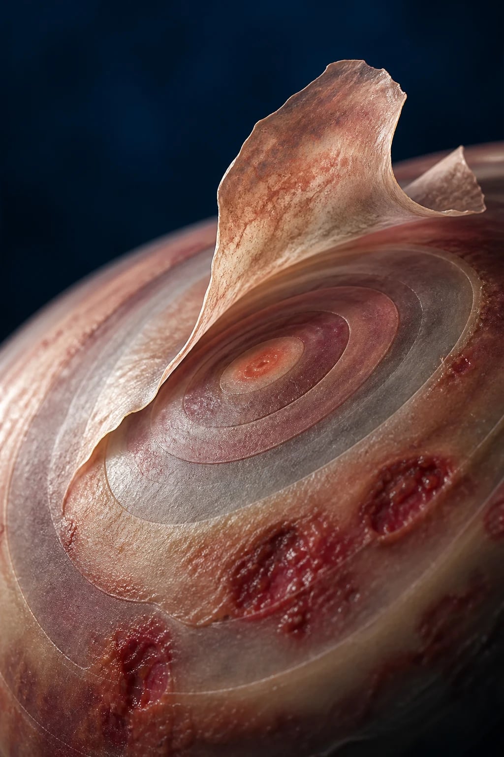
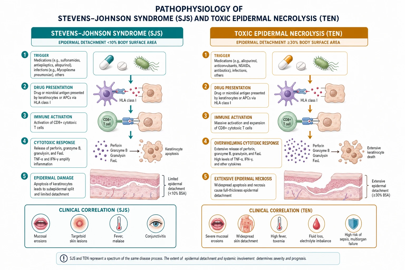
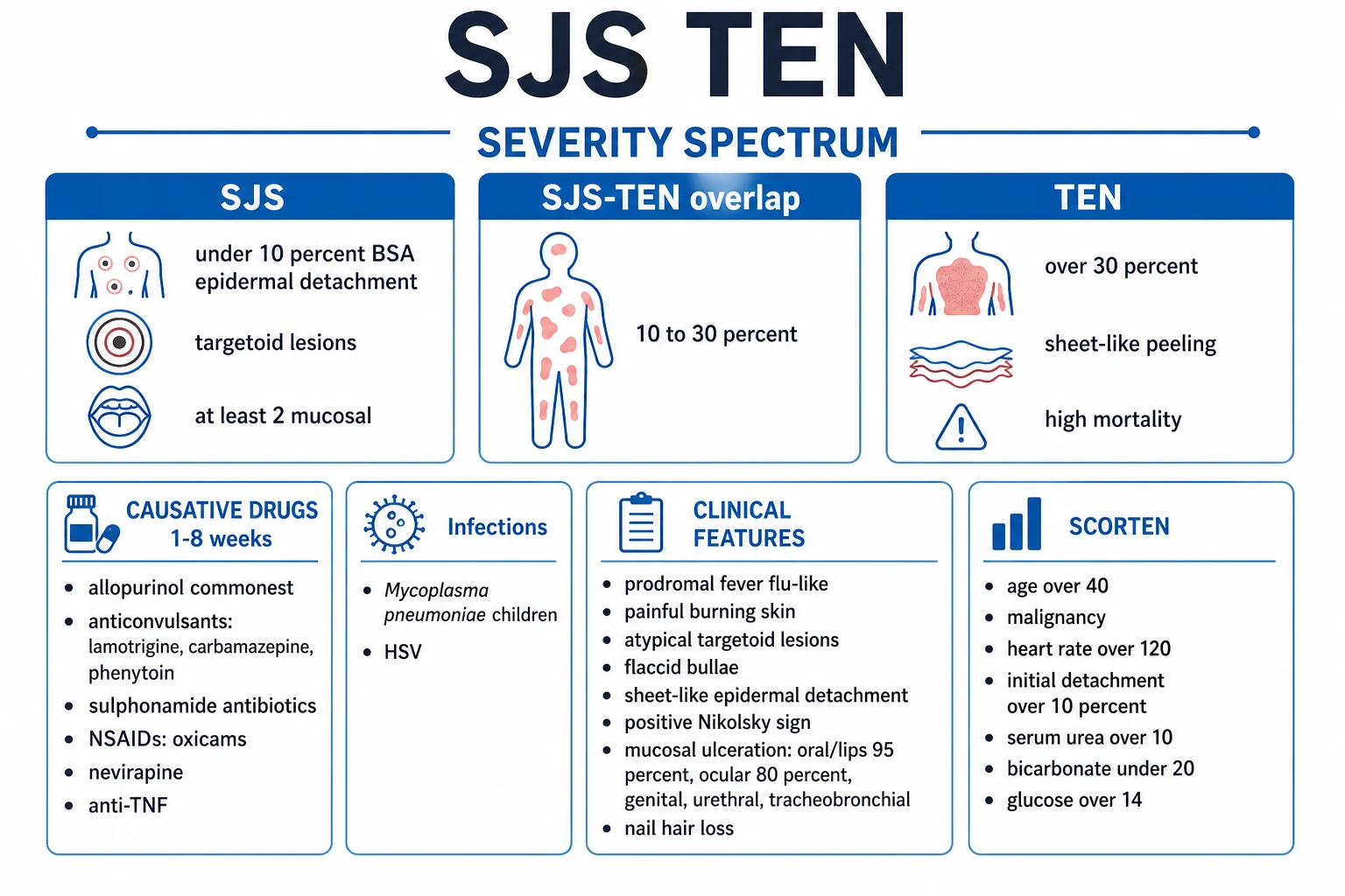
Severe cutaneous adverse reactions (SCAR) requiring ICU admission. SJS/TEN: immune-mediated epidermal necrolysis triggered by drugs (80%) or infections (Mycoplasma, HSV). Mucocutaneous: target lesions, bullae, sheet-like epidermal detachment. SJS <10% BSA detachment, SJS-TEN overlap 10-30%, TEN 30%. High mortality (SJS ~10%, TEN ~30%). DRESS (DIHS): delayed hypersensitivity (2-8 weeks post-exposure), rash + fever + lymphadenopathy + eosinophilia + organ dysfunction (hepatitis, nephritis, pneumonitis). Treatment: STOP culprit drug, supportive care (fluid, nutrition, temperature control, analgesia, infection prevention), ICU for TEN (fluid loss like burn, airway involvement), IVIG controversial, debridement controversial, ophthalmology early.
On this page & tools
Your progress
Saved locally on this device.
Target exams
Red flags
SJS vs SJS-TEN overlap vs TEN vs DRESS
| Feature | SJS | SJS-TEN overlap | TEN | DRESS (DIHS) |
|---|---|---|---|---|
| Detached BSA | <10% | 10-30% | >30% | Variable (often morbilliform, may exfoliate) |
| Onset | 1-4 weeks post-drug | 1-4 weeks | 1-4 weeks | 2-8 weeks |
| Mucosal involvement | Yes (≥2 sites) | Yes | Yes | Variable (less severe) |
| Target lesions | Atypical | Atypical | Rare | No |
| Fever | Yes | Yes | Yes | High (38-40°C) |
| Eosinophilia | No | No | No | Yes (hallmark) |
| Organ dysfunction | Rare | Variable | Common (liver, kidney) | Yes (liver, kidney, lung, heart) |
| Lymphadenopathy | No | No | No | Yes (hallmark) |
| Mortality | ~10% | ~25% | ~30% (up to 10%) | ~5-10% |
| Fluid loss | Minimal | Moderate | Massive (like burn) | Minimal |
| Recurrence | Drug-specific | Drug-specific | Drug-specific | Can reactivate months later |
Acute management of suspected SJS/TEN
- STOP all suspected drugs — antibiotics (sulphonamides), anticonvulsants (lamotrigine, carbamazepine, phenytoin), allopurinol, NSAIDs (oxicam). Withdraw ALL non-essential drugs
- Assess severity — %BSA detached, mucosal sites, organ function. Calculate SCORTEN (within 24h and 72h)
- ICU admission if: TEN (fluid loss like burn), airway involvement, organ failure, extensive mucosal disease
- Fluid resuscitation — TEN: crystalloid 2-4 mL/kg/%BSA (LESS than burn — Parkland overestimates). Monitor urine output 0.5-1 mL/kg/h
- Nutrition — EARLY enteral feeding within 24-48h. Nasogastric tube (oral mucosa too painful). Caloric needs ~1.5x basal
- Analgesia — multimodal: paracetamol, opioids (fentanyl PCA). Mucosal pain severe
- Infection prevention — reverse isolation, aseptic technique, SURVEILLANCE cultures (skin, urine, blood). Avoid prophylactic antibiotics. Treat confirmed infection promptly
- Ophthalmology review WITHIN 24h — ocular involvement causes blindness. Topical steroids, lubricants, amniotic membrane
- Skin care — leave intact blisters, non-adherent dressings (Mepitel, Jelonet), silicone dressings. Avoid aggressive debridement (controversial)
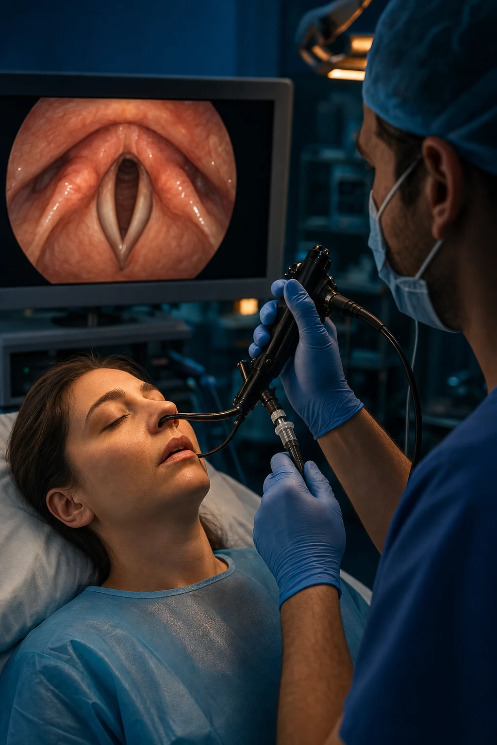
- Airway — if tracheobronchial involvement: may need intubation. Approach carefully (mucosal sloughing → difficult airway, bleeding)
Pathophysiology
Clinical features
Epidemiology
SAQ — Toxic epidermal necrolysis on allopurinol
10 minutes · 10 marks
A 68-year-old Han Chinese man started allopurinol 3 weeks ago for gout. He now has fever (39.2°C), painful oral/ocular/urethral mucositis, atypical target lesions and sheet-like epidermal detachment of 35% BSA, with a positive Nikolsky sign. SCORTEN at 24 h = 4 (age >40, HR 124, %BSA detached >10, urea 14 mmol/L, bicarbonate 18 mmol/L; no malignancy, glucose normal). LFTs ALT 220, creatinine 145.
SAQ — DRESS (drug reaction with eosinophilia and systemic symptoms)
10 minutes · 10 marks
A 44-year-old woman is admitted 5 weeks after starting carbamazepine for trigeminal neuralgia with a diffuse morbilliform rash, fever 39.5°C, cervical lymphadenopathy, facial oedema, eosinophils 2.8 ×10⁹/L, atypical lymphocytes on the blood film, ALT 480 U/L, creatinine 200 μmol/L and a normal INR. The team plans to discharge her on antihistamines once the rash fades.
Clinical pearls
Red flags
Prognosis
Long-term sequelae of SJS/TEN (Zhang 2021)
Cohort study. Long-term complications affect up to 70% of survivors:
- Ocular: dry eye, symblepharon, corneal scarring, blindness (3-10%)
- Skin: dyspigmentation (post-inflammatory), nail dystrophy, chronic pruritus, new melanocytic naevi
- Mucosal: oral synechiae, vaginal stenosis, urethral stricture, phimosis
- Respiratory: bronchiolitis obliterans, chronic bronchitis, tracheal stenosis
- Psychological: PTSD, depression, anxiety (survivors of severe acute illness)
- Reproductive: sexual dysfunction (genital scarring) [1]
Mortality best predicted by SCORTEN (72h) and ABCD-10.
AGEP — Acute Generalised Exanthematous Pustulosis
AGEP is the fourth member of the SCAR family and a high-yield discriminator in any exam stem describing hundreds of non-follicular, sterile, subcorneal pustules erupting on erythematous skin within hours-to-days of a drug. It is self-limiting once the trigger is withdrawn (resolves in <15 days with superficial desquamation) and mortality is low (<5%), but it can mimic pustular psoriasis and is the prototype drug-induced neutrophilic pustulosis.[21] }
AGEP vs SJS/TEN vs DRESS vs generalised pustular psoriasis
| Feature | AGEP | SJS/TEN | DRESS | Generalised pustular psoriasis |
|---|---|---|---|---|
| Lesion | Hundreds of small non-follicular sterile pustules on erythema | Target lesions, bullae, sheet-like detachment | Morbilliform maculopapular, facial oedema, later desquamation | Sheets of pustules on plaques of psoriasis |
| Onset after drug | <4 days (fast) | 1-4 weeks | 2-8 weeks | Not drug-related (IL-36RN/CARD14 mutation) |
| Mucosa | Usually spared (atypical: only 1 site) | Always, ≥2 sites | Variable, mild | Often |
| Fever | Yes (≥38°C, often ≥39°C) | Yes | Yes (38-40°C) | Yes |
| Eosinophilia / atypical lymphs | Mild eosinophilia possible | No | Hallmark eosinophilia + atypical lymphs | No |
| Blister fluid | Sterile pus (neutrophils) | Sterile (necrotic epidermis) | Not a blistering disorder | Sterile pus |
| Histology | Subcorneal pustule, eosinophils, no full-thickness necrosis | Full-thickness keratinocyte necrosis, sparse infiltrate | Lymphocytic interface dermatitis, eosinophils, necrotic keratinocytes | Spongiform pustule of Kogoj |
| Nikolsky | Negative | Positive | Negative | Negative |
| BSA detachment | None (pustules only) | SJS <10% / overlap 10-30% / TEN >30% | None (exfoliation late) | None |
| Mortality | <5% | ~10-30% | ~5-10% | Up to 20% |
| Treatment | Stop drug + supportive. Topical steroids. Steroids if severe | Stop drug + burn-unit supportive care | Stop drug + systemic steroids (1 mg/kg) | Anti-IL-36 (spesolimab), anti-TNF, retinoids |
Management of AGEP in ICU
- STOP the culprit drug. Classic triggers: aminopenicillins (amoxicillin/ampicillin, >50% of cases), pristinamycin (France), macrolides (pristinamycin, spiramycin), quinolones, hydroxychloroquine, terbinafine, diltiazem, sulphonamides. Withdraw ALL non-essential drugs
- Confirm sterility — unroof a pustule, send Gram stain and culture (bacterial AND viral — rule out disseminated herpes/varicella). Pustules are sterile in true AGEP
- Skin biopsy — subcorneal pustule with neutrophils ± eosinophils, spongiosis, no full-thickness necrosis
- Supportive care — antipyretics, fluid balance (skin barrier disrupted by extensive pustules), emollients. Reverse isolation not usually required (much less disruptive than TEN)
- Topical corticosteroid (e.g. clobetasol 0.05% to active areas) for symptomatic control
- Systemic corticosteroid only if severe / extensive / systemic toxicity — prednisolone 0.5-1 mg/kg/day, short taper (5-10 days). Most cases resolve without steroids
- Watch for superimposed sepsis — the ruptured pustules leave a denuded skin barrier; staphylococcal and gram-negative bacteraemia described. Treat confirmed infection promptly, never prophylactic
- Expect resolution in 1-2 weeks with superficial brawny desquamation — reassure the patient. Explain the diagnosis and warn against future re-exposure to the culprit drug (carry an alert card)
Pharmacogenomics — HLA screening (high-yield exam topic)
[1]HLA-allele × drug × population × screen-recommendation
| HLA allele | Culprit drug | Risk population | Screening recommendation |
|---|---|---|---|
| HLA-B*57:01 | Abacavir | All | Universal screen (any population) before starting abacavir |
| HLA-B*15:02 | Carbamazepine, oxcarbazepine, phenytoin | Han Chinese, Thai, SE Asian, South Asian | Screen before starting carbamazepine; avoid if positive |
| HLA-B*58:01 | Allopurinol | Han Chinese, Korean, Thai, Vietnamese | Screen before starting allopurinol (esp. CKD/low starting dose does not eliminate risk) |
| HLA-A*31:01 | Carbamazepine | Northern European, Japanese | Consider screen (Japanese guideline) |
| HLA-B*73:01 | Allopurinol | European | Consider |
| HLA-A*02:01 | Lamotrigine | European | Research / not routine |
| HLA-B*15:13, HLA-Cw*04:01 | Anti-TB drugs (rifampicin, isoniazid) | Vietnamese / SE Asian | Consider in SE Asian starting anti-TB |
| HLA-B*59:01 | Allopurinol (DRESS phenotype) | Japanese | Consider |
Test performance of pre-prescription HLA screening
Chung 2004 — HLA-B*1502 marks carbamazepine-induced SJS
Landmark case-control study in Han Chinese (Taiwan): 100% of 44 patients with carbamazepine-induced SJS carried HLA-B*1502 vs ~8% of carbamazepine-tolerant controls (OR >2500). The strongest gene-drug association in dermatology. Triggered the FDA black-box warning and a national pre-prescription screening programme in Taiwan, Hong Kong and Thailand that has demonstrably reduced carbamazepine-SJS incidence — proof that pharmacogenomic prevention of SCAR is feasible at population scale.[24] }
Chang/Chung 2020 — Pharmacogenetic testing for prevention of SCAR
Narrative review. Synthesises the case for pre-prescription HLA screening for abacavir (HLA-B*57:01, universal), carbamazepine (HLA-B*15:02, Asian), allopurinol (HLA-B*58:01, Asian). Conclusion: screening is cost-effective when the allele frequency in the target population is >1% and the gene-drug association is strong (OR >10). Remaining gap: implementing screening in low-resource settings and extending panels to additional high-risk genes (HLA-A*31:01, HLA-B*73:01).[19] }
Evidence on specific therapies in SJS/TEN
Corticosteroid / IVIG / ciclosporin / etanercept / thalidomide / plasma exchange across SCAR
| Therapy | SJS/TEN — efficacy | SJS/TEN — safety | DRESS | AGEP |
|---|---|---|---|---|
| Withdraw culprit drug | ✓✓✓ single most important | n/a | ✓✓✓ mandatory | ✓✓✓ mandatory |
| Systemic corticosteroid | ✗ Not recommended in established TEN (↑infection, ↑mortality); debated short course in early SJS | ↑infection risk | ✓✓ Prednisolone 1 mg/kg, taper 6-8 wk | Reserve for severe/extensive |
| IVIG | ⚠ Controversial; mixed trials; not routine. May use 1 g/kg × 3d in specialist centre | Renal failure, thrombosis, TRALI | Limited evidence; case series only | Not used |
| Ciclosporin | Promising: Lee 2017 retrospective showed ↓mortality vs IVIG | Immunosuppression, nephrotoxicity, hypertension | Limited evidence | Not used |
| Etanercept (anti-TNF) | Phase-II/observational signals (Paradisi 2014); faster re-epithelialisation. Not standard | Infection, TB reactivation | Not used | Not used |
| Thalidomide | ✗✗ HARMFUL (Wolkenstein 1998: ↑mortality, terminated early) | Teratogenic, neuropathy | Not used | Not used |
| Plasma exchange | Anecdotal; some centres add in refractory high-SCORTEN | Line/sepsis/bleeding risk | Not used | Not used |
| Anti-IL-36 / ciclosporin / NFKB-targeted | Research | — | — | Research |
Wolkenstein 1998 — Thalidomide INCREASED mortality in TEN (Lancet)
Randomised double-blind placebo-controlled trial, n=22. Thalidomide was hypothesised to block TNF-α-mediated keratinocyte apoptosis. Result: mortality 10/12 (83%) with thalidomide vs 3/10 (30%) placebo — trial terminated early for harm. Thalidomide paradoxically INCREASED TNF-α in TEN patients. Take-home: never give thalidomide in TEN, and never assume a biologically plausible therapy is safe without controlled evidence. A canonical example of harm from a logical-sounding treatment.[25] }
Lee 2017 — Ciclosporin reduced mortality vs IVIG (Singapore nationwide cohort)
Retrospective nationwide cohort of 105 SJS/TEN/overlap patients. Ciclosporin (n=29) vs IVIG (n=76). Standardised mortality ratio vs SCORTEN-predicted: ciclosporin 0.42 (i.e. observed mortality LOWER than predicted) vs IVIG 1.18 (observed mortality HIGHER than predicted). Ciclosporin group had less progression of BSA detachment and faster re-epithelialisation. Caveats: retrospective, non-randomised, single-centre cohort. Supports ciclosporin as a leading investigational therapy and the relative fall from favour of IVIG.[12] }
Kirchhof 2014 — IVIG vs cyclosporine retrospective (Canada)
Single-centre retrospective review comparing IVIG-treated (n=16) vs cyclosporine-treated (n=12) SJS/TEN patients. No significant difference in mortality (IVIG 38% vs ciclosporin 8%, p=0.21 — trend favouring ciclosporin, underpowered). Both treatments had comparable safety profiles in this cohort. Conclusion: ciclosporin is a reasonable alternative to IVIG; prospective randomised trial needed (still not done).[26] }
Aihara/Aoyama 2015 — IVIG added to corticosteroid in SJS/TEN (Japan)
Multicentre randomised controlled trial, n=110, SJS/TEN patients already on systemic corticosteroid. Addition of IVIG (1 g/kg/day × 3 days) vs no IVIG. Primary outcome: no significant difference in 4-week mortality or Vanderbilt wound-healing score. Modest signal favouring IVIG only in SJS (not TEN) subgroup. Conclusion: IVIG on top of steroids does NOT improve outcomes — further weakens the routine-IVIG case.[14] }
Roujeau/Bastuji-Garin 2011 — Systematic review of SJS/TEN treatments
Therapeutic Advances in Drug Safety. Comprehensive systematic review of every reported treatment (corticosteroids, IVIG, ciclosporin, anti-TNF, plasma exchange, thalidomide, N-acetylcysteine). Conclusion: NO therapy has level-1 evidence of benefit. Withdrawal of the culprit drug and expert supportive/burn-unit care remain the only proven interventions. Calls for prospective randomised trials (still largely unanswered a decade later).[8] }
Creamer 2016 — UK national guidelines for SJS/TEN in adults
Multidisciplinary consensus (dermatology, burns, ICU, ophthalmology, maxillofacial). Recommendations: (1) immediate drug withdrawal, (2) early senior review, (3) transfer to ICU/burns centre if >10% BSA or airway/mucosal involvement, (4) fluid management using <burn formula (2-4 mL/kg/%BSA), (5) early nutritional support (NG within 24-48 h), (6) daily ophthalmology review with topical steroid + lubricant, amniotic membrane within 5 days for ocular involvement, (7) skin: conservative debridement only, non-adherent silicone dressings, (8) do NOT use prophylactic antibiotics, treat confirmed infection. Specific immunomodulatory therapy is left to specialist discretion pending better evidence.[13] }
Ingen-Housz-Oro 2025 — French TOXIBUL national protocol for epidermal necrolysis
Multicentre recommendations from the French National Reference Centre for Toxic Bullous Diseases (TOXIBUL). Reaffirms: (1) early withdrawal of all suspected drugs, (2) ABCD-10 ≥2 = poor prognosis → transfer to reference centre, (3) intensive supportive care modelled on severe burns, (4) systematic ophthalmology review within 24 h and amniotic membrane transplantation for any ocular involvement <5 days from onset, (5) cautious intubation with video laryngoscopy and smaller ETT given mucosal fragility, (6) avoid corticosteroid monotherapy in established TEN, (7) ciclosporin considered in select patients under protocol.[15] }
Montmeat/de Prost 2026 — Ventilator-associated pneumonia in epidermal necrolysis (ICU)
Retrospective multicentre ICU cohort of mechanically ventilated epidermal necrolysis patients. VAP incidence was high (~30-40% of intubated TEN patients), driven by mucosal sloughing of the tracheobronchial tree impairing mucociliary clearance, prolonged sedation, and immunosuppression from systemic therapy. Late-onset VAP and Pseudomonas aeruginosa predominated. Practice point: prefer oral over nasal intubation (nasal mucosa sloughs and bleeds), use subglottic secretion drainage, early enteral nutrition, and de-escalate antibiotics rapidly after VAP treatment to limit superinfection in an already immunocompromised host.[16] }
Shay/Tseng 2010 — Sutureless amniotic membrane transplantation for acute TEN ocular disease
Case series. Amniotic membrane transplantation (AMT) delivered as sutureless PROKERA® devices within 5 days of onset of ocular TEN substantially reduced symblepharon, corneal vascularisation and visual loss at 6 months compared with historical controls managed with topical therapy alone. Time-critical — AMT after day 5 had markedly reduced benefit. Supports the Creamer/TOXIBUL recommendation for early ophthalmology referral and AMT in any TEN patient with ocular involvement.[17] } [18] }
Guegan/Bastuji-Garin 2006 — SCORTEN performance across first five days
Evaluated SCORTEN at days 0, 1, 3, 5 of hospitalisation. SCORTEN at admission under-predicts mortality in patients who progress rapidly; SCORTEN at day 3 has the best discrimination. Recommends calculating SCORTEN on admission AND on day 3 (this is the source of the "two SCORTENs" rule). Patients whose day-3 SCORTEN has risen >2 points from baseline have particularly poor prognosis.[9] }
Bouvresse/Roujeau 2012 — Overlap cases (TEN/DRESS/AGEP) exist
Reviews the existence of overlapping SCAR phenotypes: AGEP-SJS overlap (lamotrigine, sulphonamides), DRESS-SJS overlap (minocycline, diltiazem, carbamazepine, phenytoin) presenting as DRESS that evolves into TEN after several days. Practical implication: diagnose the dominant phenotype, withdraw all suspected drugs, and treat to the more severe end of the spectrum (i.e. if DRESS-TEN overlap, treat as TEN).[11] }
Pathophysiology — deeper detail
DRESS — extended clinical content
DRESS — organ involvement frequency
DRESS work-up and steroid taper (UK/Japanese consensus)
- Confirm diagnosis — RegiSCAR score ≥4 (definite DRESS); Japanese DIHS criteria include HHV-6 serology
- STOP culprit drug AND structurally related drugs (cross-reactivity: aromatic anticonvulsants share a common arene-oxide metabolite — phenytoin ↔ carbamazepine ↔ phenobarbital — avoid all three; sulphonamides cross-react broadly)
- Screen for organ involvement on day 0: FBC + differential, LFTs, creatinine/eGFR, CRP, troponin, ECG, echo, urinalysis, CXR. Consider skin biopsy (lymphocytic interface dermatitis, eosinophils, necrotic keratinocytes)
- Check baseline viral serology: HHV-6 IgG titre, CMV, EBV (to detect later reactivation by titre rise)
- Systemic corticosteroid — prednisolone 1 mg/kg/day (max 60-80 mg). Equivalent methylprednisolone 0.8 mg/kg/day if IV needed. Expect response within 24-72 h (fever and rash improve first)
- If refractory or steroid-dependent — ciclosporin 3-5 mg/kg/day (especially for DRESS with eosinophilic myocarditis or hepatitis), or mycophenolate, or IVIG. Consider JAK-inhibitor / anti-IL-5 in specialist centres
- Monitor daily: LFTs, creatinine, eosinophil count, CRP, temperature. Weekly troponin/echo if cardiac involvement
- Taper SLOWLY — prednisolone 1 mg/kg for 2-4 weeks, then reduce by 5-10 mg every 1-2 weeks. Total taper 6-8 weeks minimum; up to 3-6 months for severe/HHV-6-positive DRESS. Premature taper → relapse
- Watch for autoimmune phase at 2-6 months: type 1 diabetes (check HbA1c/fasting glucose at 3 months), autoimmune thyroiditis (TSH at 3 and 6 months), autoimmune haemolysis, SLE-like syndrome, Guillain-Barré. Counsel patient
- NEVER re-challenge — re-exposure can be fatal. Document culprit drug on the patient's chart, alert card, and medicalert jewellery
Ophthalmology staged pathway in SJS/TEN
- Day 0 (any ocular sign) — eyelid oedema, conjunctival hyperaemia, chemosis, discharge, pseudomembrane, corneal epithelial defect → urgent ophthalmology review (<24 h)
- Day 0-2 — slit-lamp exam; assess for pseudomembranes, symblepharon, fornix shortening, corneal erosion
- Topical therapy — preservative-free lubricant hourly, prednisolone acetate 1% q1-2h while active inflammation, antibiotic prophylaxis (chloramphenicol 0.5% qid or ofloxacin 0.3% qid), cycloplegia for comfort
- Fornix sweep / symblepharon lysis — daily by ophthalmology; a glass rod or moistened swab to break early synechiae; consider amniotic membrane as PROKERA® or sutured graft within 5 days
- Amniotic membrane transplantation (AMT) — within 5 days for any ocular involvement beyond mild conjunctivitis; reduces symblepharon, corneal ulceration and blindness. Sutureless PROKERA® preferred for ease
- Daily review by ophthalmology until epithelialisation complete; then weekly for 6 weeks; long-term follow-up for chronic ocular sequelae (dry eye, limbal stem cell deficiency)
- Late sequelae management — punctal occlusion, scleral lenses, mucous membrane grafting, keratoprosthesis in end-stage disease
Airway and ventilatory management of TEN with tracheobronchial involvement
- Identify airway involvement early — hoarseness, dysphonia, stridor, productive cough with mucosal shreds, hypoxia, wheeze. Tracheobronchial TEN → sloughing casts that obstruct the airway, ARDS, pneumothorax
- ANTICIPATE the difficult airway: mucosal bleeding on laryngoscopy, friable lips/tongue, oedema, limited mouth opening, possible anaphylactoid reaction to induction agents if drug still circulating
- Pre-oxygenate, position head-up, have two senior operators + surgical airway tray ready
- Prefer ORAL video laryngoscopy with a SMALLER ETT (e.g. 6.0-6.5 mm in adult) — the tracheal mucosa is inflamed and a tight ETT causes ischaemia and later tracheal stenosis; avoid nasal intubation (nasal mucosa sloughs and bleeds)
- Use a soft, low-pressure, low-pressure-cuff ETT, secure carefully (avoid circumferential ties around denuded skin — use a holder). Padding under the ETT at lip/cheek with non-adherent dressing
- Ventilation strategy: lung-protective (Vt 6 mL/kg PBW, plateau pressure <30 cmH₂O, PEEP individualised — high PEEP may worsen pneumothorax risk if alveolar involvement). Consider HFOV or prone ventilation if refractory hypoxia / ARDS
- Airway toilet is HIGH-RISK — mucosal casts plug the ETT; use gentle saline instillation, soft suction catheter; have a replacement ETT at bedside at all times. Subglottic secretion drainage device preferred
- Sedation/analgesia: fentanyl + propofol/midazolam infusions; mucosal pain is severe — multimodal with paracetamol, ketamine bolus/infusion, gabapentin, opioid PCA. Avoid NSAIDs (potential ongoing drug trigger; renal/platelet effects)
- Tracheostomy: only if prolonged ventilation (>10-14 days). Surgical technique, careful haemostasis of friable tracheal mucosa, stoma care with non-adherent dressings
- Extubation criteria: re-epithelialisation of oropharyngeal mucosa on bronchoscopy, minimal secretions, adequate cuff leak, low FiO₂, successful spontaneous breathing trial. Anticipate post-extubation stridor from laryngeal oedema — consider dexamethasone 4 mg q6h × 48 h pre-extubation (controversial in established TEN, but a single short course around extubation is reasonable)
Extended clinical pearls (15-29): deeper layers
Prognostic calculators and follow-up
SCORTEN vs ABCD-10 vs DigestQ in TEN
| Score | Year | Variables | Max | Discrimination (AUC) | Comment |
|---|---|---|---|---|---|
| SCORTEN | 2000 | Age, malignancy, HR, BSA, urea, bicarb, glucose (7) | 7 | ~0.78-0.83 | Standard; calculate at 24 h and 72 h |
| ABCD-10 | 2018 | SCORTEN variables + age (decimalised) + known culprit drug (10) | 10 | ~0.83-0.88 | Marginally better; less validated outside Burn Units |
| DigestQ | 2019 | Digestive tract involvement (anorexia, dysphagia, diarrhoea) | 1 | n/a | Independent predictor; if positive adds to SCORTEN |
| Epidermolysis prognostic score | ongoing | multi-domain | varies | varies | Research |
Discharge and long-term follow-up after SCAR
- Before discharge — confirm culprit drug and structurally related class; issue written alert card and Medicalert recommendation; document in EHR with allergy flag; notify primary care and pharmacy; arrange dermatology + ophthalmology + relevant subspecialty follow-up
- At 2 weeks — wound review (re-epithelialisation, residual erosions), ophthalmology, psychological screen (PTSD/depression common), nutritional status, functional ADL assessment
- At 1 month — repeat LFT/renal/eosinophils in DRESS; troponin/echo if cardiac involvement; HIV/hepatitis serology in allopurinol-induced DRESS (these patients often have undiagnosed renal/cardiovascular comorbidity)
- At 3 and 6 months — TSH (autoimmune thyroiditis), fasting glucose/HbA1c (T1DM), repeat echo if myocarditis; screen for autoimmune phenomena in DRESS (ANA, anti-dsDNA, complements); sexual function / urogenital sequelae (urology/gynaecology)
- At 12 months — formal psychological review, cognitive screen if prolonged ICU/delirium, formal visual assessment, lung function if respiratory involvement
- Indefinite — avoid culprit and cross-reactive drugs; pharmacogenetic testing of family members if HLA-associated SCAR (autosomal co-dominant inheritance); genetic counselling
Special situations
SCAR in special populations
| Population | Feature | Adjustment |
|---|---|---|
| Pregnancy | SJS/TEN in pregnancy rare; fetal loss risk if extensive maternal disease. Corticosteroids safe in pregnancy for DRESS. Avoid ciclosporin if possible (1st trimester). Avoid IVIG preparations with high thrombotic risk | Multidisciplinary with obstetrics + neonatology |
| Children | SJS/TEN most often Mycoplasma-driven (MIRM). IVIG used more liberally. Lower mortality than adults. Family HLA screening important (HLA-B*15:02 carbamazepine) | Pediatric burns unit preferred |
| Elderly | Higher baseline SCORTEN (age >40), more comorbidity, more polypharmacy (harder to identify culprit), higher mortality | Aggressive supportive care; deprescribe |
| HIV | High incidence of SJS/TEN and DRESS (sulphonamides — cotrimoxazole for PCP; nevirapine; abacavir — HLA-B*57:01 screen). DRESS often to cotrimoxazole. Reduced dose IVIG if renal impairment | Lower threshold for HLA screening; abacavir universal screen |
| Renal failure / dialysis | Urea is a SCORTEN variable — already elevated → over-predict mortality. Adjust drug doses; ciclosporin nephrotoxicity limits use. IVIG volume-load concern | Use ABCD-10 alongside SCORTEN |
| Transplant | Calcineurin-inhibitor-induced DRESS/AGEP (rare). Withdrawal of immunosuppression risks rejection. Multidisciplinary with transplant team | Switch immunosuppression class rather than withdraw |
| SLE/autoimmune | Hydroxychloroquine-AGEP, sulfasalazine-DRESS overlap. Active autoimmune disease itself a confounder | Biopsy; involve rheumatology/dermatology |
Diagnostic workflow summary
Bedside diagnostic algorithm for an acutely ill patient with a blistering/pustular drug rash
- Is this SJS/TEN, DRESS, AGEP, or overlap? Determine the dominant phenotype by lesion morphology, BSA detached, mucosal involvement, time since drug, fever, eosinophilia
- Time since drug start: <4 days → AGEP or acute generalised exanthem; 1-4 weeks → SJS/TEN; 2-8 weeks → DRESS. Late reactivation without drug re-exposure → DRESS viral reactivation
- Lesion: hundreds of small pustules → AGEP; target/atypical target + bullae + detachment → SJS/TEN; morbilliform macules + facial oedema + desquamation → DRESS
- Mucosa: ≥2 sites → SJS/TEN; 0-1 → AGEP; variable, mild → DRESS
- Eosinophilia / atypical lymphs: absent → SJS/TEN; mild → AGEP; hallmark → DRESS
- Skin biopsy + direct immunofluorescence (rule out pemphigus, pemphigoid, linear IgA)
- Apply diagnostic score: RegiSCAR (DRESS), EuroSCAR-AGEP validation (AGEP), Bastuji-Garin SCORTEN (severity in TEN)
- Stop ALL suspected drugs + cross-reactive class — do this BEFORE waiting for biopsy results if SJS/TEN suspected
- Stratify severity: SCORTEN + ABCD-10 at 24 h and 72 h; organ involvement screen; airway involvement assessment
- Triage: ICU vs burns/dermatology specialist centre vs ward. TEN, airway/mucosal involvement, SCORTEN ≥2-3 → transfer to specialist centre
Key takeaways
[1]References
- [1]Zhang S, et al. Amino Acids Enhance Polyubiquitination of Rheb and Its Binding to mTORC1 by Blocking Lysosomal ATXN3 Deubiquitinase Activity Mol Cell, 2020.PMID 33157014
- [2]Harr T, French LE. A qualitative exploration of attitudes to walking in the retirement life change BMC Public Health, 2022.PMID 35264126
- [3]Chung WH, et al. Hydrothermal impacts on trace element and isotope ocean biogeochemistry Philos Trans A Math Phys Eng Sci, 2016.PMID 29035265
- [4]Mockenhaupt M. Adding a protective screw improves hinge's axial and torsional stability in High Tibial Osteotomy Clin Biomech (Bristol), 2020.PMID 32151903
- [5]Kardaun SH, et al. Can sand nourishment material affect dune vegetation through nutrient addition? Sci Total Environ, 2020.PMID 32278174
- [6]Hsu DY, et al. Intraoperative Complications and Visual Outcomes of Cataract Surgery in Diabetes Mellitus: A Multicenter Database Study Am J Ophthalmol, 2021.PMID 33422465
- [7]Yang MS, et al. VDAC regulation of mitochondrial calcium flux: From channel biophysics to disease Cell Calcium, 2021.PMID 33529977
- [8]Roujeau JC, Bastuji-Garin S. Systematic review of treatments for Stevens-Johnson syndrome and toxic epidermal necrolysis using the SCORTEN score as a tool for evaluating mortality Ther Adv Drug Saf, 2011.PMID 25083204
- [9]Guégan S, Bastuji-Garin S, Poszepczynska-Guigné E, Roujeau JC, Revuz J. Performance of the SCORTEN during the first five days of hospitalization to predict the prognosis of epidermal necrolysis J Invest Dermatol, 2006.PMID 16374461
- [10]Kardaun SH, Sidoroff A, Valeyrie-Allanore L, et al. Drug reaction with eosinophilia and systemic symptoms (DRESS): an original multisystem adverse drug reaction. Results from the prospective RegiSCAR study Br J Dermatol, 2013.PMID 23855313
- [11]Bouvresse S, Valeyrie-Allanore L, Ortonne N, et al. Toxic epidermal necrolysis, DRESS, AGEP: do overlap cases exist? Orphanet J Rare Dis, 2012.PMID 23009177
- [12]Lee HY, Fook-Chong S, Koh HY, Pang SM, Thirumoorthy T. Cyclosporine treatment for Stevens-Johnson syndrome/toxic epidermal necrolysis: Retrospective analysis of a cohort treated in a specialized referral center J Am Acad Dermatol, 2017.PMID 27717620
- [13]Creamer D, Walsh SA, Dziewulski P, et al. U.K. guidelines for the management of Stevens-Johnson syndrome/toxic epidermal necrolysis in adults 2016 Br J Dermatol, 2016.PMID 27317286
- [14]Aihara Y, Iwai I, Aihara M, Kobayashi Y, Fujita H, Yamada H, Tohma S, Aoyama Y, SJS/TEN Study Group. Efficacy of additional i.v. immunoglobulin to steroid therapy in Stevens-Johnson syndrome and toxic epidermal necrolysis J Dermatol, 2015.PMID 25982480
- [15]Ingen-Housz-Oro S, Haddad C, Baton L, et al.; French National Reference Center for Toxic Bullous Diseases (TOXIBUL). Diagnosing and Managing Stevens-Johnson Syndrome and Toxic Epidermal Necrolysis in Adults: Review of Evidence 2017-2023 J Invest Dermatol, 2025.PMID 40019457
- [16]Montmeat V, Razzaghi A, Ingen-Housz-Oro S, et al.; Epidermolysis Study Group. Ventilator-associated pneumonia in adult ICU patients with epidermal necrolysis: A retrospective cohort study from the French National Reference Centre Burns, 2026.PMID 41775027
- [17]Shay E, Kheirkhah A, Liang L, Sheha H, Gregory DG, Tseng SC. Efficacy and limitation of sutureless amniotic membrane transplantation for acute toxic epidermal necrolysis Cornea, 2010.PMID 20098313
- [18]Shay E, Kheirkhah A, Liang L, et al. Amniotic membrane transplantation as a new therapy for the acute ocular manifestations of Stevens-Johnson syndrome and toxic epidermal necrolysis Surv Ophthalmol, 2009.PMID 19699503
- [19]Chang CJ, Schlapbach C, Chew AL, Hashim PW, Thakrar R, Hung SI, Chung WH. Pharmacogenetic Testing for Prevention of Severe Cutaneous Adverse Drug Reactions Front Pharmacol, 2020.PMID 32714190
- [20]Su SC, Hung SI, Fan WL, Chung WH. Severe Cutaneous Adverse Reactions: The Pharmacogenomics from Research to Clinical Implementation Int J Mol Sci, 2016.PMID 27854302
- [21]Sidoroff A, Halevy S, Bavinck JN, Vaillant L, Roujeau JC. Acute generalized exanthematous pustulosis (AGEP)--a clinical reaction pattern J Cutan Pathol, 2001.PMID 11168761
- [22]Sidoroff A, Dunant A, Viboud C, et al. Risk factors for acute generalized exanthematous pustulosis (AGEP)-results of a multinational case-control study (EuroSCAR) Br J Dermatol, 2007.PMID 17854366
- [23]Halevy S. Acute generalized exanthematous pustulosis Curr Opin Allergy Clin Immunol, 2009.PMID 19458527
- [24]Chung WH, Hung SI, Hong HS, Hsih MS, Yang LC, Ho HC, Wu JY, Chen YT. Medical genetics: a marker for Stevens-Johnson syndrome Nature, 2004.PMID 15057820
- [25]Wolkenstein P, Latarjet J, Roujeau JC, et al. Randomised comparison of thalidomide versus placebo in toxic epidermal necrolysis Lancet, 1998.PMID 9843104
- [26]Kirchhof MG, Miliszewski MA, Sikora S, Papp A, Dutz JP. Retrospective review of Stevens-Johnson syndrome/toxic epidermal necrolysis treatment comparing intravenous immunoglobulin with cyclosporine J Am Acad Dermatol, 2014.PMID 25087214
- [27]Chung WH, Hung SI, Yang JY, Su SC, Huang SP, Wei AL, Chin SI, Wu CC, Chen JZ, Chen XL, Chang CC, Chang YC, Yang CH, Chang LC, Ho HC, Yang CH, Chu NY, Lin TY, Chang TY, Lee KM, Yang CY, Hui RC, Chu CY, Chen CB, Chen WC, Hu CP, Lee WR, Tung YY, Hsiao YL, Dao RL, Chen YT. Granulysin is a key mediator for disseminated keratinocyte death in Stevens-Johnson syndrome and toxic epidermal necrolysis Nat Med, 2008.PMID 19029983
- [28]Cacoub P, Musette P, Descamps V, et al. The DRESS syndrome: a literature review Am J Med, 2011.PMID 21592453
- [29]Chung WH, Hung SI. Recent advances in the genetics and immunology of Stevens-Johnson syndrome and toxic epidermal necrosis J Dermatol Sci, 2012.PMID 22541332
- [30]Su SC, Chung WH. Cytotoxic proteins and therapeutic targets in severe cutaneous adverse reactions Toxins (Basel), 2014.PMID 24394640