ICU · Toxicology
Opioid overdose and toxicity
Also known as Opioid intoxication · Naloxone reversal · Fentanyl toxicity · Opioid-induced respiratory depression
Opioid overdose causes respiratory depression (medullary respiratory centre depression), miosis (pinpoint pupils), and decreased level of consciousness — the classic triad. Fentanyl is 50-100x more potent than morphine and increasingly implicated in overdose deaths. Management: naloxone (opioid receptor antagonist) — titrate to respiratory rate (NOT full alertness). Avoid precipitating acute withdrawal in chronic users. Long-acting opioids (methadone, fentanyl patches) require prolonged monitoring or naloxone infusion. Non-cardiogenic pulmonary oedema can occur after naloxone reversal.
On this page & tools
Your progress
Saved locally on this device.
Target exams
Red flags
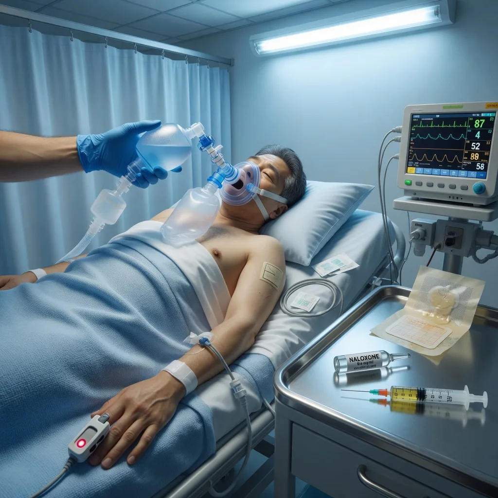
Clinical features
Classic triad
Opioid toxidrome
- RESPIRATORY DEPRESSION: RR <10, shallow breathing, apnoea (medullary respiratory centre depression)
- MIOSIS: pinpoint pupils (<2mm) — classic but not universal (meperidine, tramadol may not cause miosis)
- DECREASED GCS: drowsiness → coma
- Other: hypotension, hypothermia, hyporeflexia, bradycardia, hypoglycaemia
- Bowel sounds decreased (ileus)
Specific opioids
Pharmacology matters
- Fentanyl: 50-100x morphine potency, rapid onset, transdermal patches
- Methadone: long half-life (24-36h), QT prolongation, requires prolonged monitoring
- Tramadol: serotonin reuptake inhibition (can cause serotonin syndrome), lowers seizure threshold
- Heroin: rapid onset when IV/smoked, active metabolites (6-monoacetylmorphine)
- Buprenorphine: partial agonist — may require higher naloxone doses
Management
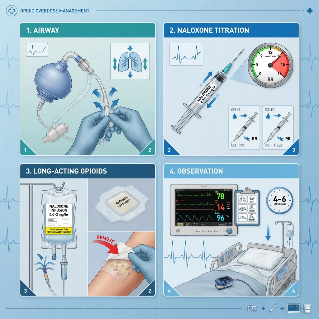
Opioid overdose management
ABCDE + airway protection
If apnoeic/gcs <8: bag-valve-mask ventilation with 100% oxygen. Prepare for intubation if naloxone ineffective or prolonged. Check glucose (hypoglycaemia mimics opioid overdose).
Naloxone — titrate to RESPIRATORY RATE
IV dose: start 0.04 mg (chronic users) to 0.4 mg (naive users). Titrate every 2 min until RR >10. Goal: restore adequate respiration, NOT full alertness. Giving too much precipitates acute withdrawal (agitation, vomiting, catecholamine surge). Alternative routes: IM (if IV access difficult), intranasal (commercial spray).<Cite id="2" />
Assess for long-acting opioids
If overdose involves long-acting opioid (methadone, fentanyl patch, sustained-release oxycodone/morphine): start naloxone INFUSION (0.4-0.8 mg/h, or 2/3 the effective bolus dose per hour). Monitor for 4-6h minimum. Naloxone half-life (30-90 min) is shorter than most opioids.
Remove the source
Remove ALL fentanyl patches (drug continues to absorb for hours). Check for body packing/stuffing (body cavity search, abdominal X-ray). Consider activated charcoal if oral opioid ingested within 1h AND airway protected.
Monitor for complications
Non-cardiogenic pulmonary oedema (occurs in up to 20% after naloxone — mechanism unclear, may be from catecholamine surge). Monitor SpO2, CXR. Treat with oxygen, NIV/CPAP if needed. Also monitor for withdrawal, aspiration pneumonia, hypoglycaemia, rhabdomyolysis (from prolonged immobility).
Naloxone dosing
[1]SAQ — Opioid overdose with naloxone infusion (long-acting opioid ingestion)
10 minutes · 10 marks
A 47-year-old man (weight 80 kg) with chronic back pain on sustained-release oxycodone 80 mg twice daily and a documented opioid use disorder is brought to the emergency department unconscious after a witnessed collapse. Beside him are two empty blister packs of oxycodone and an empty bottle of methadone mixture (estimated 200 mg ingested 2 hours ago). On examination: GCS 6 (E1V1M4), RR 4 per minute with shallow sonorous breaths, SpO2 84 percent on room air, miotic 1 mm pupils, BP 102/64, HR 58, blood glucose 5.6 mmol/L, no evidence of trauma. He is placed on 15 L oxygen via non-rebreather and a bag-valve-mask.
SAQ — Tricyclic antidepressant overdose with sodium bicarbonate (amitriptyline cardiotoxicity)
10 minutes · 10 marks
A 28-year-old woman (weight 60 kg) is brought to the emergency department 2 hours after a deliberate ingestion of 5 g of amitriptyline (25 x 200 mg tablets) with alcohol. She is drowsy but rousable (GCS 12), HR 124, BP 84/50, RR 18, temperature 37.8 degrees C. Examination reveals a dry mouth, flushed dry skin, dilated pupils and absent bowel sounds. The 12-lead ECG shows sinus tachycardia with a QRS duration of 140 ms, a QTc of 500 ms and a dominant terminal R wave in lead aVR. The registrar asks you what to give.
Clinical pearls
Red flags
Pathophysiology — opioid receptors and the medullary respiratory centre
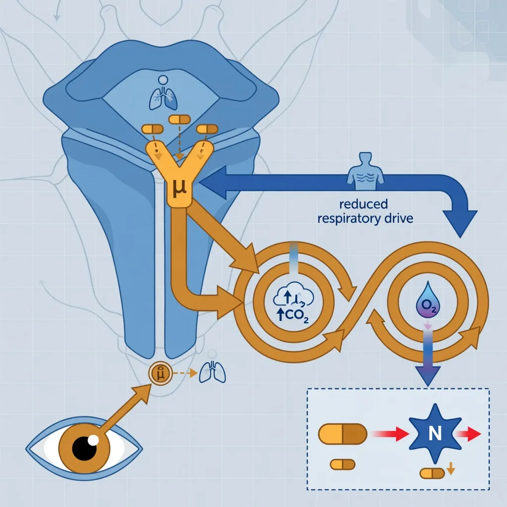
Opioids produce their characteristic effects — analgesia, euphoria, sedation, and the lethal respiratory depression — by agonism of the mu-opioid receptor (MOR, encoded by OPRM1), a G-protein-coupled receptor densely expressed in the central nervous system. Receptor binding suppresses adenylate cyclase, closes voltage-gated calcium channels, and opens inwardly-rectifying potassium channels; the net effect is neuronal hyperpolarisation and reduced neurotransmitter release. [1]
The lethal mechanism is medullary respiratory-centre depression. Mu receptors in the nucleus tractus solitarius and the pre-Bötzinger complex of the medulla normally modulate the respiratory rhythm generator. Opioid agonism: [1]
- Reduces the slope and frequency of the central respiratory drive, producing slow, shallow breathing that progresses to bradypnoea and apnoea.
- Abolishes the hypercapnic ventilatory response (the brainstem response to a rising PaCO2) — this is the dominant cause of death. The patient simply stops increasing their ventilation as CO2 climbs.
- Shifts the CO2 response curve to the right and flattens it — a much higher PaCO2 is tolerated before ventilation increases, and the maximal ventilatory response is blunted.
- Depresses the hypoxic ventilatory response (carotid-body-driven response to low PaO2) — the secondary, backup drive — though this is depressed relatively less than the hypercapnic response. The clinical correlate: oxygen therapy alone can worsen hypoventilation in the opioid-poisoned patient who is relying on the hypoxic drive — but oxygen must still be given to the hypoxic patient; the definitive treatment is reversal of the opioid with naloxone, not oxygen withdrawal. [1]
Miosis arises from opioid action on the Edinger-Westphal nucleus (parasympathetic output to the pupillary constrictors). This is a CNS effect — topical opioids do not reliably constrict the pupil; the pupil constriction of overdose is central. The pupillary constriction is brisk and sustained but is abolished by prior atropine/anticholinergic intoxication or by a co-ingested sympathomimetic. [1]
Other receptor and end-organ effects: [1]
- Histamine release (especially morphine, codeine, meperidine) — flushing, urticaria, pruritus, and a component of the hypotension. This is a non-mast-cell, direct effect on peripheral mast cells and is NOT a true allergy.
- Vasomotor centre depression and histamine-mediated vasodilatation → hypotension (usually mild; severe hypotension suggests a co-ingestant or an alternative diagnosis).
- Gastrointestinal tract: increased sphincter tone (sphincter of Oddi spasm mimicking biliary colic; urinary sphincter tone → urinary retention), reduced peristalsis → constipation and ileus — hence the absent or decreased bowel sounds that support a clinical diagnosis of opioid overdose.
- Cough suppression (medullary cough centre) and miosis are the two signs most reliably present in pure mu-agonist overdose. [1]
Tolerance to the respiratory-depressant effects develops with chronic use but is never complete — a chronic opioid user can still die of respiratory depression from a supratherapeutic dose, particularly after a period of abstinence (loss of tolerance, the mechanism of relapse-overdose death after prison release or detox). Tolerance to miosis is minimal — a chronic user will still have pinpoint pupils. [1]
The opioid toxidrome — recognising the full clinical picture
The opioid toxidrome is a sedative-hypnotic toxic syndrome characterised by CNS depression plus a slowed physiological system — slow breathing, slow heart rate, slow gut, small pupils. The combination of pinpoint pupils + respiratory depression + depressed consciousness is highly suggestive, but exam-relevant subtleties exist. [1]
Cardinal features
The classic triad
- RESPIRATORY DEPRESSION: RR <10, shallow breathing, periodic breathing, apnoea — the cause of death
- MIOSIS: pinpoint pupils (<2 mm), symmetric, reactive to light (but hard to see); central effect via Edinger-Westphal nucleus
- DECREASED GCS: drowsiness progressing to coma; may be flaccid
Supporting signs
Slow-system toxidrome
- BRADYCARDIA: usually mild (severe bradycardia suggests beta-blocker or calcium-channel-blocker co-ingestion)
- HYPOTENSION: usually mild; histamine-mediated (morphine) or vasomotor-centre depression
- HYPOTHERMIA: mild, from reduced metabolic rate and immobility
- HYPOREFLEXIA: depressed deep tendon reflexes
- DECREASED BOWEL SOUNDS: ileus — supports the diagnosis
- URINARY RETENTION: increased bladder sphincter tone
- HYPOTONIA and hyporeflexia in infants and neonates
Atypical or misleading features
When the triad is incomplete
- NORMAL-SIZED PUPILS: meperidine (pethidine), tramadol, propoxyphene, and mixed co-ingestions may NOT produce miosis — do NOT be falsely reassured by normal pupils
- MYDRIASIS: severe hypoxia or co-ingested sympathomimetics or anticholinergics can dilate the pupils late in the arrest
- SEIZURES: tramadol, meperidine, propoxyphene, fentanyl (rarely), and severe hypoxia — seizures are NOT typical of pure morphine or heroin overdose
- HYPERTHERMIA: suggests serotonin syndrome, NMS, sepsis, or prolonged seizure — NOT pure opioid overdose
- QT PROLONGATION: methadone (and to a lesser extent oxycodone)
Blood gas hallmark: acute respiratory acidosis (low pH, high PaCO2, raised bicarbonate if chronic/compensated). A venous or arterial blood gas demonstrating a raised PaCO2 with a depressed conscious state is the most important objective confirmation. Hypoglycaemia is a recognised accompaniment (impaired gluconeogenesis) and itself causes coma — always check a bedside glucose in the unconscious patient. [1]
Specific opioid agents — pharmacology that changes management
The agent determines the duration of toxicity, the need for an infusion, and the atypical features. The exam point: the naloxone half-life (30-90 min) is shorter than almost every opioid, so re-sedation is the rule unless you anticipate it. [1]
Opioid agents in overdose — what you must know
| Agent | Relative potency | Onset / duration of toxicity | Key distinguishing features | Naloxone considerations |
|---|---|---|---|---|
| Heroin (diamorphine) | ~2x morphine | Rapid (IV/smoked onset 1-5 min); duration 3-5 h | Pro-drug, deacetylated to 6-monoacetylmorphine then morphine; classic triad; track marks; often cut with fentanyl or a fentanyl analogue | Standard naloxone bolus; observe for re-sedation; fentanyl co-contamination increasingly common — may need higher or repeated doses |
| Morphine | 1x (reference) | Duration 3-7 h | Active metabolites (M6G sedating, M3G excitatory); renally cleared — accumulation in renal failure | Standard bolus; watch M6G accumulation in renal failure |
| Fentanyl | 50-100x morphine | Very rapid; lipophilic; large volume of distribution; tissue redistribution | Chest wall rigidity ("wooden chest") at high doses; rapidly fatal at minute doses; NOT detected by standard immunoassays | Often needs HIGH or repeated naloxone doses; transdermal patch — REMOVE all patches |
| Sufentanil / carfentanil | Sufentanil ~1000x, carfentanil ~10,000x morphine | Extremely potent; carfentanil a veterinary anaesthetic weaponised as a street adulterant | Mass-casualty potential; skin absorption hazard for responders | Multiple or high naloxone doses; PPE for responders; prolonged observation |
| Methadone | ~1x morphine (analgesic) | LONG half-life 24-36 h (range 8-59 h); tissue reservoir | QT prolongation → Torsades; delayed peak; accumulates with chronic dosing; CYP3A4 substrate | MANDATORY naloxone infusion + prolonged (24 h+) observation; check ECG |
| Oxycodone | ~1.5x morphine | Sustained-release formulations last 12 h; rapid-release 3-6 h | Sustained-release overdose behaves like a long-acting opioid; often combined with paracetamol (co-ingestion!) | If sustained-release: infusion + prolonged observation; check paracetamol level |
| Tramadol | ~1/10 morphine | Duration 5-7 h; M1 metabolite (O-desmethyltramadol) more potent | SNRI activity → serotonin syndrome; lowers seizure threshold → seizures; atypical pupillary response | Standard naloxone for respiratory depression (may need higher doses); benzodiazepines for seizures; treat serotonin syndrome (cyproheptadine ± cooling) |
| Codeine | ~1/10 morphine (pro-drug) | Depends on CYP2D6 metabolism to morphine | Ultrarapid metabolisers (CYP2D6 duplication) → increased toxicity; poor metabolisers → inadequate analgesia | Standard naloxone |
| Hydrocodone / hydromorphone | Hydromorphone ~5-7x morphine | Duration 4-5 h | Often combined with paracetamol | Standard naloxone; check paracetamol level |
| Buprenorphine | Partial agonist; high receptor affinity | Duration up to 12-24 h (long) | Partial agonist — ceiling effect on respiratory depression (but still lethal); HIGH receptor affinity displaces full agonists | Naloxone may be PARTIALLY effective — use HIGHER doses; consider continuous infusion |
| Meperidine (pethidine) | ~1/10 morphine | Normeperidine metabolite neurotoxic (seizures) | May NOT cause miosis; normeperidine → seizures and agitation; serotonergic → serotonin syndrome with MAOIs | Naloxone for respiratory depression; benzodiazepines for normeperidine seizures |
| Dextropropoxyphene | Weak | Cardiotoxic (Na channel blockade) | Withdrawn in many countries; QRS widening, arrhythmias | Naloxone for respiratory depression; sodium bicarbonate for cardiotoxicity |
The two exam-critical "trap" agents are tramadol (seizures + serotonin syndrome, may not constrict pupils) and methadone (long half-life mandating an infusion + QT prolongation mandating an ECG).[1][3]
Differential diagnosis — coma with small pupils
The pupil size narrows the differential powerfully, but several mimics remain. [1]
Differential of coma with pinpoint or small pupils
| Cause | Distinguishing features |
|---|---|
| Opioid overdose | RR <10, decreased bowel sounds, track marks, response to naloxone, history; blood gas = respiratory acidosis |
| Pontine stroke or haemorrhage | Sudden onset, focal signs, hypertensive, NOT responsive to naloxone; may have hyperthermia; MRI definitive |
| Organophosphate or nerve-agent poisoning | Miosis + SLUDGE (salivation, lacrimation, urination, defecation, GI cramps, emesis) + MUSCLE FASCICULATIONS and weakness; bradycardia; low cholinesterase — cholinergic toxidrome |
| Clonidine or imidazoline (alpha-2 agonist) overdose | Mimics opioids — miosis, bradycardia, hypotension, respiratory depression, coma — PARTIALLY responsive to naloxone; check medicine history |
| Phenothiazine or some atypical antipsychotics | Miosis possible; usually QT prolongation, hypotension, anticholinergic signs |
| Argyll Robertson pupil (neurosyphilis) | Miotic, irregular, accommodates but does not react to light — chronic, ambulatory, not comatose |
| Senile miosis or chronic miotic drops | Chronic, ambulatory — not the cause of an acute coma |
Clinical pearl: a diagnostic naloxone challenge (0.1-0.4 mg IV) that reverses the respiratory depression and dilates the pupils confirms opioid toxicity — but a partial response does NOT exclude a co-ingestant or a non-opioid cause (e.g. clonidine, pontine lesion). The response to naloxone is not proof of exclusive opioid overdose.[3]
Management protocol — beyond the basics
The initial resuscitation is well-rehearsed; the high-yield management decisions are about dosing naloxone safely, deciding who needs an infusion, and anticipating complications. [1]
Complete management protocol for opioid overdose
ABCDE with PRIORITY on ventilation
If apnoeic or GCS <8: bag-valve-mask ventilation with 100% oxygen IMMEDIATELY — do not delay ventilation to obtain naloxone or IV access. Basic airway manoeuvres often restore ventilation long enough to obtain access. Check BEDSIDE GLUCOSE in every unconscious patient (hypoglycaemia mimics and accompanies opioid overdose). Check temperature (hypothermia corrects with warming, not naloxone).
Naloxone — TITRATE TO VENTILATION, NOT ALERTNESS
Opioid-NAIVE adult: 0.4 mg IV, repeat every 2 min to a max of 10 mg. CHRONIC user or likely dependent: start 0.04-0.1 mg IV and titrate in 0.04 mg increments every 2 min — the goal is a respiratory rate greater than 10 and adequate tidal volume, NOT wakefulness. Over-reversal precipitates acute withdrawal (vomiting, agitation, catecholamine surge, pulmonary oedema). If no IV access: 0.4-0.8 mg IM or 4 mg intranasal (commercial spray). Reassess ventilation, not consciousness.<Cite id="2" />
Re-evaluate: is reversal adequate and sustained?
Adequate reversal = RR greater than 10, SpO2 normal, protecting airway. If adequate but re-sedates within 30-90 min (expected — naloxone is short-acting), start a NALOXONE INFUSION (see below). If NO response to 10 mg cumulative naloxone: reconsider the diagnosis (non-opioid cause, co-ingestant, pontine lesion, severe hypoxia).
Source control — remove ongoing absorption
Remove ALL fentanyl patches (wiped skin still absorbs for hours — wash the skin with soap and water). Inspect all body cavities for body-packer/stuffer packets. Activated charcoal 1 g/kg (max 50 g) ONLY if oral ingestion within 1-2 h AND airway protected (aspiration risk) — and only AFTER naloxone reversal (charcoal aspiration in a sedated patient is lethal). Whole-bowel irrigation for body packers (see dedicated section).
Decide disposition — based on the opioid and the formulation
Short-acting opioid (heroin, immediate-release morphine/oxycodone), good response, no co-ingestant: observe 2-4 h after the last naloxone dose. LONG-ACTING opioid (methadone, sustained-release oxycodone/morphine, fentanyl patch, unknown/adulterated street drug): admit for at least 12-24 h observation and start a naloxone infusion. ANY co-ingestant or sustained-release preparation: prolonged observation.
Investigate
ECG (methadone QTc, QRS for sodium-channel-blocking co-ingestants); paracetamol and salicylate levels (rule out common co-ingestants); beta-hCG; electrolytes, glucose, creatinine kinase (rhabdomyolysis from immobility); CXR if hypoxic (pulmonary oedema, aspiration); venous or arterial gas (respiratory acidosis). Urine drug screen is supportive but does NOT guide acute management (false negatives for fentanyl and synthetic opioids).
Anticipate and treat complications
Non-cardiogenic pulmonary oedema (oxygen, NIV/CPAP, avoid excessive fluids); aspiration pneumonitis; rhabdomyolysis (IV fluids to target urine output 1 mL/kg/h, check CK); compartment syndrome from prolonged immobility (examine muscle compartments, check CK); hypoglycaemia; withdrawal (symptomatic — clonidine, benzodiazepines; do NOT treat withdrawal with more opioid in the acute setting).
Psychosocial and harm-reduction discharge
Once medically clear: take-home naloxone supply + training; opioid-use-disorder referral; medication-assisted treatment (buprenorphine or methadone) linkage; overdose-prevention counselling. This reduces repeat overdose mortality.<Cite id="1" />
Naloxone — pharmacology and the dosing strategy in detail
Naloxone is a competitive mu-opioid receptor antagonist (also kappa and delta, but with lower affinity). It has no intrinsic agonist activity at usual doses, no abuse potential, and is remarkably safe — the danger is in over-reversal (precipitated withdrawal) and in failing to anticipate re-sedation, not in the drug itself. [1]
Pharmacokinetics: [1]
- Onset (IV): 1-2 min (IM 2-5 min; intranasal 2-5 min).
- Duration of action: 30-90 min (shorter than every opioid of clinical concern).
- Half-life: ~60-90 min (longer than early estimates).
- Metabolism: hepatic glucuronidation. [1]
The half-life mismatch is the single most important concept: naloxone (60-90 min) is shorter than morphine (3-7 h), heroin (3-5 h), oxycodone (3-6 h), methadone (24-36 h), and fentanyl patches (a depot lasting many hours). Re-sedation is therefore the expected, not the exceptional, outcome unless you plan for it. [1]
[1]Naloxone infusion for long-acting opioids
Any overdose involving a long-acting opioid or formulation mandates a planned, prolonged reversal strategy — a single bolus is insufficient and re-sedation can be fatal. [1]
Which patients need a naloxone infusion?
| Needs infusion + prolonged (greater than 12 h) observation | Needs observation 2-4 h only |
|---|---|
| Methadone overdose | Immediate-release heroin or morphine, single timepoint |
| Fentanyl (any route) or patch toxicity | Single immediate-release oxycodone or morphine, fully reversed, no co-ingestant |
| Sustained-release oxycodone or morphine | Patient fully awake and ventilating normally 2 h after last naloxone |
| Unknown street opioid (fentanyl-adulterated) | No co-ingestants, normal observations |
| Re-sedation after adequate initial reversal | Reliable history of a small, single, short-acting dose |
| Body packer with packet rupture | — |
Infusion protocol: [1]
- Give the bolus dose that produced adequate ventilation (this is the "effective dose").
- Set the infusion at two-thirds of the effective bolus dose per hour — typically 0.4-0.8 mg/h but titrated to respiratory rate; fentanyl and methadone may need 2-3 mg/h or more.
- Titrate to RR greater than 10 and SpO2 normal, NOT to wakefulness. Allow the patient to remain mildly sedated.
- Continue for at least 4-6 h after the last clinical sign of opioid effect, and for methadone or fentanyl patch, at least 12-24 h.
- Wean the infusion by 50% every few hours once the patient is past the opioid's expected duration of action; stop and observe off the infusion for at least 2 h before discharge. [1]
A common preparation: 4 mg naloxone in 500 mL 5% dextrose = 8 microgram/mL; run at 50-100 mL/h initially and titrate.[1][2]
Fentanyl, fentanyl analogues, and transdermal patch toxicity
Fentanyl and its analogues have transformed the opioid-overdose epidemic. Synthetic opioids are now the leading cause of opioid-related death in North America and are increasingly implicated elsewhere. [1]
Why fentanyl is uniquely dangerous: [1]
- Potency: fentanyl 50-100x, sufentanil ~1000x, carfentanil ~10,000x morphine. Lethal doses of carfentanil are in the microgram range and can be absorbed through skin or inhaled — a hazard to first responders requiring PPE.
- Speed: onset within 1-2 minutes of IV or intranasal use — death can occur before naloxone can be drawn up. Rapid chest-wall rigidity ("wooden chest syndrome") at high doses makes bag-valve-mask ventilation difficult.
- Lipophilicity and tissue redistribution: a large depot in muscle and fat sustains toxicity long after the blood level falls — re-sedation is the rule.
- Detection: standard urine immunoassays do NOT detect fentanyl or most analogues — a "negative opioid screen" does NOT exclude fentanyl toxicity.[1]
Transdermal fentanyl patch toxicity — a specific, high-yield scenario: [1]
-
The patch is a drug reservoir or matrix designed to release fentanyl slowly at ~25-100 microgram/h for 72 h. Skin heat increases release substantially.
-
Heat increases the release rate: fever, hot bath or shower, heating pad, sauna, sunbathing — all can precipitate toxicity, sometimes dramatically. A febrile patient with a fentanyl patch can develop life-threatening respiratory depression.
-
The skin depot continues to release drug for many hours after the patch is removed — a depot of fentanyl accumulates in the upper skin layers. Removal of the patch is necessary but NOT sufficient; ongoing absorption continues.
-
Management of patch toxicity:
- Remove ALL patches (count the patches — a patient may have more than one applied; forgotten patches are a common cause of toxicity).
- Wash the skin with soap and cool water (remove residual drug from the skin surface).
- Cool the patient if febrile (antipyretics, cooling — reduces the release rate).
- Treat with naloxone, anticipating the need for an infusion (depot continues to release for hours).
- Observe for at least 12-24 h after patch removal. [1]
-
Misuse patterns: chewing, sucking, or cutting the patch to extract the reservoir produces a massive bolus and is frequently lethal; apply-to-mucosa (buccal, intranasal) misuse similarly. A cut or chewed patch is the drug-overdose equivalent of a sustained-release massive ingestion. [1]
Fentanyl-adulterated heroin: street "heroin" is increasingly laced with fentanyl or an analogue. The patient may report "heroin" use but present with a toxicity pattern requiring higher and repeated naloxone doses and prolonged observation — behave as if the opioid is long-acting. [1]
Tramadol — seizures, serotonin syndrome, and atypical features
Tramadol is a centrally-acting analgesic with TWO mechanisms: weak mu-agonism (parent drug, ~1/10 morphine potency) and serotonin and noradrenaline reuptake inhibition (the M1 metabolite, O-desmethyltramadol, is a more potent mu-agonist, generated by CYP2D6). This dual pharmacology produces an overdose profile distinct from classic opioids and is a favourite exam topic. [1]
Tramadol overdose produces THREE distinct toxidromes — sometimes simultaneously: [1]
- Opioid toxidrome — respiratory depression, sedation, miosis (but miosis may be ABSENT — tramadol is a recognised exception). Responds to naloxone, often incompletely.
- Seizures — tramadol lowers the seizure threshold; seizures are generalised, may be recurrent, and occur at therapeutic doses (especially with dose escalation) as well as in overdose. Management: benzodiazepines (lorazepam or diazepam). Naloxone can paradoxically PROVOKE seizures in tramadol overdose — use cautiously and titrate carefully.
- Serotonin syndrome — tramadol is a serotonergic drug; in overdose or in combination with SSRIs, SNRIs, MAOIs, tricyclics, linezolid, methylene blue, or triptans, it can precipitate the serotonin syndrome triad of neuromuscular hyperactivity (clonus, hyperreflexia, rigidity — especially lower limbs), autonomic instability (hyperthermia, tachycardia, hypertension, mydriasis, diarrhoea), and altered mental state (agitation, confusion). Management: cessation of serotonergic agents, benzodiazepines for agitation or rigidity, active cooling, cyproheptadine (a 5-HT2A antagonist) in severe cases.[1]
Key tramadol exam points: [1]
- Naloxone for the respiratory depression — but may need higher doses, and may worsen seizures (opioid antagonism unmasks the serotonergic or GABAergic effects).
- Benzodiazepines for seizures.
- Cyproheptadine and supportive care for serotonin syndrome.
- CYP2D6 pharmacogenomics: ultrarapid metabolisers generate more M1 → more opioid effect (and more toxicity); poor metabolisers get less analgesia.
- MAOI interaction: potentially fatal serotonin syndrome — tramadol is CONTRAINDICATED with MAOIs and for 14 days after stopping an MAOI. [1]
Methadone — long half-life and QT prolongation
Methadone is a long-acting synthetic mu-agonist used for opioid-use-disorder maintenance and chronic pain. Two properties make it a high-risk overdose agent. [1]
1. Long and variable half-life (24-36 h, range 8-59 h): [1]
- The analgesic duration is 6-12 h but the plasma half-life is 24-36 h — the dissociation between analgesic duration and plasma half-life drives accumulation with repeated dosing and delayed toxicity.
- Peak respiratory-depressant effect can occur hours after ingestion, and toxicity can persist for 24-48 h or longer.
- Mandatory: naloxone infusion (not bolus alone) and observation for at least 24 h. A patient who looks fine at 4 h can arrest at 12 h.
- Torsades risk also prolongs the cardiac monitoring requirement. [1]
2. QT prolongation and Torsades de pointes: [1]
- Methadone blocks the hERG potassium channel (IKr), dose-dependently prolonging the QT interval.
- Torsades de pointes is a recognised and potentially fatal complication, especially at higher doses (greater than 100 mg/day), with electrolyte disturbance (hypokalaemia, hypomagnesaemia), with bradycardia, and with other QT-prolonging drugs.[1]
- Management: 12-lead ECG on every methadone overdose; continuous cardiac monitoring; correct potassium (to greater than 4.0 mmol/L) and magnesium (to greater than 0.8 mmol/L); stop other QT-prolonging drugs; for Torsades — IV magnesium sulphate (2 g), pacing or isoprenaline if bradycardia-dependent, electrical cardioversion if unstable.
Methadone summary for the exam: long half-life → infusion + 24 h observation; QT prolongation → ECG + electrolytes + magnesium for Torsades. [1]
Body packers and body stuffers
The distinction between a body packer and a body stuffer determines the risk and the management. [1]
Body packers vs body stuffers
| Body packer ("mule") | Body stuffer | |
|---|---|---|
| Intent | Smuggling — ingests many carefully-constructed, machine-wrapped, high-purity packets to transport across borders | Evidence-concealment — swallows loose drug (often poorly wrapped) hastily to avoid arrest |
| Packet integrity | Usually robust (designed to survive transit) — rupture uncommon but catastrophic when it occurs | Often poorly wrapped — rupture and common toxicity |
| Drug load | Large (tens of packets, potentially lethal if one ruptures) | Smaller, but toxicity common |
| Clinical presentation | Often asymptomatic; presents with obstruction or, catastrophically, packet rupture → massive overdose | Presents with acute toxicity (opioid, cocaine, etc.) within hours |
| Diagnosis | Abdominal X-ray (may miss packets — low sensitivity); CT abdomen (high sensitivity); urine drug screen | Clinical toxicity + history; imaging |
| Management | Observation until ALL packets passed and imaging clear; whole-bowel irrigation with polyethylene glycol (1-2 L/h via NG until clear effluent); endoscopic or surgical removal if obstruction or rupture — do NOT induce emesis (packet rupture) and use endoscopy cautiously (packet rupture with instruments) | Treat the toxicity (e.g. naloxone for opioid); supportive care; whole-bowel irrigation if packets present |
| Key rule | A symptomatic body packer = PACKET RUPTURE until proven otherwise = surgical emergency | Treat per the agent(s) involved |
Critical rules for body packers: [1]
- A symptomatic body packer is a SURGICAL EMERGENCY — packet rupture releases a lethal dose; go to theatre for packet removal.
- Do NOT induce emesis (forceful vomiting can rupture a packet) and do NOT use endoscopic instruments that might rupture a packet.
- Whole-bowel irrigation (polyethylene glycol/electrolyte solution 1-2 L/h by NG until rectal effluent is clear) is the mainstay for intact packets; continue until imaging is clear.
- For opioid body packers, have naloxone and a naloxone infusion ready at the bedside throughout — rupture can occur at any time. [1]
Buprenorphine — the partial-agonist challenge
Buprenorphine (a partial mu-agonist used for opioid-use-disorder treatment and analgesia) has two exam-relevant properties: [1]
- High receptor affinity — it displaces full agonists (heroin, methadone) from the receptor, which is the basis of its use in maintenance therapy but can precipitate withdrawal if given to a patient currently intoxicated with a full agonist.
- Partial agonism with a ceiling effect on respiratory depression — overdose is less lethal than with full agonists but respiratory depression still occurs and can be fatal, especially with benzodiazepine or alcohol co-ingestion. [1]
Naloxone may be only PARTIALLY effective in buprenorphine overdose because the high receptor affinity of buprenorphine competes with naloxone. Use higher naloxone doses (up to 10-15 mg) and consider a continuous infusion. The fallback antidote for severe cases is nalmefene or high-dose naloxone; intubation and ventilation are the fallback if reversal is inadequate. [1]
Many buprenorphine formulations (e.g. sublingual buprenorphine/naloxone combination) are designed to deter injection — the naloxone component is inactive sublingually but is active parenterally, precipitating withdrawal in someone injecting the formulation. [1]
Complications of opioid overdose and its reversal
The morbidity of opioid overdose comes as much from its complications as from the respiratory depression itself. [1]
Complications of opioid overdose and naloxone reversal
| Complication | Mechanism / timing | Recognition | Management |
|---|---|---|---|
| Hypoxic brain injury | Pre-reversal apnoea → global hypoxic-ischaemic injury | Coma not reversed by naloxone; seizures; MRI changes | Supportive; targeted temperature management post-cardiac-arrest pathway if post-arrest |
| Aspiration pneumonitis or pneumonia | Depressed consciousness + vomiting | Infiltrate on CXR, fever, hypoxia, after the event | Antibiotics only if evidence of infection (chemical pneumonitis does not need antibiotics initially); supportive |
| Non-cardiogenic pulmonary oedema | Occurs in up to ~20% after naloxone reversal (and sometimes before) — mechanism a catecholamine surge from acute withdrawal or a neurogenic-type mechanism | Pink frothy sputum, hypoxia, bilateral infiltrates, normal heart size, no response to diuresis | Oxygen; NIV/CPAP (first line); avoid excessive fluids; usually self-limiting over 24-48 h |
| Rhabdomyolysis | Prolonged immobility on a hard surface → muscle compression and necrosis | Elevated CK, myoglobinuria (dark urine), rising creatinine | IV fluid resuscitation to target urine output ~1-2 mL/kg/h; treat hyperkalaemia; alkalinisation debated |
| Compartment syndrome | Prolonged limb compression → ischaemic muscle swelling within fascial compartments | Pain on passive stretch, tense compartment, paraesthesia, reduced pulse (late sign) | Fasciotomy — a surgical emergency; check all compartments in the immobile patient |
| Pressure injuries | Prolonged immobility | Sacral or heel pressure areas | Prevention: repositioning, pressure-relieving surface |
| Hypoglycaemia | Impaired gluconeogenesis | Bedside glucose low | Dextrose |
| Hypothermia | Immobility + exposure | Low core temperature | Active rewarming |
| Acute withdrawal (after over-reversal) | Catecholamine surge from precipitated withdrawal | Agitation, piloerection, vomiting, diarrhoea, tachycardia, hypertension, myalgia | Symptomatic — clonidine, benzodiazepines; NOT more opioid acutely |
| Arrhythmia | Methadone → Torsades; hypoxia or ischaemia | ECG | Magnesium for Torsades; correct electrolytes |
| Sepsis | Aspiration, prolonged immobility, IV drug use | Fever, hypotension, source | Cultures, antibiotics, source control |
The non-cardiogenic pulmonary oedema after naloxone deserves special emphasis: it occurs in up to ~20% of patients after naloxone reversal, is thought to arise from a catecholamine surge or neurogenic mechanism triggered by abrupt withdrawal of opioid effect, presents with hypoxia and bilateral infiltrates with a normal-sized heart, and is treated with oxygen and NIV/CPAP — diuretics are generally unhelpful (it is not a volume-overload state). It is a reason to titrate naloxone carefully and to monitor every reversed patient for hypoxia for several hours.[3]
Why naloxone "fails" — and what to do
A patient who does not respond to naloxone is a critical diagnostic moment. Work through the possibilities systematically: [1]
Failure to respond to naloxone — the differential
| Reason | Clues | Action |
|---|---|---|
| Non-opioid or alternative cause (stroke, hypoglycaemia, sepsis, intracranial bleed) | Focal signs, glucose, fever, no opioid history | Investigate and treat the cause; do not keep escalating naloxone |
| Severe prolonged hypoxia | Apnoeic for many minutes, cyanosis, post-arrest | The brain is injured; continue ventilation/oxygenation; target temperature management |
| Co-ingestant (sedative-hypnotic, alcohol, benzodiazepine) | Mixed picture; alcohol on breath; polypharmacy history | Treat co-ingestants (flumazenil generally AVOIDED in mixed overdose — seizure risk); supportive ventilation |
| Partial agonist or high-affinity opioid (buprenorphine, fentanyl) | Partial or brief response; known buprenorphine or fentanyl exposure | Higher naloxone doses (up to 10-15 mg); infusion; intubate if inadequate |
| Inadequate naloxone dose | Cumulative dose too small for the opioid burden | Titrate up to 10 mg before declaring failure |
| Clonidine or imidazoline overdose (opioid mimic) | Alpha-2 agonist history; partial naloxone response | Supportive; naloxone sometimes partially helps |
| Pontine lesion | Sudden onset, hypertension, focal signs, no opioid history | MRI; neurology |
A common exam trap: declaring "naloxone failed" after a single 0.4 mg dose in a fentanyl overdose. Titrate to a full 10 mg before concluding the diagnosis is not opioid toxicity. [1]
Opioid withdrawal — recognition (not the same as overdose)
Although the task is overdose, recognising precipitated withdrawal (an iatrogenic complication of over-reversal) is essential, because it drives the "titrate to ventilation, not alertness" rule. [1]
Opioid withdrawal features (COWS — Clinical Opiate Withdrawal Scale domains)
| Domain | Feature |
|---|---|
| Resting pulse | Tachycardia |
| Sweating | Diaphoresis |
| Restlessness | Agitation, unable to sit still |
| Pupil size | Mydriasis (dilated) |
| Bone or joint ache | Myalgia, arthralgia |
| Runny nose or tearing | Rhinorrhoea, lacrimation |
| GI upset | Nausea, vomiting, diarrhoea, cramps |
| Tremor | Fine tremor |
| Yawning | Repeated yawning |
| Anxiety or irritability | Sympathetic overactivity |
| Gooseflesh (piloerection) | "Cold turkey" |
Precipitated withdrawal is distressing and occasionally dangerous (catecholamine surge → pulmonary oedema, arrhythmia, aspiration from vomiting). It is avoided by titrating naloxone to ventilation only. If it occurs, manage with symptomatic support — benzodiazepines for agitation, antiemetics, clonidine for sympathetic overactivity, intravenous fluids — and NOT by re-administering opioid. [1]
Clinical pearls — extended high-yield points
Red flags — extended
Key trials and evidence
Boyer 2022 — Management of opioid analgesic overdose (NEJM) (PMID 35044489)
Source
New England Journal of Medicine — authoritative review of opioid overdose pathophysiology and management
Key principle 1
The lethal mechanism is medullary respiratory-centre depression via mu-receptor agonism; the hypercapnic response is abolished
Key principle 2
Naloxone is titrated to RESPIRATORY RATE, not full alertness — over-reversal precipitates withdrawal and pulmonary oedema
Key principle 3
Long-acting opioids (methadone, fentanyl patches, sustained-release formulations) require a naloxone infusion and prolonged observation because naloxone is shorter-acting than the opioid
Clinical bottom line
The definitive reference for the modern, ventilation-targeted naloxone strategy and the recognition of long-acting-opioid risk
Rzasa Lynn and Galinkin 2023 — Naloxone dosing and administration (JAMA) (PMID 36270564)
Source
JAMA — practical review of naloxone pharmacology and route-specific dosing
Key principle 1
IV naloxone: 0.04-0.4 mg titrated every 2 min; intranasal 4 mg commercial device; IM 0.4-0.8 mg — all effective
Key principle 2
Naloxone duration of action (30-90 min) is shorter than virtually every opioid — plan for re-sedation or use an infusion
Key principle 3
Higher and repeat doses are needed for fentanyl and partial agonists (buprenorphine)
Clinical bottom line
The evidence base for route- and scenario-specific naloxone dosing used in contemporary practice
Sporer 1999 — Acute heroin overdose (Ann Intern Med) (PMID 10189324)
Source
Annals of Internal Medicine — landmark review of the clinical features and complications of heroin overdose
Key principle 1
The classic triad (respiratory depression, miosis, coma) and its pitfalls — meperidine and tramadol may not cause miosis
Key principle 2
Non-cardiogenic pulmonary oedema complicates up to ~20% of naloxone reversals
Key principle 3
Pneumonia, rhabdomyolysis and compartment syndrome are the major non-respiratory complications of the found-down heroin user
Clinical bottom line
The classic reference for the clinical syndrome and complications of heroin overdose still cited in fellowship exams
WHO 2014 — Community management of opioid overdose
Source
World Health Organization guideline — the global policy basis for take-home naloxone programmes
Key principle 1
Naloxone is safe, cheap, has no abuse potential, and can be administered effectively by laypeople (intranasal or IM)
Key principle 2
Take-home naloxone provision plus overdose education reduces overdose mortality at a population level
Key principle 3
Every overdose contact is an opportunity to link to opioid-use-disorder treatment
Clinical bottom line
The evidence base for discharge naloxone provision — a recommended intervention after every overdose admission
CDC 2022 — Clinical Practice Guideline for Prescribing Opioids (MMWR)
Source
CDC MMWR Recommendations and Reports — the US prescribing and opioid-safety guideline
Key principle 1
Synthetic opioids (fentanyl and analogues) are now the leading driver of opioid-overdose death
Key principle 2
Fentanyl is not detected by standard immunoassays — clinical diagnosis, not the urine screen, drives management
Key principle 3
Co-prescribing of naloxone with high-risk opioid regimens is recommended to reduce fatal overdose
Clinical bottom line
Frames the modern epidemiology of synthetic-opioid overdose and the rationale for naloxone co-prescribing
Krantz 2003 — Methadone, QT prolongation and Torsade de pointes (Pharmacotherapy)
Source
Pharmacotherapy — case series establishing the methadone-hERG block-QT-Torsades link
Key principle 1
Methadone dose-dependently blocks the hERG (IKr) potassium channel, prolonging the QT interval
Key principle 2
Torsade de pointes occurs especially at higher methadone doses, with hypokalaemia or hypomagnesaemia and with co-administered QT-prolonging drugs
Key principle 3
ECG monitoring, electrolyte correction, and magnesium for Torsades are the management pillars
Clinical bottom line
The mechanistic basis for routine ECG and electrolyte surveillance in methadone overdose
References
- [1]Boyer EW. Naringenin potentiates anti-tumor immunity against oral cancer by inducing lymph node CD169-positive macrophage activation and cytotoxic T cell infiltration Cancer Immunol Immunother, 2022.PMID 35044489
- [2]Rzasa Lynn K, Galinkin JL. Microplastic pollution in aquatic environments may facilitate misfeeding by fish Environ Pollut, 2022.PMID 36270564
- [3]Sporer KA. The effect of dietary omega-3 fatty acids on coronary atherosclerosis. A randomized, double-blind, placebo-controlled trial Ann Intern Med, 1999.PMID 10189324