ICU · Toxicology
Iron, Lead & Heavy Metal Poisoning
Also known as Iron overdose · Lead poisoning · Heavy metal poisoning · Deferoxamine · Chelation · Succimer · Dimercaprol
The heavy-metal poisonings — the iron (the four phases, the deferoxamine), the lead (the abdominal pain, the microcytic anaemia with the basophilic stippling, the peripheral neuropathy, the encephalopathy; the succimer, the EDTA, the dimercaprol), the arsenic (the garlic breath, the GI, the neuropathy, the QT), the mercury (the neuro, the tremor, the GI), the cadmium (the pneumonitis). The common theme the chelation (the deferoxamine for the iron; the dimercaprol/BAL, the succimer/DMSA, the CaNa2EDTA for the lead and the others).
On this page & tools
Your progress
Saved locally on this device.
Target exams
Overview & definition
The heavy-metal poisonings — the iron, the lead, the arsenic, the mercury, the cadmium — share the chelation as the specific therapy (the chelator binds the metal, the forms the water-soluble complex, the excreted). The recognition (the source, the toxidrome) and the timely chelation are the core.[2][1]

The iron
The iron overdose (the deliberate — the adult-supplement tablets, the paediatric — the prenatal vitamins).[1][1]
The four phases.[1]
- The gastrointestinal (the 0 to 6 h) — the nausea, the vomiting, the diarrhoea, the haematemesis (the direct corrosive effect). The volume loss, the dehydration.
- The quiescent (the 6 to 24 h) — the apparent improvement (the danger — the patient may the discharged, then the deteriorate). The deceptive.
- The shock and the metabolic acidosis (the 12 to 24 h) — the shock (the vasodilation — the free iron the stimulates the NO; the volume loss), the metabolic acidosis (the mitochondrial oxidative damage — the lactic, the disruption), the hepatic failure (the free iron the concentrates in the Kupffer cells and the hepatocytes — the necrosis), the coagulopathy. The lethal.
- The gastric scarring (the weeks) — the gastric-outlet obstruction (the fibrotic scarring of the corrosive injury).
- The whole-bowel irrigation (the polyethylene glycol) for the tablets seen on the X-ray (the iron tablets the radiopaque; the charcoal the not-bind).
- The deferoxamine (the desferrioxamine) — the IV chelator; the binds the free iron to the ferrioxamine (the water-soluble, the excreted — the vin-rose / the pinkish-red urine). The continuous infusion for the severe (the level above 500 to 1000, the systemic toxicity).[1]
- The supportive — the fluid, the vasopressor, the treat the hepatic failure, the coagulopathy.
The lead
The chronic exposure (the old paint, the batteries, the occupational — the smelting, the recycling, the shooting-range). The acute encephalopathy (the children).[2][1]
The clinical.[2]
- The abdominal pain, the constipation (the "the lead colic").
- The microcytic anaemia with the basophilic stippling (the lead the inhibits the aminolaevulinic-acid dehydratase and the ferrochelatase — the haem synthesis).
- The peripheral neuropathy (the "the wrist drop" — the motor, the radial nerve).
- The encephalopathy (the children esp.; the adults the cognitive, the headache, the seizures).
- The nephropathy (the chronic tubulointerstitial; the "the lead gout"), the gingival lead line (the blue, the gum-tooth margin), the hypertension.[1]
The treatment — the chelation.[2]
- The succimer (the DMSA) oral — the mild-to-moderate.
- The CaNa2EDTA IV — the moderate-to-severe.
- The dimercaprol (the BAL) IM — the severe (the encephalopathy) — the GIVEN WITH the EDTA (the dimercaprol first to the bind the lead in the blood, then the EDTA to the enhance the excretion; the EDTA alone the redistributes the lead from the soft tissues to the brain).[2]
The arsenic
The sources (the contaminated water, the pesticide, the herbicide, the intentional).[1][1]
- The garlic breath (the characteristic).
- The GI (the nausea, the vomiting, the rice-water diarrhoea), the hypotension, the QT prolongation and the torsades, the encephalopathy, the multi-organ failure.
- The neuropathy (the sensory-motor, the weeks).
- The skin (the chronic — the hyperpigmentation, the palmar-plantar keratosis, the skin cancers).[1]
The treatment. The dimercaprol (the BAL) IM, the succimer (the DMSA), the DMPS — the chelation. The supportive (the QT — the electrolytes, the magnesium).[1]
The mercury
The forms — the elemental (the vapour — the thermometer, the dental amalgam, the occupational), the inorganic (the salts), the organic (the methylmercury — the fish, the Minamata).[3][1]
- The elemental vapour — the neuro (the tremor, the emotional lability / the "the erethism", the cognitive), the GI, the renal, the pulmonary (the pneumonitis).[3]
- The organic — the neuro (the ataxia, the dysarthria, the visual-field constriction), the fetus (the neuro-developmental).
- The inorganic — the GI (the corrosive), the renal.
The treatment. The succimer (the DMSA), the dimercaprol (the BAL) for the inorganic. AVOID the dimercaprol for the elemental vapour (the redistributes to the brain). The succimer the preferred for the long-term.[3]
The cadmium
The inhalation (the welding, the smelting, the "the silver solder").[1]
- The acute — the pneumonitis (the pulmonary oedema, the chemical — the inhalation), the metal fume fever (the self-limiting — the flu-like after the metal-oxide inhalation).
- The chronic — the renal (the tubular — the Fanconi), the bone (the Itai-itai — the osteomalacia), the lung cancer. [1]
The treatment. The supportive (the no effective chelator — the EDTA the nephrotoxic). The remove from the exposure.[1]
Prognosis
The iron poisoning is the survivable with the deferoxamine; the poor-prognostic features the severe acidosis, the shock, the hepatic failure. The lead encephalopathy the serious (the children). The arsenic the QT / the torsades the life-threatening. The mercury the fetal neuro-developmental. The cadmium the chronic — the cancer.[1][2][1]
Red flags
Iron — the examiner-favourite deep dive
Iron is one of the few toxins with a staged, time-dependent clinical course, a radiopaque tablet on plain film, a specific chelator, and a characteristic urine colour that confirms the chelation is working. The Fellowship examiner rewards the candidate who can recite all five stages, the elemental-dose triage threshold, and the three serious adverse effects of deferoxamine.[4][6]
The dose and the elemental-iron calculation
Triage is driven by the mg per kg of elemental iron, not the milligram of the salt. The three oral salts carry very different elemental payloads, and confusing them is the classic exam trap.[4][6]
The oral iron salts — elemental iron content per 325 mg tablet
| Salt | Elemental iron | Typical preparation |
|---|---|---|
| Ferrous sulphate | 65 mg (20%) | Adult haematinic — the commonest overdose |
| Ferrous fumarate | 106 mg (33%) | "Palafer" — high-payload, dangerous in overdose |
| Ferrous gluconate | 38 mg (12%) | Lower payload, but still toxic in large numbers |
| Carbonyl iron | ~100% (elemental) | Poorly absorbed — lower acute toxicity per mg |
| Prenatal / multivitamin | Variable (often 27-65 mg) | The classic paediatric ingestion |
The Manoguerra / EAPCCT-AAPCT consensus thresholds for out-of-hospital triage are: under 20 mg/kg elemental observe at home; 20-40 mg/kg medical evaluation; over 40 mg/kg acute medical assessment with a serum iron level at 4-6 h. Over 60 mg/kg is associated with severe toxicity. A child who ingests a handful of ferrous sulphate can easily exceed 40 mg/kg.[4]
The five-stage clinical course
The classical description is the five-stage model; the bedside relevance is that stage 2 is a trap and stage 3 is the killer.[1][4]
The five stages of acute iron poisoning
| Stage | Timing | Mechanism | Clinical | Action |
|---|---|---|---|---|
| 1 — Gastrointestinal | 0-6 h | Direct corrosive injury of gastric and proximal small-bowel mucosa | Nausea, vomiting, haematemesis, diarrhoea (often bloody), abdominal pain, volume depletion | ABC, IV access, fluids, send 4-6 h level, abdominal X-ray |
| 2 — Quiescent (the deceptive phase) | 6-24 h | Apparent improvement as absorption slows; the patient "looks well" | Resolution of GI symptoms; may look deceptively well for 6-12 h | DO NOT DISCHARGE — observe, monitor for stage 3 |
| 3 — Shock and metabolic acidosis (the killer) | 6-48 h (peak 12-24 h) | Free (unbound) iron — (a) vasodilation via NO and direct vascular smooth-muscle relax-ation; (b) mitochondrial oxidative phosphorylation uncoupling → lactic acidosis; (c) hepatocellular necrosis (Kupffer cells and zone-1 hepatocytes); (d) coagulopathy (inhibited thrombin formation) | Hypotension / refractory shock, high-anion-gap metabolic acidosis, coagulopathy, lethargy, seizures; hepatic failure at 2-5 d | Deferoxamine IV infusion, aggressive shock resuscitation, treat coagulopathy and hepatic failure |
| 4 — Hepatic failure | 2-5 d | Free iron concentrated in the portal system → zone-1 (periportal) hepatocyte necrosis | AST/ALT often >10,000, jaundice, coagulopathy, hypoglycaemia, encephalopathy | Hepatology / transplant unit referral; King's College-style criteria |
| 5 — Gastrointestinal obstruction | 2-6 weeks | Fibrotic scarring and cicatrisation of the corrosive gastric and pyloric injury | Gastric-outlet obstruction, pyloric stricture — recurrent vomiting weeks after the ingestion | Surgical / endoscopic management; the late, deceptive complication |
The stage-3 shock is the direct cause of early death; the stage-4 hepatic failure is the cause of late death. The stage-2 quiescent phase is the medicolegal trap — discharging a patient who is about to develop shock and hepatic failure.[1][4]
The serum iron level — interpretation and timing
The serum iron level peaks at 4-6 h after ingestion and is the cornerstone of triage. A level drawn after 8 h can be falsely reassuring as iron has redistributed into tissues. Use the level together with the clinical picture, never in isolation.[4][6]
Serum iron level (mcg/dL / micromol/L) and the clinical threshold
| Serum iron | Interpretation | Action |
|---|---|---|
| < 300 mcg/dL (< 54 micromol/L) | Unlikely to be toxic | Observe; discharge if asymptomatic at 6 h with normal level |
| 300-500 mcg/dL (54-90 micromol/L) | Mild-to-moderate toxicity | Observe; consider deferoxamine if symptomatic |
| 500-1000 mcg/dL (90-180 micromol/L) | Significant toxicity — chelation threshold | Deferoxamine IV infusion |
| > 1000 mcg/dL (> 180 micromol/L) | Severe — high mortality | Deferoxamine IV + intensive-care support |
The transferrin saturation (iron divided by total iron-binding capacity, TIBC) over 100% indicates free, unbound iron — the toxic fraction. In practice the TIBC assay is unreliable in the acute setting and the serum iron level with the clinical picture drives treatment.[6] The anion gap and the lactate are excellent bedside surrogates for systemic toxicity when the level is pending or unreliable.[4]
The abdominal X-ray — radiopaque tablets and pill concretions
Iron tablets are radiopaque on plain abdominal X-ray (the dense ferrous salt). A film taken early can (a) confirm the ingestion, (b) estimate the tablet load, and (c) detect pill concretions / bezoars — a mass of tablets in the stomach that behaves as a reservoir for ongoing absorption.[5]
Deferoxamine (desferrioxamine) — the chelator
Deferoxamine is a siderophore-derived hexadentate chelator that binds free (unbound) iron — and only free iron; it does not remove iron already bound to transferrin, ferritin or haemosiderin. The ferrioxamine complex is water-soluble and excreted renally, producing the characteristic vin-rose (pinkish-red) urine that confirms active chelation.[1][6]
Dosing: 15 mg/kg/hour IV initially (slow — give the first 90 mg/kg over 6-8 h, then reassess). The older 15 mg/kg/h continuous infusion for 24 h is still used. Hypotension is rate-related — slow the infusion if it occurs. Continue until the patient is clinically stable, the metabolic acidosis has resolved, and the vin-rose urine has cleared (often 12-24 h; longer in massive overdose).[6]
The three serious adverse effects of deferoxamine
| Adverse effect | Mechanism / context | Mitigation |
|---|---|---|
| Hypotension (rate-related) | Rapid infusion → histamine release + direct vasodilation | Slow the infusion rate; give as a controlled infusion over 6-8 h, not as a bolus; fluid resuscitate first |
| Acute respiratory distress syndrome (ARDS) / pulmonary toxicity | Reported with infusions over 24 h; the deferoxamine-iron complex is itself toxic to lung tissue, or deferoxamine chelates iron needed by pulmonary defences | Limit total infusion duration; do not exceed 24 h routinely; watch for hypoxia and new infiltrates |
| Yersinia enterocolitica sepsis | Deferoxamine acts as a siderophore for Yersinia — the bacterium cannot acquire iron on its own but uses ferrioxamine → fulminant sepsis with the tell-tale right-lower-quadrant pain mimicking appendicitis | Maintain a high index of suspicion during and after chelation; culture blood and stool; treat with fluoroquinolone / third-generation cephalosporin; consider empirical cover if febrile |
The Yersinia association is the single highest-yield deferoxamine-adverse-effect exam fact. The mechanism — deferoxamine "feeds" an otherwise iron-starved organism — is the answer to almost every "why does deferoxamine predispose to Yersinia?" question.[6]
When to stop deferoxamine: clinical recovery (resolution of shock and acidosis), the serum iron level falling back toward the reference range, and the disappearance of the vin-rose urine. Do not run the infusion to a fixed clock — run it to the clinical endpoint.[6]
Lead — pathophysiology and chelation deep dive
Lead poisoning is overwhelmingly chronic in adults (occupational, hobbyist, retained bullet fragments, traditional remedies, "surma"/"kohl" cosmetics) and acute in children (pica on leaded paint dust in pre-1970s housing). The paediatric acute encephalopathy is the feared presentation and the one with the highest mortality.[2][11]
Pathophysiology — three enzyme targets in haem synthesis
Lead disrupts haem synthesis at three points, which is why the anaemia is microcytic and why the surrogate markers (ALA, zinc protoporphyrin) are diagnostically useful:[2]
Lead and haem synthesis — the three enzyme targets
| Enzyme | Effect of lead | Consequence | Marker |
|---|---|---|---|
| ALA dehydratase (porphobilinogen synthase) | Inhibited → ALA accumulates | ↑ urinary and plasma delta-aminolaevulinic acid (ALA); ALA is neurotoxic (contributes to encephalopathy and neuropathy) | Urinary ALA (historical) |
| Ferrochelatase | Inhibited → iron cannot be inserted into protoporphyrin → protoporphyrin accumulates | ↑ zinc protoporphyrin (ZPP) / free erythrocyte protoporphyrin; reflects integrated lead exposure over the red-cell lifespan (≈ 120 days) | Blood ZPP |
| (Indirect) — mitochondrial iron-handling | Iron trapped as ZPP → functional iron deficiency in the marrow | Microcytic, hypochromic anaemia with basophilic stippling (residual ribosomal RNA from impaired red-cell maturation) | Blood film |
Basophilic stippling — coarse blue-grey granules on the red cell — is the classic blood-film finding. It arises because lead denatures the enzymes that degrade ribosomes as reticulocytes mature, leaving residual RNA. It is not specific to lead (also seen in thalassaemia, sideroblastic anaemia, pyrimidine-5'-nucleotidase deficiency) but in the right context it is pathognomonic.[2][11]
Blood lead level and clinical correlation
The whole-blood lead level (micrograms per decilitre, mcg/dL) is the diagnostic standard. Levels correlate with the clinical picture and drive the chelation decision.[2]
Blood lead level (mcg/dL) — severity, features and chelation threshold
| Blood lead (mcg/dL) | Adults — features | Children — features | Chelation |
|---|---|---|---|
| < 10 | Reference range (occupational removal often at 30) | Increasing concern; CDC reference value for action is 3.5 | No chelation; remove from exposure |
| 10-25 | Hypertension, mild cognitive effects, reduced libido | Reduced IQ, decreased hearing | Remove from exposure; environmental investigation |
| 25-50 | Abdominal pain, constipation, fatigue, peripheral neuropathy (wrist-drop) | Iron-deficiency-type anaemia, slowed nerve conduction | Succimer (DMSA) oral; EDTA if severe |
| 50-70 | Lead colic, overt neuropathy, anaemia | Symptomatic; early encephalopathy | CaNa2EDTA IV ± succimer |
| > 70 (children > 50) | Encephalopathy signs | Acute lead encephalopathy — ataxia, vomiting, seizures, coma | Dimercaprol IM + CaNa2EDTA IV (combination) |
The CDC reference value for paediatric public-health action is currently 3.5 mcg/dL (the 97.5th percentile of NHANES blood leads in young children). There is no safe blood lead level in a child.[2]
Acute lead encephalopathy — the emergency
Acute lead encephalopathy is a medical emergency with a mortality of 5-25% and a high rate of permanent neurological sequelae in survivors. It presents with ataxia, lethargy, vomiting (the lead-induced ↑ ICP), seizures and coma, typically in a child with a blood lead over 70-100 mcg/dL. The treatment is dimercaprol (BAL) IM FIRST, then CaNa2EDTA IV — the order is non-negotiable.[2]
The chelation drugs for lead — a comparison
Lead chelation drugs — succimer (DMSA), CaNa2EDTA, dimercaprol (BAL)
| Drug | Class / route | Dose | Indication | Key adverse effects |
|---|---|---|---|---|
| Succimer (DMSA) | Water-soluble dithiol, oral | 10 mg/kg TID × 5 d then BID × 14 d | Mild-moderate (blood lead 45-70 mcg/dL); outpatient step-down after parenteral therapy | GI upset, transient transaminase rise, neutropenia (rare); well-tolerated |
| CaNa2EDTA (edetate calcium disodium) | Polyaminocarboxylic acid, IV (IM alternative) | 1000-1500 mg/m²/day × 5 d (continuous or divided) | Moderate-severe (blood lead > 50, no encephalopathy) | Renal toxicity (tubular), trace-metal depletion (zinc, copper, manganese); must be the calcium salt — Na EDTA chelates calcium → fatal hypocalcaemia |
| Dimercaprol (BAL / British anti-Lewisite) | Lipid-soluble dithiol, IM | 75 mg/m² IM q4h × 48 h, then q6-12h | Severe — encephalopathy or blood lead > 70; given before EDTA | Painful injection, fever, tachycardia, nausea/vomiting/hypertension (transient), contraindicated in iron, selenium, cadmium poisoning (forms toxic complexes), G6PD haemolysis, peanut allergy (formulated in peanut oil) |
The single most dangerous pharmacy error is dispensing disodium EDTA (the anticoagulant chelator used in tube collection) instead of calcium-disodium EDTA — disodium EDTA chelates serum calcium and has caused fatal hypocalcaemic arrhythmias. The "EDTA safety" naming conventions (edetate calcium disodium vs edetate disodium) exist precisely because of these deaths.[2][10]
Other clinical features of chronic lead poisoning
- Lead colic — refractory abdominal pain with constipation, classically without signs of peritonism; resolves with chelation, not analgesia.
- Peripheral neuropathy — motor > sensory; wrist-drop (radial nerve) is the classic adult occupational finding; foot-drop and extensor weakness of the fingers follow.
- Nephropathy — chronic tubulointerstitial disease with Fanconi-type proximal tubular dysfunction (glycosuria, aminoaciduria, phosphaturia); gout ("saturnine gout") from lead-induced hyperuricaemia.
- Reproductive — reduced sperm count, reduced libido, miscarriage.
- Cardiovascular — hypertension (lead directly increases vascular smooth-muscle tone and reduces NO availability).
- Gingival "lead line" — a blue-grey line at the gum-tooth margin, seen only in those with poor dental hygiene; reflects lead sulphide deposition. Exam trap: the line reflects exposure duration and hygiene, NOT the blood lead level.[2][11]
Arsenic — acute, chronic and chelation
Arsenic poisoning remains a major global health problem from contaminated groundwater (the Ganges delta, parts of South America and the south-western US), occupational exposure (smelting, semiconductor manufacture), pesticides and herbicides, and the intentional ingestion (the classic "inheritance powder"). Acute ingestion is dramatic; chronic exposure is insidious and carcinogenic.[7]
Acute arsenic poisoning
Acute ingestion produces a severe gastroenteritis with rapid progression to multi-organ failure within hours. The garlic breath and the QT prolongation with torsades are the two highest-yield exam facts.[7]
Acute arsenic poisoning — system by system
| System | Features | Mechanism |
|---|---|---|
| GI (within 1 h) | Severe nausea, vomiting, profuse rice-water diarrhoea (cholera-like), abdominal cramping, haemorrhagic gastritis | Direct mucosal toxicity; massive capillary transudation |
| Cardiovascular | Hypotension, refractory shock, QT prolongation and torsades de pointes | Myocardial depression + prolonged repolarisation (arsenic inhibits myocardial repolarising channels); volume loss |
| CNS | Encephalopathy, seizures, coma | Direct neurotoxicity; cerebral oedema from capillary leak |
| Renal / hepatic | Acute tubular necrosis, hepatic failure (later) | Direct tubular and hepatocellular toxicity |
| Haematological | Pancytopenia (days later) | Bone-marrow suppression |
| Skin / hair | Garlic breath and garlic body odour | Methylated arsenic metabolites excreted via lungs and skin |
The QT prolongation and torsades is the single most feared cardiac complication. Treat aggressively with IV magnesium sulphate (2 g over 10 min) for torsades, isoprenaline infusion and overdrive pacing for refractory cases, and meticulous correction of potassium and magnesium — arsenic-induced torsades is notoriously provoked by hypokalaemia and hypomagnesaemia.[7]
Chronic arsenic poisoning — the dermatological and neurological signature
Chronic exposure (drinking-water, occupational) produces a characteristic, slowly-evolving syndrome with skin, neurological, cardiovascular and carcinogenic features.[7]
Chronic arsenic poisoning — the classic features
| Feature | Description | Timing |
|---|---|---|
| Skin — hyperpigmentation | "Raindrop" bronze hyperpigmentation, especially trunk and palms | Months |
| Skin — palmoplantar keratosis | Punctate thickened hyperkeratotic patches on palms and soles | Months-years |
| Skin cancers | Bowen disease (intraepidermal SCC), basal-cell carcinoma, squamous-cell carcinoma — arsenic is a class-1 carcinogen | Decades (latency 10-40 years) |
| Mee's lines (Aldrich-Mees lines) | Transverse white bands across all nails — appear 4-6 weeks after acute or chronic exposure; reflect transient arsenic-induced nail-matrix toxicity during a growth-arrest episode | 4-6 weeks after the exposure peak |
| Peripheral neuropathy | Symmetrical sensorimotor, distal, painful — often the dominant chronic symptom | Weeks-months |
| Cardiovascular | "Blackfoot disease" (endemic peripheral vascular disease / gangrene in Taiwan); hypertension; ischaemic heart disease | Years |
| Malignancy | Cancers of skin, lung, bladder, kidney, liver — even decades after exposure ceases | Decades |
Mee's lines are the chronic analogue of Aldrich-Mees lines (synonyms). They are pathognomonic in the right context but also appear in thallium poisoning, severe chemotherapy-induced cytopenia, carbon monoxide, mees lines of trauma ( Beau's lines — nail trauma, not arsenic). The distinction from thallium is critical: thallium produces a painful ascending neuropathy PLUS alopecia; arsenic produces Mee's lines but NOT alopecia.[7]
Arsenic chelation
- Dimercaprol (BAL) IM — the traditional first-line for acute, severe arsenic; given 3-5 mg/kg IM every 4-12 h.
- Succimer (DMSA) oral — preferred for subacute and chronic; better tolerated than BAL.
- DMPS (2,3-dimercapto-1-propanesulfonate, unithiol) — water-soluble dithiol available orally and IV in many countries; the modern agent of choice where available; better adverse-effect profile than BAL.[7][10]
Supportive care is decisive: aggressive volume resuscitation for the cholera-like GI losses, vasopressors for shock, magnesium and potassium replacement, QT-prolongation management, and renal-replacement therapy for the acute tubular necrosis.[7]
Mercury — the three forms and the chelation rules
Mercury exists in three chemical forms with completely different toxicokinetics, target organs, and chelation rules. Confusing them is the single most common and dangerous exam error.[3][8]
The three forms of mercury — source, target organ and chelation
| Form | Source / exposure | Target organ | Clinical | Chelation |
|---|---|---|---|---|
| Elemental (Hg⁰) — the vapour | Broken thermometer, sphygmomanometer, dental amalgam, "gold mining" amalgam, fluorescent lamps; the vapour is inhaled | Brain (lipid-soluble vapour crosses BBB), lung, kidney | Erethism (anxiety, irritability, emotional lability, the "mad hatter"), intention tremor, gingivitis, salivation, acute chemical pneumonitis from heavy vapour exposure, renal tubular injury | Succimer (DMSA) or DMPS. AVOID dimercaprol (BAL) — it redistributes elemental mercury TO the brain and worsens the neurotoxicity |
| Inorganic (Hg⁺, Hg²⁺ salts) — mercuric chloride, mercurous chloride (calomel, in old skin-lightening creams and traditional remedies) | Ingestion of salts; occupational (fur/hat felting historically) | GI tract (corrosive), kidney (acute tubular necrosis) | Severe corrosive gastroenteritis, haemorrhagic colitis, acute renal failure, tremor, erethism | Dimercaprol (BAL) IM is acceptable, or succimer / DMPS. BAL is NOT contraindicated here (no elemental-vapour-to-brain redistribution) |
| Organic (methylmercury, ethylmercury) — methylmercury in predatory fish (tuna, swordfish, shark), Minamata disease; ethylmercury in thimerosal preservative | Diet (bioaccumulated in fish), industrial discharge into waterways | Brain (especially the developing fetal brain) and sensory cortex | Ataxia, dysarthria, constricted visual fields (tunnel vision), paraesthesiae, hearing loss; fetal neuro-developmental injury (cerebral palsy, mental retardation) at far lower doses than the maternal syndrome | Succimer (DMSA) or DMPS — long courses; AVOID dimercaprol (same brain-redistribution concern). Treat the source (stop the fish). Whole-blood mercury is the preferred sample (methylmercury is in red cells) |
Erethism and the "mad hatter"
Erethism (from Greek erethizo, to irritate) is the constellation of insomnia, memory loss, emotional lability, extreme shyness, irritability, and pathological fear of criticism — the personality change of chronic elemental-mercury vapour exposure. Combined with the intention (mercurial) tremor it produces the historical "mad hatter" of Danbury, Connecticut, where 19th-century felt-hat workers used mercuric nitrate to cure felt. The tremor is fine, regular, abolished by movement onset then exaggerated by sustained posture, and dramatically worsened by emotional stress.[3][8]
Minamata disease
The paradigm organic-mercury epidemic: industrial discharge of methylmercury into Minamata Bay, Japan (1956), bioaccumulated in fish, then consumed by the local population. Adults developed ataxia, dysarthria, constricted visual fields and paraesthesiae; children exposed in utero developed catastrophic cerebral palsy and mental retardation at maternal doses that produced no maternal symptoms — the fetal brain is the most mercury-sensitive tissue in the body. The epidemic established methylmercury as a transplacental neurotoxin.[3][8]
Sample collection and monitoring
The whole-blood mercury reflects organic (methylmercury) exposure (methylmercury is concentrated in red cells); the urinary mercury reflects inorganic and elemental exposure. Hair mercury reflects integrated long-term organic exposure (like a retrospective exposure diary, segmented by hair length). Use the right sample for the suspected source.[11]
The chelation pharmacology — the one-stop reference
Chelation is the unifying specific therapy for the heavy metals. Each chelator has a chemistry (the number and arrangement of its donor atoms — O, N, S), a metal preference (driven by HSAB theory: hard acids bind hard bases), a route, and a characteristic adverse-effect profile.[10]
The chelators — chemistry, metals, route and key adverse effects
| Chelator | Chemistry | Metals chelated | Route | Key adverse effect |
|---|---|---|---|---|
| Deferoxamine (desferrioxamine, DFO) | Siderophore derivative; hexadentate (6 donor atoms, mostly O) | Iron (free, unbound) — also aluminium | IV (IM in chronic iron overload) | Hypotension (rate), ARDS (>24 h infusion), Yersinia enterocolitica sepsis (siderophore effect) |
| Deferasirox | Oral tridentate (3 O donors) | Iron (chronic transfusional overload) | Oral | Renal, hepatic, GI; not used in acute overdose |
| Succimer (DMSA — 2,3-dimercaptosuccinic acid) | Water-soluble dithiol (2 S donors) | Lead (oral, outpatient), mercury (elemental, organic), arsenic | Oral | Well-tolerated; transient transaminase rise, GI upset, neutropenia (rare) |
| DMPS (unithiol — 2,3-dimercapto-1-propanesulfonate) | Water-soluble dithiol | Mercury (all forms), arsenic, lead | Oral and IV | Well-tolerated; not universally licensed (available under named-patient in many countries) |
| Dimercaprol (BAL — British anti-Lewisite) | Lipid-soluble dithiol | Lead (severe, with EDTA), arsenic, inorganic mercury | IM only | Painful injection, fever, tachycardia, hypertension, vomiting; contraindicated in iron, cadmium, selenium (forms toxic complexes) and in elemental/organic mercury (redistributes to brain); peanut allergy (peanut-oil vehicle); G6PD haemolysis |
| CaNa2EDTA (edetate calcium disodium) | Polyaminocarboxylic acid | Lead (moderate-severe, no encephalopathy); historically cadmium | IV / IM | Renal tubular; trace-metal depletion (zinc, copper, manganese, iron); NEVER use disodium EDTA — fatal hypocalcaemia |
| Penicillamine | Monothiol (copper) | Copper (Wilson disease), lead (oral, mild), mercury | Oral | Nephrotic syndrome, aplastic anaemia, hypersensitivity; cross-reacts with penicillin allergy |
What NOT to chelate — the metals with no effective chelator
| Metal | Chelator status | Why |
|---|---|---|
| Cadmium | No effective chelator | EDTA is nephrotoxic and may worsen cadmium nephropathy; BAL forms a toxic Cd-BAL complex. Management is supportive + remove from exposure |
| Iron already bound (transferrin, ferritin) | Deferoxamine only chelates FREE iron | The chelator must catch iron in the unbound, toxic state — once tissue-bound, little can be removed |
| Elemental / organic mercury | BAL contraindicated | BAL redistributes mercury to the brain — use succimer / DMPS |
Iron overdose — the management algorithm
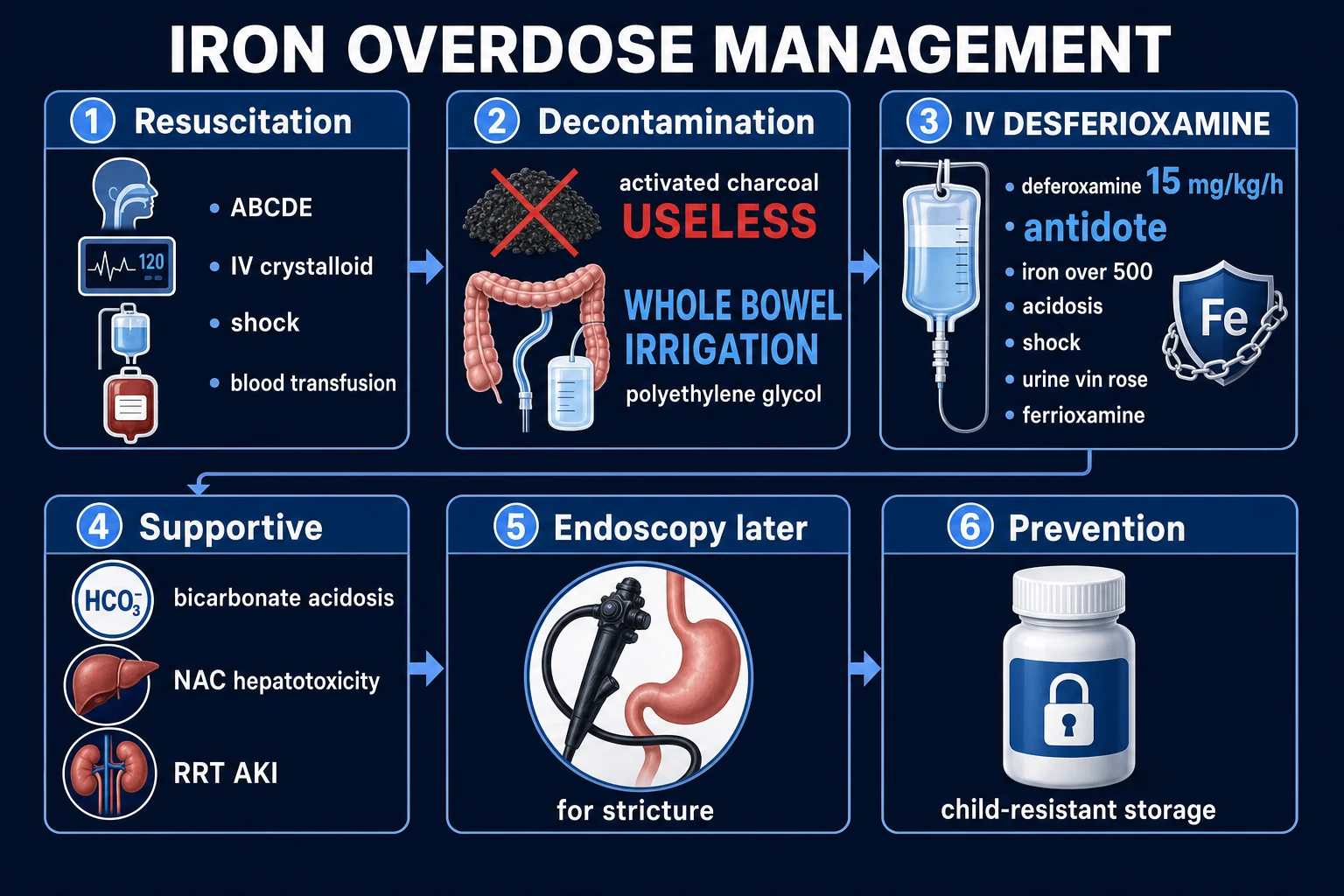
The acute iron ingestion — a step-by-step management pathway
1
2
3
4
5
6
7
8
9
10
Lead encephalopathy — the emergency algorithm
Acute lead encephalopathy (child or adult) — the chelation sequence
1
2
3
4
5
6
7
Arsenic acute poisoning — the algorithm
Acute arsenic ingestion — from resuscitation to chelation
1
2
3
4
5
6
The toxicology references — trial cards
Intentional iron overdose in pregnancy — management and outcome
Journal of Emergency Medicine (2000)
Case series
Population: Pregnant women with intentional iron overdose
Key finding
Established that deferoxamine is SAFE in pregnancy and that withholding chelation for fear of fetal effects is the greater danger; the maternal phase-2 quiescent window still applies and observation through it is mandatory.
Iron poisoning — a literature-based review of epidemiology, diagnosis, and management
Pediatric Emergency Care (2011)
Narrative review
Population: Paediatric and adult iron ingestions
Key finding
Codified the five-stage clinical course, the elemental-iron dose triage, and the central role of the 4-6 h serum iron level and deferoxamine; the most-cited modern review of iron poisoning.
The role of whole bowel irrigation in the treatment of toxic ingestions
British Journal of Clinical Pharmacology (2023)
Position-statement review (EAPCCT)
Population: Toxic ingestions amenable to mechanical gut decontamination
Key finding
Reaffirmed whole-bowel irrigation with polyethylene glycol as the decontamination of choice for sustained-release and enteric-coated iron, for iron-tablet concretions (bezoar), and for any radiopaque-tablet ingestion where charcoal is ineffective.
Acute and chronic arsenic toxicity
Postgraduate Medical Journal (2003)
Narrative review
Population: Acute and chronic arsenic exposure
Key finding
The definitive single-author synthesis of arsenic toxicology — garlic breath, rice-water diarrhoea, QT prolongation, Mee's lines, palmoplantar keratosis, skin and visceral cancers; the chelation hierarchy (BAL then succimer/DMPS).
Mercury toxicity and treatment — a review of the literature
Journal of Environmental and Public Health (2012)
Narrative review
Population: Elemental, inorganic and organic mercury exposure
Key finding
Established the form-specific chelation rules: succimer/DMPS for elemental and organic mercury; the BAL contraindication for elemental mercury on the basis of brain redistribution; the fetal neuro-developmental catastrophe of Minamata.
A review of pitfalls and progress in chelation treatment of metal poisonings
Journal of Trace Elements in Medicine and Biology (2016)
Narrative review
Population: Metal poisonings and chelation therapy
Key finding
Synthesised the modern chelator pharmacology — HSAB-theory matching, the BAL contraindications (iron, cadmium, selenium, elemental mercury), the disodium-EDTA fatal-hypocalcaemia error, and the rise of succimer and DMPS as better-tolerated oral chelators.
SAQ — Iron overdose with five-stage clinical course
10 minutes · 10 marks
A 3-year-old girl (14 kg) is brought to ED 3 hours after the babysitter found her with an open bottle of prenatal ferrous sulphate 325 mg tablets; ~30 tablets missing. She has vomited three times (including tablet fragments) and is now drowsy. HR 130, BP 88/55, capillary refill 4 s, abdomen mildly tender. Serum iron pending. Abdominal X-ray shows multiple radiopaque tablets in the stomach.
SAQ — Acute arsenic poisoning
10 minutes · 10 marks
A 35-year-old agricultural worker presents 4 hours after ingesting an unknown amount of arsenic trioxide pesticide with severe abdominal pain, profuse rice-water diarrhoea and vomiting. He is hypotensive (BP 76/40), tachycardic (HR 132), and confused. ECG shows QTc 540 ms with T-wave inversion. The breath smells of garlic.
Clinical pearls — the 22 high-yield facts
Mnemonics
The iron five stages — 'Go Quiet, Shock Hurts, Obstruct'
Heavy-metal chelation — the metal-to-drug pairings
Garlic-breath poisons — 'PASTA-D'
What BAL must NOT be given for — 'ICEM'
Additional red flags
Additional answer card — the deep-dive synthesis
References
- [1]Minocha A, et al. Intentional iron overdose in pregnancy--management and outcome J Emerg Med, 2000.PMID 10699527
- [2]Bradberry S, Vale A. Lead toxicity and chelation therapy Am J Health Syst Pharm, 2007.PMID 17189579
- [3]Bose-O'Reilly S, et al. Acute mercury intoxication and use of chelating agents J Biol Regul Homeost Agents, 2009.PMID 20003760
- [4]Chang TP, Rangan C Iron poisoning: a literature-based review of epidemiology, diagnosis, and management Pediatr Emerg Care, 2011.PMID 21975503
- [5]Tenenbein M, Lheureux P The role of whole bowel irrigation in the treatment of toxic ingestions Br J Clin Pharmacol, 2023.PMID 36639859
- [6]Mann KV, Picciotti MA, Spevack TA, Durbin M Management of acute iron overdose Clin Pharm, 1989.PMID 2663331
- [7]Ratnaike RN Acute and chronic arsenic toxicity Postgrad Med J, 2003.PMID 12897217
- [8]Bernhoft RA Mercury toxicity and treatment: a review of the literature J Environ Public Health, 2012.PMID 22235210
- [9]Risher JF, De Rosa CT Inorganic: the other mercury J Environ Health, 2007.PMID 18044248
- [10]Andersen O, Aaseth J A review of pitfalls and progress in chelation treatment of metal poisonings J Trace Elem Med Biol, 2016.PMID 27150911
- [11]Barlow NL, Bradberry SM Investigation and monitoring of heavy metal poisoning J Clin Pathol, 2023.PMID 36600633