ICU · Transplant / pharmacology
Post-Transplant Immunosuppression Complications — Toxicity, PTLD & Metabolic
Also known as Post-transplant immunosuppression complications · Calcineurin inhibitor toxicity · Tacrolimus nephrotoxicity · PRES · Posterior reversible encephalopathy · PTLD · Post-transplant lymphoproliferative disorder · Steroid diabetes · EBV lymphoma · Induction therapy · Basiliximab · Antithymocyte globulin · Mycophenolate · Sirolimus · Therapeutic drug monitoring
Post-transplant immunosuppression complications: the immunosuppression principles (induction — ATG/basiliximab/alemtuzumab; maintenance — tacrolimus + mycophenolate ± steroids; rescue for rejection), the drug pharmacology (the tacrolimus — the nephrotoxicity, the neurotoxicity [PRES], the diabetes; the ciclosporin — the nephrotoxicity, the hypertension, the gingival hyperplasia; the mycophenolate — the leucopenia; the azathioprine — the TPMT; the sirolimus — the wound healing, the pneumonitis; the steroids), the calcineurin inhibitor toxicity, the infection (the opportunistic — the CMV, the PCP, the EBV/PTLD), the metabolic (the steroid), the malignancy (the PTLD — the EBV-driven; the skin cancer — the SCC), the TDM (the tacrolimus the trough the levels), and the CYP3A4 drug interactions. The management: the balance the immunosuppression (the rejection vs the toxicity), the reduce/switch the agent, the monitor the levels.
On this page & tools
Your progress
Saved locally on this device.
8 MCQs with explanations
Target exams
Overview & definition
The post-transplant immunosuppression complications — the price of the graft survival. Four categories: the calcineurin inhibitor toxicity, the infection (the opportunistic), the metabolic, and the malignancy (the PTLD). Each requires the balance of the immunosuppression (the reduce the toxicity without the triggering the rejection).[1]
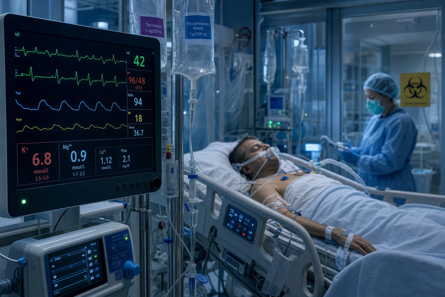
Immunosuppression is delivered in three conceptual phases — induction (intense, peri-operative, to prevent early rejection), maintenance (lifelong, lower intensity, to preserve the graft), and rescue/antirejection (intensified therapy for a rejection episode). Every transplant recipient the intensivist encounters is on some combination of agents drawn from these phases, and every ICU complication — infection, drug toxicity, malignancy, graft dysfunction — must be interpreted through the lens of which agents, at what intensity, for how long. Halloran (2004) provides the canonical framework for classifying these drugs by their mechanism and target in the T-cell activation cascade.[1]
The complications
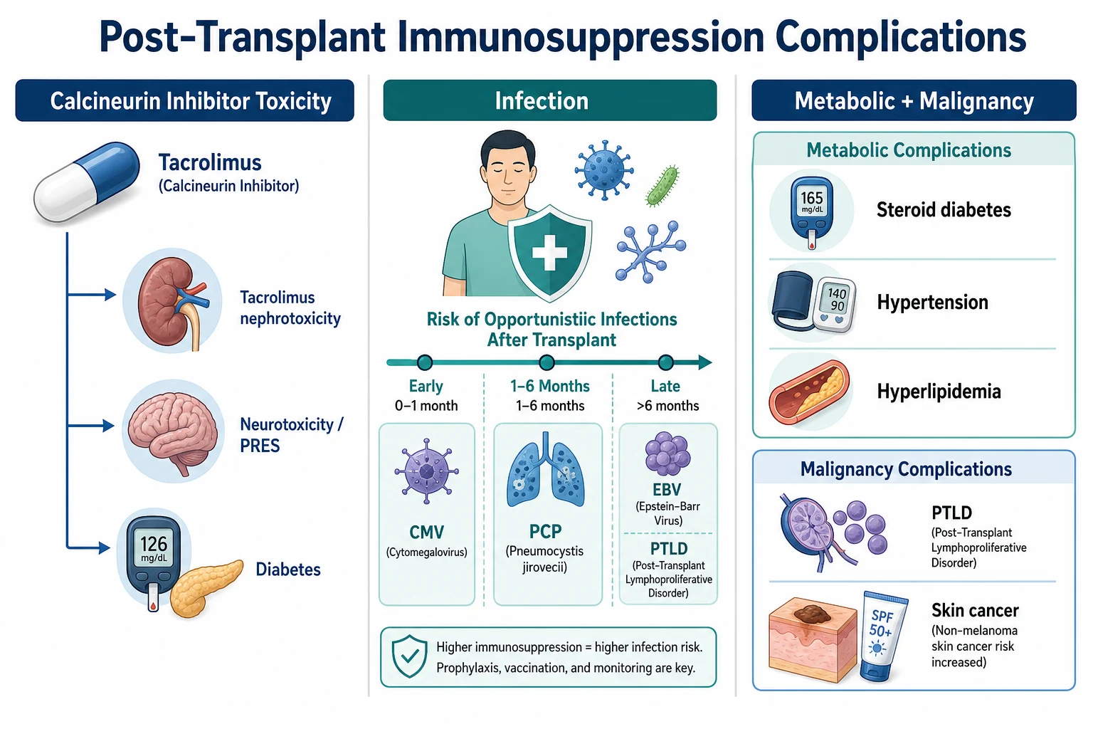
Calcineurin inhibitor toxicity
- The tacrolimus — the nephrotoxicity (the afferent the arteriolar the vasoconstriction; the AKI; the chronic — the interstitial the fibrosis), the neurotoxicity (the PRES — the posterior the reversible the encephalopathy the syndrome; the seizures, the visual, the headache; the MRI), the diabetes (the beta-cell the toxicity), the hyperkalaemia.[1]
- The ciclosporin — the nephrotoxicity, the hypertension, the gingival the hyperplasia, the hirsutism.[1]
Opportunistic infection
- The CMV (the fever, the leucopenia, the colitis, the pneumonitis; the valganciclovir).[1]
- The PCP (the cotrimoxazole).[1]
- The EBV → the PTLD (the B-cell the lymphoma; the driven by the EBV; the risk the higher the with the T-cell the depleting the agents).[1]
Metabolic
- The steroid — the diabetes, the hypertension, the hyperlipidaemia, the osteoporosis, the adrenal the suppression.[1]
- The mTOR — the hyperlipidaemia, the proteinuria, the impaired the wound the healing, the pneumonitis.[1]
Malignancy
- The PTLD (the post-transplant the lymphoproliferative the disorder) — the EBV-driven the B-cell the lymphoma. The risk higher with the T-cell the depleting agents (the ATG, the OKT3). The diagnosis: the tissue the biopsy (the CD20, the EBV-EBER). The treatment: the reduce the immunosuppression (the first-line), the rituximab (the anti-CD20), the chemotherapy (the refractory).[1]
- The skin cancer (the SCC — the immunosuppression; the sun the protection).[1]
Immunosuppression principles — induction, maintenance, and rescue
A working knowledge of how transplant immunosuppression is constructed is essential before one can take it apart at the bedside in the ICU. The modern regimen is built around the calcineurin inhibitor (CNI) cornerstone — almost always tacrolimus — paired with an antimetabolite — almost always mycophenolate — and a variable dose of corticosteroid. This triple therapy suppresses T-cell activation at three distinct points: signal 1 (antigen recognition), signal 2 (co-stimulation), and signal 3 (cytokine-driven proliferation/differentiation).[1]
The T-cell activation cascade — where each immunosuppressant acts
| Signal / target | What happens | Drug class | Prototypical agent |
|---|---|---|---|
| Signal 1 — antigen recognition | T-cell receptor binds antigen presented on MHC; calcineurin dephosphorylates NF-AT, which enters the nucleus to drive IL-2 transcription | Calcineurin inhibitors | Tacrolimus (binds FKBP-12), ciclosporin (binds cyclophilin) |
| Signal 2 — co-stimulation | CD28 on the T-cell binds B7 (CD80/86) on the antigen-presenting cell; without it the T-cell becomes anergic | Co-stimulation blocker | Belatacept (CTLA4-Ig fusion protein) |
| Signal 3 — cytokine response / proliferation | IL-2 drives T-cell proliferation (via mTOR) and clonal expansion | Antimetabolites (inhibit purine synthesis → block DNA synthesis) and mTOR inhibitors | Mycophenolate, azathioprine; sirolimus/everolimus |
| Depletion / lymphocyte reduction | Non-specific removal of T-cells (and sometimes B-cells) at induction | Depleting antibodies | Antithymocyte globulin (ATG), alemtuzumab |
| Non-depleting blockade | IL-2 receptor blockade on activated T-cells | IL-2 receptor antagonists | Basiliximab, daclizumab |
| Broad anti-inflammatory | Globally dampens cytokine transcription, migration, effector function | Corticosteroids | Methylprednisolone, prednisolone |
Induction therapy — the intense peri-operative phase
Induction is a short course of potent antibody therapy given at or around the time of transplantation to profoundly suppress the recipient's immune response during the period of maximum graft immunogenicity (ischaemia-reperfusion injury, donor antigen load). Induction is used in nearly all deceased-donor transplants today and is divided into depleting (lymphocyte-depleting polyclonal or monoclonal antibodies) and non-depleting (IL-2 receptor antagonists) strategies.[1]
- Antithymocyte globulin (ATG) — polyclonal antibodies raised in rabbits (rATG, thymoglobulin) or horses against human thymocytes. Depleting. Causes prolonged lymphopenia (months). Used for high immunological risk (sensitised recipients, re-transplant, African ancestry, ABO-incompatible) and for CNI-minimisation protocols (allowing delayed CNI introduction when there is delayed graft function). Brennan et al. (NEJM 2006) showed rATG reduced acute rejection and CMV disease compared with basiliximab in high-risk renal recipients, at the cost of more infection and malignancy.[2]
- Alemtuzumab (Campath-1H) — monoclonal anti-CD52 antibody causing profound, prolonged depletion of lymphocytes and monocytes. Single-dose induction. Hanaway et al. (NEJM 2011) showed lower rejection rates than basiliximab in low-risk and rATG in high-risk recipients, but no graft-survival advantage and a trend to more late infection — it is used selectively, not universally.[3]
- Basiliximab — chimeric monoclonal anti-CD25 (IL-2 receptor α-chain) antibody. Non-depleting. Two doses (day 0 and day 4). Far fewer side effects than depleting agents; used in low-immunological-risk recipients. Does not cause cytokine release, leucopenia, or significant infection.
- OKT3 (muromonab) — first-generation murine anti-CD3 monoclonal. Largely abandoned due to severe cytokine release syndrome and a high PTLD risk; included for historical/exam context.
Induction agents — depleting vs non-depleting
| Agent | Class | Mechanism | Depletion | Typical use | Key adverse effects |
|---|---|---|---|---|---|
| rATG (thymoglobulin) | Polyclonal Ab | Anti-T-cell antibodies → complement/ADCC lysis | Yes (profound, lasting months) | High-risk recipients; CNI-minimisation; delayed graft function | Cytokine release, serum sickness, leucopenia, thrombocytopenia, infection, PTLD |
| Alemtuzumab | Monoclonal anti-CD52 | Depletes lymphocytes + monocytes | Yes (profound, lasting >1 year) | Single-dose induction; steroid-free protocols | Cytokine release, profound lymphopenia, late infection, autoimmune disease (ITP, thyroid) |
| Basiliximab | Monoclonal anti-CD25 (IL-2R) | Blocks IL-2 receptor on activated T-cells | No | Low-risk recipients | Minimal — hypersensitivity, rare cytokine release |
| OKT3 (muromonab) | Murine anti-CD3 | Binds CD3 → blocks TCR signalling | Yes | Largely obsolete | Severe cytokine release syndrome, pulmonary oedema, aseptic meningitis, high PTLD risk |
Maintenance therapy — lifelong, lower intensity
Maintenance is the lifelong regimen that keeps the graft viable. The contemporary standard across most solid organs is tacrolimus + mycophenolate ± low-dose prednisolone. The choice of regimen is tailored by organ and by patient risk (nephrotoxicity, metabolic, infection, malignancy, pregnancy intent).[1]
Typical maintenance regimens by organ
| Organ | Backbone | Antimetabolite | Steroid | Notes |
|---|---|---|---|---|
| Kidney | Tacrolimus | Mycophenolate | ± (many steroid-free/withdrawal) | Steroid withdrawal risks rejection in some; belatacept an alternative to avoid CNI nephrotoxicity |
| Liver | Tacrolimus | Mycophenolate | ± (early withdrawal common) | Liver less CNI-toxicity-sensitive; tacrolimus preferred over ciclosporin |
| Heart | Tacrolimus or ciclosporin | Mycophenolate | Yes (usually continued) | Higher immunosuppression than kidney/liver; mTOR sometimes added |
| Lung | Tacrolimus | Mycophenolate or azathioprine | Yes | Highest immunosuppression burden; highest infection/malignancy risk |
| Pancreas | Tacrolimus | Mycophenolate or azathioprine | Yes | Often combined with kidney (SPK); sirolimus sometimes used |
Rescue (antirejection) therapy
When biopsy-proven rejection occurs, immunosuppression is intensified (rescue therapy). The approach depends on the rejection mechanism: [1]
- T-cell mediated (cellular) rejection — first-line is high-dose corticosteroid pulse (methylprednisolone 250–1000 mg IV daily for 3 days); steroid-resistant cases receive depleting antibody (rATG).
- Antibody-mediated (humoral) rejection — plasmapheresis to remove donor-specific antibodies, intravenous immunoglobulin (IVIG), rituximab (anti-CD20, to deplete B-cells), and sometimes bortezomib (anti-plasma cell) or complement blockade (eculizumab). Webster et al. (Cochrane 2017) reviewed antibody-based treatment of acute rejection episodes.[11]
Drug pharmacology — agent by agent
This is the core knowledge the examiner expects. Each agent has a mechanism, a target level, a characteristic toxicity profile, and a set of interactions. Know all four for every drug. [1]
Tacrolimus (FK506) — the cornerstone calcineurin inhibitor
- Mechanism — binds FKBP-12 (an immunophilin); the complex inhibits calcineurin, preventing dephosphorylation of NF-AT and so blocking IL-2 transcription → no T-cell activation. (Ciclosporin binds a different immunophilin, cyclophilin, but converges on the same calcineurin target.)
- Toxicity profile (the exam staples):
- Nephrotoxicity — afferent arteriolar vasoconstriction (acute, reversible, dose/level-dependent) → rising creatinine; chronic interstitial fibrosis and tubular atrophy with long-term use (a major cause of late graft loss). Synergistic with other nephrotoxins (aminoglycosides, NSAIDs, contrast, amphotericin) and with sepsis-related AKI.
- Neurotoxicity — tremor (very common), headache, insomnia; severe forms include posterior reversible encephalopathy syndrome (PRES), seizures, and coma. See the dedicated PRES section below.[9]
- Diabetes mellitus — direct pancreatic β-cell toxicity; new-onset diabetes after transplant (NODAT); dose-dependent; partly reversible on dose reduction.
- Electrolytes — hyperkalaemia (type 4 RTA picture), hypomagnesaemia, hyperuricaemia/gout.
- TDM — trough level monitoring (see TDM section). Therapeutic window is narrow.
- Interactions — CYP3A4 and P-glycoprotein substrate (see interactions table).
Cyclosporin (ciclosporin) — the original CNI
- Mechanism — binds cyclophilin → calcineurin inhibition (same downstream effect as tacrolimus, different binding protein).
- Toxicity profile — the distinctive ones the examiner loves:
- Gingival hyperplasia — hallmark, cosmetically significant; related to the drug's effect on gingival fibroblasts.
- Hirsutism — increased hair growth.
- Hypertension — more pronounced than tacrolimus, via vasoconstriction and sodium retention.
- Nephrotoxicity — same afferent arteriolar vasoconstriction and chronic fibrosis as tacrolimus.
- Neurotoxicity — PRES also occurs, though slightly less than tacrolimus.
- Cosmetic burden (gingival hyperplasia, hirsutism) is a major reason ciclosporin has been displaced by tacrolimus as first-line in most organs.
- Bioequivalence — the microemulsion formulation (Neoral) has more predictable absorption than the original Sandimmune. [1]
Tacrolimus vs cyclosporin — the head-to-head exam comparison
| Feature | Tacrolimus | Ciclosporin |
|---|---|---|
| Binding protein | FKBP-12 | Cyclophilin |
| Target | Calcineurin | Calcineurin |
| Nephrotoxicity | Yes (acute + chronic) | Yes (acute + chronic) — comparable |
| Neurotoxicity / PRES | Yes (more tremor; PRES) | Yes (slightly less) |
| New-onset diabetes | More (β-cell toxicity) | Less |
| Hypertension | Less | More |
| Dyslipidaemia | Less | More |
| Gingival hyperplasia | No | Yes — hallmark |
| Hirsutism | No | Yes |
| Alopecia | Yes | No |
| Current role | First-line in most organs | Second-line; used where tacrolimus not tolerated |
Mycophenolate mofetil (MMF) — the dominant antimetabolite
- Mechanism — prodrug of mycophenolic acid; reversibly inhibits inosine monophosphate dehydrogenase (IMPDH), the rate-limiting enzyme in de novo purine synthesis. Lymphocytes rely on the de novo pathway (unlike most cells, which can use salvage), so MMF selectively inhibits lymphocyte proliferation.
- Toxicity profile:
- Leucopenia / cytopenia — dose-dependent marrow suppression (leucopenia, anaemia, thrombocytopenia); monitor CBC; dose-reduce or switch to azathioprine.
- Gastrointestinal — diarrhoea, nausea, vomiting, gastritis; may improve with the enteric-coated formulation (mycophenolate sodium).
- Teratogenicity — contraindicated in pregnancy (switch to azathioprine pre-conception); required pregnancy prevention programmes.
- Infection — contributes to overall immunosuppression burden.
- TDM — generally not routine (unlike CNIs); guided by clinical response and CBC. [1]
Azathioprine — the older antimetabolite (and its TPMT trap)
- Mechanism — prodrug converted to 6-mercaptopurine (6-MP) then to thioguanine nucleotides, which incorporate into DNA and inhibit purine synthesis. Non-selective (affects all dividing cells).
- Toxicity profile:
- Myelosuppression — the big one; especially dangerous in patients with TPMT deficiency (see below).
- Hepatotoxicity — veno-occlusive disease, cholestasis.
- Pancreatitis.
- Increased malignancy with long-term use.
- TPMT (thiopurine methyltransferase) — the critical pharmacogenomic safety check. TPMT inactivates 6-MP. About 0.3% of people are homozygous deficient (no enzyme) — they cannot metabolise 6-MP, accumulate lethal thioguanine levels, and develop severe, potentially fatal myelosuppression with standard dosing. ~10% are heterozygous (intermediate activity, need dose reduction). Check TPMT activity/genotype BEFORE starting azathioprine. Deficient patients must receive a drastically reduced dose (or a different agent).
- The lethal interaction — azathioprine + allopurinol. Allopurinol inhibits xanthine oxidase (XO), the other route of 6-MP inactivation. Combining them causes 6-MP accumulation → profound pancytopenia. Reduce azathioprine dose by 70–75% if allopurinol is essential, or switch mycophenolate. This is a classic exam question and a real-world fatal error. [1]
Sirolimus / everolimus — the mTOR inhibitors
- Mechanism — bind FKBP-12 (like tacrolimus) but the complex inhibits mTOR (mammalian target of rapamycin), blocking the response of T-cells to IL-2 (signal 3 — proliferation) rather than IL-2 production. Therefore synergistic with, and non-nephrotoxic alternative to, calcineurin inhibitors.
- Toxicity profile (the exam favourites):
- Impaired wound healing — surgical wound dehiscence, lymphocele, incisional hernia; avoid in the early post-operative period (typically hold >1 month post-transplant). Manzia et al. (2020) confirmed delayed (vs early) everolimus introduction reduces wound complications.[10]
- Hyperlipidaemia — significant hypertriglyceridaemia and hypercholesterolaemia.
- Proteinuria — can unmask or worsen proteinuria; monitor urine PCR.
- mTOR pneumonitis — interstitial pneumonitis, a rare but important differential in the dyspnoeic transplant recipient.
- Mouth ulcers / stomatitis.
- Myelosuppression (anaemia, thrombocytopenia, leucopenia).
- Clinical niche — CNI-sparing (to avoid nephrotoxicity), anti-proliferative (reduces malignancy risk, e.g. in transplant recipients with skin cancer or Kaposi sarcoma), and in cardiac transplant to slow vasculopathy.
Corticosteroids — the broad anti-inflammatory backbone
- Mechanism — bind glucocorticoid receptor → broadly suppress cytokine transcription (TNF, IL-1, IL-2, IL-6), lymphocyte migration, and effector function.
- Toxicity profile — the familiar list:
- Diabetes mellitus (steroid-induced hyperglycaemia/NODAT).
- Hypertension, fluid retention.
- Dyslipidaemia.
- Osteoporosis and avascular necrosis (especially femoral head).
- Adrenal suppression — the critical ICU point: chronic steroid recipients cannot mount a stress response; in critical illness convert to IV hydrocortisone (e.g. 50–100 mg IV tds or a stress-dose infusion) and never abruptly stop.
- Myopathy (critical-illness and steroid myopathy overlap in the ICU).
- Infection susceptibility, peptic ulcer disease (synergistic with NSAIDs), cataracts, glaucoma, mood/psychosis, hyperglycaemia.
- Steroid withdrawal/minimisation is common (especially kidney, liver) to reduce metabolic toxicity, but carries a rejection risk — weigh per recipient. [1]
Therapeutic drug monitoring (TDM) — levels, sampling, and the narrow window
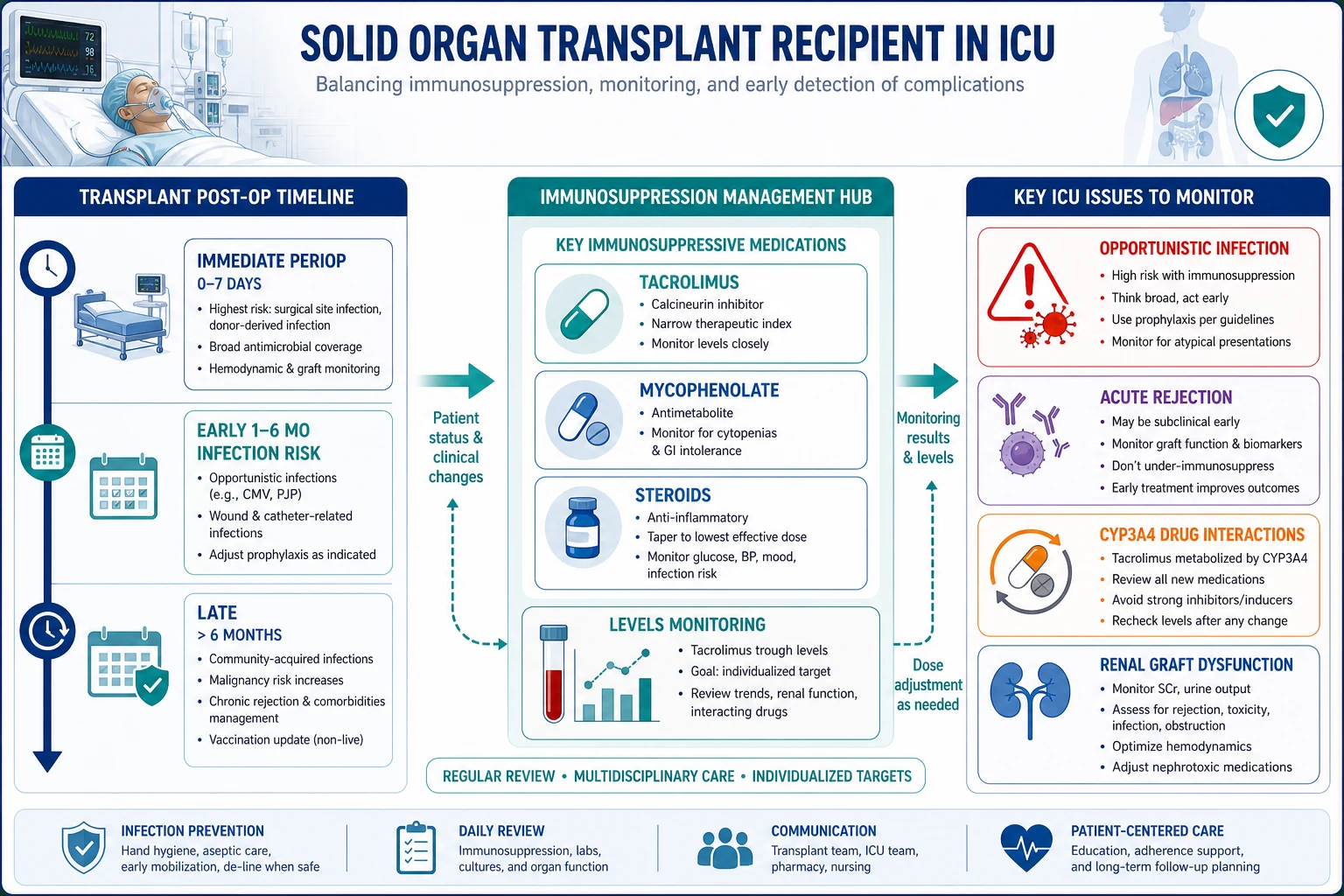
Tacrolimus and ciclosporin have narrow therapeutic windows and enormous inter-patient variability in absorption and metabolism — TDM is mandatory. Sirolimus and everolimus are also monitored (slower onset, longer half-life). Mycophenolate and azathioprine are generally not routinely monitored (guided by clinical effect and CBC). [1]
Therapeutic drug monitoring — targets and sampling
| Drug | Sample | Timing | Typical trough target (kidney) | Why monitor |
|---|---|---|---|---|
| Tacrolimus | Whole blood (EDTA) | Trough — just before the next dose (C0) | Early post-transplant 8–12 ng/mL; maintenance 4–8 ng/mL (lower in CNI-minimisation) | Narrow window; sub-therapeutic → rejection; supra-therapeutic → nephro/neurotoxicity |
| Ciclosporin | Whole blood | Trough (C0) or C2 (2 h post-dose) | C0 100–300 ng/mL (varies) | Same narrow window rationale |
| Sirolimus | Whole blood | Trough (C0) | 4–12 ng/mL (alone); 3–8 ng/mL (with reduced CNI) | Long half-life (~60 h); take 5–7 days to steady state |
| Everolimus | Whole blood | Trough (C0) | 3–8 ng/mL | Similar to sirolimus |
| Mycophenolate | Plasma | Not routine (some centres use MPA AUC) | — | Guided by CBC and clinical response |
| Azathioprine | — | None (TPMT pre-dose + CBC) | — | Marrow toxicity via CBC |
Managing tacrolimus levels in the ICU — a practical sequence
- DRAW THE LEVEL CORRECTLY — a true trough (C0), taken immediately before the next dose, in an EDTA whole-blood tube. A level drawn at the wrong time is misleading — high if too soon after the dose, falsely low if delayed. Record the dose and time on the request.
- INTERPRET IN CONTEXT — correlate the level with the clinical picture, not the number alone. A "therapeutic" level in a patient with rising creatinine and tremor still suggests toxicity; a low level with graft dysfunction suggests rejection. Levels guide, they do not decide.
- CHECK FOR INTERACTIONS FIRST — before changing the tacrolimus dose, review every drug added or stopped in the last 1–2 weeks (see interactions table). A newly started clarithromycin or fluconazole will raise the level dramatically; simply stopping the interacting drug may be the fix rather than altering the tacrolimus dose.
- ADJUST THE DOSE IN SMALL INCREMENTS — tacrolimus dose changes produce non-linear level responses; adjust by ~25% and re-check in 2–3 days (sooner if toxicity is severe).
- CONSIDER THE ROUTE — absorption is via the gut; diarrhoea, vomiting, nasogastric administration, or gut oedema all alter levels. In the critically ill with gut failure, consider IV tacrolimus (continuous infusion, ~1/3 of the oral dose, with close level monitoring).
- MONITOR END-ORGANS — alongside levels, track creatinine/eGFR (nephrotoxicity), magnesium and potassium (CNI-induced renal wasting), glucose (NODAT), CBC (if on mycophenolate/azathioprine), and neurological examination (tremor, PRES).
- COMMUNICATE WITH THE TRANSPLANT TEAM — dose changes, drug switches, and especially holding immunosuppression must be made with the transplant unit, who hold the long-term context and the organ-specific targets.
CYP3A4 and P-glycoprotein drug interactions — the single highest-yield ICU topic
Tacrolimus and ciclosporin are metabolised by CYP3A4 and are substrates for P-glycoprotein. Almost every ICU drug interaction in a transplant recipient flows from this. The rule: CYP3A4 inhibitors RAISE CNI levels → toxicity; CYP3A4 inducers LOWER CNI levels → rejection. Always check, always involve a transplant pharmacist.[1]
CYP3A4 drug interactions with calcineurin inhibitors — the ICU reference
| Effect on CNI level | Mechanism | Common ICU culprits | Consequence |
|---|---|---|---|
| RAISES the level → TOXICITY | CYP3A4 / P-gp inhibition | Macrolides (clarithromycin, erythromycin — not azithromycin); azoles (fluconazole, voriconazole, itraconazole, ketoconazole, posaconazole); diltiazem, verapamil, nicardipine; amiodarone; protease inhibitors / cobicistat; grapefruit juice; metoclopramide | Nephrotoxicity, neurotoxicity/PRES, hyperkalaemia, diabetes |
| LOWERS the level → REJECTION | CYP3A4 / P-gp induction | Rifampicin (potent — always expect a big drop); phenytoin, carbamazepine, phenobarbitone; St John's wort; efavirenz, nevirapine; high-dose glucocorticoids (transiently) | Sub-therapeutic CNI → acute rejection, graft loss |
| Safe alternatives | Minimal CYP3A4 effect | Azithromycin (instead of clarithromycin); β-lactams, vancomycin, aminoglycosides, linezolid, doxycycline; antifungal terbinafine (or topical agents) | No significant level perturbation |
- Practical rules: (1) When you must use a strong inhibitor (e.g. fluconazole for fungal prophylaxis), anticipate the rise — pre-emptively reduce the tacrolimus dose by ~50% and re-check the level in 48–72 h. (2) When you start rifampicin, expect a dramatic fall — increase the tacrolimus dose substantially (often 3–5×) and re-check in 2–3 days; switch to an alternative anti-TB agent if possible. (3) Use azithromycin, not clarithromycin/erythromycin, when a macrolide is needed. (4) Ban grapefruit juice in transplant recipients. [1]
The infection timeline — opportunistic risk over time
The single most useful framing of infection in a transplant recipient is the timeline — the dominant pathogens change in a predictable sequence because the net state of immunosuppression and the epidemiological exposures evolve. Fishman's classic framework divides infection into three periods.[1]
The post-transplant infection timeline — what to suspect when
| Period | Time after transplant | Dominant infection pattern | Typical pathogens | Rationale |
|---|---|---|---|---|
| 1 — First month | 0–30 days | Nosocomial / donor-derived / surgical | Wound/line/urinary infection, donor-derived infection (rare), anastomotic leak, C. difficile | Same nosocomial flora as any post-operative ICU patient; immunosuppression not yet maximal; technical/surgical complications dominate |
| 2 — 1 to 6 months | 1–6 months | OPPORTUNISTIC (the classic transplant period) | CMV, EBV→PTLD, PCP, Listeria, Toxoplasma, Aspergillus, Cryptococcus, mycobacteria, BK virus, HSV, VZV | Peak net immunosuppression; opportunistic pathogens that require cell-mediated immunity to control emerge; this is the period prophylaxis targets |
| 3 — Late (>6 months) | >6 months | Community-acquired + late opportunistic | CAP, UTI, influenza; Cryptococcus, late CMV, PTLD, chronic viral hepatitis (HBV/HCV); Nocardia, Rhodococcus | Immunosuppression reduced toward maintenance; infection resembles the general community plus a residual risk of opportunists in those over-immunosuppressed |
The period-2 opportunists — the ICU must-knows
- Cytomegalovirus (CMV) — the most important opportunistic pathogen in solid-organ transplant. Syndrome: fever, malaise, leucopenia/thrombocytopenia, tissue-invasive disease (colitis with diarrhoea/blood, pneumonitis, hepatitis, retinitis). Diagnosis: CMV PCR (quantitative), tissue biopsy with immunohistochemistry. Highest risk in D+/R− mismatch (donor positive, recipient negative). Prophylaxis: valganciclovir for 3–6 months (longer in high-risk D+/R−). Hodson et al. (Cochrane 2008) confirmed antiviral prophylaxis reduces CMV disease.[7]
- Pneumocystis jirovecii pneumonia (PCP/PJP) — diffuse bilateral pneumonitis, hypoxia out of proportion to examination, elevated LDH, characteristic "ground-glass" CT. Prophylaxis: trimethoprim–sulfamethoxazole (cotrimoxazole) daily for 6–12 months (longer in lung transplant). Stern et al. (Cochrane 2014) confirmed cotrimoxazole prophylaxis prevents PCP in non-HIV immunocompromised patients.[8]
- EBV → PTLD — see dedicated section; risk peaks 1–6 months with T-cell-depleting induction.
- Listeria monocytogenes — meningitis/encephalitis, bacteraemia; food-borne (soft cheese, deli meats).
- Aspergillus — invasive pulmonary aspergillosis (especially lung transplant, neutropenia, heavy immunosuppression); galactomannan, CT halo/air-crescent signs.
- BK polyomavirus — nephropathy (kidney transplant), rising creatinine; diagnosis by urine cytology (decoy cells), plasma BK PCR; management is immunosuppression reduction (no proven antiviral — ciprofloxacin and cidofovir have limited evidence).
- Cryptococcus — pneumonia or meningitis (often indolent); late as well as period 2.
Routine prophylaxis after solid-organ transplant
| Pathogen | Prophylactic agent | Duration (typical) | Notes |
|---|---|---|---|
| PCP (Pneumocystis) | Trimethoprim–sulfamethoxazole (cotrimoxazole) | 6–12 months (lifelong in lung) | Dapsone or atovaquone if sulpha-allergic |
| CMV | Valganciclovir (or IV ganciclovir) | 3–6 months (longer D+/R−) | Universal or pre-emptive depending on centre |
| Oropharyngeal candidiasis | Nystatin mouthwash or fluconazole | 1–3 months | |
| HSV | Aciclovir (often via CMV prophylaxis) | Variable | Reactivation prevention |
| HBV (if recipient at risk) | Antiviral (entecavir/tenofovir) | Per protocol |
Acute rejection — cellular vs antibody-mediated
Graft dysfunction in a transplant recipient demands consideration of rejection alongside infection, drug toxicity, and surgical/structural causes. The diagnosis is tissue biopsy (kidney — Banff classification; liver, heart, lung each have their own schemes). Rejection is fundamentally divided by mechanism.[1][1]
T-cell mediated (cellular) vs antibody-mediated (humoral) rejection
| Feature | T-cell mediated rejection (TCMR) | Antibody-mediated rejection (AMBR) |
|---|---|---|
| Effector | T-cells (CD4+, CD8+) attacking graft parenchyma and vasculature | Preformed or de novo donor-specific antibodies (DSAs) binding graft endothelium |
| Onset | Days to weeks; commonest form overall | Often early (preformed Ab — hyperacute/accelerated) or late (de novo DSA) |
| Histology | Tubulitis, interstitial inflammation, endotheliitis (Banff grading IA/IB/IIA/IIB/III) | Capillary inflammation, glomerulitis, C4d deposition in peritubular capillaries; microthrombi |
| Serology | No specific antibody | Donor-specific anti-HLA antibodies (DSA) detectable |
| First-line treatment | High-dose IV corticosteroid pulse (methylprednisolone 250–1000 mg daily × 3 days) | Plasmapheresis + IVIG ± rituximab (anti-CD20); bortezomib / eculizumab in refractory |
| Steroid-resistant | Depleting antibody (rATG) | Intensified apheresis, rituximab, proteasome inhibitor |
Diagnostic approach to graft dysfunction in the ICU — the four questions
- IS IT REJECTION, INFECTION, DRUG TOXICITY, OR STRUCTURAL? — rising creatinine (kidney), abnormal LFTs (liver), falling ejection fraction (heart), worsening gas exchange (lung). Distinguish: drug levels (toxicity), inflammatory markers and cultures (infection), ultrasound Doppler (vascular/anastomotic problems), biopsy (rejection). Often more than one coexist.
- EXCLUDE A SURGICAL / STRUCTURAL CAUSE FIRST — ultrasound with Doppler for vascular thrombosis, obstruction (hydro), collection/abscess; for the kidney, exclude urinary obstruction and volume depletion. Avascular necrosis of the graft from vascular thrombosis is a surgical emergency.
- CHECK THE DRUG LEVELS AND THE INTERACTIONS — tacrolimus level, recent drug changes, nephrotoxin co-administration. CNI nephrotoxicity is common and reversible with dose reduction — do not biopsy before excluding it.
- BIOPSY IF UNEXPLAINED OR TO CONFIRM REJECTION — the gold standard. Kidney: percutaneous, Banff-classified. Distinguishes TCMR (treat with steroid), AMBR (treat with apheresis/IVIG/rituximab), CNI toxicity (striped interstitial fibrosis), and recurrent disease. Always involve the transplant team before biopsy and before changing immunosuppression.
Calcineurin inhibitor toxicity — deep dive (nephrotoxicity and PRES)
CNI nephrotoxicity — the most common drug-induced kidney injury in transplant recipients
CNI nephrotoxicity is the rule, not the exception, and exists in acute and chronic forms:[1]
- Acute — afferent arteriolar vasoconstriction (the CNIs upregulate endothelin and downregulate nitric oxide/prostacyclin) → reduced glomerular blood flow → falling eGFR, rising creatinine, often with hyperkalaemia and hyperuricaemia. Reversible with dose reduction. Concentration-dependent.
- Chronic — interstitial fibrosis and tubular atrophy ("striped fibrosis"), arteriolar hyalinosis. Irreversible; a leading cause of late graft loss. Driven by cumulative exposure and by episodes of acute toxicity plus ischaemia.
- Management — reduce the CNI dose (monitor the level); address cofactors (volume status, avoid NSAIDs/aminoglycosides/contrast); consider CNI-minimisation (switch the antimetabolite to mTOR, or convert to belatacept, the co-stimulation blocker). Vincenti et al. (NEJM 2016) showed belatacept-based regimens preserve renal function and reduce nephrotoxicity versus ciclosporin-based regimens, with a higher cardiovascular-event-free survival — at the cost of higher early acute rejection and a small PTLD risk in EBV-seronegative recipients (belatacept is contraindicated in EBV-seronegative patients).[5][6]
PRES — posterior reversible encephalopathy syndrome (tacrolimus neurotoxicity)
PRES is the dramatic neurotoxic manifestation of calcineurin inhibitors (tacrolimus > ciclosporin). Bartynski and Boardman (2008) defined its fundamental imaging and clinical features.[9]
- Clinical features — headache, visual disturbance (cortical blindness, hemianopia), seizures (often the presenting feature), altered consciousness, nausea/vomiting. May occur at "therapeutic" levels — toxicity is idiosyncratic and not strictly level-dependent, though high levels increase risk.
- Imaging — MRI (more sensitive than CT) shows vasogenic oedema, predominantly in the parieto-occipital white matter (the posterior circulation has less sympathetic autoregulation, hence vulnerability). T2/FLAIR hyperintensity. Usually bilateral, symmetrical. Less commonly frontal, basal ganglia, brainstem, cerebellum. DWI differentiates vasogenic (PRES) from cytotoxic (infarct) oedema.
- Reversibility — typically fully reversible (clinically and radiologically) on CNI reduction or switch — hence "reversible" in the name. Persistent deficits suggest infarction or haemorrhage (PRES can haemorrhage in ~15%).
- Differentials to exclude — CNS infection (lumbar puncture, MRI with contrast), CNS malignancy (PTLD, metastases), posterior circulation stroke, hypertensive encephalopathy, electrolyte disturbance (the transplant recipient often has hyponatraemia, hypomagnesaemia).
- Management — reduce or switch the CNI (reduce tacrolimus; or switch tacrolimus↔ciclosporin, or convert to belatacept/mTOR); control seizures (levetiracetam — avoid enzyme-inducers that perturb immunosuppression); control blood pressure; correct electrolytes. [1]
PTLD — post-transplant lymphoproliferative disorder
PTLD is a spectrum of abnormal lymphoid proliferation driven by Epstein–Barr virus (EBV) in the setting of pharmacological immunosuppression. It is the most serious malignancy specific to transplantation and one of the few potentially reversible with immunosuppression reduction alone.[1][1]
Pathogenesis and risk
EBV infects B-cells and normally is held in check by EBV-specific cytotoxic T-cells. Immunosuppression — particularly T-cell-depleting agents (rATG, OKT3, alemtuzumab) — removes that T-cell surveillance, allowing EBV-driven polyclonal, then monoclonal, B-cell proliferation → lymphoma. The vast majority (>80%) of PTLD is of B-cell origin; a minority are T-cell or NK-cell. [1]
Risk factors:
- EBV mismatch (D+/R−) — the highest single risk factor: an EBV-seronegative recipient receiving an EBV-positive graft (or, with primary EBV infection post-transplant) has no pre-existing immunity.
- T-cell-depleting induction (rATG, OKT3, alemtuzumab) — particularly cumulative dose/number of courses.
- Intensity of maintenance immunosuppression — higher burden = higher risk.
- Recipient age — children (more likely EBV-seronegative) and primary EBV infection post-transplant.
- Organ — highest in intestine/multi-visceral > lung/heart > kidney/liver. [1]
Classification (WHO)
PTLD is histologically heterogeneous: (1) non-destructive (plasmacytic hyperplasia, infectious mononucleosis-like); (2) polymorphic PTLD (destructive but heterogeneous); (3) monomorphic PTLD (frank lymphoma — diffuse large B-cell lymphoma, Burkitt, plasma-cell myeloma, T/NK-cell); (4) classic Hodgkin lymphoma-type PTLD. Monomorphic is the commonest clinically significant form. [1]
Presentation
Variable and often non-specific: fever, night sweats, weight loss (B-symptoms); lymphadenopathy; organ infiltration — gut (mass, obstruction, perforation, bleeding), liver (hepatomegaly, dysfunction), lung (nodules, effusion), and CNS (a particularly feared and hard-to-treat site); rapid deterioration. Extranodal disease is more common than in immunocompetent lymphoma. [1]
Diagnosis
- Tissue biopsy is essential — histology, immunophenotyping (CD20+ for most B-cell PTLD), and EBV-EBER in-situ hybridisation (the gold standard for demonstrating EBV in the tumour cells). Excision biopsy > core > FNA.
- EBV viral load (quantitative PCR, whole blood or plasma) — useful for monitoring (risk, response to treatment) but not diagnostic alone (load correlates imperfectly with disease; some PTLD is EBV-negative).
- Staging — CT/PET-CT, marrow biopsy, CNS imaging (and LP if neurological), careful examination of Waldeyer's ring and gut. [1]
Management
Treatment of PTLD — the staged approach
- REDUCE IMMUNOSUPPRESSION — FIRST-LINE, ALWAYS — the single most important step; many early/low-burden lesions regress with reduction alone. Typically halve the calcineurin inhibitor and stop the antimetabolite, accepting some rejection risk. Monitor graft function closely. Response is gauged over weeks.
- RITUXIMAB (anti-CD20) — for CD20-positive B-cell PTLD not regressing with immunosuppression reduction alone. Often given in combination with reduction; some regimens use a step-wise "R-then-CHOP" approach.
- SYSTEMIC CHEMOTHERAPY — for refractory, high-burden, or monomorphic aggressive disease; CNS PTLD; or failure of rituximab. Regimens such as R-CHOP. Significant toxicity and infection risk in the already-immunosuppressed.
- SUPPORTIVE AND ADJUNCTIVE — treat the tumour lysis syndrome risk (rasburicase/allopurinol, hydration — recall the azathioprine + allopurinol interaction if the patient is on azathioprine); infection prophylaxis during chemotherapy; surveillance of the graft for rejection as immunosuppression is reduced; EBV viral load monitoring for response/relapse.
- NOVEL/EMERGING — adoptive immunotherapy with EBV-specific cytotoxic T-lymphocytes (EBV-CTL), brentuximab in CD30+ disease, and consideration of the graft-versus-lymphoma effect.
PTLD — the quick-reference exam summary
| Feature | Detail |
|---|---|
| Definition | Abnormal lymphoid proliferation (usually EBV-driven B-cell) after solid-organ or stem-cell transplant |
| Driver | EBV (EBER in-situ hybridisation in tumour cells); loss of T-cell surveillance |
| Highest risk | EBV D+/R− mismatch; T-cell-depleting induction (rATG, OKT3, alemtuzumab); intestine/multi-visceral > lung > heart > kidney/liver |
| Presentation | B-symptoms, lymphadenopathy, extranodal mass (gut/liver/lung/CNS) |
| Diagnosis | Tissue biopsy + immunophenotype (CD20) + EBER; EBV PCR for monitoring (not diagnostic alone) |
| First-line treatment | Reduce immunosuppression (often regresses early disease) |
| Second-line | Rituximab (anti-CD20) ± chemotherapy (R-CHOP) |
| Prognosis | Variable; early EBV-positive polyclonal disease responds best; CNS/monomorphic/refractory disease worse |
The management
- The balance — the reduce the toxicity without the triggering the rejection. The transplant the team the input.[1]
- The reduce/switch — the reduce the calcineurin (the monitor the levels); the switch to the alternative (the belatacept — the co-stimulation the blocker; the mTOR).[1]
- The monitor — the levels (the tacrolimus), the renal function, the glucose, the CBC (the PTLD — the lymphocytosis), the LDH.[1]
- The prophylaxis — the PCP (the cotrimoxazole), the CMV (the valganciclovir), the fungal (the nystatin).[1]
How to adjust immunosuppression in the critically ill transplant recipient — by scenario
| ICU scenario | Antimetabolite (MMF/AZA) | Calcineurin inhibitor (tacrolimus) | Steroid | Rationale |
|---|---|---|---|---|
| Severe sepsis / septic shock | STOP (major contributor to immunoparesis; myelosuppression) | REDUCE (monitor levels; CNI nephrotoxicity synergistic with septic AKI) | KEEP / convert to IV hydrocortisone (adrenal suppression; relative adrenal insufficiency in shock) | Reverse the net immunosuppression to fight infection; the steroid protects against adrenal crisis |
| Life-threatening infection (PCP, CMV, invasive fungal) | STOP | REDUCE or hold | KEEP | As above; resume once infection controlled |
| Neutropenic sepsis (e.g. post-chemo for PTLD) | STOP | REDUCE | KEEP / stress-dose | Minimise further myelosuppression |
| CNI nephrotoxicity (rising creatinine, toxic level) | Continue | REDUCE the dose; consider switch to belatacept or add mTOR | Continue | Address the offending agent directly |
| Tacrolimus PRES / severe neurotoxicity | Continue | SWITCH (tacrolimus↔ciclosporin, or convert to belatacept/mTOR) | Continue | The toxicity is class but partly drug-specific; switching helps |
| Suspected rejection | Continue / increase | Continue / increase to target | Steroid pulse | Intensify, do not reduce |
| Emergency surgery (wound-healing concern) | Continue | Continue | Continue | mTOR (sirolimus/everolimus) is the wound-healing offender — consider holding it peri-operatively if recently started |
Red flags
[1] [1] [1]SAQ — Tacrolimus toxicity with PRES in a kidney transplant recipient
10 minutes · 10 marks
A 42-year-old man 6 weeks post-renal transplant (tacrolimus 4 mg BD, mycophenolate 1 g BD, prednisolone 20 mg/day) is admitted with seizures, tremor, headache and confusion. BP 168/96, creatinine 220 μmol/L (baseline 140). Tacrolimus trough 22 ng/mL (target 8–12). MRI brain shows T2/FLAIR hyperintensity in the parieto-occipital white matter, consistent with posterior reversible encephalopathy syndrome (PRES).
SAQ — Antibody-mediated rejection after kidney transplant
10 minutes · 10 marks
A 35-year-old woman 8 weeks post-renal transplant presents with fever, graft tenderness, oliguria and a creatinine that has risen from 130 to 290 μmol/L over 4 days. Biopsy shows glomerulitis and peritubular capillaritis with C4d staining; donor-specific antibodies (DSA) are detected.
Clinical pearls
Prognosis and evidence
Post-transplant immunosuppression — landmark evidence
Halloran, N Engl J Med 2004 (PMID 15616206) — the canonical review classifying immunosuppressive drugs by their target in the T-cell activation cascade (signal 1/2/3). The framework examiners expect you to use when describing mechanisms (CNI = signal 1; belatacept = signal 2; antimetabolite/mTOR = signal 3). The single best overview reference for transplant immunosuppression pharmacology.[1]
Brennan et al., N Engl J Med 2006 (PMID 17093248) — the Thymoglobulin Induction Study: randomised trial of rabbit antithymocyte globulin (rATG) versus basiliximab in renal transplant recipients. rATG reduced the incidence of acute rejection and CMV disease versus basiliximab, at the cost of more infectious complications and malignancy. The key evidence for choosing depleting induction in higher-risk recipients and for the infection/malignancy trade-off.[2]
Hanaway et al., N Engl J Med 2011 (PMID 21591943) — the INTAC Study: randomised comparison of alemtuzumab (single dose) versus basiliximab (low-risk) and versus rATG (high-risk) induction in renal transplantation. Alemtuzumab lowered acute rejection rates in both groups but did not improve graft or patient survival and was associated with more late infectious complications — supporting its selective, not universal, use.[3]
Vincenti et al., N Engl J Med 2005 (PMID 16120857) — the phase 2 Belatacept Study Group trial establishing costimulation blockade (CTLA4-Ig) as a feasible, non-nephrotoxic alternative to ciclosporin in renal transplantation — the foundation of the BENEFIT programme.[4]
Vincenti et al., N Engl J Med 2016 (PMID 26816011) — long-term outcomes of belatacept-based versus ciclosporin-based immunosuppression: better preserved renal function, lower blood pressure and lipid profile, and a higher cardiovascular-event-free survival, with a higher rate of early acute rejection and a small PTLD risk in EBV-seronegative recipients (hence belatacept's contraindication in EBV-seronegative patients). The evidence base for CNI-minimisation/CNI-free strategies.[5]
Wojciechowski & Vincenti, Clin Transplant 2017 (PMID 28190259) — early post-transplant conversion from tacrolimus to belatacept for prolonged delayed graft function improved renal function, supporting belatacept as a CNI-sparing strategy when nephrotoxicity is the dominant problem.[6]
Hodson et al., Cochrane Database Syst Rev 2008 (PMID 18425894) — antiviral medications (aciclovir, ganciclovir, valganciclovir) for preventing CMV disease in solid-organ transplant recipients. Confirmed that antiviral prophylaxis reduces CMV disease and CMV-related mortality — the evidence base for routine valganciclovir prophylaxis (3–6 months; longer in D+/R−).[7]
Stern et al., Cochrane Database Syst Rev 2014 (PMID 25269391) — prophylaxis for Pneumocystis pneumonia (PCP) in non-HIV immunocompromised patients. Confirmed that trimethoprim–sulfamethoxazole (cotrimoxazole) prophylaxis significantly reduces PCP — the evidence base for routine cotrimoxazole prophylaxis (6–12 months; lifelong in lung transplant).[8]
Bartynski & Boardman, AJNR Am J Neuroradiol 2008 (PMID 18356474) — the definitive imaging and clinical description of posterior reversible encephalopathy syndrome (PRES), including the classic parieto-occipital vasogenic oedema pattern, the spectrum of clinical features (headache, visual disturbance, seizures), and reversibility. The reference to cite for tacrolimus/ciclosporin neurotoxicity.[9]
Manzia et al., Transplantation 2020 (PMID 31335776) — randomised study of the impact on wound healing of early versus delayed introduction of everolimus in de novo kidney transplant recipients. Delayed introduction reduced wound-healing complications, supporting the practice of withholding mTOR inhibitors in the early post-operative period.[10]
Webster et al., Cochrane Database Syst Rev 2017 (PMID 28731207) — polyclonal and monoclonal antibodies (including rATG and OKT3) for treating acute rejection episodes in kidney transplant recipients. The evidence base for antibody-based rescue therapy in steroid-resistant or severe rejection.[11]
Outcomes: With contemporary tacrolimus-based triple therapy and tailored induction, one-year graft survival exceeds 90–95% for kidney and liver transplant. The leading causes of late graft loss and death are CNI nephrotoxicity / chronic allograft nephropathy, cardiovascular disease (driven by the metabolic burden of steroids and CNIs), infection, and malignancy (PTLD, skin cancer). CNI-minimisation (belatacept, mTOR) and prophylaxis (cotrimoxazole, valganciclovir) have shifted the balance toward preserving long-term renal function and reducing opportunistic infection, while PTLD management (immunosuppression reduction + rituximab) can be curative in early disease.
References
- [1]Halloran PF Immunosuppressive drugs for kidney transplantation N Engl J Med, 2004.PMID 15616206
- [2]Brennan DC, Daller JA, Lake KD, Cibrik D, Castillo Dela O, Thymoglobulin Induction Study Group Rabbit antithymocyte globulin versus basiliximab in renal transplantation N Engl J Med, 2006.PMID 17093248
- [3]Hanaway MJ, Woodle ES, Mulgaonkar S, et al., INTAC Study Group Alemtuzumab induction in renal transplantation N Engl J Med, 2011.PMID 21591943
- [4]Vincenti F, Larsen C, Durrbach A, et al., Belatacept Study Group Costimulation blockade with belatacept in renal transplantation N Engl J Med, 2005.PMID 16120857
- [5]Vincenti F, Rostaing L, Grinyo J, et al. Belatacept and Long-Term Outcomes in Kidney Transplantation N Engl J Med, 2016.PMID 26816011
- [6]Wojciechowski D, Vincenti F Early post-transplant conversion from tacrolimus to belatacept for prolonged delayed graft function improves renal function in kidney transplant recipients Clin Transplant, 2017.PMID 28190259
- [7]Hodson EM, Jones CA, Webster AC, et al. Antiviral medications for preventing cytomegalovirus disease in solid organ transplant recipients Cochrane Database Syst Rev, 2008.PMID 18425894
- [8]Stern A, Green H, Paul M, Vidal L, Leibovici L Prophylaxis for Pneumocystis pneumonia (PCP) in non-HIV immunocompromised patients Cochrane Database Syst Rev, 2014.PMID 25269391
- [9]Bartynski WS, Boardman JF Posterior reversible encephalopathy syndrome, part 1: fundamental imaging and clinical features AJNR Am J Neuroradiol, 2008.PMID 18356474
- [10]Manzia TM, Sforza D, Tariciotti L, et al. A 3-month, Multicenter, Randomized, Open-label Study to Evaluate the Impact on Wound Healing of the Early (vs Delayed) Introduction of Everolimus in De Novo Kidney Transplant Recipients, With a Follow-up Evaluation at 12 Months After Transplant (NEVERWOUND Study) Transplantation, 2020.PMID 31335776
- [11]Webster AC, Taylor RS, Chapman JR, Craig JC, Carr SJ Polyclonal and monoclonal antibodies for treating acute rejection episodes in kidney transplant recipients Cochrane Database Syst Rev, 2017.PMID 28731207