ICU · Transplant / immunology
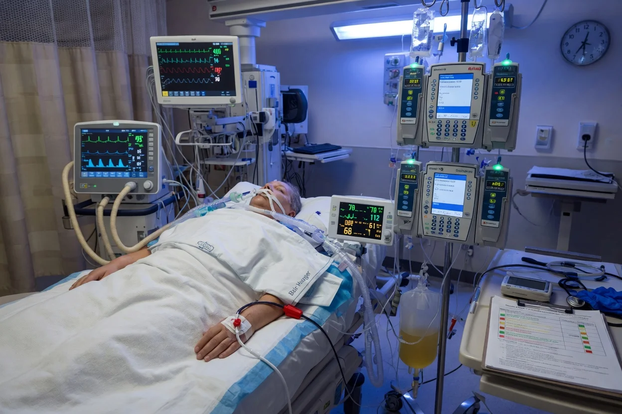
Solid-Organ Transplant Recipient in the ICU — Immunosuppression, Infection & Rejection
Also known as Solid-organ transplant · Transplant recipient · Immunosuppression · Tacrolimus · Ciclosporin · Mycophenolate · Acute rejection · Opportunistic infection · Calcineurin inhibitor
The solid-organ transplant recipient in the ICU: the immunosuppression (the calcineurin inhibitors — the tacrolimus, the ciclosporin; the antimetabolites — the mycophenolate; the mTOR — the sirolimus; the steroids), the infection risk (the timeline — the first month the nosocomial; the 1 to 6 months the opportunistic — the CMV, the PCP, the Listeria, the Aspergillus; the late the community-acquired), the acute rejection (the T-cell mediated — the cellular; the antibody-mediated — the humoral), the drug interactions (the CYP3A4 — the tacrolimus levels), and the graft dysfunction (the diagnostic — the ultrasound, the biopsy).
On this page & tools
Your progress
Saved locally on this device.
8 MCQs with explanations
Target exams
Overview & definition
The solid-organ transplant recipient in the ICU faces three interlinked threats: infection (the immunosuppression), rejection (the immune response to the graft), and drug toxicity (the immunosuppressants). The timeline matters (the infection pattern changes over time). The immunosuppression regimen — the manage carefully in the ICU.[1]
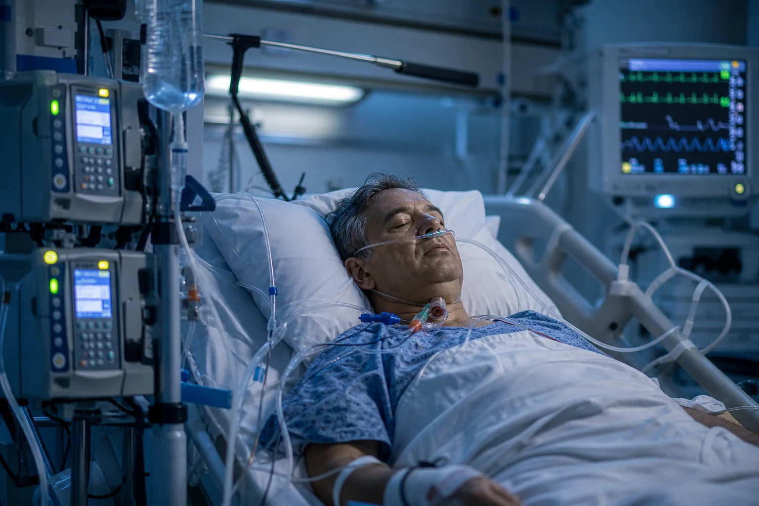
The immunosuppression
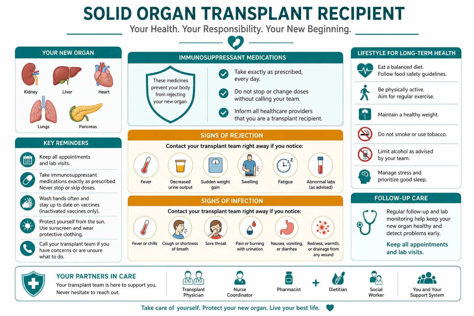
The regimen
- The calcineurin inhibitors (the tacrolimus preferred; the ciclosporin) — inhibit IL-2. Levels monitoring (trough). Adverse: nephrotoxicity, hypertension, neurotoxicity (PRES), diabetes.[1]
- The antimetabolites (the mycophenolate mofetil, the azathioprine). Adverse: cytopenia, GI.[1]
- The mTOR inhibitors (the sirolimus). Adverse: impaired wound healing, pneumonitis.[1]
- The steroids (the prednisolone). Adverse: diabetes, osteoporosis, infection.[1]
Drug interactions (the CYP3A4)
- Inhibitors (the macrolides, the azoles, the diltiazem, the grapefruit) → the RAISE the tacrolimus levels → the toxicity. Inducers (the rifampicin, the phenytoin, the St John wort) → the LOWER → the rejection. Check ALL the drugs.[1]
The infection timeline
- The first month — the nosocomial (same as the non-transplant).[1]
- The 1 to 6 months — the opportunistic: the CMV (the valganciclovir prophylaxis), the PCP (the cotrimoxazole), the Listeria (the meningitis), the Aspergillus (the invasive).[1]
- The late (over 6 months) — the community-acquired.[1]
The acute rejection
- The T-cell mediated (cellular) — the biopsy (Banff). The treatment: the steroid pulse (the methylprednisolone 500 to 1000 mg daily for 3 days).[1]
- The antibody-mediated (humoral) — the donor-specific antibodies. The treatment: the plasmapheresis + IVIG + rituximab.[1]
The ICU management
- The sepsis — the broad-spectrum (the cover the opportunistic); the reduce the immunosuppression (stop the antimetabolite; reduce the calcineurin; keep the steroid).[1]
- The drug levels — the monitor the tacrolimus.[1]
- The prophylaxis — the PCP (the cotrimoxazole), the CMV (the valganciclovir).[1]
Red flags
The Fishman infection calendar — the timeline that drives everything
The single most examinable concept in transplant infectious disease is the calendar — the post-transplant timeline that predicts which infections are most likely at each phase. Fishman's framework (NEJM 2007) divides the post-transplant course into three periods, each with a characteristic infection profile determined by the net state of immunosuppression and the epidemiological exposures of the donor and recipient.[1]
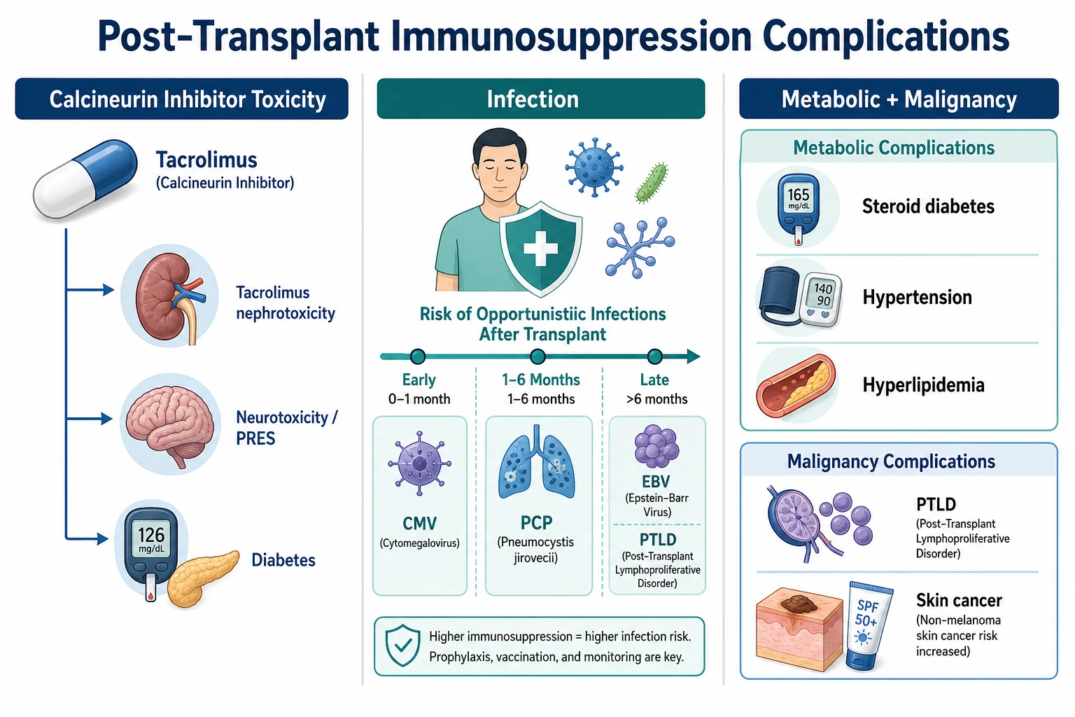
The Fishman calendar — infection risk by post-transplant phase
| Phase | Post-transplant timing | Net immunosuppression | Characteristic infections | Rationale |
|---|---|---|---|---|
| Phase 1 — early (technical / nosocomial) | 0–1 month | Lower (wound healing, induction wearing off) | Nosocomial — line-related bacteraemia (Staph, Enterococcus, Gram-negatives), wound infection, UTI, anastomotic leak, C. difficile; donor-derived infection (transmitted with the graft); recipient-derived recurrent infection (e.g. pre-existing TB, HBV, HCV) | The same pathogens as any postoperative ICU patient PLUS donor-derived and recipient-derived. The immunosuppression is not yet maximal. Technical/surgical complications dominate |
| Phase 2 — the opportunistic window | 1–6 months | Maximum (full maintenance immunosuppression; cumulative effect) | Opportunistic — CMV (the most important), Pneumocystis (PCP), Listeria monocytogenes (meningitis), Aspergillus (invasive pulmonary), Nocardia, Toxoplasma gondii, EBV (drives PTLD), BK polyomavirus (nephropathy), mycobacteria (TB and NTM), Strongyloides hyperinfection | The classic opportunistic window — the net state of immunosuppression is maximal and the prophylaxis (cotrimoxazole, valganciclovir) is masking some pathogens; pathogens NOT covered by prophylaxis emerge (Aspergillus, Listeria, Nocardia) |
| Phase 3 — late (community / chronic) | >6 months | Lower (maintenance weaned) | Community-acquired (pneumococcal pneumonia, influenza, UTI); chronic viral (HBV/HCV progression, BK); late opportunistic only if augmented immunosuppression given for rejection (CMV, Aspergillus return); malignancy-related infection (PTLD) | The recipient resembles a mildly immunocompromised host — most infections are community-acquired. BUT if augmented immunosuppression is given for a rejection episode, the recipient 'moves back' to phase 2 opportunistic risk |
- The calendar is not rigid — the timeline shifts with the intensity of immunosuppression and the use of prophylaxis. A recipient treated with augmented immunosuppression for a late rejection episode 'moves back' into the phase 2 opportunistic window regardless of the calendar date.[1]
- Donor-derived infection is the great mimicker in phase 1 — pathogens transmitted with the graft (e.g. donor-derived CMV, HBV, HCV, HIV, West Nile virus, rabies, lymphocytic choriomeningitis virus, mycobacteria, Trypanosoma cruzi) can present as unexplained sepsis, meningitis, or graft dysfunction in the first month. Always consider donor-derived infection when an unusual pathogen is isolated.
- The net state of immunosuppression — the qualitative measure of how immunocompromised the recipient is. Increased by: high-dose steroids, T-cell depleting antibodies (ATG, OKT3, alemtuzumab), rejection treatment, neutropenia, underlying immunodeficiency, malnutrition, extremes of age. The higher the net state, the earlier and more severe the opportunistic infection.[1]
Phase 1 (0–1 month) — technical and nosocomial
In the first month post-transplant, infections are dominated by the same nosocomial pathogens that afflict any ICU patient, PLUS donor-derived and recipient-derived infections. The opportunistic infections of phase 2 have not yet emerged because the immunosuppression has not had time to exert its full suppressive effect, and prophylaxis (cotrimoxazole, valganciclovir) is providing cover. [1]
Phase 1 (0–1 month) — the diagnostic approach to early post-transplant fever
- EXCLUDE TECHNICAL/SURGICAL COMPLICATIONS — the first priority. Anastomotic leak (kidney ureteric, hepaticojejunostomy, bronchial, vascular), vascular thrombosis (hepatic artery, portal vein, renal artery, pancreatic graft), wound infection/dehiscence, intra-abdominal collection, biliary leak/stricture. Doppler ultrasound / CT angiogram of the graft vasculature is mandatory for graft dysfunction + fever in phase 1. A thrombosed hepatic artery causing biliary necrosis is a surgical emergency.[1]
- EXCLUDE NOSOCOMIAL INFECTION (same as any ICU patient) — line-related bacteraemia (Staphylococcus aureus, coagulase-negative staph, Enterococcus, Gram-negative bacilli), ventilator-associated pneumonia, catheter-related UTI, Clostridioides difficile colitis (the transplant recipient is high-risk). Send blood cultures (peripheral + from every line), urine, sputum, and a C. diff toxin assay on any diarrhoea.
- CONSIDER DONOR-DERIVED INFECTION — pathogens transmitted with the graft. Classic donor-derived: CMV (D+/R− mismatch), HBV/HCV/HIV (window period), West Nile virus, rabies, lymphocytic choriomeningitis virus (LCMV), Trypanosoma cruzi (Chagas), mycobacteria. If an unusual or unexpected pathogen is isolated, OR if multiple recipients from the same donor are unwell, suspect donor-derived transmission and notify the organ procurement organisation IMMEDIATELY.[1]
- CONSIDER RECIPIENT-DERIVED RECURRENT INFECTION — pre-existing latent infection reactivating or progressing: latent TB, chronic HBV/HCV, Strongyloides stercoralis (hyperinfection syndrome with Gram-negative bacteraemia — risk with steroids), endemic mycoses (Histoplasma, Coccidioides, Blastomyces), Toxoplasma gondii (in heart transplant — the cysts reside in cardiac muscle). Pre-transplant screening should have identified these, but always re-ask.
- EMPIRICAL THERAPY — broad-spectrum as for any septic ICU patient (the local sepsis protocol) — cover MRSA, Pseudomonas, Gram-negatives. Add a donor-derived / opportunistic screen (CMV PCR, BK PCR if kidney, T. gondii IgG/PCR if heart). Do NOT empirically add antifungal or antiviral coverage unless specifically indicated (e.g. fluconazole if hepatobiliary or pancreatic transplant with high fungal risk; ganciclovir if CMV syndrome suspected). Adjust the immunosuppression in consultation with the transplant team.
Phase 2 (1–6 months) — the opportunistic window
This is the most examinable and clinically dangerous phase. The net state of immunosuppression is at its maximum, and the opportunistic infections emerge. The prophylaxis given (cotrimoxazole for PCP, valganciclovir for CMV) suppresses SOME pathogens but NOT others — the organisms NOT covered by prophylaxis (Aspergillus, Listeria, Nocardia, endemic mycoses) emerge during prophylaxis.[1]
The phase 2 opportunistic infections — the canonical list with the distinguishing features
| Pathogen | Typical syndrome | Risk factors / clues | Diagnosis | Treatment |
|---|---|---|---|---|
| Cytomegalovirus (CMV) | The most important — CMV syndrome (fever, leucopenia, thrombocytopenia, malaise) OR tissue-invasive disease (colitis with diarrhoea/blood, pneumonitis especially in lung transplant, hepatitis especially in liver transplant, retinitis, encephalitis) | D+/R− mismatch (donor CMV-positive, recipient CMV-negative — highest risk); lung and kidney-pancreas transplant; T-cell depleting induction | Quantitative CMV PCR (whole blood or plasma); tissue biopsy with immunohistochemistry (intranuclear inclusions — 'owl's eye') for tissue-invasive disease | IV ganciclovir (or valganciclovir for less severe); valganciclovir prophylaxis for D+/R− (3–6 months); reduce immunosuppression; CMV-Ig in severe pneumonitis |
| Pneumocystis jirovecii (PCP) | Pneumocystis pneumonia — subacute dyspnoea, dry cough, fever, hypoxia out of proportion to chest findings; pneumothorax | Absence of cotrimoxazole prophylaxis (the single biggest risk factor — every transplant recipient should be on cotrimoxazole); augmented immunosuppression | BAL — immunofluorescence / silver stain or PCR; serum β-D-glucan (supportive, not specific); LDH elevated; chest CT — bilateral ground-glass opacities | High-dose cotrimoxazole (TMP-SMX 15–20 mg/kg/day TMP); steroids if hypoxic (PaO₂ <70 mmHg — prednisolone 40 mg BD); reduce immunosuppression |
| Listeria monocytogenes | Meningoencephalitis (can be rhombencephalitis — brainstem); bacteraemia; gastroenteritis | Cell-mediated immune defect; contaminated food (soft cheese, deli meats, smoked seafood); can occur any time but peak phase 2 | Blood cultures; CSF (Gram-positive rods, lymphocytic pleocytosis); CSF/blood PCR | IV ampicillin (penicillin allergy — cotrimoxazole); add gentamicin for severe CNS / endocarditis (synergy); listeria is INHERENTLY resistant to cephalosporins including ceftriaxone |
| Aspergillus | Invasive pulmonary aspergillosis (cough, haemoptysis, pleuritic pain, fever, nodules with halo sign and cavitation); disseminated (brain abscess — seizures, focal deficit); tracheobronchitis in lung transplant | Lung and liver transplant (highest risk); neutropenia; high-dose steroids; renal failure; construction/dust exposure; CMV co-infection (CMV is a co-factor); augmented immunosuppression | BAL galactomannan; serum galactomannan (less sensitive in non-neutropenic); CT chest — nodules, halo sign, air crescent; biopsy (septate acute-angle hyphae) — the gold standard | Voriconazole (first-line — but check CYP3A4 interaction with tacrolimus — reduce tacrolimus by ~50%); liposomal amphotericin B alternative; isavuconazole (fewer interactions); reduce immunosuppression; surgical resection for localised disease |
| Nocardia | Pulmonary nocardiosis (subacute pneumonia, nodules, cavitation); CNS abscess (multiple); cutaneous (primary skin inoculation) | Chronic steroids; T-cell depleting therapy; lung transplant | BAL / sputum — Gram-positive branching, beaded filaments (weakly acid-fast); culture; biopsy | Cotrimoxazole (first-line); imipenem + amikacin for severe/disseminated; prolonged therapy (6–12 months); reduce immunosuppression |
| Toxoplasma gondii | Toxoplasmosis — disseminated infection, pneumonitis, myocarditis, encephalitis (multiple ring-enhancing lesions); MOST common in heart transplant (the cysts reside in cardiac muscle — the donor heart can transmit toxoplasma) | D+/R− heart transplant (donor IgG-positive, recipient IgG-negative); no cotrimoxazole prophylaxis (cotrimoxazole covers toxoplasma — this is why PCP prophylaxis also prevents toxoplasma) | PCR (blood, BAL, CSF); serology (IgG — but can be negative in immunosuppressed); brain biopsy | Pyrimethamine + sulfadiazine + folinic acid (NOT folic acid — folinic acid to spare marrow); cotrimoxazole is an alternative |
| EBV → PTLD | Post-transplant lymphoproliferative disorder — fever, lymphadenopathy, organ infiltration (gut bleeding/obstruction, liver, CNS); see the dedicated PTLD section below | EBV D+/R− mismatch; paediatric recipient (EBV-naive); T-cell depleting induction (ATG, OKT3, alemtuzumab); intestine/multi-visceral transplant | Tissue biopsy — CD20+, EBV-EBER in situ hybridisation; EBV viral load (PCR); LDH elevated; PET-CT for staging | Reduce immunosuppression (first-line — can induce regression alone); rituximab (anti-CD20); R-CHOP chemotherapy for refractory/aggressive |
| BK polyomavirus | BK nephropathy (kidney transplant) — gradual rise in creatinine mimicking rejection; ureteric stenosis; haemorrhagic cystitis | Kidney transplant; intensity of immunosuppression; no effective antiviral | Quantitative BK PCR (plasma — >10,000 copies/mL suggests nephropathy); kidney biopsy — SV40 large-T antigen immunostain in tubular epithelial cells | Reduce immunosuppression (the mainstay — reduce/stop MMF, lower tacrolimus); fluoroquinolone (ciprofloxacin — modest effect); IVIG; switch MMF → leflunomide; cidofovir (nephrotoxic — rarely used) |
| Mycobacteria (TB and NTM) | Tuberculosis (pulmonary, disseminated, meningitis); non-tuberculous mycobacteria (M. avium complex, M. abscessus, M. kansasii) — pulmonary, cutaneous, disseminated | Pre-existing latent TB reactivation (steroid-induced); donor-derived; high-prevalence region | AFB smear and culture (sputum, BAL); NAAT/Xpert MTB/RIF; interferon-gamma release assay (IGRA — less sensitive in immunosuppressed); biopsy | Standard RIPE therapy for TB (check drug interactions — rifampicin is a POTENT CYP3A4 inducer — will crash tacrolimus levels → rejection; use rifabutin instead); NTM per species |
| Strongyloides stercoralis | Hyperinfection syndrome — Gram-negative (E. coli, Klebsiella) bacteraemia, meningitis (the larvae carry gut bacteria); ARDS; enteric bacterial sepsis; can be fatal | Pre-existing infection (endemic areas — tropical/subtropical, plus rural southern USA); steroid therapy is the classic trigger; can occur phase 1 or with augmented immunosuppression | Stool microscopy (multiple samples — larvae); serology (IgG); sputum/BAL larvae in hyperinfection | Ivermectin (daily for 2 days, repeat at 2 weeks for hyperinfection); albendazole alternative; broad-spectrum antibiotics for the enteric bacterial sepsis |
- Why CMV is the most important — CMV is the most common and clinically significant opportunistic infection after solid-organ transplant. It causes: (1) direct effects — CMV syndrome, tissue-invasive disease (colitis, pneumonitis, hepatitis, retinitis, encephalitis); (2) indirect effects — CMV is an immunomodulator that increases the risk of OTHER opportunistic infections (Aspergillus, PCP, EBV/PTLD, bacterial sepsis), acute and chronic rejection, and graft loss. CMV D+/R− mismatch is the highest-risk configuration. The Kotton 2013 international consensus (Transplantation Society) provides the prophylaxis and pre-emptive therapy algorithms.[2]
Phase 3 (>6 months) — community and chronic
Beyond six months, most transplant recipients are on lower maintenance immunosuppression and resemble a mildly immunocompromised host — the bulk of infections are community-acquired (pneumococcal pneumonia, influenza, UTI, community respiratory viruses). However, several late threats remain: [1]
- Chronic viral infections — BK nephropathy (kidney), HBV/HCV progression to cirrhosis/hepatocellular carcinoma, EBV/PTLD (late), recurrent CMV.
- Late opportunistic infection — ONLY if the recipient has received augmented immunosuppression for a rejection episode (the recipient 'moves back' to phase 2 risk). CMV, Aspergillus, and PCP can all present late in this context.
- Malignancy-related infection — PTLD (EBV-driven), and solid-organ malignancies (skin SCC, post-transplant lymphomas) that themselves predispose to infection.
- Cardiovascular disease — by far the leading cause of late mortality in kidney and heart transplant recipients (see below). [1]
The acute rejection — cellular vs antibody-mediated
Acute rejection is the immune response against the graft. It is divided into T-cell mediated (cellular) rejection — the more common, generally responsive to steroids — and antibody-mediated (humoral) rejection — more aggressive, associated with donor-specific antibodies (DSA), and requiring plasmapheresis + IVIG ± rituximab. The diagnosis is tissue biopsy (kidney — Banff; liver — Banff; heart — ISHLT; lung — ISHLT working formulation).[1]
T-cell mediated (cellular) vs antibody-mediated (humoral) rejection
| Feature | T-cell mediated rejection (TCMR / cellular) | Antibody-mediated rejection (AMR / humoral) |
|---|---|---|
| Immunology | CD4⁺ and CD8⁺ T-cells infiltrate the graft → tubulitis, interstitial inflammation, endothelialitis | Preformed or de novo donor-specific antibodies (DSA) bind graft endothelium → complement activation → endothelial injury, thrombosis, ischaemia |
| Timing | Typically 1 week to 6 months (peak 1–3 months); can occur later with non-adherence | Hyperacute (minutes — preformed antibody, prevented by cross-match); early acute (days–weeks — preformed low-level antibody); late (months–years — de novo antibody, often with non-adherence) |
| Clinical | Graft dysfunction (rising creatinine, abnormal LFTs, heart failure, ↑ dyspnoea/FEV₁ decline) ± fever, graft tenderness, oliguria | Often more severe — graft dysfunction + (kidney) graft tenderness, oliguria, haematuria; (heart) haemodynamic compromise, cardiogenic shock; (lung) rapid gas exchange deterioration |
| Biopsy | Interstitial lymphocytic infiltrate, tubulitis, intimal arteritis/endarteritis; graded by Banff (kidney, liver) or ISHLT (heart, lung) | Microvascular inflammation (glomerulitis, peritubular capillaritis), C4d staining in peritubular capillaries (kidney) — marker of complement activation; DSA in serum |
| Serology | Not diagnostic | Donor-specific anti-HLA antibodies (DSA) — single-antigen bead assay; C1q-binding assays detect complement-binding (more pathogenic) antibodies |
| Treatment | IV methylprednisolone pulse (500–1000 mg daily × 3 days) — usually responsive; refractory → ATG (anti-thymocyte globulin) | Plasmapheresis + IVIG + rituximab (anti-CD20); ** bortezomib** (proteasome inhibitor — depletes plasma cells) for refractory; eculizumab (complement blockade) in selected; treat any trigger (non-adherence, infection) |
| Prognosis | Usually responsive; worse if recurrent or severe (Banff grade) | Worse — AMR is a major cause of graft loss; de novo DSA and late AMR carry the worst prognosis |
- The Banff classification (kidney) — the standardised histological grading of renal allograft pathology. TCMR: Borderline (i1 t1), IA (i2 t2), IB (i2 t3 or i3 t1-3), IIA (mild intimal arteritis v1), IIB (severe transmural arteritis v2), III (arterial fibrinoid necrosis/neointimal formation v3). AMR: requires (1) histological evidence of tissue injury (microvascular inflammation, acute tubular injury, arteritis), (2) C4d deposition or moderate microvascular inflammation, and (3) serological evidence of DSA. Higher grades correlate with worse outcomes and dictate treatment intensity.[1]
- The ISHLT heart biopsy grading (Stewart 2005 revision) — endomyocardial biopsy remains the gold standard for heart rejection surveillance, performed routinely (weekly → monthly → quarterly) in the first year. Grades: 0R (none), 1R (mild — interstitial/perivascular infiltrate, no myocyte damage), 2R (moderate — TWO or more foci with associated myocyte damage), 3R (severe — diffuse infiltrate with multifocal myocyte damage ± oedema/haemorrhage/vasculitis). 2R and 3R warrant treatment.[3]
The diagnostic workup of suspected acute rejection in the ICU
- SUSPECT REJECTION WHEN — graft dysfunction without another explanation (kidney: rising creatinine; liver: rising bilirubin/AST/ALT; heart: new diastolic dysfunction, arrhythmia, low output; lung: decline in FEV₁, infiltrates); fever, graft tenderness, oliguria; recent non-adherence, infection, or drug level subtherapeutic. Always exclude infection first — rejection and infection can co-exist and their treatments oppose each other (steroids worsen infection).[1]
- BLOODS — graft function (creatinine/eGFR, LFTs, troponin, BNP/NT-proBNP), CBC (eosinophilia?), CRP, and DSA (donor-specific anti-HLA antibodies — single-antigen bead Luminex assay) if AMR suspected.
- IMAGING — Doppler ultrasound of the graft (vascular patency — exclude thrombosis; assess for collection/obstruction); echocardiogram (heart); CT chest (lung — exclude infection, airway dehiscence). Imaging supports but does NOT diagnose rejection.
- BIOPSY — the gold standard — kidney (percutaneous core biopsy — Banff grading); liver (transjugular/percutaneous — Banff schema); heart (endomyocardial biopsy — ISHLT grading); lung (transbronchial biopsy — ISHLT working formulation). Biopsy differentiates TCMR vs AMR, excludes recurrent disease, and guides treatment. C4d immunostaining and DSA serology confirm AMR.[3][10]
- TREAT — TCMR: IV methylprednisolone 500–1000 mg daily × 3 days (responsive in most); refractory or severe → ATG (anti-thymocyte globulin). AMR: plasmapheresis + IVIG + rituximab; bortezomib or eculizumab for refractory. Address any trigger (non-adherence, infection, subtherapeutic CNI). Reduce immunosuppression is NOT done for rejection — rather, AUGMENT it (but exclude infection first).
- RE-ASSESS — repeat graft function (creatinine, LFTs, troponin, FEV₁); consider repeat biopsy to confirm resolution. Augmented immunosuppression moves the recipient BACK into the phase 2 opportunistic window — restart CMV monitoring and ensure PCP prophylaxis.[1]
Complications by organ — the transplant-specific ICU considerations
Each solid organ has a distinctive post-transplant complication profile. The intensivist must know the organ-specific surgical complications, rejection patterns, infection risks, and long-term threats for each graft type.[1]
Complications by transplanted organ — the ICU-relevant summary
| Organ | Most common in ICU | Rejection / diagnosis | Distinctive infection risk | Distinctive late threat |
|---|---|---|---|---|
| Kidney (most common SOT) | Fluid/electrolyte (hyperkalaemia from CNI, fluid overload), delayed graft function (DGF), ATN | Biopsy (Banff); DSA for AMR | BK nephropathy (gradual creatinine rise); UTI; CMV | Chronic allograft injury (interstitial fibrosis/tubular atrophy); CV disease (leading cause of death) |
| Liver | Early graft dysfunction / primary non-function (PNF); biliary (leak/stricture/ischaemic cholangiopathy); hepatic artery thrombosis (catastrophic — biliary necrosis) | Banff schema biopsy (Demetris); transjugular preferred if coagulopathy | Bacterial cholangitis (biliary complication); CMV hepatitis; invasive fungal (candida, aspergillus) | Disease recurrence (HCV, HBV, PBC, PSC, autoimmune hepatitis, NASH); de novo malignancy |
| Heart | Acute heart failure (graft dysfunction), arrhythmia, tamponade (post-op bleed), RV failure (pulmonary hypertension) | Endomyocardial biopsy (ISHLT) — routine surveillance in year 1; DSA for AMR | Toxoplasma (D+/R− heart transplant — cysts in myocardium); CMV myocarditis; Aspergillus | Cardiac allograft vasculopathy (CAV) — chronic diffuse coronary vasculopathy; malignancy |
| Lung (highest infection risk) | Primary graft dysfunction (PGD) — Reperfusion injury → ARDS-like; anastomotic dehiscence; airway complications | Transbronchial biopsy (ISHLT); FEV₁ decline as marker | HIGHEST infection risk of any SOT — bacterial pneumonia, CMV pneumonitis, Aspergillus, PCP; anastomotic infection | Bronchiolitis obliterans syndrome (BOS) — chronic lung allograft dysfunction; the main long-term threat |
| Pancreas | Graft thrombosis (venous — catastrophic; the leading cause of early graft loss); pancreatitis; leak (exocrine drainage — bladder vs enteric) | Difficult biopsy; hyperglycaemia (loss of endocrine function) as marker; DSA | Intra-abdominal infection (leak, abscess); candida (pancreatic flora); UTI (bladder drainage) | Recurrent autoimmune diabetes; chronic rejection |
Kidney transplant — the most common SOT
The kidney is the most commonly transplanted solid organ, and kidney transplant recipients form the largest single group of SOT recipients in the ICU. The unique ICU issues are fluid and electrolyte management, delayed graft function (DGF), BK nephropathy, and chronic allograft injury. [1]
Kidney transplant recipient — ICU management priorities
- FLUID AND ELECTROLYTE MANAGEMENT — the kidney allograft is exquisitely sensitive to volume status. Hypovolaemia → ATN, delayed graft function. Hypervolaemia → pulmonary oedema, hypertension. Target euvolaemia — assess with passive leg raise, ultrasound IVC, or Swan-Ganz if shocked. Hyperkalaemia is common — calcineurin inhibitors (tacrolimus/ciclosporin) cause type IV RTA (hyporeninaemic hypoaldosteronism) and tubular toxicity → K⁺ rises. Treat with standard hyperkalaemia therapy; avoid/dose-adjust spironolactone and ACE-inhibitors. Hypomagnesaemia is also common (CNI renal magnesium wasting) — replete (IV magnesium sulfate).
- DELAYED GRAFT FUNCTION (DGF) — the need for dialysis in the first week post-transplant. More common in DCD donors, prolonged cold ischaemia, older donors. Usually resolves (ATN recovery) but prolongs hospital stay and may slightly worsen long-term graft survival. Exclude vascular thrombosis (renal artery/vein — Doppler urgent — surgical emergency) and acute rejection (biopsy). Manage with dialysis as needed; optimise haemodynamics; minimise nephrotoxins.
- BK NEPHROPATHY — gradual rise in creatinine 1–12 months post-transplant, mimicking rejection. Quantitative BK PCR (plasma >10,000 copies/mL suggests nephropathy); kidney biopsy with SV40 large-T antigen immunostain. Treatment: reduce immunosuppression (the mainstay — reduce/stop MMF, lower tacrolimus); ciprofloxacin; IVIG; switch MMF → leflunomide. There is NO effective specific antiviral. ALWAYS check BK before treating 'rejection' — treating BK nephropathy with steroids WORSENS it.
- ACUTE REJECTION — biopsy (Banff). TCMR → steroid pulse. AMR → plasmapheresis + IVIG + rituximab. Check DSA. Always exclude BK and infection first (steroids worsen both).
- CALCINEURIN INHIBITOR NEPHROTOXICITY — the CNI (tacrolimus, ciclosporin) causes afferent arteriolar vasoconstriction → AKI; chronic interstitial fibrosis and tubular atrophy ('striped fibrosis') → chronic allograft injury. Monitor trough levels (tacrolimus 5–10 ng/mL typical post-kidney). The nephrotoxicity is SYNERGISTIC with sepsis-related AKI — reduce the dose in AKI (transplant team). Belatacept (a co-stimulation blocker) avoids CNI nephrotoxicity and improves long-term renal function — used in selected patients (the BENEFIT trial showed sustained efficacy and renal benefit at 5 years).[6]
- CHRONIC ALLOGRAFT INJURY — the leading cause of late kidney graft loss. Multifactorial — antibody-mediated injury, CNI nephrotoxicity, BK, recurrent disease, hypertension, hyperlipidaemia. Histology: interstitial fibrosis, tubular atrophy, glomerulosclerosis, transplant glomerulopathy. No specific therapy — minimisation/withdrawal of CNI, blood pressure and lipid control, treat DSA.[7][12]
Liver transplant — graft dysfunction and biliary
The liver transplant recipient in the ICU poses the questions: (1) is the graft working? (early graft dysfunction / primary non-function); (2) is the hepatic artery patent? (HAT — see red flag); (3) is there a biliary complication? (leak, stricture, ischaemic cholangiopathy); (4) is there infection? (bacterial cholangitis, CMV hepatitis, fungal). [1]
Patterns of early liver graft dysfunction — the differential
| Pattern | Timing | Hallmark | Cause | Action |
|---|---|---|---|---|
| Primary non-function (PNF) | <7 days (often <48 h) | Fulminant graft failure — AST/ALT >2000–3000, rising INR, falling glucose (failure of gluconeogenesis), lactate rising, encephalopathy, haemodynamic instability | Poor donor quality (steatosis, DCD, prolonged ischaemia), preservation injury, reperfusion | Re-transplantation — PNF is the indication for urgent re-transplant; supportive care (metabolic, coagulation, haemodynamics) while awaiting |
| Initial poor function (IPF) / early graft dysfunction (EGD) | First week | Slow recovery — AST/ALT high but falling, INR improving, bile production present but slow | Mild preservation/reperfusion injury; donor factors | Supportive — usually recovers; minimise nephrotoxins; monitor for progression to PNF |
| Hepatic artery thrombosis (HAT) | Any time, peak early | Rising bilirubin/ALP out of proportion to AST/ALT; bile leak; biliary sepsis; sometimes fulminant necrosis | Vascular — small vessel, hypercoagulable, technical | Urgent Doppler → revascularisation or re-transplant; the biliary tree depends entirely on hepatic arterial flow |
| Portal vein thrombosis (PVT) | Early | Ascites, gastrointestinal bleeding (varices), bowel congestion; less dramatic than HAT | Hypercoagulability, technical, recurrent PSC | Revascularisation; anticoagulation; usually graft survives |
| Acute rejection | 5–30 days (peak) | Rising bilirubin, AST/ALT, ALP, fever, graft tenderness, eosinophilia | T-cell mediated (usually); AMR less common | Liver biopsy (Banff schema); steroid pulse (TCMR); plasmapheresis + IVIG + rituximab (AMR)[10] |
| Biliary complication (leak/stricture) | Days–months | Leak — bilious drain output, peritonitis, rising bilirubin; stricture — cholangitis, rising ALP/bilirubin | Ischaemic (HAT, ischaemic cholangiopathy), technical (anastomotic) | ERCP/MRCP — stenting, drainage; revise anastomosis if needed; antibiotics for cholangitis |
| Ischaemic cholangiopathy | Weeks–months (DCD grafts) | Non-anastomotic biliary strictures, casts, dilatation; recurrent cholangitis | Microvascular injury to peribiliary plexus (DCD grafts, HAT, prolonged ischaemia) | Often requires re-transplantation; percutaneous/endoscopic drainage is temporising |
- Liver biopsy (Banff schema) — the gold standard for diagnosing liver rejection. The Demetris et al. (2002) Banff schema grades acute rejection on a Rejection Activity Index (RAI) composed of portal inflammation, bile duct damage, and venous endothelial inflammation — each 0–3, total 0–9. RAI >4 (moderate–severe) warrants treatment. Transjugular biopsy is preferred when coagulopathy/ascites are present (lower bleeding risk than percutaneous).[10]
Heart transplant — rejection, biopsy, and infection
The heart transplant recipient has unique surveillance requirements — routine endomyocardial biopsy in the first year is standard, because acute rejection is often clinically silent until advanced. The ISHLT biopsy grading (Stewart 2005 revision) classifies cellular rejection from 0R (none) to 3R (severe). The distinctive infection threats are toxoplasmosis (D+/R− heart transplant — the cysts reside in the donor myocardium) and Aspergillus. The distinctive late threat is cardiac allograft vasculopathy (CAV) — a chronic, diffuse, concentric coronary vasculopathy that is the leading cause of late graft loss and re-transplantation after heart transplant.[3][5]
Heart transplant — the ISHLT endomyocardial biopsy grading (Stewart 2005 revision)
| Grade | Histology | Clinical significance | Treatment |
|---|---|---|---|
| 0R | No rejection | None | Continue maintenance immunosuppression |
| 1R (mild) | Interstitial and/or perivascular infiltrate with up to ONE focus of myocyte damage | Usually asymptomatic; does not require treatment in the absence of haemodynamic compromise | Continue maintenance; observe |
| 2R (moderate) | Two or more foci of infiltrate with associated myocyte damage | Treat — risk of progression; can present with heart failure, arrhythmia | IV methylprednisolone pulse; optimise maintenance |
| 3R (severe) | Diffuse infiltrate with multifocal myocyte damage ± oedema, interstitial haemorrhage, vasculitis | Treat aggressively — high risk of haemodynamic compromise, graft loss | IV methylprednisolone + ATG; haemodynamic support; consider mechanical support |
| AMR (antibody-mediated) | Intravascular macrophages (CD68+), interstitial oedema, haemorrhage; ± C4d/CD68 immunostaining | Often with haemodynamic compromise; DSA positive | Plasmapheresis + IVIG + rituximab; consider eculizumab/bortezomib |
- The denervated heart — the transplanted heart is denervated (no vagal or sympathetic input). Consequences: (1) resting tachycardia (~90–100 bpm — no vagal tone); (2) blunted response to exercise initially (slow rise in heart rate — relies on circulating catecholamines); (3) atypical presentation of ischaemia (no anginal pain — CAV presents as silent ischaemia, heart failure, arrhythmia, or sudden death); (4) denervation hypersensitivity to direct-acting vasopressors (e.g. metaraminol) but reduced response to indirect (e.g. ephedrine). Atropine is INEFFECTIVE for bradycardia (no vagal tone to block) — use isoprenaline or theophylline.[5]
- Cardiac allograft vasculopathy (CAV) — chronic, diffuse, concentric intimal hyperplasia of the coronary arteries (distinct from atherosclerosis, which is focal and proximal). Driven by immune injury (DSA, TCMR) and traditional risk factors (hypertension, hyperlipidaemia, diabetes, CMV). Presents as silent ischaemia, heart failure, arrhythmia, or sudden death (no angina — denervated). Surveillance: annual coronary angiography or intravascular ultrasound (IVUS — more sensitive). Treatment: statins (all heart recipients), mTOR inhibitors (sirolimus/everolimus slow CAV progression), treat modifiable risk factors, percutaneous/surgical revascularisation is rarely feasible (diffuse disease) — re-transplantation for severe CAV.[5]
Lung transplant — the highest infection risk and BOS
The lung is unique among solid organs because it is directly exposed to the environment via the airway — it has the highest infection risk of any transplanted organ. The lung transplant recipient also faces primary graft dysfunction (PGD) in the early post-op period (a form of ischaemia-reperfusion injury resembling ARDS), airway anastomotic complications (dehiscence, stenosis), and bronchiolitis obliterans syndrome (BOS) — the chronic lung allograft dysfunction that is the leading cause of late death. [1]
Lung transplant — the complications spectrum
| Phase | Complication | Clinical | Diagnosis | Management |
|---|---|---|---|---|
| Immediate (0–72 h) | Primary graft dysfunction (PGD) — ischaemia-reperfusion injury | ARDS-like picture — hypoxia, bilateral infiltrates within 72 h, reduced compliance, pulmonary oedema; severity graded PGD 0–3 by PaO₂/FiO₂ | Clinical + CXR/CT (exclude rejection, infection, volume overload, venous anastomotic obstruction); NO specific diagnostic test — a diagnosis of exclusion | Lung-protective ventilation; minimise fluids; iNO / inhaled prostacyclin (selective pulmonary vasodilator); ECMO for severe (PGD 3); PGD predicts BOS (Daud 2007 — recipients with PGD have higher BOS rates)[8] |
| Early | Airway anastomotic complications — dehiscence (early, days–weeks), stenosis (later) | Dehiscence — air leak, mediastinitis, sepsis; stenosis — stridor, recurrent infection, ↓FEV₁ | Bronchoscopy | Dehiscence — surgical/stent; stenosis — balloon dilatation, stent |
| 1–6 months | Infection — the highest risk of any SOT | Bacterial pneumonia, CMV pneumonitis, Aspergillus, PCP | BAL (microbiology + cell count); galactomannan | Pathogen-directed; reduce immunosuppression; prophylaxis (cotrimoxazole, valganciclovir, inhaled amphotericin) |
| 1–6 months | Acute rejection — cellular | Decline in FEV₁, fever, infiltrates, dyspnoea | Transbronchial biopsy (ISHLT working formulation — perivascular/peribronchiolar infiltrate); exclude infection first | Steroid pulse (usually responsive) |
| Months–years | Bronchiolitis obliterans syndrome (BOS) — chronic lung allograft dysfunction | Progressive, irreversible airflow obstruction (↓FEV₁ >20% from baseline) WITHOUT another cause; dry cough, dyspnoea; recurrent 'chest infections' | Clinical diagnosis — sustained FEV₁ decline (BOS stage 1: 66–80% baseline; 2: 51–65%; 3: ≤50%); biopsy (obliterative bronchiolitis) often not needed and may be non-diagnostic (patchy) | Augmented immunosuppression (ATG, azithromycin — anti-inflammatory); re-transplant for end-stage; azithromycin (some respond — neutrophilic reversible alloplegia); outcomes poor |
- BOS — the leading cause of late death after lung transplant — bronchiolitis obliterans is a fibroproliferative obliteration of the small airways (the bronchioles) driven by alloimmune injury (acute rejection episodes, AMR, DSA) and non-alloimmune injury (PGD, infection — especially CMV and bacterial pneumonia, gastro-oesophageal reflux with microaspiration). It presents as progressive, irreversible airflow obstruction and is staged by the sustained decline in FEV₁ from the best post-transplant baseline. The histology (obliterative bronchiolitis) is patchy and transbronchial biopsy is often non-diagnostic — BOS is therefore a clinical diagnosis of exclusion (defined by the ISHLT working formulation). There is no effective treatment — augmented immunosuppression, azithromycin (a subset have neutrophilic reversible allograft dysfunction that responds), and re-transplantation. Primary graft dysfunction (PGD) at transplant is the strongest predictor of later BOS (Daud 2007 — recipients with severe PGD had significantly higher BOS rates).[8][9]
Pancreas transplant — thrombosis and leak
The pancreas transplant (usually simultaneous kidney-pancreas, SPK, for type 1 diabetes with renal failure) has the highest surgical complication rate of any solid-organ transplant. The leading cause of early graft loss is vascular thrombosis (venous — the pancreatic venous drainage is low-flow and prone to thrombosis). Other complications include graft pancreatitis, anastomotic leak (exocrine drainage — bladder vs enteric), and intra-abdominal infection. [1]
Pancreas transplant recipient — the early post-op ICU priorities
- GLYCAEMIC MONITORING — the transplanted pancreas should restore normoglycaemia and C-peptide independence from insulin within hours. Hyperglycaemia in the early post-op period suggests graft thrombosis, pancreatitis, or rejection. Monitor blood glucose hourly; once euglycaemic, the patient is insulin-independent. Persistent hyperglycaemia + rising amylase/lipase → suspect graft thrombosis or pancreatitis → urgent CT angiogram.
- GRAFT THROMBOSIS — the leading cause of early graft loss — the pancreatic venous drainage is low-flow and prone to thrombosis (especially venous — splenic vein/SMV). Presents as sudden hyperglycaemia, rising amylase/lipase, graft tenderness, fever, sometimes haemodynamic compromise (venous infarction → haemorrhagic pancreatitis). Diagnosis: CT angiogram (venous phase). Treatment: urgent surgical thrombectomy / thrombolysis — if not salvageable, graft pancreatectomy. Time is critical — delay = graft loss. Most centres use early post-op anticoagulation (heparin then warfarin/aspirin) to prevent this.
- GRAFT PANCREATITIS — preservation/reperfusion injury → raised amylase/lipase, graft tenderness, peripancreatic fluid. Usually mild and self-limiting; severe → necrosis, infection. Manage as for any pancreatitis (fluid resuscitation, analgesia, nil-by-mouth); exclude thrombosis.
- ANASTOMOTIC LEAK — the exocrine drainage is either bladder (older technique — duodenocystostomy; complications: UTI, haematuria, dehydration from bicarbonate loss, reflux pancreatitis) or enteric (modern standard — duodenojejunostomy; complications: leak → intra-abdominal sepsis, abscess). Leak presents with abdominal pain, fever, rising inflammatory markers, intra-abdominal collection. Diagnosis: CT with contrast (extravasation). Treatment: drainage (percutaneous or surgical), antibiotics, repair/revision.
- INFECTION — intra-abdominal infection (leak, abscess — often Candida from the pancreatic flora, or enteric Gram-negatives/anaerobes); UTI (especially bladder drainage); standard opportunistic prophylaxis (cotrimoxazole, valganciclovir). Have a low threshold for CT abdomen and broad-spectrum antibiotics including antifungal (candida coverage) in any septic pancreas recipient.[1]
Immunosuppression complications — infection, malignancy (PTLD), metabolic
The price of graft survival is the burden of immunosuppression. Four categories of complication dominate: infection (covered above — the timeline), malignancy (PTLD, skin cancer, solid tumours), metabolic (steroid and CNI toxicity), and specific drug toxicity (covered in the drug interactions section).[1]
Malignancy — PTLD and the rest
Post-transplant malignancy is driven by chronic immunosuppression — impaired immune surveillance allows oncogenic viruses (EBV → PTLD; HPV → cervical/ano-genital cancer; HHV-8 → Kaposi sarcoma; HCV/HBV → hepatocellular carcinoma) and UV-induced skin cancers to flourish. The most feared and examinable is post-transplant lymphoproliferative disorder (PTLD). [1]
The post-transplant malignancies — the spectrum
| Malignancy | Relative risk vs general population | Driven by | Presentation | Management |
|---|---|---|---|---|
| PTLD (post-transplant lymphoproliferative disorder) | 10–100× (highest of any malignancy) — varies by organ (highest: intestine/multi-visceral > heart/lung > kidney/liver) | EBV (the B-cell is immortalised by EBV; immunosuppression removes T-cell surveillance) — EBV D+/R− mismatch and paediatric (EBV-naive) recipients are highest risk; T-cell depleting induction (ATG, OKT3, alemtuzumab) increases risk | Fever, lymphadenopathy, organ infiltration (gut — bleeding/obstruction; liver — hepatitis, mass; CNS — seizures, focal deficit; lung — nodules; marrow — cytopenia); LDH elevated; EBV viral load elevated | Reduce immunosuppression first-line (can induce regression alone in polyclonal/early disease); rituximab (anti-CD20) — the PTLD-1 trial established rituximab ± chemo; R-CHOP chemotherapy for refractory/aggressive/monomorphic; EBV viral load monitoring for high-risk (D+/R−) recipients |
| Non-melanoma skin cancer (SCC > BCC) | 65–100× for SCC — the MOST COMMON post-transplant malignancy overall | UV radiation + immunosuppression (azathioprine photosensitises — switch to MMF); HPV | Sun-exposed skin — face, scalp, hands; multiple lesions; aggressive course (metastasis more common than in immunocompetent) | Sun protection (sunscreen, clothing, avoidance); regular dermatology surveillance; switch azathioprine → MMF or CNI → mTOR (sirolimus/everolimus reduce skin cancer risk); excision; consider reduced immunosuppression |
| Kaposi sarcoma | 100–500× | HHV-8 (human herpesvirus 8); Mediterranean/African ancestry | Cutaneous plaques/nodules (lower limbs); visceral (lung, GI) | Reduce immunosuppression; switch CNI → mTOR (sirolimus has anti-KS activity); chemotherapy if extensive |
| Solid tumours (lung, colorectal, breast, renal) | 2–3× (modest) | Chronic immunosuppression | Standard presentation | Standard oncology; maintain immunosuppression (do not stop); age-appropriate screening |
Metabolic and drug-specific complications
The metabolic and drug-specific toxicities of immunosuppression are chronic, insidious, and contribute substantially to long-term morbidity and mortality. The intensivist must recognise them because they often manifest acutely in the ICU setting (e.g. tacrolimus PRES, steroid hyperglycaemia, CNI nephrotoxicity in AKI). [1]
Drug-specific immunosuppression toxicities — the ICU-relevant recognition
| Drug class / drug | Toxicity | Clinical / ICU presentation | Management |
|---|---|---|---|
| Calcineurin inhibitors (tacrolimus, ciclosporin) | Nephrotoxicity (afferent arteriolar vasoconstriction → AKI; chronic interstitial fibrosis/tubular atrophy) | Rising creatinine, hyperkalaemia, hypomagnesaemia, hyperuricaemia/gout, hypertension | Monitor trough levels; reduce dose in AKI; minimise nephrotoxins; belatacept (co-stimulation blocker) avoids this — BENEFIT trial[6] |
| Tacrolimus — neurotoxicity (PRES) | Posterior reversible encephalopathy syndrome | Seizures, visual disturbance, headache, altered consciousness, hypertension; MRI: T2/FLAIR hyperintensity in parieto-occipital white matter (vasogenic oedema) — usually reversible | Reduce/switch tacrolimus (reduce dose; or switch to ciclosporin/belatacept/mTOR); control blood pressure and seizures; exclude CNS infection (LP, MRI with contrast) and CNS PTLD |
| Tacrolimus — diabetes | Beta-cell toxicity; insulin resistance | New-onset diabetes after transplant (NODAT) — hyperglycaemia, sometimes DKA | Insulin; consider switch tacrolimus → ciclosporin (less diabetogenic) or belatacept |
| Ciclosporin | Gingival hyperplasia, hirsutism, hypertension, hyperuridaemia | Distinctive cosmetic effects; hypertension | Switch to tacrolimus (preferred — fewer cosmetic effects, equivalent/better efficacy) |
| mTOR inhibitors (sirolimus, everolimus) | Impaired wound healing (avoid in early post-op — dehiscence, lymphocoele); interstitial pneumonitis (dry cough, dyspnoea, bilateral infiltrates — drug-induced); proteinuria; hyperlipidaemia; mouth ulcers | Wound complications (delay initiation until wound healed — typically >1 month post-op); pneumonitis (diagnosis of exclusion — BAL to exclude infection then drug cessation); proteinuria (monitor urine PCR) | Withhold in early post-op; for pneumonitis — stop the mTOR (usually resolves); reduce dose / switch for proteinuria |
| Antimetabolites (mycophenolate MMF, azathioprine) | Cytopenia (leucopenia, anaemia, thrombocytopenia — marrow suppression); GI (diarrhoea, gastritis, colitis — MMF-induced colitis mimics IBD/CMV colitis); hepatotoxicity (azathioprine); pancreatitis (azathioprine) | Cytopenia (monitor CBC weekly early); MMF colitis (diarrhoea — biopsy shows crypt cell apoptosis without viral inclusions — distinguish from CMV) | Reduce/stop the antimetabolite; in sepsis — STOP first-line (see sepsis red flag); azathioprine — check TPMT activity before starting (deficiency → fatal marrow aplasia) |
| Steroids (prednisolone, methylprednisolone) | Diabetes (steroid-induced hyperglycaemia); osteoporosis (vertebral fractures); adrenal suppression (can't mount stress response — stress-dose steroids in critical illness); hypertension; dyslipidaemia; myopathy (critical illness weakness); infection; cataracts; psychosis | Stress-dose hydrocortisone in any transplant recipient with critical illness/adrenal suppression; steroid myopathy complicating weaning | Minimise (steroid-sparing protocols — induction with basiliximab/alemtuzumab allows early steroid withdrawal); bisphosphonate + vitamin D for bone protection; PJP prophylaxis if >20 mg prednisolone for >4 weeks |
| T-cell depleting induction (ATG, OKT3, alemtuzumab) | Cytokine release syndrome (fever, rigors, hypotension, pulmonary oedema on first dose); prolonged lymphopenia (increases opportunistic infection and PTLD risk); serum sickness | Cytokine release syndrome — premedicate with steroid + antihistamine + paracetamol; the prolonged lymphopenia is the rationale for selective use | Pre-medicate; reserve for high immunological risk; the lymphopenia drives opportunistic infection risk in phase 2 |
Drug interactions — the CYP3A4 minefield
The calcineurin inhibitors (tacrolimus and ciclosporin) and the mTOR inhibitors (sirolimus, everolimus) are metabolised by cytochrome P450 3A4 (CYP3A4) in the liver and gut, and are substrates for P-glycoprotein (the efflux pump). Drugs that inhibit CYP3A4/P-glycoprotein raise the CNI/mTOR levels → toxicity (nephrotoxicity, neurotoxicity, immunosuppression-related infection). Drugs that induce CYP3A4 lower the levels → rejection. Every drug prescribed to a transplant recipient must be checked against the CNI/mTOR interaction list.[1]
The CYP3A4/P-glycoprotein interactions with tacrolimus/ciclosporin/mTOR inhibitors
| Interaction type | Drugs | Effect on tacrolimus/ciclosporin/mTOR level | Clinical consequence | Management |
|---|---|---|---|---|
| CYP3A4 inhibitors (RAISE levels) | Macrolides — clarithromycin, erythromycin (NOT azithromycin — minimal CYP effect); azoles — fluconazole, voriconazole, itraconazole, posaconazole, ketoconazole; non-DHP calcium channel blockers — diltiazem, verapamil; grapefruit juice (irreversible gut CYP3A4 inhibition); protease inhibitors (HIV — ritonavir, darunavir); cobicistat; amiodarone; metoclopramide (mild); oral contraceptives (mild) | RAISE tacrolimus/ciclosporin levels — often 2–5× within days | Nephrotoxicity (rising creatinine), neurotoxicity (PRES — seizures, headache), hyperkalaemia, hypertension, over-immunosuppression (infection) | Avoid if possible; if essential — pre-emptively reduce tacrolimus by 25–50% and monitor trough levels closely (every 2–3 days); use azithromycin instead of clarithromycin; use isavuconazole instead of voriconazole (fewer interactions); transplant pharmacist to adjust |
| CYP3A4 inducers (LOWER levels) | Rifampicin (the most potent — can drop tacrolimus by 80–90% → acute rejection); phenytoin; carbamazepine; phenobarbital; St John's wort (often undisclosed by patient — ALWAYS ASK); glucocorticoids (chronic high-dose — auto-induction); efavirenz, nevirapine (antiretrovirals); enzalutamide, mitotane | LOWER tacrolimus/ciclosporin levels — often dramatically | Acute rejection — rising creatinine, graft dysfunction; the drug can be stopped or started WITHOUT the transplant team being informed (e.g. a course of rifampicin for TB) | Avoid if possible; if essential — pre-emptively increase tacrolimus (often 3–5×) and monitor trough levels daily; use rifabutin instead of rifampicin (less CYP induction) for MAC prophylaxis; transplant pharmacist to adjust; educate patient to disclose ALL medications including herbal |
| Synergistic nephrotoxicity (not CYP — additive renal insult) | Aminoglycosides (gentamicin, tobramycin, amikacin); amphotericin B (all formulations — additive to CNI nephrotoxicity); NSAIDs (block prostaglandin-mediated afferent arteriolar vasodilation — dangerous in CNI vasoconstriction); ACE-inhibitors/ARBs (efferent arteriolar vasodilation — synergistic with CNI); contrast (iodinated); tenofovir; foscarnet; cidofovir | Levels unchanged but additive nephrotoxicity | AKI, additive to the CNI's intrinsic nephrotoxicity | Avoid/minimise; if essential — dose-adjust, monitor renal function daily, maintain adequate hydration; the CNI nephrotoxicity is SYNERGISTIC with sepsis-related AKI — reduce CNI in AKI |
| Myelosuppression (additive with antimetabolites) | Cotrimoxazole (TMP component — additive marrow suppression with MMF/azathioprine); ganciclovir/valganciclovir; allopurinol (profound — with azathioprine, the TPMT pathway — reduce azathioprine dose by 65–75%); chloramphenicol | Levels unchanged but additive marrow suppression | Leucopenia, anaemia, thrombocytopenia — infection, bleeding | Monitor CBC weekly; reduce MMF/azathioprine dose; the allopurinol-azathioprine interaction is FATAL if missed — must reduce azathioprine by 65–75% or switch azathioprine → MMF; consider folic acid for cotrimoxazole |
SAQ — Febrile neutropenic lung transplant recipient with CMV pneumonitis
10 minutes · 10 marks
A 55-year-old man 4 months post bilateral lung transplant (tacrolimus, mycophenolate, prednisolone; CMV D+/R−) presents with fever, dyspnoea, leucopenia (WCC 1.8 ×10⁹/L), and bilateral interstitial infiltrates on chest X-ray. He has been on valganciclovir prophylaxis which ran out 3 weeks ago. CMV PCR 850,000 IU/mL. Bronchoalveolar lavage shows CMV inclusions on histology.
SAQ — Heart transplant recipient in cardiogenic shock
10 minutes · 10 marks
A 60-year-old man 18 months post-orthotopic heart transplant (tacrolimus, mycophenolate, prednisolone 5 mg/day) presents with 3 days of progressive dyspnoea, orthopnoea and a productive cough. He is in cardiogenic shock: BP 78/45, JVP raised, bilateral crackles, cool peripheries, lactate 4.8. Echocardiography shows globally reduced LV function (EF 25%), previously 60%. Troponin 0.6 μg/L. Tacrolimus trough 8 ng/mL.
Clinical pearls
Additional red flags
Prognosis and evidence
Solid-organ transplant recipient — landmark evidence on infection, rejection, and outcomes
Fishman JA, N Engl J Med 2007 (PMID 18094380) — the landmark review "Infection in solid-organ transplant recipients" that established the three-phase infection calendar (month 1 = nosocomial/donor-derived/recipient-derived; 1–6 months = opportunistic; >6 months = community-acquired). The single most cited reference in transplant infectious disease and the framework for every ICU fellow's approach to the febrile transplant recipient. The net state of immunosuppression and the epidemiological exposures (donor, recipient, environment) shape the timeline.[1]
Kotton CN et al., Transplantation 2013 (PMID 23896556) — the Updated International Consensus Guidelines on the Management of Cytomegalovirus in Solid-Organ Transplantation (The Transplantation Society). The definitive guidance on CMV prophylaxis vs pre-emptive therapy, the duration of prophylaxis by organ and D/R serostatus (D+/R− = highest risk), the diagnosis (quantitative PCR; tissue-invasive disease requires biopsy), and treatment (IV ganciclovir for tissue-invasive disease; valganciclovir for less severe). The reference for every CMV question in the exam.[2]
Stewart S et al., J Heart Lung Transplant 2005 (PMID 16297770) — the Revision of the 1990 Working Formulation for the Standardization of Nomenclature in the Diagnosis of Heart Rejection (the ISHLT 2004 revision). Replaced the 1990 0/1/2/3A/3B/4 grades with the modern 0R/1R/2R/3R scheme for endomyocardial biopsy grading of acute cellular rejection. The basis of heart rejection surveillance worldwide. (The 1990 original working formulation is Berry et al., PMID 2277294.)[3][14]
Yousem SA et al., J Heart Lung Transplant 1996 (PMID 8820078) — the Revision of the 1990 Working Formulation for the Classification of Pulmonary Allograft Rejection (Lung Rejection Study Group). The standardised histological classification of acute and chronic lung allograft rejection — the perivascular/peribronchiolar infiltrate of acute cellular rejection, the airway inflammation of lymphocytic bronchiolitis, and the obliterative bronchiolitis of chronic rejection. The basis of lung rejection diagnosis.[4]
Costanzo MR et al., J Heart Lung Transplant 2010 (PMID 20643330) — the ISHLT Guidelines for the Care of Heart Transplant Recipients. The comprehensive practice guideline covering immunosuppression, rejection surveillance (endomyocardial biopsy schedule), infection prophylaxis (PCP, CMV, toxoplasma D+/R−), cardiac allograft vasculopathy (CAV) surveillance and management, and long-term follow-up. The reference for heart transplant ICU management.[5]
Rostaing L, Vincenti F et al., Am J Transplant 2013 (PMID 24047110) — the 5-year long-term extension of the BENEFIT study of belatacept (a CTLA4-Ig co-stimulation blocker) vs ciclosporin in kidney transplant. Demonstrated sustained efficacy and a superior renal profile (better eGFR, lower blood pressure) for belatacept — the evidence for CNI-sparing regimens to mitigate chronic allograft injury. Caveat: belatacept is contraindicated in EBV-seronegative recipients (PTLD risk, especially CNS).[6]
Langewisch E, Mannon RB, Clin J Am Soc Nephrol 2021 (PMID 33820759) — the contemporary review "Chronic Allograft Injury" — the pathogenesis (antibody-mediated, CNI nephrotoxicity, BK, recurrent disease, metabolic), histology (interstitial fibrosis, tubular atrophy, transplant glomerulopathy), and treatment strategies (CNI minimisation/withdrawal, belatacept, mTOR, DSA management). The reference for the leading cause of late kidney graft loss.[7]
Madariaga HM, Riella LV, Contrib Nephrol 2017 (PMID 28535528) — "Chronic Allograft Injury: An Overview of Pathogenesis and Treatment Strategies" — a complementary overview of the mechanisms (immune, non-immune, infectious) and the rationale for the minimisation and conversion strategies that slow progression.[12]
Daud SA et al., Am J Respir Crit Care Med 2007 (PMID 17158279) — the landmark study showing that primary graft dysfunction (PGD) after lung transplant is the strongest predictor of later bronchiolitis obliterans syndrome (BOS). Recipients with severe PGD (grade 3) had significantly higher BOS rates at 3 and 5 years. The evidence that the early post-transplant reperfusion injury has long-term consequences — minimising PGD (donor management, short ischaemia time, careful fluid strategy) pays long-term dividends.[8]
Woodrow JP et al., J Heart Lung Transplant 2010 (PMID 20580267) — comparison of BOS to other forms of chronic lung allograft dysfunction (CLAD) — established that the restrictive allograft syndrome (RAS) and other CLAD phenotypes have even worse prognosis than classic BOS. The reference for the CLAD spectrum and the recognition that not all post-lung-transplant FEV₁ decline is BOS.[9]
Demetris AJ et al., Transplantation 2002 (PMID 12451268) — the Banff schema for grading liver allograft rejection (the Rejection Activity Index — RAI — composed of portal inflammation, bile duct damage, venous endothelial inflammation). The basis of liver rejection diagnosis; transjugular biopsy is preferred when coagulopathy or ascites is present.[10]
Stehlik J, Christie JD et al., J Heart Lung Transplant 2021 (PMID 34657795) — "The Evolution of the ISHLT Transplant Registry" — the contemporary summary of the international heart and lung transplant registry. Median survival: adult heart ~12.5 years (conditional on surviving 1 year, ~14 years); adult lung ~6.5 years (conditional, ~9 years). The leading causes of death (year 1 = graft failure, infection, multi-organ failure; beyond 1 year = BOS for lung, CAV for heart, malignancy, infection). The evidence base for counselling and risk stratification.[11]
Lefaucheur C et al., Kidney Int 2019 (PMID 31005275) — non-HLA agonistic anti-angiotensin II type 1 receptor (AT1R) antibodies induce a distinctive phenotype of antibody-mediated rejection in kidney transplant recipients. Demonstrates that AMR is not solely an anti-HLA phenomenon — non-HLA antibodies (anti-AT1R, anti-MICA, anti-endothelial cell antibodies) contribute to graft injury and expand the AMR spectrum. The reference for the contemporary understanding of AMR pathogenesis.[13]
Outcomes: One-year patient survival after solid-organ transplant is excellent (kidney ~95–98%, liver ~90–95%, heart ~90%, lung ~85–90%, pancreas ~95%). Long-term survival is more variable — kidney and liver recipients have the longest median survival (10–15+ years); lung recipients the shortest (~6.5 years, dominated by BOS/CLAD). Beyond the first year, cardiovascular disease is the leading cause of death in kidney recipients; CAV in heart recipients; BOS in lung recipients; disease recurrence and de novo malignancy in liver recipients. Infection remains a threat throughout — the timeline (Fishman calendar) dictates the differential. The intensivist's role: rapid diagnosis (biopsy the graft, BAL the lung), broad-spectrum empiric coverage that includes the opportunistic pathogens, judicious reduction of immunosuppression in sepsis (stop antimetabolite, reduce CNI, keep steroid), and meticulous attention to CYP3A4 drug interactions. Always involve the transplant team.[11]
References
- [1]Fishman JA Infection in solid-organ transplant recipients N Engl J Med, 2007.PMID 18094380
- [2]Kotton CN, Kumar D, Caliendo AM, et al. Updated international consensus guidelines on the management of cytomegalovirus in solid-organ transplantation Transplantation, 2013.PMID 23896556
- [3]Stewart S, Winters GL, Fishbein MC, et al. Revision of the 1990 working formulation for the standardization of nomenclature in the diagnosis of heart rejection J Heart Lung Transplant, 2005.PMID 16297770
- [4]Yousem SA, Berry GJ, Cagle PT, et al. Revision of the 1990 working formulation for the classification of pulmonary allograft rejection: Lung Rejection Study Group J Heart Lung Transplant, 1996.PMID 8820078
- [5]Costanzo MR, Dipchand A, Starling R, et al. The International Society of Heart and Lung Transplantation Guidelines for the care of heart transplant recipients J Heart Lung Transplant, 2010.PMID 20643330
- [6]Rostaing L, Vincenti F, Grinyo J, et al. Long-term belatacept exposure maintains efficacy and safety at 5 years: results from the long-term extension of the BENEFIT study Am J Transplant, 2013.PMID 24047110
- [7]Langewisch E, Mannon RB Chronic Allograft Injury Clin J Am Soc Nephrol, 2021.PMID 33820759
- [8]Daud SA, Yusen RD, Meyers BF, et al. Impact of immediate primary lung allograft dysfunction on bronchiolitis obliterans syndrome Am J Respir Crit Care Med, 2007.PMID 17158279
- [9]Woodrow JP, Shlobin OA, Barnett SD, et al. Comparison of bronchiolitis obliterans syndrome to other forms of chronic lung allograft dysfunction after lung transplantation J Heart Lung Transplant, 2010.PMID 20580267
- [10]Demetris AJ, Ruppert K, Dvorchik I, et al. Real-time monitoring of acute liver-allograft rejection using the Banff schema Transplantation, 2002.PMID 12451268
- [11]Stehlik J, Christie JD, Goldstein DR, et al. The evolution of the ISHLT transplant registry. Preparing for the future J Heart Lung Transplant, 2021.PMID 34657795
- [12]Madariaga HM, Riella LV Chronic Allograft Injury: An Overview of Pathogenesis and Treatment Strategies Contrib Nephrol, 2017.PMID 28535528
- [13]Lefaucheur C, Viglietti D, Bouatou Y, et al. Non-HLA agonistic anti-angiotensin II type 1 receptor antibodies induce a distinctive phenotype of antibody-mediated rejection in kidney transplant recipients Kidney Int, 2019.PMID 31005275
- [14]Berry GJ, Brunt EM, Chamberlain D, et al. A working formulation for the standardization of nomenclature in the diagnosis of heart and lung rejection: Lung Rejection Study Group. The International Society for Heart Transplantation J Heart Transplant, 1990.PMID 2277294