ICU · Trauma
Abdominal Trauma — Blunt, Penetrating, FAST & Non-Operative Management
Also known as Abdominal trauma · Blunt abdominal trauma · Penetrating abdominal trauma · FAST scan · Splenic injury · Liver injury · Hollow viscus injury · Damage-control laparotomy · Non-operative management · Angioembolisation · Solid organ injury · Mesenteric injury · Pancreatic trauma · Genitourinary trauma
The abdominal trauma: the blunt (the solid organ — the spleen, the liver; the hollow viscus — the small bowel, the colon; the mesenteric) and the penetrating (the stab, the gunshot). The FAST scan (the bedside — the free fluid in the 4 views) — the rapid triage. The CT with the IV contrast (the stable patient — the grading). The laparotomy indications: the peritonitis, the hypotension unresponsive, the penetrating with the peritonitis, the evisceration. The non-operative management (the NOM) for the blunt solid organ (the spleen, the liver) if the haemodynamically stable. The angioembolisation for the contrast extravasation (the blush). The damage-control laparotomy (the physiological extremis — the open abdomen, the packing, the planned re-operation) for the lethal triad.
On this page & tools
Your progress
Saved locally on this device.
8 MCQs with explanations
Target exams
Overview & definition
The abdominal trauma — the blunt (the motor vehicle, the fall, the crush — the solid organ, the hollow viscus) and the penetrating (the stab, the gunshot — the hollow viscus, the vascular). The FAST scan (the bedside — the rapid triage for the free fluid). The CT with the IV contrast (the stable patient — the grading). The laparotomy for the peritonitis, the unresponsive hypotension, the penetrating with the peritonitis. The non-operative management for the stable blunt solid organ.[1]
The single most important triage decision in abdominal trauma is haemodynamic stability: the unstable patient is resuscitated and goes to theatre (or angioembolisation) without a CT; the stable patient gets a contrast-enhanced CT — the gold standard for grading solid-organ injury, identifying a contrast "blush" (active extravasation), and detecting hollow-viscus and mesenteric injury. The FAST is a triage tool, never a definitive investigation: it detects free fluid (not the organ injury itself), and its sensitivity for hollow-viscus, retroperitoneal, and diaphragmatic injury is poor.[1][2]
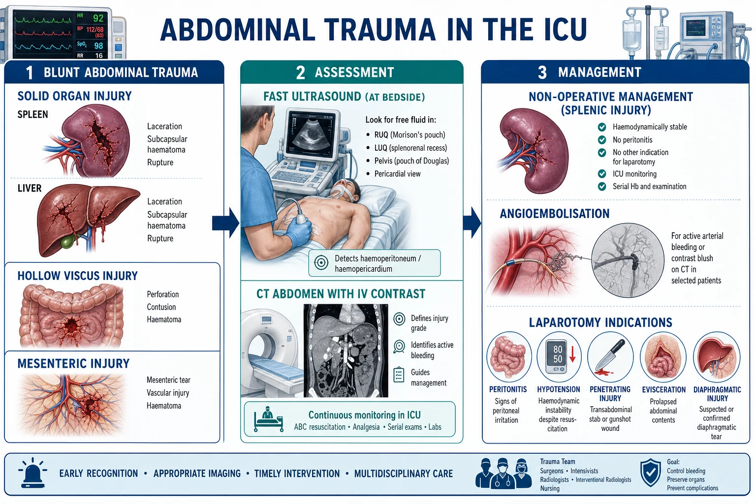
Mechanisms of injury — blunt vs penetrating
The mechanism determines the likely injury pattern, the pre-test probability, and the index of suspicion for specific organs. The abdomen is divided into three zones — intrathoracic (above the costal margin, behind the ribs), true abdomen (between costal margin and pelvis), and pelvic/retroperitoneal — and a penetrating wound is also classified by its tract relative to the peritoneum (anterior abdomen, flank, back, thoracoabdominal).[1]
Blunt trauma
MVC, fall, crush, assault
- Mechanisms: motor vehicle collision (steering wheel, seatbelt, airbag), fall from height, crush injury, direct blow, bicycle handlebar
- Pattern: SOLID organs dominate — SPLEEN is the #1 injured organ, then liver; mesenteric and hollow-viscus injury less common but missed
- Seatbelt sign (ecchymosis across lower abdomen) → high risk of mesenteric and hollow-viscus (small bowel) injury and Chance fracture of lumbar spine
- Retroperitoneal injuries (duodenum, kidney, pancreas, IVC) often occult — FAST misses them
- Associated injuries: rib fractures (spleen/liver), pelvic fracture (bladder, rectum, retroperitoneal bleed), thoracic and TBI
- Solid organ injury bleeds into the peritoneum → FAST may be positive; hollow viscus leaks gas/bowel content → peritonitis develops late (6–12 h)
Penetrating trauma
Stab, gunshot, impalement
- Stab wound: low energy, narrow tract, injury confined to structures in the wound path; ~30% of anterior abdominal stabs penetrate the peritoneum, ~1/3 of those injure an organ
- Gunshot wound (GSW): high energy, cavitation, unpredictable tract; intra-abdominal injury in ~80% of GSWs that traverse the peritoneum
- Pattern: HOLLOW VISCUS (small bowel, colon) and VASCULAR (aorta, IVC, mesenteric vessels) dominate; solid organs also injured
- Thoracoabdominal wounds (nipple line to costal margin) → diaphragm, liver, spleen, heart, lung
- Evisceration of omentum/bowel, blood from NG/rectal tube, or impalement in situ → theatre
- Mandatory laparotomy historically; now SELECTIVE non-operative management for stable patients with CT + serial exams (Demetriades)
The assessment
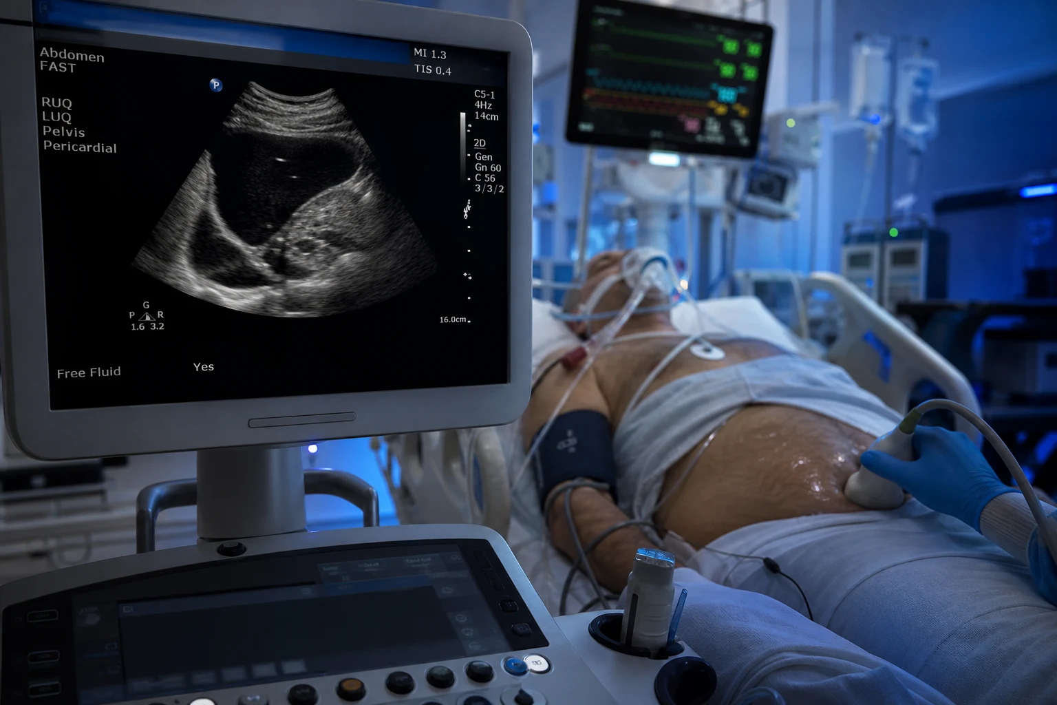
The FAST scan (Focused Assessment with Sonography for Trauma)
The FAST is the bedside ultrasound examination performed during the secondary survey to detect free intraperitoneal or pericardial fluid (blood). It is rapid (2–5 minutes), repeatable, radiation-free, and operator-dependent. It answers ONE question: is there free fluid? It does NOT grade organ injury, does NOT identify the source, and does NOT visualise the retroperitoneum or hollow viscus reliably.[1][2]
- The 4 views — the RUQ (Morrison pouch — hepatorenal recess, the most sensitive single view), the LUQ (perisplenic — splenorenal recess), the suprapubic (pelvic — pouch of Douglas / rectovesicular), and the subxiphoid pericardial (pericardial effusion / tamponade).[1]
- The free fluid = the positive (the anechoic/black stripe between organs; ~200 mL needed in the Morrison pouch to be reliably seen).[2]
- The rapid (the minutes), the repeatable, the no radiation. The triage the tool.[1]
- The limitations — the low sensitivity for the hollow viscus, the retroperitoneal, the diaphragmatic; the operator-dependent; the false negative if the small the volume, the obesity, the subcutaneous emphysema, the pelvic fracture retroperitoneal haematoma. The serial the FAST (the repeat at the 30 min if the negative but the clinical the concern) increases the sensitivity.[2]
Sensitivity
For free intraperitoneal fluid
- Pooled sensitivity ~60–90% depending on volume of fluid, operator skill, and mechanism
- Highest in hypotensive blunt trauma patients with large haemoperitoneum (~80–95%)
- Lower in stable patients with small volumes, in penetrating trauma, and in isolated retroperitoneal/hollow viscus injury
- Specificity is HIGH (~95–99%) — a positive FAST in an unstable patient mandates laparotomy
What FAST CANNOT do
The dangerous false sense of security
- Does NOT detect solid organ injury grade — only free fluid
- Does NOT see retroperitoneal injury (kidney, duodenum, pancreas, IVC, pelvic fracture haematoma)
- Does NOT see hollow viscus / mesenteric injury reliably (often little free fluid early)
- Does NOT see diaphragmatic injury (especially small left-sided post-penetrating)
- Does NOT see bladder injury if intraperitoneal volume small; bowel content looks like fluid
- A NEGATIVE FAST does NOT exclude intra-abdominal injury — serial exam + CT in the stable patient
The CT with the IV contrast
For the haemodynamically stable patient, the contrast-enhanced CT (venous phase; arterial phase added if vascular injury suspected) is the gold standard — it grades solid organ injury (AAST scale), identifies hollow-viscus injury (free air, wall thickening, mesenteric stranding, contrast extravasation into bowel wall), detects active bleeding (the contrast blush = active extravasation), and characterises vascular and retroperitoneal injuries.[1][12]
- The grading the solid organ (the AAST — the spleen I to V, the liver I to VI, the kidney I to V). The grade guides the management and predicts the failure of NOM.[6]
- The identify the hollow viscus (the free air, the wall thickening, the mesenteric stranding, the mesenteric contrast extravasation — the "vascular beading"). The oral/rectal contrast if the concern the high.[12]
- The vascular (the contrast extravasation — the "blush"). The blush = the active the bleeding → the angioembolisation or the theatre.[1]
- The NOT for the unstable (the delay — the to the CT the dangerous; the to the theatre).[1]
FAST
Bedside triage
- Indication: EVERY trauma patient during the secondary survey; the unstable patient who cannot go to CT
- Question answered: is there free intraperitoneal/pericardial fluid?
- Sensitivity ~60–90% for free fluid; misses retroperitoneal, hollow viscus, diaphragm
- Radiation: none; time: minutes; can be repeated serially
- A POSITIVE FAST in a haemodynamically UNSTABLE patient → immediate laparotomy
- A NEGATIVE FAST does NOT exclude injury — serial exam ± CT
CT with IV contrast
Gold standard — stable patient
- Indication: haemodynamically STABLE patient with suspected intra-abdominal injury
- Question answered: which organ, what grade, is there active bleeding, is there hollow viscus injury?
- Sensitivity ~95%+ for solid organ injury; detects blush, free air, mesenteric stranding
- Radiation: significant; time: 15–30 min; requires transport off the resus bay
- Contrast blush → angioembolisation; high-grade injury + blush → theatre or angio
- CONTRAINDICATED in the unstable patient — the radiation/transport risk is irrelevant; they bleed to death
Diagnostic peritoneal aspiration/lavage (DPA/DPL)
Historical, rarely used
- Largely supplanted by FAST and CT in modern trauma centres
- May still have a role in the unstable patient with an equivocal FAST (e.g. pelvic fracture retroperitoneal haematoma obscuring the pelvis)
- Aspiration of >10 mL frank blood → laparotomy; lavage fluid with bile, bowel content, or elevated amylase/alkaline phosphatase → bowel injury
- Bowel enters the umbilical trocar site — the open (Hasson) technique preferred to avoid iatrogenic injury
The laparotomy indications
The mandatory laparotomy for:[1]
- The peritonitis (the rigid abdomen, the rebound — the hollow viscus perforation).[1]
- The hypotension unresponsive to the fluid resuscitation with a positive FAST (the ongoing the bleeding).[1]
- The penetrating trauma with the peritonitis or the hypotension, the evisceration, the diaphragmatic injury, the impalement, the blood from the NG or the rectal tube.[1]
- The transabdominal gunshot wound (most traverse the peritoneum).[8]
- The iatrogenic — the haemodynamically unstable with the clinical deterioration despite the resuscitation.[1]
The decision algorithm — blunt abdominal trauma
Step 1 — Primary survey (ABCDE) and resuscitation
Airway with cervical spine control, breathing, CIRCULATION with haemorrhage control (apply direct pressure to external bleeding, pelvic binder for pelvic fracture, splint long-bone fractures). Two large-bore IV cannulae, bloods including crossmatch, lactate, venous gas, coagulation, β-hCG in women of reproductive age. Begin permissive hypotension (SBP 80–90) in the bleeding patient without TBI.
Step 2 — FAST during the secondary survey
Perform the 4-view FAST as part of the secondary survey. INTERPRET WITH THE HAEMODYNAMIC STATUS: a POSITIVE FAST in an UNSTABLE patient (SBP <90 not responding to 1–2 L crystalloid or blood) → immediate laparotomy (no CT). A POSITIVE FAST in a STABLE patient → CT abdomen with IV contrast for grading. A NEGATIVE FAST does NOT exclude injury — proceed based on mechanism and clinical exam.
Step 3 — Stability determines the next step
STABLE: CT abdomen with IV contrast (gold standard) — grade solid organs, identify blush, look for hollow viscus (free air, wall thickening, mesenteric stranding). UNSTABLE: resuscitate with blood (MTP), proceed to theatre for damage-control laparotomy (or angioembolisation if isolated pelvic fracture blush). NEVER take an unstable patient to CT.
Step 4 — Serial examination and disposition
Admit for serial abdominal examinations (every 1–2 h for the first 24 h), serial haemoglobin, vital signs, and lactate. Deterioration (new peritonitis, falling Hb, rising lactate, haemodynamic instability) triggers repeat imaging or laparotomy. Persistent tachycardia, falling blood pressure, or a rising lactate after resuscitation is organ injury until proven otherwise.
Step 5 — Definitive management
NOM for stable blunt solid organ injury (spleen, liver, kidney) with ICU monitoring and angioembolisation for the blush. Laparotomy for peritonitis, unresponsive hypotension, hollow viscus injury, or NOM failure. Damage-control laparotomy for the patient in physiological extremis (the lethal triad).
Specific organ injuries
The pattern of organ injury follows the mechanism and the anatomy. Solid organs (spleen, liver, kidney) dominate blunt trauma; hollow viscus and vascular structures dominate penetrating trauma. Retroperitoneal organs (duodenum, pancreas, kidney, IVC/aorta) are occult — FAST misses them, and they declare themselves late.[1]
The spleen — the #1 blunt solid organ
The spleen is the most commonly injured abdominal organ in blunt trauma (MVC handlebars, left lower rib fractures, falls). The grading is the AAST I–V (I = subcapsular tear <10%/capsular laceration <1 cm; V = shattered spleen or hilar vascular injury).[6][9]
- Non-operative management (NOM) is the standard for the haemodynamically stable patient with grades I–IV — successful in 70–90% of adults and up to 95% of children. Admission to a monitored setting, serial abdominal exams (q1–2h × 24h), serial haemoglobin, bed rest initially, and ICU if high grade or transfusion required.[4][9]
- Angioembolisation for the contrast blush (active extravasation), the pseudoaneurysm, the AV fistula, or the high-grade (IV–V) injury even without blush in some protocols. Proximal splenic artery embolisation (reduces the arterial pressure head into the spleen, allows haemostasis); distal selective embolisation for the focal bleed.[15]
- Failure of NOM (ongoing hypotension, falling Hb requiring transfusion, new peritonitis) → laparotomy. Predictors: high grade (IV–V), blush, haemodynamic instability on arrival, ISS, age >55.[9]
- Splenectomy → the overwhelming post-splenectomy infection (OPSI) risk — encapsulated organisms (pneumococcus, meningococcus, Haemophilus influenzae b). Post-splenectomy vaccination (pneumococcal, meningococcal ACWY + B, Hib) ideally >14 days pre-discharge, annual influenza, lifelong penicillin V prophylaxis (or at minimum patient education + alert bracelet).[7]
The liver — packing and damage control
The liver is the second most common blunt solid organ injury and the most common cause of death from abdominal trauma (deep lacerations, juxtahepatic venous injuries — IVC, hepatic veins). AAST grades I–VI (I = subcapsular haematoma <10% / capsular laceration <1 cm; VI = hepatic avulsion — universally fatal).[6]
- NOM for the stable patient with grades I–III is the standard (success >80%). Higher grades (IV–V) may still attempt NOM if stable, with ICU monitoring and angioembolisation for the blush.[3]
- Damage-control surgery for the unstable: perihepatic packing (the classic technique — laparotomy pads packed around the liver to tamponade, particularly for the deep laceration and the juxtahepatic venous injury), the Pringle manoeuvre (clamping the hepatoduodenal ligament — hepatic artery + portal vein — for up to 60 minutes warm ischaemia to control inflow bleeding), and the temporary closure. The definitive repair (resection, venous reconstruction, selective ligation) deferred to the re-operation.[5]
- Angioembolisation for the hepatic artery blush; selective hepatic artery ligation for uncontrolled arterial bleeding in theatre; atriocaval shunt for the retrohepatic IVC / hepatic vein injury (the most lethal).[3]
- Complications — the biloma, the hepatic necrosis (post-embolisation), the abscess, the haemobilia (triad: jaundice, RUQ pain, UGI bleed — treated by angioembolisation of the hepatic artery).[3]
The kidney
The kidney is a retroperitoneal organ — FAST misses it. Suspected with haematuria (gross or microscopic), flank pain/haematoma, or the mechanism (direct flank blow, deceleration — renal pedicle injury). CT with IV contrast (the excretory/nephrogenic phase) grades the injury (AAST I–V; V = shattered kidney or renal pedicle avulsion).[1]
- NOM is the standard for grades I–IV in the stable patient (success >90%); the kidney has a robust collateral circulation and most injuries heal.
- Operative exploration for the grade V (pedicle avulsion, expanding/ pulsatile retroperitoneal haematoma), the unstable, or the renal vein avulsion. Exploration risks nephrectomy (devascularisation from the hilar control).
- The renal pedicle injury (intimal tear of the renal artery from deceleration) — the delayed diagnosis (hours), the devascularisation, the salvage only if the revascularisation within 6 hours. [1]
The small bowel and mesentery — hard to diagnose
The small bowel is the most common HOLLOW viscus injured in blunt trauma (the crush against the spine, the deceleration tear at fixed points — ligament of Treitz, ileocaecal valve). The mesenteric injury bleeds and may devascularise the bowel. These are the most commonly missed abdominal injuries — FAST is usually negative (little free fluid early), CT findings are subtle, and peritonitis evolves over 6–12 hours.[12]
- The CT signs — the free air (ruptured hollow viscus), the bowel wall thickening (>3 mm), the mesenteric stranding/haematoma, the mesenteric contrast extravasation (the "vascular beading"), the "triangle sign" (free air between loops), the intramural haematoma, the unexplained free fluid WITHOUT solid organ injury (highly suspicious for mesenteric/bowel injury).[12]
- The DPL — the elevated amylase/alkaline phosphatase in the lavage fluid (the bowel content).
- The management — the perforation (the primary repair, the resection with the primary anastomosis if stable, the resection in discontinuity if the damage-control), the mesenteric devascularisation (the resection of the non-viable segment). The delayed presentation (peritonitis at 12–24 h) — the increased morbidity/mortality; the high index of suspicion, the serial exam, the repeat CT.[16]
The pancreas — high morbidity, occult
The pancreas is a retroperitoneal organ, injured by the direct epigastric blow (the steering wheel, the bicycle handlebar — the compression against the spine). The mechanism is classic; the injury is occult — the FAST is negative, the initial amylase/lipase may be normal (rises over 24–48 h), and the CT may miss the ductal injury. The ductal integrity is the determinant of management.[11]
- The grade — the I/II (no or minor ductal injury — the contusion, the superficial laceration) the NOM; the III/IV (ductal injury in the body/tail — the distal pancreatectomy; in the head — the complex, the Roux-en-Y, the damage-control drainage); the V (the massive disruption of the pancreatic head — the pancreaticoduodenectomy rarely, the damage-control).[11]
- The MRCP/ERCP for the ductal assessment if the CT indeterminate.
- The complications — the pseudocyst, the pancreatic fistula, the pancreatic abscess, the delayed haemorrhage. The high morbidity (the prolonged ICU, the multi-organ failure).[11]
- The associated — the duodenal injury (the pancreaticoduodenal complex — the high morbidity, the damage-control).
The bladder — pelvic fracture association
The bladder is injured in 5–10% of pelvic fractures (the bony fragment laceration, the deceleration) and is associated with the urethral injury (especially the male, the pelvic fracture, the blood at the meatus, the high-riding prostate, the perineal haematoma — the DO NOT catheterise blindly until the urethrogram).[1]
- The extraperitoneal (the 80%, the pelvic fracture) — the NOM (the urethral catheter, the 10–14 days).
- The intraperitoneal (the 20%, the dome, the full-bladder blunt trauma) — the surgical repair (the absorbable suture, the 2-layer closure).
- The signs — the gross haematuria (the 95%), the suprapubic pain, the inability to void, the elevated creatinine (the peritoneal reabsorption). The cystography (the CT or the flat-plate) for the diagnosis (the extravasation).[1]
The non-operative management (NOM)
For the blunt solid organ injury (the spleen, the liver, the kidney) if the haemodynamically stable and the no peritonitis:[4][3]
- The ICU admission, the serial the examinations (the vitals, the abdomen, the q1–2 h for the 24 h), the serial the Hb (the q6 h initially), the serial the lactate.[1]
- The angioembolisation for the CT contrast extravasation (the "blush") — the splenic artery embolisation (the proximal — the reduce the pressure; the distal — the selective), the hepatic artery, the renal artery.[15]
- The failure of the NOM → the laparotomy. The predictors: the high grade (the IV–V), the blush, the haemodynamic instability on the arrival, the ISS, the age >55.[9]
- The splenic — the NOM successful in the 70 to 90 per cent; the post-splenectomy the vaccination (the pneumococcal, the meningococcal, the Hib) if the splenectomy.[7]
Criteria for NOM (spleen, liver, kidney)
The stable blunt solid organ injury
- Haemodynamically STABLE (no sustained hypotension, responds to resuscitation, stable Hb)
- NO peritonitis (soft, non-tender abdomen) — peritonitis = hollow viscus injury = laparotomy
- NO other indication for laparotomy (e.g. pneumoperitoneum, diaphragmatic injury)
- Documented AAST grade on CT; blush managed by angioembolisation
- ICU/HDU monitoring capacity, serial exam possible, immediate theatre access if failure
- Transfusion requirement kept minimal (<2 units) — ongoing transfusion = failure
Contraindications to NOM / criteria for laparotomy
Operate now
- Haemodynamic instability unresponsive to resuscitation (ongoing bleeding)
- Peritonitis (rigid, tender abdomen — hollow viscus injury)
- Pneumoperitoneum (free air — perforated hollow viscus)
- Diaphragmatic injury, intraperitoneal bladder injury
- NOM failure — falling Hb, rising lactate, new hypotension, new peritonitis
- Penetrating trauma with peritonitis, evisceration, hypotension (selective NOM possible in stable)
Penetrating trauma — selective non-operative management
Historically, all penetrating abdominal wounds underwent mandatory laparotomy, with a high negative laparotomy rate. Modern trauma practice (Demetriades and the LAC+USC group) supports selective NOM for the haemodynamically stable patient with a penetrating wound (especially stab wounds and selected GSWs) managed by CT + serial abdominal examinations + local wound exploration, reserving laparotomy for the unstable, the peritonitic, the eviscerated, and the impaled.[8]
Mandatory laparotomy (traditional)
Historical standard
- All penetrating abdominal wounds → laparotomy
- High negative laparotomy rate (~20–40% for stabs, ~10–20% for GSWs)
- Justified in an era without CT and reliable serial examination
- Negative laparotomy carries morbidity (ileus, adhesions, wound complications) and cost
Selective NOM (modern)
Demetriades, LAC+USC
- Stable stab wound — local wound exploration to determine peritoneal penetration; if non-penetrating, discharge/observe
- Stable penetrating with CT showing no significant injury — serial exams + observe
- Stable GSW with tangential tract and no peritoneal violation — observe
- Reduces negative laparotomy rate dramatically while maintaining safety
- Requires reliable serial abdominal examination, CT, and immediate theatre access
- FAILURE (new peritonitis, hypotension) → immediate laparotomy
The damage-control laparotomy
For the physiological extremis (the lethal triad — acidosis pH under 7.2, hypothermia under 35, coagulopathy INR >1.5), the damage-control laparotomy is the abbreviated operation — the only goal is to deliver the patient alive to the ICU with correctable physiology, NOT the definitive repair. The lethal triad is self-perpetuating: each component worsens the others, and once established the mortality approaches 100%.[5]
- The rapid control — the packing (the control the bleeding — the liver, the spleen, the pelvis, the retroperitoneum), the ligation (the vessel), the temporary shunt (the vessel the repair the deferred — the carotid, the femoral, the mesenteric), the resection in discontinuity (the ischaemic bowel — the NOT the primary anastomosis), the temporary closure (the Bogota bag, the negative-pressure dressing — the VAC, the Wittmann patch — the fascia the OPEN).[5]
- The planned re-operation (the 24 to 48 hours) — the definitive repair, the anastomosis, the packing removal, the closure. The transition is the physiology-driven, NOT the time-driven (the lethal triad the corrected first).[5]
- The open abdomen — the manage the intra-abdominal hypertension (the IAP; the APP = MAP − IAP >50–60), the entero-atmospheric fistula, the massive third-space the fluid losses, the failed the closure (the planned ventral hernia + the split-thickness the skin graft).[1]
- The correct the lethal triad in the ICU between the operations — the warm (the forced-air, the warmed the fluids, the warm the ambient), the correct the acidosis (the resuscitation; the NaHCO3 the refractory only), the correct the coagulopathy (the MTP 1:1:1 — the PROPPR; the TXA — the CRASH-2 within the 3 h; the calcium; the cryoprecipitate for the fibrinogen; the viscoelastic the guided).[17]
Damage-control laparotomy — the index operation
Step 1 — Rapid entry and control of inflow
Long midline incision, rapid evacuation of blood and clot. Control major vascular inflow: aortic compression / supracoeliac aortic clamping (or REBOA pre-operatively in the resus bay) for the haemodynamically collapsed patient; Pringle manoeuvre (clamping the hepatoduodenal ligament) for liver bleeding. Pack all four quadrants systematically (RUQ, LUQ, pelvis, small bowel mesentery).
Step 2 — Haemostasis
Pack solid organ bleeding (liver perihepatic packing, splenic bed, pelvis). Ligate accessible bleeding vessels. Place temporary intraluminal shunt (e.g. Pruitt-Inahara, Argyle) for vessels needed for limb/organ perfusion (common/external iliac, femoral, mesenteric, carotid) — repair deferred. Do NOT attempt definitive vascular repair in the cold, coagulopathic patient.
Step 3 — Control of contamination
Close hollow viscus holes with rapid suture or linear stapler. Resect frankly non-viable/ischaemic bowel with a linear stapler and LEAVE IN DISCONTINUITY (do NOT perform primary anastomosis in the hostile environment — the leak rate is high). Identify and tag the injury sites for the re-operation. Place closed-suction drains where needed.
Step 4 — Temporary abdominal closure
Close the SKIN only or use a temporary closure device — Bogota bag (sterile 3L IV bag sutured to skin), commercial negative-pressure dressing (V.A.C. Abdominal), or Wittmann patch (Velcro-like temporary fascial closure). Leave the FASCIA OPEN to prevent abdominal compartment syndrome. The goal is a "tension-free" closure over the packed, oedematous bowel.
Step 5 — Transfer to ICU for physiological resuscitation
Do NOT continue operating. Transfer to ICU for rewarming, correction of acidosis, correction of coagulopathy (viscoelastic-guided — FFP, fibrinogen, platelets, TXA, calcium), and ventilatory/renal support. Monitor intra-abdominal pressure. The patient returns to theatre at 24–48 h when the lethal triad is corrected.
Definitive (primary) laparotomy
The stable patient
- Performed in the haemodynamically stable, physiologically normal patient
- Goals: complete exploration, repair all injuries, primary anastomosis, formal fascial closure
- Indications: isolated solid organ injury manageable at one operation, stable penetrating with hollow viscus
- Risk: low, when performed in the right patient
- Outcome: single operation, shorter ICU stay
Damage-control laparotomy
The lethal triad patient
- Performed in the patient in physiological extremis (pH <7.2, temp <35°C, INR >1.5)
- Goals: ABBREVIATED — haemostasis (pack/ligate/shunt), contamination control (staple in discontinuity), temporary closure
- Indications: exsanguination, lethal triad, massive transfusion, the "blood vessel" patient
- Risk: high — but LESS than attempting definitive repair in the hostile environment (which kills)
- Outcome: planned re-operation at 24–48 h for definitive repair once physiology corrected
The lethal triad of damage control
The damage-control philosophy exists to break the lethal triad — the self-perpetuating cycle of acidosis, hypothermia, and coagulopathy that develops in the exsanguinating trauma patient and drives mortality toward 100% once established.[5]
- The acidosis (pH under 7.2) — the clotting factor dysfunction (the enzymatic, the >50% reduction of the activity below the pH 7.2; virtually no clot below the pH 7.0). The correct by the resuscitation (the stop the bleed, the blood).[5]
- The hypothermia (under 35 degrees C) — the coagulation enzyme and the platelet dysfunction (the temperature-dependent; the functional the failure below the 34 degrees C). The prevent (the active the warming, the warmed the fluids, the warm the ambient).[5]
- The coagulopathy — the trauma-induced coagulopathy (the TIC — the endogenous, the present on the arrival in ~25% of the severe trauma; the tissue factor, the protein C, the hyperfibrinolysis) PLUS the dilutional (the iatrogenic, the crystalloid) PLUS the consumption. The INR >1.5 on the arrival the defines. The MTP 1:1:1 (the PROPPR), the TXA (the CRASH-2), the calcium, the fibrinogen.[17]
The lethal triad of damage-control laparotomy (click each)
INR >1.5
Trauma-induced coagulopathy (TIC, endogenous) + dilutional coagulopathy (iatrogenic, from crystalloid) + consumption. INR >1.5 on arrival defines TIC and multiplies mortality 3–4×. Treat with MTP 1:1:1, TXA (within 3 h), fibrinogen (cryo/concentrate), platelets, calcium, and viscoelastic-guided therapy.
Complications of abdominal trauma and the open abdomen
Early (0–72 h)
The acute complications
- Ongoing/ re-bleeding — most common cause of NOM failure; falling Hb, rising lactate, hypotension
- Abdominal compartment syndrome — rising IAP, falling APP, oliguria, falling tidal volume, shock; surgical decompression
- Missed injury (small bowel, mesenteric, pancreatic) — new peritonitis at 12–24 h; repeat CT and laparotomy
- Haemorrhage from solid organ (liver, spleen) despite NOM — angioembolisation or theatre
- Coagulopathy, hypothermia, acidosis — the lethal triad persists until corrected
Intermediate (days–weeks)
The ICU complications
- Intra-abdominal collection/abscess — fever, raised WCC, CT-guided drainage
- Biloma (liver), pancreatic fistula, pseudocyst — the pancreatic/biliary complications
- Entero-atmospheric fistula (open abdomen) — extremely difficult to manage; protect exposed bowel
- Bowel obstruction (adhesions), ileus, anastomotic leak
- Splenic/ hepatic infarction or abscess post-embolisation
Late (months–years)
The long-term sequelae
- Overwhelming post-splenectomy infection (OPSI) — encapsulated organisms; lifelong risk
- Incisional/ventral hernia (especially after open abdomen) — delayed repair
- Adhesions, chronic small bowel obstruction
- Pancreatic endocrine/exocrine insufficiency (after distal pancreatectomy / severe injury)
- Renal impairment / loss of kidney (after high-grade injury or nephrectomy)
Special situations
The pregnant trauma patient
In pregnancy, the gravid uterus displaces the bowel cephalad (shielding small bowel but exposing the liver/spleen), compresses the IVC in the supine position (left lateral tilt essential), and the fundal height determines the approach — uterus at/above the umbilicus increases the risk of abruption and uterine rupture. Rh-negative mother → anti-D immunogloblobulin (even minor trauma can cause feto-maternal haemorrhage). Continuous cardiotocography (CTG) monitoring for at least 4–6 hours (longer if contractions/abruption signs). The FAST includes the 4 standard views plus the foetal heart rate. Perimortem Caesarean section if maternal arrest >20 weeks gestation (within 5 minutes of arrest — improves both maternal and foetal survival).[1]
The anticoagulated patient
Patients on warfarin, direct oral anticoagulants (DOACs), or with pre-existing coagulopathy have a markedly lower threshold for operative management and a higher NOM failure rate. The INR >1.5 doubles the mortality from blunt liver/spleen injury. Reverse early — vitamin K + PCC (or FFP) for warfarin; specific reversal agents (idarucizumab for dabigatran, andexanet alfa for anti-Xa) where indicated. The isolated minor injury can become life-threatening.[1]
The paediatric patient
Children have a higher NOM success rate for solid organ injury (>90%) because of more compliant ribs and a better physiological reserve. The ABCDE paediatric adjustments (weight-based fluids/blood, Broselow tape). Selective non-operative management is the default for stable children with spleen/liver injury — transfuse for low threshold but operate for instability. Avoid the splenectomy (the OPSI risk higher in children).[13]
[1]Red flags
Exam practice
SAQ — Blunt abdominal trauma with splenic injury
12 minutes · 12 marks
A 32-year-old man is brought to the emergency department after a high-speed motor vehicle collision in which he was the unrestrained driver. He is drowsy (GCS 14), BP 96/60, HR 118, sats 97% on 8 L O₂. He has left lower rib fractures (ribs 8–10), left upper quadrant tenderness, and a seatbelt abrasion across the lower abdomen. FAST is positive in the left upper quadrant. He is taken to CT because he transiently responds to 1 L of warmed crystalloid and 1 unit of O-negative RBC.
Clinical pearls
References
- [1]American College of Surgeons Committee on Trauma Dissociation Pattern of Sodiated Amide Peptides as a Tool for De Novo Sequencing J Am Soc Mass Spectrom, 2020.PMID 33064467
- [2]Richards JR, Knopf NA, Wang L, McGahan JP Association of reduced carbon monoxide diffusing capacity with moderate or severe left ventricular diastolic dysfunction in obese persons Chest, 2005.PMID 16162766
- [3]Stassen NA, Bhullar I, Cheng JD, et al. (Eastern Association for the Surgery of Trauma) The Baux score is dead. Long live the Baux score: a 27-year retrospective cohort study of mortality at a regional burns service J Trauma Acute Care Surg, 2012.PMID 22310134
- [4]Stassen NA, Bhullar I, Cheng JD, et al. (Eastern Association for the Surgery of Trauma) Blunt splenic trauma: splenectomy increases early infectious complications: a prospective multicenter study J Trauma Acute Care Surg, 2012.PMID 22310131
- [5]Rotondo MF, Schwab CW, McGonigal MD, et al. 'Damage control': an approach for improved survival in exsanguinating penetrating abdominal injury J Trauma, 1993.PMID 8371295
- [6]Moore EE, Cogbill TH, Jurkovich GJ, et al. BW373U86, a delta-opioid receptor agonist, reverses bradykinin-induced thermal allodynia in rhesus monkeys Eur J Pharmacol, 1995.PMID 7493622
- [7]Kortbeek JB, Al Turki SA, Ali J, et al. Acoustic field assisted demixing of aqueous two-phase systems J Chromatogr A, 2002.PMID 12456106
- [8]Demetriades D, Hadjizacharia P, Constantinou C, et al. Antiphospholipid syndrome presenting as unilateral renal artery occlusion: case report and literature review Rheumatol Int, 2009.PMID 19037608
- [9]Peitzman AB, Heil B, Rivera L, et al. Somatostatin receptor-mediated arachidonic acid mobilization: evidence for partial agonism of synthetic peptides Br J Pharmacol, 2001.PMID 11159729
- [10]Kortbeek JB, Al Turki SA, Ali J, et al. Theileria equi merozoite antigen-2 interacts with actin molecule of equine erythrocyte during their asexual development Exp Parasitol, 2012.PMID 23047133
- [11]Biffl WL, Moore EE, Croce M, et al. New echocardiographic technologies in the study of acute myocardial infarction Ital Heart J, 2004.PMID 15185913
- [12]Malhotra AK, Fabian TC, Katsis SB, et al. Liver transplantation for hilar cholangiocarcinoma: Spanish experience Transplant Proc, 2003.PMID 12962808
- [13]Holmes JF, Wiebe JD [Adaptation, innovation or research?] Rev Port Cir Cardiotorac Vasc, 2006.PMID 16862258
- [14]Velmahos GC, Toutouzas KG, Radin R, et al. Purification and characterization of Mycobacterium tuberculosis KatG, KatG(S315T), and Mycobacterium bovis KatG(R463L) Protein Expr Purif, 2004.PMID 15249045
- [15]Raikhlin A, Baerlocher MO, Asch MR, Myers A The stress of internship and interactions with stress Arch Gen Psychiatry, 2010.PMID 20530005
- [16]Sangthong B, Demetriades D, Martin M, et al. Systematic review of the evidence underlying the association between mineral metabolism disturbances and risk of all-cause mortality, cardiovascular mortality and cardiovascular events in chronic kidney disease Nephrol Dial Transplant, 2009.PMID 19001560
- [17]Holcomb JB, Tilley BC, Baraniuk S, et al. (PROPPR Study Group) Transfusion of plasma, platelets, and red blood cells in a 1:1:1 vs a 1:1:2 ratio and mortality in patients with severe trauma: the PROPPR randomized clinical trial JAMA, 2015.PMID 25647203