ICU · Trauma
Damage-Control Resuscitation & Surgery — The Lethal Triad & Planned Re-operation
Also known as Damage-control resuscitation · DCR · Damage-control surgery · DCS · Lethal triad · Permissive hypotension · Open abdomen · Planned re-operation · Abbreviated laparotomy
Damage-control resuscitation (DCR) and damage-control surgery (DCS) — the integrated approach to the exsanguinating trauma patient. DCR: the permissive hypotension (SBP 80 to 90 until the bleeding controlled), the MTP 1:1:1 (RBC:plasma:platelets), the TXA within 3 hours, the minimise the crystalloid (the worsens the dilutional the coagulopathy and the acidosis), the warm. DCS: the abbreviated the laparotomy (the packing to control the bleeding, the temporary the closure, the NOT the definitive the repair), the correct the lethal triad (the acidosis, the hypothermia, the coagulopathy), and the planned the re-operation at the 24 to 48 hours (the definitive the repair, the anastomosis, the closure). The open the abdomen (the manage the intra-abdominal the hypertension, the fistula, the fluid the losses).
On this page & tools
Your progress
Saved locally on this device.
8 MCQs with explanations
Target exams
Overview & definition
Damage-control resuscitation (DCR) and damage-control surgery (DCS) — the integrated approach to the exsanguinating trauma patient. The principle: stop the bleeding FAST, correct the lethal triad, and DEFER the definitive repair. The lethal triad (the acidosis, the hypothermia, the coagulopathy) drives the mortality. The DCR addresses the physiology; the DCS the anatomy. The planned re-operation at 24 to 48 hours for the definitive.[1]

The lethal triad
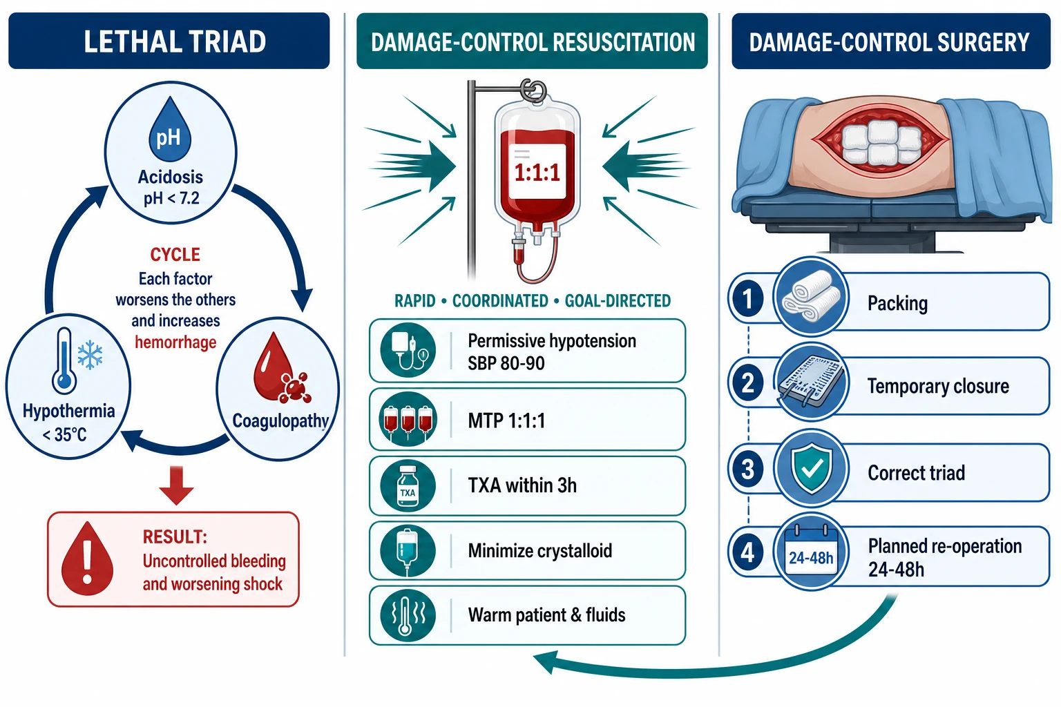
- The acidosis (pH under 7.2) — the clotting factor dysfunction (the enzymatic).[1]
- The hypothermia (temp under 35 degrees C) — the platelet and the enzyme the dysfunction.[1]
- The coagulopathy — the more the bleeding → the more the acidosis and the hypothermia → the vicious the cycle.[1]
DCR (the physiology)
- The permissive hypotension — the SBP 80 to 90 mmHg (the MAP 65) until the bleeding controlled. The avoid the fluid the over-load (the dislodges the clot, the dilutes the factors, the worsens the acidosis). The NOT for the TBI (the need the CPP — the SBP over 110).[1]
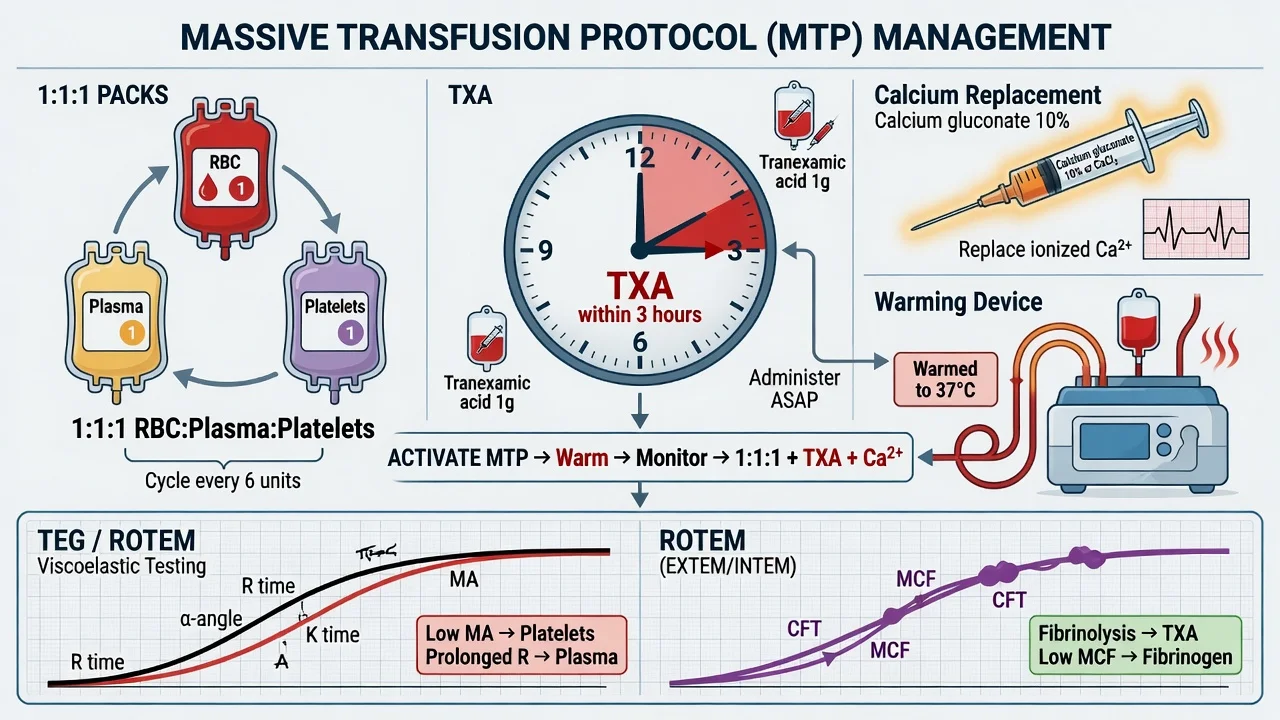
- The MTP 1:1:1 (the RBC: the plasma: the platelets) — the PROPPR trial. The rapid the infuser; the warm.[1]
- The TXA (the 1 g plus 1 g within 3 hours — the CRASH-2).[1]
- The minimise the crystalloid (the worsens the dilutional the coagulopathy and the acidosis).[1]
- The warm (the fluid the warmer; the active the warming; the blankets).[1]
- The calcium (the citrate the chelation — the ionized the Ca over 1.0; the empiric the 1 g calcium the chloride the every 4 units RBC).[1]
DCS (the anatomy)
- The abbreviated laparotomy — the NOT the definitive. The rapid the control:[1]
- The packing (the control the bleeding — the liver, the spleen, the pelvis, the retroperitoneum).[1]
- The ligation (the major the vessel — the shunt the if the limb the perfusion the needed).[1]
- The temporary closure (the Bogota bag, the negative-pressure the dressing, the Wittmann the patch). The NOT the fascial the closure (the risk the abdominal the compartment the syndrome).[1]
- The NOT the anastomosis (the defer to the re-operation; the stapled the - the resect the ischaemic the bowel; the stapled the - leave in discontinuity).[1]
- The correct the lethal triad (the warm, the blood, the TXA, the calcium; the - the in the ICU between the operations).[1]
- The planned re-operation at 24 to 48 hours — the definitive repair (the anastomosis, the - the packing the removal, the closure).[1]
The open abdomen
- The manage the intra-abdominal hypertension (the IAP; the APP).[1]
- The fistula (the entero-atmospheric; the difficult the manage).[1]
- The fluid losses (the large; the replace).[1]
- The closure — the early if the feasible (the 5 to 7 days); the skin the graft if the not (the planned the ventral the hernia the repair).[1]
The three pillars of damage-control resuscitation
DCR rests on three pillars that run concurrently from the point of injury: a physiological pillar, a haematological pillar, and a surgical pillar. Each pillar targets a different driver of the lethal triad. The pillars are not sequential — they are activated together the moment massive haemorrhage is recognised, and they are deactivated together once haemorrhage control is achieved.[7]
The three pillars of damage-control resuscitation (DCR)
Pillar 1 — Permissive hypotension (the physiology)
Resuscitate to a LOW blood pressure until haemorrhage control: SBP 80-90 mmHg (MAP ~50-60) until surgical or endovascular control of bleeding. Rationale: restoring full pressure before haemostasis blows off clots ("pop the clot"), dilutes clotting factors, and accelerates bleeding. Give blood (not crystalloid) only to maintain consciousness and perfusion; once the bleed is controlled, restore normotension. EXCEPTIONS (permissive hypotension is harmful): traumatic brain injury with shock (need CPP >60 → MAP >80, SBP >110); non-trauma bleeding (variceal, ruptured AAA) where evidence is absent.
Pillar 2 — Ratio-based (haemostatic) transfusion (the blood)
Replace lost blood with blood — not crystalloid. During ACTIVE massive haemorrhage give RBC:FFP:platelets in approximately 1:1:1 (delivered as predefined MTP packs). PROPPR (2015): 1:1:1 was safe, achieved earlier haemostasis, and reduced death from exsanguination at 24h versus 1:1:2, with no difference in ARDS or overall mortality. Once the immediate bleed is controlled, switch from blind ratios to viscoelastic (ROTEM/TEG)-guided, goal-directed component therapy to avoid over-transfusion of plasma and platelets.
Pillar 3 — Damage-control surgery (the anatomy)
The index operation is ABBREVIATED — its only goals are to (a) stop surgical bleeding (pack, ligate, shunt), (b) control contamination (staple/resect ischaemic bowel in discontinuity, close hollow viscus holes), and (c) achieve TEMPORARY abdominal closure (Bogota bag, negative-pressure dressing, Wittmann patch). Definitive repairs — anastomoses, packing removal, formal closure — are deferred to a planned re-operation at 24-48h once the lethal triad is corrected in the ICU.
Pillar 1 in depth — permissive hypotension
Permissive hypotension means deliberately tolerating a sub-normal blood pressure in the actively bleeding patient. The single physiological target is SBP 80-90 mmHg (or a MAP around 50-60 mmHg) until haemorrhage control. The mechanism is mechanical: a clot is a friable plug, and the harder the arterial pressure pushing against it, the more likely it is to be dislodged ("pop the clot"). Premature normalisation of pressure therefore converts a controllable venous/oozing bleed into a torrential arterial one.[7]
The trade-off is deliberate, temporary under-perfusion. Most young trauma patients tolerate SBP 80-90 well, because the goal of resuscitation in this window is perfusion sufficient to maintain consciousness and end-organ oxygen delivery, not a normal number. Give blood, not crystalloid, to hold this floor; once surgical, endovascular (REBOA) or angiographic control is achieved, restore normotension.[1]
[1]Pillar 2 in depth — haemostatic, ratio-based transfusion
The second pillar replaces shed blood with blood, in a ratio that mimics whole blood. During the uncontrolled phase of massive haemorrhage, give RBC:FFP:platelets ~1:1:1 (one adult dose of platelets ≈ one apheresis unit ≈ equivalent to a 6-pack of pooled platelets). The aim is to prevent the onset and deepening of coagulopathy by delivering clotting factors and platelets concurrently with red cells, rather than chasing a dilutional coagulopathy that has already developed.[2]
The evidence is PROPPR (2015): a 1:1:1 ratio did not change 24-hour or 30-day mortality compared with 1:1:2, but it achieved earlier haemostasis and fewer deaths from exsanguination at 24h, with no increase in ARDS, multi-organ failure, or thrombotic complications. The pragmatic reading: 1:1:1 is safe and reasonable to use empirically during active massive bleeding, then de-escalate.[2]
The critical transition — and the most common exam trap — is that ratio-based transfusion is a bridge, not a destination. Once the immediate haemorrhage is controlled and laboratory/viscoelastic data return, switch to goal-directed component therapy driven by ROTEM/TEG, fibrinogen, platelet count, and ionised calcium. Reflexively continuing 1:1:1 leads to over-transfusion of plasma and platelets, volume overload, and increased ARDS/MOF.[9]
Ratio-based (empiric) phase
Active uncontrolled haemorrhage
- Used during the FIRST phase — before laboratory data are available
- Deliver RBC:FFP:platelets ~1:1:1 as predefined MTP packs
- Goal: prevent the onset of coagulopathy, deliver clotting factors concurrently
- Give empiric calcium, TXA, warm all products
- Time-limited: switch out as soon as bleeding slows
Goal-directed (viscoelastic) phase
Bleeding controlled / slowing
- Used once ROTEM/TEG, fibrinogen, platelet count, Ca²⁺ are available
- Replace the SPECIFIC deficient component — not a fixed ratio
- CT/R prolonged → FFP; A10/α-angle low → fibrinogen (cryo/concentrate); MA/MCF low → platelets; ML/LY30 high → TXA
- Avoids over-transfusion of plasma/platelets → less ARDS/MOF/TACO
- De-activate MTP once haemostasis and haemodynamic stability achieved
Pillar 3 in depth — damage-control surgery
Damage-control surgery (DCS) is the abbreviated laparotomy. The index operation is intentionally incomplete: its only purpose is to deliver the patient alive to the ICU with correctable physiology, not to perform definitive repairs. Every additional minute on the operating table with an open abdomen on a cold, coagulopathic, acidotic patient deepens the lethal triad and increases mortality.[10]
The three operative goals of DCS are: (1) haemostasis — packing (liver, spleen, pelvis, retroperitoneum), suture/ligation of accessible vessels, temporary intraluminal shunt for a vessel whose repair is needed to preserve limb or organ perfusion; (2) control of contamination — close hollow-viscus injuries, resect ischaemic/non-viable bowel with a linear stapler and leave it in discontinuity (NOT a primary anastomosis); (3) temporary abdominal closure — Bogota bag, commercial negative-pressure dressing (VAC), or Wittmann patch, deliberately leaving the fascia OPEN to avoid abdominal compartment syndrome.[10]
Definitive reconstruction — bowel anastomoses, packing removal, pancreatic/biliary repairs, and formal fascial closure — is performed at a planned re-operation at 24-48 hours, after the lethal triad has been corrected in the ICU.[1]
Trauma-induced coagulopathy (TIC)
Trauma-induced coagulopathy (TIC) is an endogenous hypocoagulable state present on arrival in ~25% of severely injured patients — before any fluid has been given. This is the single most important conceptual point for the exam: TIC is NOT dilutional (it is not caused by crystalloid or stored-blood transfusion). It is driven by shock and tissue injury.[6]
Mechanism of TIC — the shock → coagulopathy axis
The unifying mechanism is tissue hypoperfusion + endothelial activation, producing a systemic anticoagulant and hyperfibrinolytic state:[6]
- Hypoperfusion + tissue injury release tissue factor (TF) from disrupted tissues and activated endothelium.
- TF drives thrombin-thrombomodulin complex formation; in shock this activates protein C (endogenous anticoagulant) rather than clot.
- Activated protein C consumes factors Va and VIIIa (anticoagulation) and degrades plasminogen activator inhibitor-1 (PAI-1) — loss of PAI-1 permits unopposed hyperfibrinolysis (clot breakdown).
- Endothelial glycocalyx shedding and sympathetically-driven platelet dysfunction compound the effect. [1]
The result is a low-clot-strength, hyperfibrinolytic picture on viscoelastic testing, with a prolonged CT/R and elevated lysis (ML/LY30). TIC multiplies mortality by 3-4×.[6]
Trauma-induced coagulopathy (TIC)
Endogenous, present on arrival
- Present BEFORE any fluid or blood given (~25% of severe trauma)
- Driven by tissue hypoperfusion + endothelial activation (NOT dilution)
- Mechanism: tissue factor + protein C activation → anticoagulation + hyperfibrinolysis
- Diagnosis: viscoelastic testing — prolonged CT/R, low clot strength, raised ML/LY30
- 3-4× mortality; the target of TXA (anti-fibrinolytic) and early plasma
Dilutional / iatrogenic coagulopathy
Caused by our resuscitation
- Develops AFTER large volumes of crystalloid or stored RBC without plasma/platelets
- Driven by dilution of clotting factors and platelets + citrate-induced hypocalcaemia
- Aggravated by hypothermia and acidosis (the lethal triad)
- Prevented by DCR: minimise crystalloid, 1:1:1 ratios, give calcium, warm
- Reversible with component therapy and correction of the triad
Detecting TIC at the bedside
Conventional coagulation tests (INR, aPTT) are slow (30-60 min), report plasma rather than whole-blood function, and correlate poorly with bleeding. Viscoelastic testing (ROTEM/TEG) gives a whole-blood clot profile in 10-20 minutes and is the diagnostic standard. Key thresholds: INR >1.5 on arrival suggests TIC; on ROTEM, a prolonged CT (clotting time) and elevated maximum lysis (>15%) are the hallmarks.[9]
Tranexamic acid (TXA) — CRASH-2 and the 3-hour rule
Tranexamic acid is an anti-fibrinolytic (lysine analogue that blocks plasminogen activation). In trauma it counteracts the hyperfibrinolytic component of TIC. The dose is 1 g IV over 10 minutes, then 1 g over 8 hours.[3]
The landmark trial is CRASH-2 (2010, 20,211 patients): TXA reduced all-cause mortality (14.5% vs 16.0%) and bleeding death (4.9% vs 5.7%), with no increase in thromboembolic events.[3] The crucial refinement came from the time-window analysis (CRASH-2, 2011): benefit is greatest when TXA is given within 1 hour of injury, present when given within 3 hours, but TXA INCREASES mortality when given more than 3 hours after injury.[4] The military MATTERs study corroborated a survival benefit in combat casualties, including in those requiring massive transfusion.[5]
CRASH-2 (Lancet 2010)
Multicentre, placebo-controlled RCT; 20,211 trauma patients with or at risk of major bleeding
Population: Adult trauma patients with significant haemorrhage, 274 hospitals across 40 countries
Key finding
TXA reduced all-cause mortality (14.5% vs 16.0%, p=0.0035) and bleeding death (4.9% vs 5.7%, p=0.0077), with no increase in vascular occlusive events.
Practice change
Give TXA 1 g IV ASAP in trauma bleeding. Benefit is greatest within 1 hour and present within 3 hours; after 3 hours it is harmful.
CRASH-2 time-window analysis (Lancet 2011)
Exploratory analysis of CRASH-2 by time from injury to treatment
Population: 13,966 patients with bleeding deaths analysed by treatment timing
Key finding
Bleeding deaths reduced: within 1h (5.3% vs 7.7%) and within 1-3h. Given >3 hours after injury, TXA INCREASED bleeding death (4.4% vs 3.1%).
Practice change
The 3-hour rule: TXA must be given within 3 hours of injury (ideally within 1 hour, pre-hospital if possible). Never give TXA >3 hours after injury.
PROPPR (Holcomb, JAMA 2015)
Multicentre RCT; 680 severely injured adults predicted to need massive transfusion
Population: Trauma patients at 12 Level I trauma centres in North America
Key finding
No significant difference in 24h mortality (12.7% vs 17.0%, p=0.09) or 30-day mortality. BUT 1:1:1 achieved earlier haemostasis and fewer exsanguination deaths at 24h, with no increase in ARDS or multi-organ failure.
Practice change
1:1:1 is safe and reasonable empirically during active massive bleeding — it improves early haemostasis. There is no overall mortality advantage, so switch to viscoelastic-guided therapy as soon as possible.
Viscoelastic testing — ROTEM/TEG-guided resuscitation
Viscoelastic tests (TEG, ROTEM) measure the kinetics and strength of whole-blood clot formation and breakdown in real time (10-20 minutes). They are the foundation of goal-directed haemostatic resuscitation and have superseded the reflexive use of conventional ratios once the immediate bleed is controlled.[9]
| TEG parameter | ROTEM equivalent | What it measures | Abnormal finding | Treatment |
|---|---|---|---|---|
| R (reaction time) | CT (clotting time) | Clotting factor initiation | Prolonged R/CT | FFP (or prothrombin complex concentrate if available) |
| α-angle, K | A10/A20, α-angle | Fibrin polymerisation (fibrinogen) | Low α / low A10 | Cryoprecipitate or fibrinogen concentrate (target fibrinogen >1.5-2.0 g/L) |
| MA (max amplitude) | MCF (max clot firmness) | Platelet contribution to clot strength | Low MA / MCF | Platelets (1 adult dose) |
| LY30, EPL | ML (max lysis) | Clot breakdown (fibrinolysis) | ML >15% | TXA 1 g IV |
Massive transfusion protocol (MTP) — activation and deactivation
Massive transfusion is defined as replacement of >1 blood volume in 24 hours, or >50% blood volume in 3 hours, or >4 units RBC in 1 hour with ongoing bleeding. The MTP is the institutional machine that delivers balanced blood products rapidly. Activate early — if you are thinking about activating it, activate it.[1]
MTP activation, delivery and deactivation
Activation criteria
Activate on any of: (a) active massive bleeding (>150 mL/min or >4 units RBC in 1h); (b) severe trauma with haemodynamic instability and predicted ongoing bleeding; (c) clinical judgement — impending exsanguination, positive FAST with shock, major pelvic fracture with haemodynamic compromise. A single activation call triggers blood bank, lab, and team mobilisation.
Pack delivery
Pre-defined packs delivered rapidly. A typical pack: 4 units RBC + 4 units FFP + 1 adult dose platelets (~1:1:1), plus cryoprecipitate (10 units) and calcium. Use group O RBC and AB plasma (or low-titre O whole blood) until type-specific products are available. Draw blood every 30-60 min for ROTEM/TEG, fibrinogen, platelets, ionised Ca²⁺, and haemoglobin.
Adjuncts given with every pack
TXA 1 g IV (within 3h of injury); empiric calcium chloride 10 mmol (1 g) per ~4 units blood products (citrate chelates Ca²⁺); warm ALL products to ~37°C (rapid infuser with integrated warmer); minimise crystalloid; if crystalloid is unavoidable, use a balanced solution (Hartmann/Plasma-Lyte), never normal saline (hyperchloraemic acidosis worsens coagulopathy).
Transition to goal-directed therapy
As soon as bleeding slows and viscoelastic/standard labs return, switch from fixed 1:1:1 ratios to ROTEM/TEG-guided component therapy (CT→FFP, A10/α→fibrinogen, MA/MCF→platelets, ML→TXA). This prevents over-transfusion of plasma and platelets and the consequent ARDS/MOF.
Deactivation
Stand the MTP down when haemorrhage is controlled AND haemodynamic stability is achieved. After deactivation, screen for transfusion complications: TRALI, TACO, hyperkalaemia (stored RBC K⁺ up to 70-80 mmol/L), citrate-induced hypocalcaemia, and recheck coagulation, electrolytes, haemoglobin, and fibrinogen.
Transition from damage-control to definitive care
A defining feature of the damage-control philosophy is the deliberate transition between three phases: a temporising resuscitative phase, a restorative ICU phase, and a definitive reconstructive phase. The handover between them is governed by physiology, not the clock.[10]
The three phases of damage control
Phase 0–1 — Index operation + DCR (minutes to <2h)
Concurrent DCR and abbreviated laparotomy. Goals: permissive hypotension, 1:1:1 transfusion, TXA, calcium, warm; operative goals are haemostasis, contamination control, temporary closure. The patient is transferred to ICU cold, acidotic and coagulopathic — physiology is the priority.
Phase 2 — ICU resuscitation (first 24-48h)
Correct the lethal triad: rewarm (forced-air warming, warmed fluids, warm ambient temperature); correct acidosis (restore perfusion — stop the bleed, give blood; avoid bicarbonate except in extremis); correct coagulopathy (viscoelastic-guided plasma, fibrinogen, platelets, TXA, calcium). Continue ventilation with lung-protective settings, treat pain, VTE prophylaxis once safe, and monitor the open abdomen for abdominal compartment syndrome.
Phase 3 — Planned re-operation (24-48h)
Definitive reconstruction once physiology restored: remove packs, perform bowel anastomoses, repair the pancreas/biliary tree/bladder, and attempt definitive fascial closure. If the abdomen is still hostile, re-pack and re-close temporarily, with a further planned re-operation. Each return to theatre is an opportunity to close the abdomen before it becomes chronically open.
Phase 4 — Definitive care & closure
After physiological recovery and source control, pursue early definitive fascial closure (ideally by days 5-7). If primary closure is not achievable, manage the open abdomen with a planned ventral hernia and split-thickness skin graft, with delayed reconstruction. The transition out of DCR is complete when haemostasis is secure, the triad is corrected, and definitive repairs are in place.
Criteria for moving from damage control to definitive surgery
The patient is ready for definitive reconstruction once all of the following are met:[1]
- Haemostasis achieved — no ongoing surgical or angiographic bleeding; stable haemoglobin off transfusion.
- Lethal triad corrected — temperature >35°C, pH >7.25 (lactate falling), INR near normal / viscoelastic tracing normal.
- Haemodynamically stable — off or on low-dose vasopressors, restoring normotension after a period of permissive hypotension.
- Adequate perfusion — improving lactate, adequate urine output, no ongoing end-organ failure. [1]
The lethal triad — severity and interactions
The lethal triad of massive haemorrhage (click each)
INR >1.5
Trauma-induced coagulopathy (endogenous) plus dilutional coagulopathy (iatrogenic) plus consumption. INR >1.5 on arrival defines TIC. The three elements of the triad reinforce one another; once fully established, mortality approaches 100%. Break the cycle with DCR: minimise crystalloid, 1:1:1 then viscoelastic-guided components, TXA, fibrinogen, calcium, and rewarming.
Complications of damage-control resuscitation and the open abdomen
Metabolic / storage-related
From stored blood and citrate
- Hypocalcaemia — citrate chelates Ca²⁺; give CaCl₂ 10 mmol per ~4 units blood; monitor ionised Ca²⁺
- Hyperkalaemia — stored RBC leak K⁺ (up to 70-80 mmol/L in old units); arrhythmia risk; monitor
- Acid–base — early acidosis (stored blood pH ~6.6-7.0), then alkalosis as citrate metabolises to bicarbonate
- Hypothermia — warm all products to ~37°C
Transfusion reactions
Immunological
- TRALI — donor antibodies vs recipient neutrophils → ARDS; supportive care
- TACO — circulatory overload; diuretics
- ABO-incompatible haemolysis — rare, fatal; stop transfusion immediately
- Transfusion-transmitted infection — rare (viral, bacterial)
Open-abdomen complications
From temporary closure
- Abdominal compartment syndrome — measure IAP; keep APP (MAP − IAP) >50-60 mmHg
- Entero-atmospheric fistula — very difficult to manage; protect exposed bowel
- Massive third-space fluid losses — replace; nutrition critical
- Failed / delayed closure — planned ventral hernia + split-thickness skin graft
Over-resuscitation
From prolonged ratio-based transfusion
- ARDS — over-transfusion of plasma/platelets and crystalloid
- Multi-organ failure — driven by overload and the inflammatory hit
- Dilutional coagulopathy — if crystalloid-led instead of blood-led
- Prevent by early transition to viscoelastic-guided therapy and prompt MTP deactivation
Red flags
Exam practice
SAQ — Damage-control resuscitation in penetrating torso trauma
12 minutes · 12 marks
A 26-year-old man is brought to the emergency department 40 minutes after a single stab wound to the abdomen. He is drowsy (GCS 13), BP 74/46, HR 132, saturations 95% on 15 L O₂. FAST is positive. He has received 2 units of O-negative RBC pre-hospital. Temp 35.0°C, pH 7.16, lactate 7.2, INR 1.9, fibrinogen 1.2 g/L, ionised Ca²⁺ 0.82 mmol/L. He is taken immediately to theatre.
Clinical pearls
References
- [1]Holcomb JB, Jenkins D, Rhee P, et al. Damage control resuscitation: directly addressing the early coagulopathy of trauma J Trauma, 2007.PMID 17297317
- [2]Holcomb JB, Tilley BC, Baraniuk S, et al. (PROPPR Study Group) Transfusion of plasma, platelets, and red blood cells in a 1:1:1 vs a 1:1:2 ratio and mortality in patients with severe trauma: the PROPPR randomized clinical trial JAMA, 2015.PMID 25647203
- [3]CRASH-2 trial collaborators Effects of tranexamic acid on death, vascular occlusive events, and blood transfusion in trauma patients with significant haemorrhage (CRASH-2): a randomised, placebo-controlled trial Lancet, 2010.PMID 20554319
- [4]CRASH-2 collaborators The importance of early treatment with tranexamic acid in bleeding trauma patients: an exploratory analysis of the CRASH-2 randomised controlled trial Lancet, 2011.PMID 21439633
- [5]Morrison JJ, Dubose JJ, Rasmussen TE, Midwinter MJ Military Application of Tranexamic Acid in Trauma Emergency Resuscitation (MATTERs) Study Arch Surg, 2012.PMID 22006852
- [6]Brohi K, Singh J, Heron M, Coats T Acute coagulopathy of trauma: hypoperfusion induces systemic anticoagulation and hyperfibrinolysis J Trauma, 2008.PMID 18469643
- [7]Cannon JW, Khan MA, Raja AS, et al. Damage control resuscitation in patients with severe traumatic hemorrhage: A practice management guideline from the Eastern Association for the Surgery of Trauma J Trauma Acute Care Surg, 2017.PMID 28225743
- [8]Spahn DR, Bouillon B, Cerny V, et al. The European guideline on management of major bleeding and coagulopathy following trauma: fifth edition Crit Care, 2019.PMID 30917843
- [9]Brill JB, Brenner M, Duchesne J, et al. The Role of TEG and ROTEM in Damage Control Resuscitation Shock, 2021.PMID 33769424
- [10]Rotondo MF, Schwab CW, McGonigal MD, et al. 'Damage control': an approach for improved survival in exsanguinating penetrating abdominal injury J Trauma, 1993.PMID 8371295