ICU · Trauma
Fat Embolism Syndrome — The 12–72h Triad, Gurd's Criteria & Lung-Protective Support
Also known as Fat embolism syndrome · FES · Fat embolism · Petechial rash · Triad fat embolism · Gurd's criteria · Cerebral fat embolism · Fat emboli · Post-fracture respiratory failure · Intramedullary nailing embolism
Fat embolism syndrome (FES) is a clinical diagnosis defined by the triad of respiratory insufficiency, neurological dysfunction and a petechial rash arising 12 to 72 hours after a major injury — classically a long bone (femur, tibia) or pelvic fracture, but also orthopaedic procedures (intramedullary nailing, joint replacement), liposuction, pancreatitis, sickle-cell crisis and bone-marrow transplant. The pathophysiology is dual — a MECHANICAL phase in which marrow fat globules embolise to the pulmonary and cerebral microvasculature, and a BIOCHEMICAL phase in which lipase hydrolyses the embolic triglyceride to free fatty acids that ignite an inflammatory cascade, injure the endothelium and produce an ARDS-like lung. The diagnosis rests on Gurd's criteria (major: respiratory insufficiency, cerebral involvement, petechial rash; minor: fever, tachycardia, retinal changes, jaundice, renal changes, thrombocytopenia). Management is SUPPORTIVE: oxygen, lung-protective ventilation (6 mL/kg, plateau <30 cmH₂O) for the ARDS-like lung, cautious fluids, vasopressors; early surgical stabilisation of the fracture (within 24 h) is the single most effective preventive measure; high-dose corticosteroid prophylaxis reduces incidence in meta-analysis but remains controversial and is not routine. Mortality is 5–15%; most deaths are from multi-organ failure, not FES itself.
On this page & tools
Your progress
Saved locally on this device.
8 MCQs with explanations
Target exams
Overview & definition
The fat embolism syndrome (FES) is a multisystem disorder characterised by the triad of respiratory insufficiency, neurological dysfunction and a petechial rash that declares itself 12 to 72 hours after the entry of medullary fat into the venous circulation — most often from a long-bone (femur, tibia) or pelvic fracture, but also from orthopaedic surgery, liposuction, pancreatitis, sickle-cell crisis, bone-marrow transplant and parenteral lipid infusion.[7][11]
The cardinal points for the exam: (1) it is a clinical diagnosis — no single laboratory test confirms it; (2) the timing is the discriminant — respiratory failure in the first 1–2 hours after injury is contusion, aspiration or haemorrhage, NOT FES; (3) marrow fat embolises to EVERY major trauma patient, but only a minority (1–5% of isolated long-bone fractures, up to 10–20% of multiple fractures) develop the syndrome; and (4) management is supportive with an ARDS lung-protective ventilatory strategy, and the most effective treatment is prevention by early fracture fixation.[2][5][9]
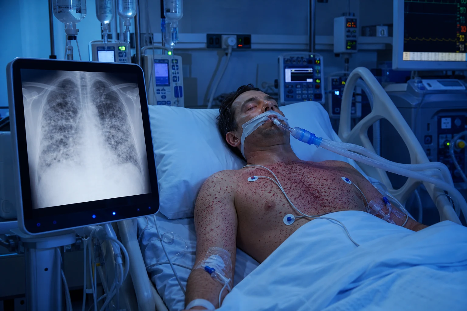
Epidemiology & incidence — fat emboli versus the fat embolism syndrome
The single most important epidemiological distinction is between fat embolism (the histological/radiological presence of marrow fat in the circulation — present to some degree in almost every major trauma patient) and the fat embolism syndrome (the clinical expression in a minority). Confusing the two leads to over-diagnosis.[7][11]
- Incidence of the syndrome: ~1–5% of isolated long-bone fractures; 10–20% with multiple long-bone fractures or a femur + pelvic combination; up to 35% historically in unplated major lower-limb trauma before early fixation was standard. Mortality 5–15%; full recovery in the majority.[2][9]
- Risk is proportional to the energy and marrow burden: closed fractures > open (the open fracture decompresses the medullary canal); femur > tibia > humerus; polytrauma and high ISS; age 10–40 (the marrow is fatty and the bones are high-energy); male sex.[8][11]
- latrogenic / non-traumatic triggers — recognise these because the patient may have NO fracture: intramedullary nailing, hip/knee arthroplasty (cemented, pressurisation), Kuntscher rod insertion, liposuction, medullary reaming, bone-marrow harvest/transplant, parenteral lipid emulsion, sickle-cell crisis (marrow infarction), acute pancreatitis, decompression sickness, and high-dose corticosteroid withdrawal.[7][14]
Triggers — the #1 is the long bone fracture
Traumatic (the #1 group)
Femur > tibia > humerus; pelvic
- LONG-BONE FRACTURE is the #1 cause — the femoral and tibial shafts hold the largest volume of fatty marrow; a closed, displaced, high-energy fracture forces marrow into the torn venous sinuses
- PELVIC FRACTURE (especially the sacroiliac region) — high marrow content, and the FES sits alongside massive haemorrhage as a competing cause of instability
- MULTIPLE long-bone fractures or femur + pelvis — the risk multiplies; this is the patient to watch on the ICU between 12 and 72 hours
- Closed > open fracture: an open fracture vents the medullary pressure, so FES is rarer; a closed high-energy shaft fracture is the classic substrate
- Less common: rib/sternal fracture, spinal fracture with marrow exposure, severe soft-tissue crush
Iatrogenic / surgical
Pressurisation of marrow
- INTRAMEDULLARY NAILING of the femur or tibia — reaming and nail insertion pressurise the canal; echocardiography during nailing shows showers of echogenic material crossing the right heart (the "snowstorm") and, with a patent foramen ovale, crossing to the left
- Cemented HIP/KNEE ARTHROPLASTY — cement and pressurisation force marrow into the venous system; the bone-cement implantation syndrome is a related but distinct entity (hypotension, hypoxia, cardiac arrest at cementation)
- Medullary reaming, Kuntscher rodding, plate fixation of long bones
- LIPOSUCTION — large-volume aspiration liberates fat; FES and lidocaine toxicity (tumescent) coexist
- Bone-marrow harvest/transplant, orthopaedic tumour reamed intramedullary work
Non-traumatic / medical
Endogenous fat release
- ACUTE PANCREATITIS — circulating lipase and the enzymatic release of free fatty acids injure distant endothelium and may produce a FES-like picture without a fracture
- SICKLE-CELL CRISIS — marrow infarction releases fat; cerebral fat embolism is a recognised cause of acute neurological deficit in sickle-cell disease
- Parenteral LIPID EMULSION (e.g. propofol infusion at high dose, TPN) — overload and embolisation of infused lipid
- Decompression sickness, severe burns (marrow necrosis), high-dose corticosteroid withdrawal, fatty liver of pregnancy
- These non-traumatic forms are easily missed because the operator is not expecting FES in a "medical" patient
Pathophysiology — the two-hit mechanical + biochemical model
FES is best understood as a two-mechanism model — a mechanical phase and a biochemical phase acting in sequence and amplifying each other. Understanding this model explains why the lungs dominate the picture, why the brain is involved despite all emboli being venous, and why the syndrome looks like ARDS.[8][11]
The mechanical phase — fat globules embolise to the pulmonary capillaries
The marrow sinusoids are torn by the fracture; the sudden rise in intramedullary pressure forces liquid fat globules (30–100 µm, too large to traverse the pulmonary capillary bed) into the draining veins. The globules travel via the right heart to the pulmonary microvasculature, where they mechanically obstruct precapillary arterioles and capillaries, producing an acute rise in pulmonary vascular resistance, a degree of right-heart strain, and an early, often mild, ventilation–perfusion mismatch. This mechanical phase accounts for the immediate (minutes to hours) hypoxaemia and the transient pulmonary hypertension seen during intramedullary nailing.[8][14]
In some patients a fraction of the fat bypasses the lung: through a patent foramen ovale (present in ~25% of adults), through pulmonary precapillary arteriovenous anastomoses, or by simple distension of capillaries under high pressure — delivering fat to the systemic (cerebral and retinal) circulation and explaining the cerebral and petechial manifestations.[13]
The biochemical phase — free fatty acids → inflammation → endothelial damage → ARDS
The embolic triglyceride is itself relatively inert. The damage is done by its hydrolysis. Pulmonary lipase (and tissue lipase) cleaves the triglyceride to free fatty acids (FFAs) — toxic, detergent-like molecules that:[8][11]
- Directly injure the pulmonary capillary endothelium → increased permeability, protein-rich oedema, haemorrhage into the alveoli — the histological picture is a haemorrhagic alveolitis.
- Activate the inflammatory cascade — complement (C5a), neutrophils, platelets, cytokines (TNF-α, IL-1, IL-6, IL-8), thromboxane and leukotrienes — producing a systemic inflammatory response and amplifying endothelial injury.
- Generate a coagulopathy — platelet activation and consumption (thrombocytopenia is a Gurd minor criterion), microvascular thrombi, and a clinical overlap with early DIC. [1]
The end result is a permeability-type pulmonary oedema that is clinically and radiologically indistinguishable from ARDS — bilateral diffuse alveolar infiltrates, refractory hypoxaemia, reduced compliance. The time lag between the fracture and the onset (12–72 h) reflects the time required for lipase hydrolysis and the inflammatory cascade to develop — this is the biological basis of the delayed presentation.[6][11]
The brain — systemic embolism, not primary lung failure
Cerebral involvement is from systemic fat emboli (paradoxical or via pulmonary AV shunts), NOT simply hypoxia. Autopsy studies show microvascular fat and petechial haemorrhages distributed in the white matter and brainstem — a pattern distinct from anoxic encephalopathy. This is why neurological signs can precede the respiratory failure, and why they can be focal or bizarre (not the symmetric picture of hypoxia).[13]
The petechial rash — the pathognomonic clue
The petechial rash is the embolisation of platelet-fibrin-fat microthrombi to the dermal capillaries, with thrombocytopenia amplifying it. Its distribution over the upper chest, axillae, neck, conjunctivae and the oral mucosa reflects dependent and gravity-independent zones with abundant capillaries — and its transient nature (resolves in 24–48 h) is the reason the diagnosis is missed if the skin is not examined early and serially.[2][9]
The two-hit pathophysiology — fracture to syndrome
Hit 1 — Mechanical embolisation (minutes to hours)
A high-energy long-bone or pelvic fracture tears the medullary sinusoids; a sharp rise in intramedullary pressure forces 30–100 µm fat globules into the torn draining veins. The globules travel via the right heart and mechanically lodge in the pulmonary capillary bed (too large to traverse). Early consequences: a transient rise in pulmonary vascular resistance and right-heart strain, mild early hypoxaemia from V/Q mismatch, and (with a patent foramen ovale in ~25%) paradoxical embolism to the brain and retina.
Latent interval (the 12–72 h window)
The patient often looks well in the first hours. During this window pulmonary and tissue lipase hydrolyse the embolic triglyceride to free fatty acids, and the inflammatory cascade is recruited (complement, neutrophils, cytokines, platelet activation, thromboxane). This biochemical amplification is why FES is delayed and progressive — and why the patient who looked fine on admission deteriorates the next day.
Hit 2 — Biochemical / inflammatory injury (12–72 h)
The free fatty acids and the inflammatory mediators strip and injure the pulmonary capillary endothelium → a permeability-type oedema that is indistinguishable from ARDS (bilateral infiltrates, refractory hypoxaemia, poor compliance). Systemic emboli produce the cerebral microhaemorrhages (confusion, seizures, coma) and the dermal capillary microthrombi (the petechial rash). Platelet consumption produces thrombocytopenia.
The full syndrome (24–72 h)
The clinical triad is now expressed: respiratory insufficiency (ARDS-like, often the most severe and the usual reason for ICU admission), neurological dysfunction (confusion to coma, occasionally focal or seizure), and the petechial rash (50–60%, transient — examine early). A minority progress to multi-organ failure; most recover over 1–2 weeks with supportive care.
The clinical triad & timing
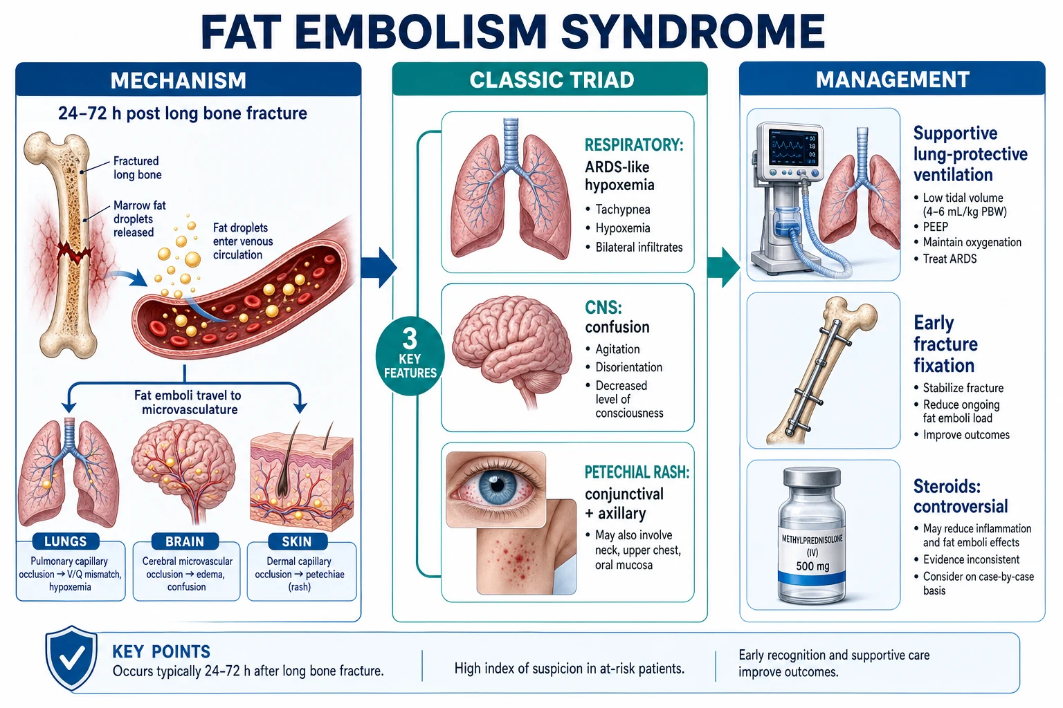
The triad, in order of frequency and importance:[2][9]
- Respiratory insufficiency (75–85%, the commonest and the leading reason for ICU admission): the earliest and most reliable sign is hypoxaemia out of proportion to the mechanism — a falling SpO₂, tachypnoea, increasing work of breathing, then frank ARDS (bilateral diffuse alveolar infiltrates on the CXR developing over hours, refractory hypoxaemia, reduced compliance). Almost every patient with FES has some degree of respiratory involvement.
- Neurological dysfunction (~80%, often second): ranges from subtle confusion and agitation to obtundation, seizures and coma. The picture is non-focal and fluctuating (a toxic-metabolic encephalopathy pattern), though focal deficits and bizarre behaviour occur from discrete cerebral microemboli. Neurological signs may precede the respiratory signs and are the source of the most exam-worthy confusion with traumatic brain injury.[13]
- Petechial rash (50–60%, the pathognomonic but transient feature): a 1–2 mm non-blanching eruption over the conjunctivae, oral mucosa, neck, upper chest, axillae (the dependent, gravity-independent skin of the upper body). It appears 24–36 h after injury, lasts 24–48 h, and may be the only sign that clinches the diagnosis — examine the conjunctivae, oral mucosa and axillae of every polytrauma patient at 24 h.[2]
Timing — the discriminant against early diagnoses
The 12–72 hour window is the single most useful clinical discriminant:[7][11]
- Immediate (0–2 h) respiratory failure after chest trauma = pulmonary contusion, tension pneumothorax, haemothorax, aspiration — NOT FES. FES is excluded by being too early.
- 2–12 h = the latent period; the patient often looks well; this is when the biochemical cascade is building.
- 12–72 h = the classical window; a previously stable polytrauma patient who now hyperventilates, desaturates and becomes confused has FES until proven otherwise.
- Beyond 72 h is unusual but recognised; late or stuttering presentations occur, particularly with delayed or repeated surgical manipulation of the fracture. [1]
Gurd's criteria — the diagnostic framework
Gurd and Wilson (1970–1974) formalised the diagnosis. The classic construct requires at least one major criterion plus four minor criteria, plus the presence of macroglobulinaemia (fat macroglobulinuria). In modern practice the criteria are used more flexibly — the triad of respiratory + CNS + petechial rash in the right temporal window is itself diagnostic, and Gurd's criteria are the structured way to defend the diagnosis in a vivva.[1][2]
MAJOR criteria (the triad)
Each alone is highly suggestive
- RESPIRATORY INSUFFICIENCY — tachypnoea, dyspnoea, hypoxaemia, bilateral infiltrates on CXR (ARDS-like). The commonest major criterion.
- CEREBRAL INVOLVEMENT — confusion, altered consciousness, drowsiness, seizures, focal deficit (non-focal encephalopathy pattern is typical).
- PETECHIAL RASH — the pathognomonic sign. Conjunctivae, oral mucosa, neck, upper chest, axillae. Transient (examine early and serially).
MINOR criteria (supporting)
Systemic & laboratory
- FEVER (typically 38–39°C, non-infective initially)
- TACHYCARDIA (HR >110, out of proportion to volume status)
- RETINAL CHANGES — petechiae, cotton-wool spots, or fat globules seen on fundoscopy (examine the fundi — a positive finding is highly specific)
- JAUNDICE (mild, from haemolysis and hepatic microemboli; usually delayed)
- RENAL CHANGES — oliguria, haematuria, proteinuria, a rising creatinine (fat and microthrombi in the renal microcirculation)
- THROMBOCYTOPENIA — a falling platelet count is one of the most useful early laboratory clues
Laboratory / ancillary markers
Supportive, not definitive
- FAT MACROGLOBULINAEMIA / fat in urine or sputum cytology (the Gurd laboratory criterion) — specific but insensitive and not routinely available
- Sudden unexplained ANAEMIA (haemolysis / haemodilution) and a falling platelet count
- Hypofibrogenaemia / fibrin degradation products (coagulopathy overlap with early DIC)
- Hypocalcaemia (calcium saponification by free fatty acids), mild hyperbilirubinaemia
- ECG changes (ischaemic-pattern T-wave changes, arrhythmia) — from myocardial microemboli and hypoxaemia
Differential diagnosis — what FES is NOT
The polytrauma patient at 24–72 hours can deteriorate from many causes; FES is one of several, and the wrong diagnosis delays the right treatment. The petechial rash and the timing are the discriminants, but several mimics must be actively excluded.[7][11]
Pulmonary contusion
Immediate, not delayed
- Develops at the moment of injury, declares itself within 0–6 hours, NOT at 24–72 h
- CXR infiltrates are localised to the area of impact (often unilateral), not the diffuse bilateral ARDS pattern of FES
- Improves over 3–5 days; FES worsens through the window then improves over 1–2 weeks
Aspiration pneumonitis
Early, focal, lobar
- Follows a period of reduced consciousness (intubation, sedation, seizure) — often in the ED
- Right lower lobe and posterior upper lobe predominance; focal rather than diffuse
- Bacterial superinfection (pneumonia) follows at 48–72 h and may cloud the picture
Pulmonary embolism
Focal, pleuritic, RV strain
- Pleuritic pain, sudden dyspnoea, RV strain on echo, regional V/Q mismatch; CXR usually clear or wedge infarct
- The 24–72 h timing can overlap; DVT signs, CTPA, and bedside echo (RV dilatation) discriminate
- A fat embolus itself can produce acute RV strain early — but the full syndrome has the rash and CNS signs PE does not
Sepsis / early ARDS
Fever + source + rising lactate
- FES produces fever and leucocytosis that mimic sepsis; the absence of a focus and the timing argue for FES
- Blood cultures, lactate, and a careful search for a source (line, wound, urine) — treat empirically with antibiotics if unclear, but do not let sepsis work-up delay the lung-protective ventilation
Traumatic brain injury
Focal, structural, on CT
- The confusion of FES is non-focal and fluctuating; TBI gives a focal, structural deficit and an abnormal head CT
- Always image the brain — a CT excludes haemorrhage and oedema; FES may show the diffuse "starfield" pattern on MRI later
- Both can coexist in polytrauma; do not attribute confusion to "just the TBI" in a femur-fracture patient
Transfusion-related acute lung injury (TRALI)
Within 6 h of transfusion
- Onset within 6 hours of transfusion (vs 12–72 h for FES); bilateral infiltrates, hypoxaemia, no elevation in left atrial pressure
- Resolves over 48–72 h; temporally linked to the blood product
Investigations — supportive, not definitive
No single test confirms FES; the diagnosis is clinical (Gurd's criteria in the right temporal context). Investigations serve to support the diagnosis, grade the severity, and exclude the mimics.[7][11]
- Arterial blood gas: the earliest and most useful single test — hypoxaemia (often PaO₂/FiO₂ <300, meeting ARDS criteria) with a respiratory alkalosis from hyperventilation, progressing to a metabolic acidosis as shock and organ failure develop.
- Chest X-ray: typically normal initially, then bilateral diffuse alveolar infiltrates develop over 12–24 h (the picture of ARDS); the radiograph lags behind the hypoxaemia (the CXR may be near-normal while the PaO₂ is dire). Cardiomegaly and effusions are absent.
- Full blood count: a falling platelet count (thrombocytopenia) and a falling haemoglobin (haemodilution/haemolysis) are useful early clues; leucocytosis is common.
- Coagulation: prolonged INR/aPTT, raised D-dimer, low fibrinogen in the more severe cases (overlap with DIC); a falling platelet count plus coagulopathy supports the diagnosis.
- Biochemistry: hypocalcaemia (saponification of free fatty acids), mild hyperbilirubinaemia, raised LDH (haemolysis), and a rising creatinine with urinary fat and protein. Serum lipase may be elevated.
- Fundoscopy: retinal petechiae, cotton-wool spots or visible fat globules (Purtscher-like retinopathy) — a positive finding is highly specific though insensitive.
- Urine and sputum cytology for fat: fat globulinuria / fat in the urine (the Gurd laboratory criterion) supports the diagnosis but is insensitive and not routinely performed.
- CT head: to exclude TBI; cerebral fat embolism may show the "starfield" pattern on MRI (scattered bright dots on diffusion-weighted imaging) — highly suggestive but usually a delayed finding.
- Bronchoalveolar lavage: staining for fat (Sudan) in alveolar macrophages supports the diagnosis but is invasive and not routinely required; positive in a majority of FES patients but also in major trauma without the syndrome (low specificity).
- Echocardiography (transoesophageal, intraoperative): during nailing, shows the "snowstorm" of echogenic material in the right heart; in established FES, look for right-heart strain, pulmonary hypertension and a patent foramen ovale.[8]
- ECG: non-specific ST/T changes, arrhythmia — from myocardial microemboli and hypoxaemia; exclude ischaemia.
Management — supportive, with an ARDS lung-protective strategy
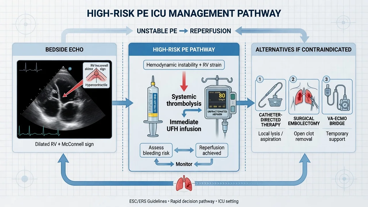
There is no specific therapy for FES. Management is supportive, focused on the respiratory failure (the usual reason for ICU admission) and the prevention of further fat release. The principles are: oxygenate and ventilate with a lung-protective strategy, circulate cautiously, treat the fracture definitively (early fixation), and avoid secondary brain and lung injury.[1][7][11]
Respiratory support — escalate along the oxygen ladder to lung-protective ventilation
- Supplemental oxygen and high-flow nasal cannula (HFNC) or non-invasive ventilation (NIV) for the mild–moderate case with alert, cooperative neurology and PaO₂/FiO₂ >200. Close monitoring — escalation to intubation should not be delayed if work of breathing rises, neurology deteriorates, or gas exchange worsens.
- Invasive mechanical ventilation for the severe case (refractory hypoxaemia, falling GCS, exhaustion, shock). Use a lung-protective ARDS strategy: tidal volume 6 mL/kg predicted body weight, plateau pressure <30 cmH₂O, moderate PEEP titrated to oxygenation, and permissive hypercapnia (pH >7.20). The ARDS Network trial (lower tidal volumes) established the mortality benefit that underpins this approach in the ARDS-like lung of FES.[6]
- Prone positioning for moderate–severe ARDS (PaO₂/FiO₂ <150): the PROSEVA trial showed a 28-day mortality benefit of sustained prone ventilation (16 h/day) in severe ARDS — applicable to the severe FES lung.[12]
- Neuromuscular blockade (48 h cisatracurium) for severe early ARDS improves oxygenation and may reduce mortality; inhaled pulmonary vasodilators (nitric oxide, prostacyclin) are rescue therapies for refractory hypoxaemia.
- ECMO (venovenous) as a last resort for refractory hypoxaemia unresponsive to prone ventilation — a bridge through the biochemical storm, which is self-limiting.
Circulation and fluids — cautious, lung-sparing
- Cautious crystalloid resuscitation — the FES lung is a permeability-type oedema; aggressive fluid deepens the ARDS. Use a balanced crystalloid, target euvolaemia, and consider early colloid or albumin and vasopressors (noradrenaline) to maintain perfusion rather than flooding the lung.
- Vasopressors for shock; inotropes if right-heart failure develops from pulmonary hypertension (sildenafil, inhaled pulmonary vasodilators in selected cases).
- Transfusion for anaemia — keep haemoglobin adequate for oxygen delivery; avoid over-transfusion. [1]
Definitive fracture management — the single most effective treatment
- Early surgical stabilisation of the long-bone or pelvic fracture (within 24 h) is the single most effective intervention to prevent and reduce the severity of FES, by stopping ongoing fat release. The Bone et al. prospective randomised study established that early fixation of femoral fractures reduced pulmonary complications (including FES and ARDS) compared with delayed fixation, especially in polytrauma.[5]
- Reamed versus unreamed intramedullary nailing — reaming pressurises the canal more and liberates more fat; in the patient with established or impending FES or severe chest injury, unreamed nailing, plating, or external fixation may be preferred. The choice is individualised by the orthopaedic and trauma team, but the principle — stabilise early, stop the fat release — is universal.
- Avoid repeated manipulation of the fracture; splint and stabilise in the ED.
Corticosteroids — prophylaxis reduces incidence but remains controversial
- High-dose methylprednisolone prophylaxis (various regimens, e.g. 1.5 mg/kg q8h for six doses, or 7.5 mg/kg/kg at admission and at 24 h) given to high-risk patients (femoral shaft fracture, multiple long-bone fractures) within 24 h of injury has been shown in several prospective studies (Schonfeld 1983; Lindeque 1987) and a meta-analysis (Bederman 2009) to reduce the incidence of FES.[3][4][10]
- However, steroids are NOT routine: the studies are heterogeneous, the definition of FES inconsistent, and there is concern about infection, wound healing and the masking of sepsis in polytrauma. Most contemporary trauma and ICU guidelines reserve steroids for the highest-risk patient on an individual basis; they are not a treatment for established FES (no benefit once the syndrome is declared).[7][10]
- The exam answer: "corticosteroid prophylaxis reduces the incidence of FES in high-risk patients in meta-analysis but is not used routinely because of methodological concerns and the risk of sepsis; it has no role once the syndrome is established."
Supportive and general ICU measures
- DVT and stress-ulcer prophylaxis (FES patients are immobilised, critically ill and often coagulopathic — balance the risk).
- Nutrition — early enteral feeding; the FES patient is catabolic.
- Venous thromboembolism prophylaxis with mechanical and pharmacological means, balancing the coagulopathy.
- Avoid secondary brain injury in the encephalopathic patient — keep PaO₂ >60 mmHg, avoid hypotension, control seizures (the cerebral fat embolism patient is at risk).
- Normoglycaemia, normothermia, infection surveillance (FES mimics sepsis; cultures are negative initially but nosocomial infection is common). [1]
Cerebral fat embolism — the neurology that confuses the viva
Cerebral fat embolism (CFE) is the neurological expression of FES, occurring in up to 80% of FES patients (mild in most). It deserves separate attention because it is the most common source of diagnostic confusion with traumatic brain injury and because the MRI finding — the "starfield" pattern — is an exam-favourite.[13]
- Clinical spectrum: mild confusion and agitation (the most common) → obtundation, decorticate/decerebrate posturing → focal deficits (hemiparesis, visual disturbance, aphasia) → seizures and coma. The picture is non-focal and fluctuating in most, but discrete focal deficits occur from discrete microemboli.
- Mechanism: systemic (paradoxical, via PFO, or via pulmonary AV shunts) fat emboli to the cerebral microcirculation → microvascular obstruction, petechial haemorrhage in the white matter and brainstem, and a vasogenic oedema. Histology shows a "purpuric" pattern of multiple petechial haemorrhages — distinct from the confluent contusion of TBI.
- Imaging: CT is often normal early or shows non-specific oedema (it is performed to exclude haemorrhage). MRI is the modality of choice: on diffusion-weighted imaging, the "starfield" pattern — scattered, discrete, bright-restricted-diffusion dots on a dark background (vasogenic + cytotoxic oedema from microemboli) — is highly suggestive of CFE. Additional findings: multiple small T2/FLAIR hyperintensities ("raindrop"), susceptibility-weighted imaging microhaemorrhages.[13]
- Management: as for FES (supportive, lung-protective ventilation, avoid secondary brain injury — normoxia, normotension, normoglycaemia, normocapnia, seizure control, head-up 30°). There is no specific therapy; most neurological deficits recover completely over days to weeks, though a minority have residual cognitive or focal deficits.
Prevention — the most effective treatment is to not let it happen
Prevention is the cornerstone of FES management, because no specific therapy reverses the syndrome once declared. The hierarchy:[5][8][10]
- Early (within 24 h) surgical stabilisation of long-bone and pelvic fractures — the single most effective intervention. Stabilise the fracture, stop ongoing fat release, mobilise the patient. The Bone et al. study showed fewer pulmonary complications with early versus delayed fixation.[5]
- Careful operative technique — unreamed or limited reaming, venting of the medullary canal, reduced pressurisation, external or plating alternatives in the highest-risk patient (severe chest injury, established FES). Intraoperative transoesophageal echo and prompt action (pause, reduce pressurisation, vasopressor) at the first sign of the snowstorm.
- Avoid repeated fracture manipulation; splint and handle gently in the ED and during transfers.
- Corticosteroid prophylaxis for the highest-risk patient on an individual basis (see Management) — reduces incidence but not routine.[3][10]
- Vigilance — monitor every polytrauma patient with a long-bone or pelvic fracture with serial oxygen saturation, respiratory rate, GCS, platelet count and a daily skin/fundi examination through the 12–72 h window.
Prognosis & outcomes
- Mortality 5–15% overall; deaths are usually from multi-organ failure (refractory ARDS, sepsis, MODS) rather than FES itself. With modern lung-protective ventilation and early fixation, mortality has fallen.[9][14]
- Recovery in the majority over 1–2 weeks; the respiratory failure resolves as the biochemical cascade exhausts itself and the endothelium recovers. The CXR clears over 7–14 days.
- Neurological recovery is usually complete, but a minority (~10%) have residual cognitive or focal deficits, particularly after a severe cerebral fat embolism.[13]
- Predictors of severity: high ISS, multiple long-bone fractures, femur + pelvic combination, delayed fixation, older age, pre-existing lung disease, and the depth of the initial hypoxaemia.
Red flags
Exam practice
SAQ — Polytrauma with fat embolism syndrome
12 minutes · 12 marks
A 24-year-old man is admitted after a high-speed motorcycle crash with a closed right femoral shaft fracture and a closed left tibial shaft fracture. He is resuscitated and admitted to the ward for operative fixation the following day. At 36 hours post-injury (the operation has been deferred) the nurse calls you: he is agitated and confused (GCS 13), respiratory rate 32, SpO₂ 88% on room air, temperature 38.6°C, heart rate 122, BP 104/64. The chest is clear on auscultation. There are a few petechiae on the right conjunctiva and in the right axilla. CXR shows bilateral diffuse alveolar infiltrates. ABG on 8 L O₂: pH 7.46, PaCO₂ 32, PaO₂ 54, HCO₃ 22, lactate 2.4. Platelets 112 (was 240 on admission), Hb 102, INR 1.3.
Trial cards
Gurd AR — The diagnostic criteria of fat embolism (J Bone Joint Surg 1970 & 1974, PMID 5487573 / 4547466)
Source
The Journal of Bone and Joint Surgery (British), 1970 and 1974 — the two foundational papers from Alfred R. Gurd (and Wilson RI in 1974)
Contribution
Gurd formalised the diagnosis: a triad of MAJOR criteria (respiratory insufficiency, cerebral involvement, petechial rash) supported by MINOR criteria (fever, tachycardia, retinal changes, jaundice, renal changes, thrombocytopenia) and the laboratory finding of fat macroglobulinaemia
Diagnostic rule
At least one major criterion plus four minor criteria plus macroglobulinaemia. In modern practice the criteria are used flexibly — the triad in the right temporal window (12–72 h post long-bone fracture) is itself diagnostic, and Gurd's framework is the structured way to defend the diagnosis in a viva
Clinical bottom line
The framework every exam answer on FES is built on. The petechial rash (major) and the falling platelet count (minor) are the two most useful individual findings at the bedside.
Schonfeld 1983 & Lindeque 1987 — corticosteroid prophylaxis of FES (Ann Intern Med PMID 6354030; JBJS PMID 3818718)
Sources
Schonfeld SA et al., Annals of Internal Medicine 1983 — a prospective study of corticosteroid prophylaxis in high-risk patients; Lindego BG et al., J Bone Joint Surg (Br) 1987 — a double-blind therapeutic study
Intervention
High-dose methylprednisolone given prophylactically (within 24 h of injury) to patients at high risk of FES (femoral shaft fracture, multiple long-bone fractures)
Findings
Both studies reported a reduction in the incidence of FES in the corticosteroid-treated group versus controls/regimen comparators, supporting a prophylactic role
Why still controversial
The studies are methodologically heterogeneous (small numbers, inconsistent diagnostic criteria, variable regimens), and there is concern about infection, wound healing and the masking of sepsis in polytrauma. Subsequent trials have been inconsistent, so most contemporary guidelines reserve steroids for the highest-risk patient on an individual basis and do NOT recommend them routinely or for established FES.
Bederman 2009 — meta-analysis of corticosteroids for FES prevention (Can J Surg, PMID 19865573)
Design
Systematic review and meta-analysis of randomised and quasi-randomised trials of corticosteroid prophylaxis versus no prophylaxis in patients with long-bone fractures at risk of FES
Primary outcome
Incidence of fat embolism syndrome
Key result
Corticosteroid prophylaxis was associated with a statistically significant reduction in the incidence of FES, although the included trials were heterogeneous and of variable quality
Clinical bottom line
The strongest aggregate evidence FOR a prophylactic effect of corticosteroids in high-risk fracture patients — but the authors caution that the benefit must be weighed against infection and sepsis risks, and that prophylaxis cannot be recommended for all fracture patients. Inform the individual decision in the highest-risk patient.
Bone et al. 1989 — early versus delayed stabilisation of femoral fractures (JBJS; classic reprint PMID 15187827)
Design
Prospective randomised study comparing early (within 24 h) versus delayed (beyond 48 h) surgical stabilisation of femoral shaft fractures in polytrauma patients
Intervention
Early intramedullary stabilisation within 24 h of injury versus delayed stabilisation
Key findings
In the polytrauma subgroup (ISS >18), delayed fixation was associated with a significantly higher rate of pulmonary complications, including ARDS and fat embolism syndrome, compared with early fixation. Patients with isolated femoral fractures did well with either timing.
Clinical bottom line
The landmark justification for EARLY (within 24 h) surgical stabilisation of femoral fractures in polytrauma — the single most effective intervention to prevent FES and ARDS. The biological rationale: early fixation stops the ongoing release of medullary fat and marrow contents into the circulation.
ARDS Network ARMA & PROSEVA — the ventilatory evidence for the FES lung (NEJM PMID 10793162; NEJM PMID 23688302)
Sources
ARDS Network (Brower RG et al.), NEJM 2000 — the ARMA trial of lower versus traditional tidal volumes; Guérin C et al. (PROSEVA), NEJM 2013 — prone positioning in severe ARDS
Relevance to FES
The FES lung is a permeability-type pulmonary oedema indistinguishable from ARDS — these two trials define the ventilatory standard of care that the FES lung must be managed with
ARMA findings
Tidal volume 6 mL/kg predicted body weight versus 12 mL/kg: lower mortality (31% vs 39.8%), more ventilator-free days, lower plateau pressures — established lung-protective ventilation as the ARDS standard
PROSEVA findings
Sustained prone ventilation (16 h/day) in severe ARDS (PaO₂/FiO₂ <150) reduced 28-day mortality (16% vs 32.8%) — apply to the severe FES lung
Clinical bottom line
Ventilate the FES patient as you would any ARDS patient — 6 mL/kg, plateau <30 cmH₂O, permissive hypercapnia, prone for the severe, ECMO as rescue. There is no FES-specific ventilator strategy.
Clinical pearls
References
- [1]Gurd AR Fat embolism: an aid to diagnosis J Bone Joint Surg Br, 1970.PMID 5487573
- [2]Gurd AR, Wilson RI The fat embolism syndrome J Bone Joint Surg Br, 1974.PMID 4547466
- [3]Schonfeld SA, Ploysongsang Y, DiLisio R, et al. Fat embolism prophylaxis with corticosteroids. A prospective study in high-risk patients Ann Intern Med, 1983.PMID 6354030
- [4]Lindeque BG, Schoeman HS, Dommisse GF, Boeyens HL, Vlok AL Fat embolism and the fat embolism syndrome. A double-blind therapeutic study J Bone Joint Surg Br, 1987.PMID 3818718
- [5]Bone LB, Johnson KD, Weigelt J, Scheinberg R Early versus delayed stabilization of femoral fractures: a prospective randomized study. 1989 Clin Orthop Relat Res, 2004.PMID 15187827
- [6]The Acute Respiratory Distress Syndrome Network (Brower RG, Matthay MA, Morris A, et al.) Ventilation with lower tidal volumes as compared with traditional tidal volumes for acute lung injury and the acute respiratory distress syndrome N Engl J Med, 2000.PMID 10793162
- [7]Mellor A, Soni N Fat embolism Anaesthesia, 2001.PMID 11167474
- [8]Hofmann S, Huemer G, Salzer M [Pathophysiology of fat embolisms in orthopedics and traumatology] Orthopade, 1995.PMID 7753543
- [9]Taviloglu K, Yanar H Fat embolism syndrome Surg Today, 2007.PMID 17186337
- [10]Bederman SS, Bhandari M, McKee MD, Schemitsch EH Do corticosteroids reduce the risk of fat embolism syndrome in patients with long-bone fractures? A meta-analysis Can J Surg, 2009.PMID 19865573
- [11]Akhtar S Fat embolism Anesthesiol Clin, 2009.PMID 19825491
- [12]Guérin C, Reignier J, Richard JC, et al. (PROSEVA trial) Prone positioning in severe acute respiratory distress syndrome N Engl J Med, 2013.PMID 23688302
- [13]Alrasheed AS, Elazrag AM, et al. Pathophysiology, clinical manifestations, and prognostic insights of cerebral fat embolism: a literature review Front Neurol, 2025.PMID 41603011
- [14]Miyake T, Okada H, et al. Advances and uncertainties in fat embolism syndrome: a review Trauma Surg Acute Care Open, 2026.PMID 42375760