ICU · Trauma
Pelvic & Extremity Trauma, Compartment Syndrome & Fat Embolism
Also known as Pelvic fracture · Pelvic binder · Young-Burgess classification · Extremity trauma · Open fracture · Gustilo classification · Compartment syndrome · Fasciotomy · Fat embolism syndrome · FES · Angioembolisation pelvic · Preperitoneal pelvic packing · External fixation · Rhabdomyolysis trauma
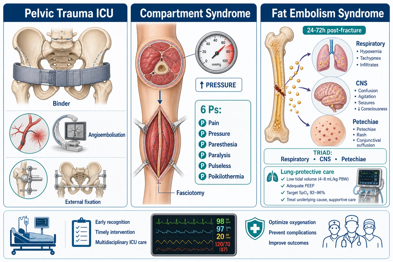
The pelvic and extremity trauma in the ICU: the pelvic fracture (the Young–Burgess mechanism classification — the anteroposterior compression or open book, the lateral compression or closed book, the vertical shear; each with a different bleeding pattern and stability), the pelvic binder (for the open book — reduces the pelvic volume → the tamponade the venous bleeding), the angiographic embolisation for the persistent arterial bleeding (after the binder + the packing), the preperitoneal packing, the external fixation, the associated injuries (the bladder 15%, the urethra 10%, the rectal, the vaginal, the sacral plexus the neurological), the extremity trauma (the open fracture — the Gustilo I/II/IIIA/IIIB/IIIC; the vascular, the nerve), the compartment syndrome (the 6 Ps — the pain disproportionate, the pressure, the paresthesia, the paralysis, the pulseless [late], the poikilothermia; the delta P under 30; the fasciotomy within 6 hours), and the fat embolism syndrome (the 24 to 72 hours post-fracture; the triad — the respiratory, the CNS, the petechial rash; the lung-protective ventilation, the supportive). The rhabdomyolysis (the crush injury — the CK, the myoglobin, the AKI; the aggressive hydration, the alkalinise the urine).
On this page & tools
Your progress
Saved locally on this device.
8 MCQs with explanations
Target exams
Overview & definition
The pelvic and extremity trauma — the pelvic fracture (the life-threatening bleeding), the extremity (the fracture, the vascular, the nerve, the compartment), and the fat embolism. The three ICU priorities: (1) the pelvic bleeding control (the binder, the angioembolisation), (2) the compartment syndrome (the early fasciotomy), and (3) the fat embolism (the lung-protective, the supportive).[1]
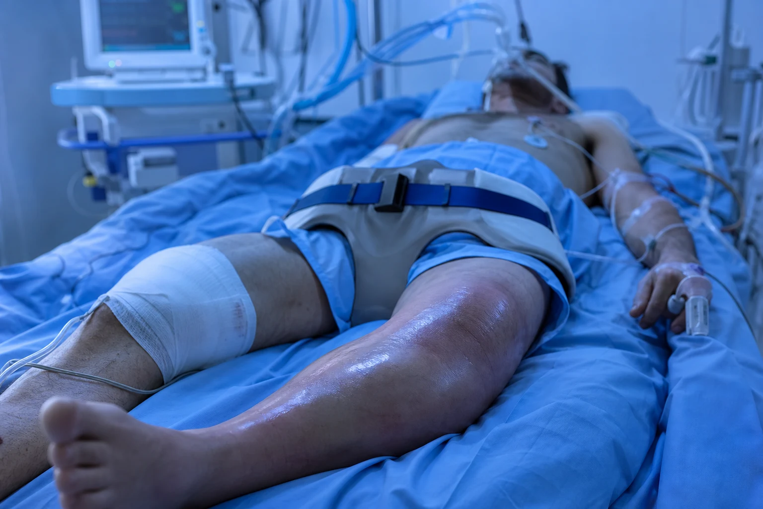
Pelvic fracture
- The bleeding — the pelvic fracture (the open book, the vertical shear) → the venous and the arterial bleeding (the internal iliac, the obturator, the pudendal). The retroperitoneal the haematoma.[1]
- The management:[1]
- The pelvic binder (the circumferential — the reduce the volume; the apply the at the level of the greater trochanters). The first-line the immediate.[1]
- The angioembolisation for the arterial bleeding (the CT blush or the persistent the hypotension despite the binder). The embolise the internal iliac branches.[1]
- The external fixation (the orthopaedic — the stabilise the fracture, the reduce the volume).[1]
- The pre-peritoneal packing (the if the angioembolisation unavailable).[1]
- The associated injuries — the bladder rupture (the haematuria; the CT cystogram), the urethral injury (the blood at the meatus; the RUG before the catheter), the rectal injury (the rectal exam; the flexible sigmoidoscopy), the sacral plexus (the neurological deficit).[1]
Pelvic fracture classification — the Young–Burgess mechanism-based system
The pelvic ring is a mechanism-driven injury, and the Young–Burgess classification (Dalal/Burgess 1989) classifies the fracture by the force vector that produced it. The mechanism is the master variable: it predicts who will bleed, how much, from where (pelvic vs non-pelvic), how stable the ring is, and which associated injuries to expect.[1][2]
The four patterns are: anteroposterior compression (APC) — the "open book"; lateral compression (LC) — the "closed book"; vertical shear (VS); and the combined mechanism (CM). Roughly 70% of lethal pelvic bleeding arises from APC and VS patterns, which is why the binder, the packing and the angioembolisation are concentrated there.[2][4]
APC — anteroposterior compression (open book)
High-volume bleeder
- Force opens the ring anterior-to-posterior (frontal impact) → the symphysis pubis springs open and the sacroiliac joint disrupts
- APC I: pubic diastasis under 2.5 cm, minimal SI widening — rotationally stable
- APC II: pubic diastasis over 2.5 cm, anterior SI ligament disruption — rotationally unstable, vertically stable
- APC III: complete posterior SI disruption — rotationally AND vertically unstable; the worst bleeder
- Increases the pelvic volume → massive venous + arterial bleeding; the highest transfusion requirement of all patterns
- THIS is the pattern the pelvic binder reverses — reduce the volume, tamponade the venous bleed
LC — lateral compression (closed book)
Low bleed, high head-injury risk
- Lateral force compresses the ring internally → overlapping pubic rami fractures, the ilium impacted against the sacrum
- LC I: pubic ramus compression/crescent fracture; LC II: iliac wing (crescent) fracture; LC III: "windswept" pelvis (LC one side + APC the other, e.g. crush)
- Internally rotates the hemipelvis → REDUCES pelvic volume, so venous bleeding self-tamponades; low transfusion need
- Beware: LC carries the highest rate of associated TRAUMATIC BRAIN INJURY (the lateral force flings the head)
- Relative contraindication to the pelvic binder — can over-reduce and worsen nerve/vessel injury; if shocked, look for a NON-pelvic bleed (abdomen, chest, long bone)
VS — vertical shear
Major bleeder, grossly unstable
- Axial load (a fall from height onto the legs) drives one hemipelvis superiorly through the SI joint
- Vertically AND rotationally unstable; the hemipelvis migrates proximally
- Tears the rich presacral venous plexus and the superior gluteal / internal iliac arteries → major haemorrhage
- Needs skeletal traction / early external fixation to restore length; the binder alone does not fully reduce the vertical displacement
- High transfusion requirement and high mortality — combine binder, packing and angiography
CM — combined mechanism
Mixed and unpredictable
- Two or more force vectors (e.g. LC + VS, APC + LC) — common in high-energy crush and pedestrian impacts
- Bleeding and stability depend on the dominant vector; assume unstable until proven otherwise
- Manage for the worst component: assume an open-book component is present and apply the binder if haemodynamically unstable
The pelvic binder — application for the open-book fracture
The pelvic binder (a circumferential compression device — commercial pelvic sheet or folded sheet) is the first-line, immediate, non-invasive haemorrhage control for the haemodynamically unstable pelvic fracture. Its mechanism is mechanical: an open-book (APC) or vertically unstable (VS) fracture increases the pelvic volume, and the binder closes the ring and restores the normal (small) pelvic volume, which tamponades the low-pressure venous and cancellous-bone bleeding that constitutes the majority of pelvic haemorrhage. It does NOT control high-pressure arterial bleeding — that is the job of angioembolisation.[4]
Pelvic binder application
Select the patient
Apply a binder to ANY haemodynamically unstable patient with a clinically suspected unstable pelvic fracture (APC open-book or vertical shear) — before imaging. Deformity, scrotal/labial swelling, leg-length discrepancy, or a mechanism suggesting a high-energy pelvic load all justify empirical application. A binder is a RESUSCITATION manoeuvre, not a definitive orthopaedic one.
Position at the level of the GREATER TROCHANTERS — not the iliac crest
The single common error. The binder sits low, centred over the femoral heads / greater trochanters and the symphysis pubis. Placing it too high (around the iliac crests) fails to close the ring and can cause pressure injury. With the patient log-rolled or lifted, slide the binder under at the trochanteric level and bring the Velcro straps anteriorly.
Tighten to close the ring
Tension the binder until the legs are gently internally rotated and the pelvis feels "closed" — typically a binder tension around 180 N (a commercial binder gives a tension indicator). The aim is anatomical reduction of the open book, not maximal force. Over-tightening an LC (closed-book) pattern can worsen displacement and injure nerve/vessel — so reserve the binder for APC/VS patterns and remove/avoid it in isolated LC.
Confirm with imaging
Plain AP pelvis and then CT (with IV contrast, portal-venous phase, looking for active extravasation / "blush"). Document the response: if shock resolves and no blush, the bleed was venous and the binder has done its job. If a blush or refractory shock persists, proceed to pelvic packing and/or angiographic embolisation.
Remove within 24 hours (ideally at definitive fixation)
The binder is a pressure device — leave it on too long and it causes pressure necrosis over the iliac crests, sacrum and greater trochanters, and skin breakdown. Plan removal within 24 hours; ideally remove it in theatre at the time of definitive external or internal fixation. If definitive fixation is delayed, release and reposition the binder and inspect the skin every few hours.
Angiographic embolisation and pre-peritoneal packing — controlling the arterial bleed
Venous and cancellous-bone bleeding (the majority) is controlled by volume reduction (binder). Arterial bleeding — ~10–20% of pelvic haemorrhage, from the internal iliac branches (superior gluteal, obturator, internal pudendal, lateral sacral) — does not tamponade and needs angiographic embolisation (selective gelfoam or coil embolisation of the bleeding branch). The trigger is a CT contrast blush or persistent shock despite a correctly applied binder and pelvic packing.[2][4]
When the patient is too unstable to leave the department for the angiography suite, the next move is pre-peritoneal (extraperitoneal) pelvic packing — a rapid suprapubic incision, dividing the linea alba but staying OUT of the peritoneum, and packing the true pelvis with three rolls on each side (behind the symphysis, along the pelvic side wall, and into the sacral concavity). This controls venous bleeding directly, buys haemodynamic stability, and bridges to angiography.[3]
Pelvic haemorrhage control — the stepwise ladder
Step 1 — Pelvic binder + damage-control resuscitation
Binder at the greater trochanters immediately on suspicion; concurrent MTP 1:1:1, permissive hypotension SBP 80-90 (NOT if TBI coexists), TXA within 3 hours, empiric calcium, warm everything, minimise crystalloid. This controls most venous bleeding.
Step 2 — FAST to exclude intraperitoneal bleeding
A positive FAST in shock means an intra-abdominal source — proceed to laparotomy. A NEGATIVE FAST with a major pelvic fracture localises the bleed to the pelvis, and the strategy shifts to pelvic packing / angiography rather than laparotomy.
Step 3 — Pre-peritoneal pelvic packing (if shocked and angio not immediately available)
Rapid extraperitoneal suprapubic packing (3 rolls each side of the true pelvis) controls venous bleeding and stabilises the patient. This is a paradigm shift from angiography-first: pack FIRST for the unstable patient, then angiography for residual arterial bleeding. Do NOT open the retroperitoneum at laparotomy — releasing the retroperitoneal tamponade causes exsanguination.
Step 4 — Angiographic embolisation for arterial bleeding
Indicated for a CT blush, an expanding pelvic haematoma, or shock persisting after binder + packing. Selective embolisation of the bleeding internal iliac branch (superior gluteal, obturator, internal pudendal) with gelfoam or coils. Repeat if recurrent. Pelvic bleeding that is purely venous needs no angio.
Step 5 — Definitive orthopaedic stabilisation
External fixation (anterior frame or C-clamp) within 24-48 hours restores ring stability, reduces ongoing bleeding, allows binder removal, and mobilises the patient. Definitive internal fixation is deferred until physiology is restored. Stabilise first, fix definitively later.
Associated injuries — the bladder, urethra, rectum, vagina and sacral plexus
A high-energy pelvic ring injury is a marker of massive energy transfer, and the organs tethered to and crossing the ring are injured in a predictable pattern. Missed associated injuries kill — every unstable pelvic fracture needs a deliberate search for genitourinary, gastrointestinal and neurological damage.[1][1]
Bladder rupture (~15%)
Look for haematuria
- The bladder sits directly behind the symphysis pubis; a shear or a bony spike lacerates it
- Extramural (extraperitoneal) rupture is more common with pelvic fracture and usually managed non-operatively (catheter)
- Intramural (intraperitoneal) rupture needs surgical repair
- Diagnosis: gross haematuria + a CT CYSTOGRAM (distend the bladder with contrast — a passive fill is falsely negative)
Urethral injury (~10%)
Posterior urethra in men
- A shear injury avulses the membranous (posterior) urethra from the prostate — classically in men
- Red flags: blood at the urethral meatus, a high-riding / non-palpable prostate on PR, perineal haematoma, scrotal swelling, inability to void
- NEVER blindly pass a urethral catheter if urethral injury is suspected — do a RETROGRADE URETHROGRAM (RUG) first; a partial tear can be converted to a complete one by blind catheterisation
- If RUG confirms injury: a suprapubic catheter (urology) rather than urethral
Rectal injury (open pelvic fracture)
High mortality
- A bony spike lacerates the rectum — this converts a closed pelvic fracture into an OPEN one with faecal contamination → catastrophic sepsis
- Diagnosis is by DIGITAL RECTAL EXAM (blood on the glove) and flexible sigmoidoscopy; a contrast enema can define the level
- Management: diverting colostomy, washout, distal rectal washout, and broad-spectrum antibiotics — these injuries carry very high mortality
Vaginal injury (in women)
Must examine to avoid missing an open fracture
- A vaginal laceration makes the pelvic fracture OPEN — a frequently missed diagnosis because the vagina is not routinely examined
- Every woman with a pelvic fracture MUST have a speculum vaginal examination to exclude a laceration
- An open fracture via the vagina needs washout, repair, and antibiotics — the same principle as an open skin wound
Sacral plexus / nerve roots
Neurological deficit
- Sacral fractures and SI joint disruption can injure the lumbosacral plexus and sacral nerve roots
- Document a baseline neurological exam of both lower limbs (and ask specifically about perineal sensation and sphincter tone — cauda equina / sacral plexus)
- L5/S1 injury gives foot drop and posterior thigh/calf sensory loss; sacral root injury gives bladder/bowel/sexual dysfunction
Pelvic ring stability by mechanism (click each)
APC III, VS, CM
Complete posterior disruption → the hemipelvis migrates. Major haemorrhage (venous + arterial), massive transfusion, the highest mortality. Binder + packing + angiography + external fixation; skeletal traction for vertical shear to restore length.
Extremity trauma and compartment syndrome
Compartment syndrome
- The 6 Ps:[1]
- Pain disproportionate to the injury (and the worse on the passive stretch — the key the early the sign).[1]
- Pressure (the tense the compartment).[1]
- Paresthesia (the early; the numbness).[1]
- Paralysis (the late).[1]
- Pulseless (the LATE — the arterial the inflow the preserved the until the very the late; the pulse the present the does the NOT the exclude).[1]
- Poikilothermia (the cold).[1]
- The diagnosis — the clinical (the pain disproportionate + the passive stretch). The compartment the pressure (the delta P — the diastolic the minus the compartment the pressure under 30 mmHg).[1]
- The management — the fasciotomy (the immediate; the decompress the all the compartments; the open the fascia; the leave the open; the delayed the closure).[1]
Open fractures — the Gustilo–Anderson classification
An open fracture is one in which the fracture haematoma communicates with the external environment through a skin wound. It is a surgical emergency: the risk of infection rises steeply with the severity of the soft-tissue damage, and the Gustilo–Anderson classification (built on Gustilo's 1984 subdivision of type III) grades that severity and guides debridement, antibiotics, stabilisation and the need for soft-tissue cover.[5]
Type I
Clean, under 1 cm
- Low-energy wound under 1 cm, clean, minimal soft-tissue damage
- First-generation cephalosporin (e.g. cefazolin); single dose to 24 hours after wound cover
- Infection rate under 2%
- Debride, irrigate, reduce and stabilise
Type II
1–10 cm, no extensive damage
- Wound over 1 cm (up to ~10 cm) without extensive soft-tissue damage, periosteal stripping, or gross contamination
- Cephalosporin; add Gram-negative cover (e.g. aminoglycoside) if contaminated
- Infection rate ~2–7%
Type IIIA
Adequate soft-tissue cover
- High-energy, over 10 cm OR any size with extensive damage, contamination, farm injury, shotgun, or segmental fracture
- IIIA: adequate soft tissue remains — the fracture can be covered WITHOUT a local or free flap
- Antibiotic cover for Gram-positive AND Gram-negative (cephalosporin + aminoglycoside); add penicillin for farm/soil (Clostridium)
- Infection rate ~10%
Type IIIB
Needs a flap
- Extensive soft-tissue loss with periosteal stripping / exposed bone — cover requires a rotational or free soft-tissue FLAP
- Multidisciplinary (orthopaedic + plastic) reconstruction; external fixation and delayed flap cover
- Infection rate ~30% — high; prolonged broad-spectrum antibiotics
Type IIIC
Vascular injury needing repair
- ANY open fracture with an associated VASCULAR injury that requires repair (regardless of soft-tissue damage)
- Limb-threatening; amputation rate historically 50% or higher; time-critical — restore perfusion within 6 hours
- Revascularise (shunt or repair), fasciotomy prophylactically after reperfusion (reperfusion oedema → compartment syndrome), broad-spectrum antibiotics
Compartment syndrome — the 6 Ps in depth, the pressure threshold, and fasciotomy
Acute compartment syndrome is a time-critical surgical emergency in which raised pressure within a fascial compartment collapses the capillary bed, producing ischaemia of muscle and nerve within the compartment while the large artery remains patent. The single most reliable early sign is pain disproportionate to the injury, made worse by passive stretch of the muscles in that compartment — the ischaemic-muscle pain. The distal pulse is present until very late; pulselessness is a pre-terminal sign and its presence does NOT exclude the diagnosis.[6]
The leg has four compartments (anterior, lateral, superficial posterior, deep posterior); the forearm, thigh, foot, hand, gluteal and abdominal compartments can all be affected. The diagnosis is primarily clinical, supported in equivocal or unconscious patients by compartment pressure measurement: the delta P (perfusion pressure) = diastolic BP − compartment pressure, with a delta P under 30 mmHg the threshold for fasciotomy (McQueen/Court-Brown). An absolute pressure above 30 mmHg is a less sensitive alternative. Untreated compartment syndrome progresses within hours to irreversible necrosis, Volkmann ischaemic contracture, and amputation.[6]
Fasciotomy for acute compartment syndrome
Recognise — pain disproportionate + passive stretch (do NOT wait for pulselessness)
The earliest, most specific sign is pain out of proportion to the injury, worse on passive stretch of the compartment muscles (e.g. passive toe extension stretches the calf in leg compartment syndrome). Paresthesia follows (early nerve ischaemia). The pulse is PRESENT. In the unconscious or multiply-injured patient, a tense woody compartment, rising pressure, or a delta P under 30 mmHg is the trigger — a high index of suspicion is mandatory after reperfusion, after reduction of displaced fractures, after crush/electrical injury, and after vascular repair.
Confirm clinically or with delta P
In the awake cooperative patient the diagnosis is CLINICAL and pressure measurement should not delay surgery. In equivocal or unconscious patients, measure the compartment pressure: delta P (diastolic BP minus compartment pressure) under 30 mmHg supports the diagnosis. Treat the patient, not the number — a convincing clinical picture mandates immediate fasciotomy.
Decompress ALL compartments within 6 hours
For the leg, the standard two-incision fasciotomy opens all four compartments (anterolateral incision for anterior and lateral; posteromedial incision for superficial and deep posterior). The fascia is opened along its full length; the skin is left OPEN. Aim to decompress within 6 hours of the onset — delay beyond 6–8 hours sharply increases irreversible necrosis, Volkmann contracture and amputation.
Leave the wound open; plan delayed closure
The skin and fascia are left open; the wounds are dressed (vacuum dressing or synthetic skin substitute). Primary closure at the index operation defeats the purpose. Re-inspect at 48–72 hours; achieve delayed primary closure or skin grafting once swelling resolves. A second-look debrides any non-viable muscle.
Treat the cause and the consequences
Reduce and stabilise the underlying fracture, relieve constrictive casts/dressings, correct hypotension/hypoxia, and watch for reperfusion hyperkalaemia, myoglobinuria and acute kidney injury (rhabdomyolysis). Document the timing meticulously — missed compartment syndrome is a leading source of litigation and preventable amputation.
Fat embolism syndrome (FES)
- The timing — 24 to 72 hours post-fracture (the long bone — the femur, the tibia; the pelvic).[1]
- The triad:[1]
- The management — the supportive (the oxygen, the lung-protective the ventilation for the severe). The early the fracture the fixation (the reduce the fat the release). The steroids the controversial.[1]
FES — pathophysiology, diagnostic criteria, and management
Fat embolism (fat in the circulation) is almost universal after a long-bone or major pelvic fracture — transoesophageal echocardiography detects echogenic fat in nearly every patient. The clinical fat embolism syndrome (FES) is far less common, occurring in ~1–5% (up to ~10% in high-risk patterns), and is the dangerous end of the spectrum. The hallmark is the 12–72 hour latent period after the fracture (or after intramedullary nailing) followed by the classical triad of respiratory failure (an ARDS-like picture), non-focal cerebral dysfunction, and a petechial rash.[7][8]
Two mechanisms act in sequence. The mechanical phase: fat and marrow debris embolise to the pulmonary capillaries, causing physical obstruction and right-heart strain. The biochemical phase: lipase hydrolyses the neutral fat to free fatty acids, which are directly toxic to the pulmonary capillary endothelium and type II pneumocytes, producing a diffuse inflammatory ARDS-like lung injury. Cerebral and systemic (petechial) manifestations arise either through fat crossing a patent foramen ovale or through direct microvascular embolisation.[8]
Gurd criteria (1970) — major + minor
The classical set
- MAJOR features: respiratory insufficiency, cerebral involvement, petechial rash
- MINOR features: fever, tachycardia, retinal changes (fat or petechiae), jaundice, renal changes (lipuria, haematuria, oliguria), a drop in haematocrit/platelets, high ESR
- Diagnosis needs at least 1 major + 4 minor features, OR 1 major + 3 minor + fat macroglobinaemia
Schonfeld / Lindeque criteria
Alternative diagnostic sets
- Schonfeld: a point score from petechial rash, hypoxaemia (PaO2 under 70), CNS changes, fever, tachycardia, tachypnoea — a score over 5 supports FES
- Lindeque: relies on respiratory criteria alone (hypoxaemia requiring intervention plus CNS/rash) at 24–72h after long-bone fracture
- No single criterion is gold-standard — diagnosis remains CLINICAL, on the triad at the expected latency
FES vs pulmonary embolism vs ARDS
Differential at the bedside
- FES: 24–72h post-fracture, ARDS-like lungs PLUS a non-focal confused brain PLUS a petechial rash — the rash is the discriminator
- Acute PE: sudden onset, focal (one lung field if submassive), pleuritic, no rash, no fever-latency pattern; CTPA shows clot
- ARDS from another cause (aspiration, sepsis, transfusion): no petechial rash, no clear fracture-latency link
Rhabdomyolysis (crush injury)
- The muscle the breakdown → the CK (the markedly), the myoglobin (the dark the urine), the AKI (the pigment the nephropathy).[1]
- The management — the aggressive hydration (the dilute the myoglobin), the alkalinise the urine (the bicarbonate — the reduce the crystallisation), the RRT if the severe AKI.[1]
Rhabdomyolysis — crush injury, myoglobin nephropathy, and fluid strategy
Crush injury (prolonged compression — entrapment, collapsing buildings, electrical injury, severe ischaemia/reperfusion, compartment syndrome) lyses skeletal muscle, releasing myoglobin, creatine kinase (CK) and potassium. Myoglobin is filtered into the tubules, where at an acidic urine pH it precipitates (cast formation) and generates free-radical injury, producing pigment nephropathy and acute kidney injury. The CK rises within hours and peaks at 24–72 hours (often above 5,000–15,000 U/L); myoglobin turns the urine dark ("tea"/"cola") and the urine dipstick is positive for "blood" in the absence of red cells (myoglobin pseudo-peroxidase activity).[1]
Management is fluid-led. The priority is aggressive isotonic crystalloid to flush myoglobin through the tubules — early and copious (target urine output 200–300 mL/h in the first 24 hours). Bicarbonate (to alkalinise the urine to pH over 6.5 and reduce myoglobin precipitation) is historically used, but modern evidence (including Cochrane review) shows no clear benefit of bicarbonate over saline alone; it remains a reasonable adjunct when there is severe metabolic acidosis or hyperkalaemia. Treat hyperkalaemia aggressively (the lysed muscle releases potassium), correct hypocalcaemia only if symptomatic, and initiate renal replacement therapy for the indications of AKI (refractory hyperkalaemia, acidosis, fluid overload, uraemia). Crucially, look for and treat the underlying compartment syndrome, which is both the cause and a complication of the crush.[1]
[1]Red flags
Landmark trials
Dalal, Burgess et al. — Young–Burgess mechanism classification (J Trauma 1989)
Retrospective cohort; multiple trauma patients with pelvic ring fractures classified by the mechanism of injury
Population: Adults with pelvic fractures sustained in high-energy multiple trauma
Key finding
The mechanism of pelvic injury correlated strongly with the associated organ injury pattern, the magnitude of bleeding and transfusion, and mortality. APC and VS patterns carried the highest transfusion requirements and haemodynamic instability; LC patterns bled less but carried a high rate of associated head injury.
Practice change
Classify pelvic fractures by MECHANISM (Young–Burgess): it predicts who will bleed, how much, from where, and which associated injuries to hunt for. The classification that governs the whole resuscitation strategy.
Eastridge, Starr, Scalea et al. (J Trauma 2002)
Retrospective review; patients with haemorrhagic shock and pelvic ring disruptions stratified by fracture pattern
Population: Multiple trauma patients in shock with pelvic ring fractures at a Level I trauma centre
Key finding
Lateral-compression patterns bled predominantly from NON-pelvic sources; anteroposterior-compression and vertical-shear patterns bled from PELVIC sources that needed intervention (binder, packing, angioembolisation). Fracture pattern reliably predicted the source and type of pelvic haemorrhage control required.
Practice change
Use the fracture PATTERN to direct bleeding control: APC/VS = a pelvic source (binder, packing, embolisation); LC = search elsewhere (abdomen, chest, long bones) for the bleed. Do not assume a shocked LC patient is bleeding from the pelvis.
Cothren, Osborn, Moore et al. — preperitoneal pelvic packing (J Trauma 2007)
Prospective evaluation of a protocolised preperitoneal pelvic packing strategy for haemodynamically unstable pelvic fractures
Population: Patients with unstable pelvic fractures and shock refractory to pelvic binder activation
Key finding
Preperitoneal pelvic packing rapidly stabilised haemodynamics and reduced transfusion requirements, with mortality lower than the historical angiography-first comparator. Packing controlled the predominantly venous pelvic bleeding that angiography does not address.
Practice change
Preperitoneal pelvic packing is a rapid, universally available first-line haemorrhage control for the unstable pelvic fracture — pursue it BEFORE angiography, then angiography for residual arterial bleeding. A genuine paradigm shift in pelvic trauma.
Schonfeld et al. — corticosteroid prophylaxis for FES (Ann Intern Med 1983)
Randomised, double-blind, placebo-controlled trial in high-risk fracture patients
Population: Patients at high risk of fat embolism syndrome after long-bone and pelvic fractures
Key finding
Corticosteroid prophylaxis significantly reduced the incidence of the fat embolism syndrome compared with placebo in high-risk fracture patients.
Practice change
Corticosteroids PROPHYLACTICALLY reduce the incidence of FES in high-risk patients. Routine use is not universally adopted because of infection and glycaemic concerns; supportive care (oxygen, lung-protective ventilation) and EARLY FRACTURE FIXATION remain the standard, with steroids an option in selected high-risk cases.
Coccolini, Stahel et al. — WSES pelvic trauma classification & guidelines (World J Emerg Surg 2017)
International expert consensus guideline (World Society of Emergency Surgery)
Population: Adults with blunt and penetrating pelvic trauma
Key finding
Provides a single severity-based classification and an algorithm that sequences pelvic binder → damage-control resuscitation → FAST → preperitoneal packing / angiographic embolisation → definitive fixation, calibrated to haemodynamic stability and fracture pattern.
Practice change
The WSES guideline is the modern reference for sequencing pelvic haemorrhage control — pack early for the unstable patient, reserve angio for the stable-but-bleeding patient, and fix definitively once physiology is restored.
Exam practice
SAQ — Unstable pelvic fracture with an associated open femoral fracture
12 minutes · 12 marks
A 24-year-old man is brought in by ambulance after a high-speed motorcycle crash. He is hypotensive (BP 78/50, HR 128) with clinical deformity of the pelvis and an open right femoral fracture with bone exposed through a 12 cm contaminated wound. A pelvic binder is applied with partial improvement in blood pressure, but he remains in refractory shock. FAST is negative. Once briefly stabilised, CT shows an APC (open-book) pelvic fracture with an arterial contrast blush, and a Gustilo type IIIB right femur fracture.
Clinical pearls
References
- [1]Dalal SA, Burgess AR, Siegel JH, Young JW, Brumback RJ, Poka A, et al. Pelvic fracture in multiple trauma: classification by mechanism is key to pattern of organ injury, resuscitative requirements, and outcome J Trauma, 1989.PMID 2746708
- [2]Eastridge BJ, Starr A, Minei JP, O'Keefe GE, Scalea TM The importance of fracture pattern in guiding therapeutic decision-making in patients with hemorrhagic shock and pelvic ring disruptions J Trauma, 2002.PMID 12352479
- [3]Cothren CC, Osborn PM, Moore EE, Morgan SJ, Johnson JL, Smith WR Preperitonal pelvic packing for hemodynamically unstable pelvic fractures: a paradigm shift J Trauma, 2007.PMID 17426537
- [4]Coccolini F, Stahel PF, Montori G, Biffl W, Horer TM, Catena F, et al. Pelvic trauma: WSES classification and guidelines World J Emerg Surg, 2017.PMID 28115984
- [5]Gustilo RB, Mendoza RM, Williams DN Problems in the management of type III (severe) open fractures: a new classification of type III open fractures J Trauma, 1984.PMID 6471139
- [6]McQueen MM, Gaston P, Court-Brown CM Acute compartment syndrome. Who is at risk? J Bone Joint Surg Br, 2000.PMID 10755426
- [7]Schonfeld SA, Ploysongsang Y, DiLisio R, Crissman JD, Miller E, Hammerschmidt DE, Jacob HS Fat embolism prophylaxis with corticosteroids. A prospective study in high-risk patients Ann Intern Med, 1983.PMID 6354030
- [8]Godoy DA, Di Napoli M, Rabinstein AA Cerebral Fat Embolism: Recognition, Complications, and Prognosis Neurocrit Care, 2018.PMID 28932982