ICU · Trauma / neurocritical care
Spinal Trauma & Spinal Shock — ASIA Scale, CT vs MRI & Steroid Controversy
Also known as Spinal cord injury · SCI · Spinal shock · Neurogenic shock · ASIA impairment scale · NASCIS · Methylprednisolone spinal injury · Cervical spine clearance · Complete cord syndrome · Anterior cord syndrome · Central cord syndrome · Brown-Séquard syndrome · NEXUS criteria · Canadian C-spine rule · STASCIS · Autonomic dysreflexia
The spinal trauma and the spinal shock in the ICU: the spinal cord injury (the SCI — from the trauma, the fracture, the dislocation) causing the motor and sensory deficit below the level. The mechanism of injury (the axial loading, the flexion, the extension, the rotation, the distraction) determines the fracture pattern and the cord injury. The ASIA impairment scale (A = complete; B = sensory incomplete; C = motor incomplete; D = motor useful; E = normal). The spinal shock (the temporary the areflexia and the flaccidity below the level — the resolves over the days to the weeks) vs the neurogenic shock (the hypotension, the bradycardia, the vasodilation from the loss of the sympathetic tone — the T6 and above). The cervical spine clearance (the NEXUS low-risk criteria vs the Canadian C-spine rule; the CT first-line for the high-risk; the MRI for the neurological deficit; the clinical clearance for the low-risk). The imaging: the CT first (the bony), the MRI for the cord (the oedema, the haematoma, the compression). The steroids (the methylprednisolone — the NASCIS trials; the controversial; the not the standard of the care).
On this page & tools
Your progress
Saved locally on this device.
Target exams
Overview & definition
The spinal cord injury (SCI) from the trauma causes the motor and sensory deficit below the level. The two ICU priorities: (1) the prevent the secondary injury (the hypoperfusion, the hypoxia, the hypotension) and (2) the MAP over 85 to 90 (the cord the perfusion). The ASIA impairment scale the grades the severity. The neurogenic shock (the hypotension + the bradycardia from the loss of the sympathetic the tone — the T6 and above) requires the vasopressors. The steroids are the controversial (the NASCIS trials; the not the standard of care in the most the units).[1]
The primary injury — the mechanical cord damage at the moment of impact (the compression, the contusion, the shear, the laceration) — is irreversible; the ICU cannot undo it. Everything in the first week is directed at the secondary injury cascade: the ischaemia, the oedema, the excitotoxicity (glutamate), the calcium influx, the free-radical lipid peroxidation, the apoptosis — all amplified by hypotension, hypoxia, hypoglycaemia and hyperthermia. Each MAP under 85 mmHg and each SpO₂ under 90 per cent converts salvageable cord to infarcted cord. This is why the resuscitation target is higher in SCI than in almost any other shock state, and why a single hypotensive episode is treated as a neurological emergency.[1][13]
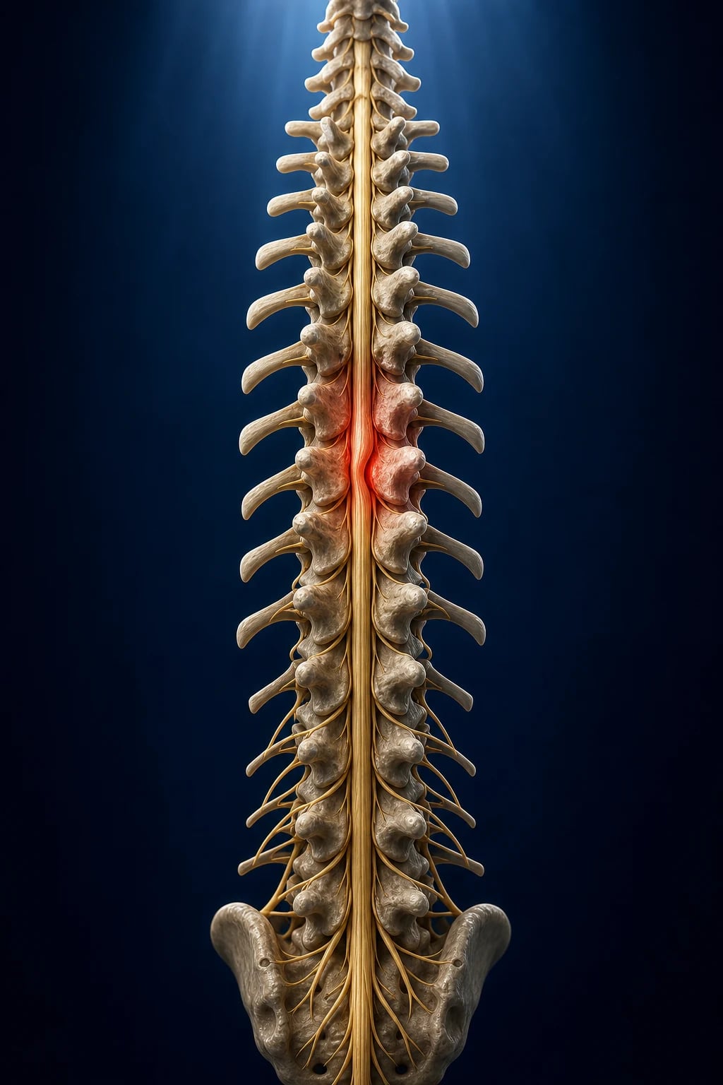
Mechanism of injury — how the force vector predicts the fracture
The mechanism determines the fracture pattern, the stability, the likelihood and the pattern of cord injury, and the associated injuries. The spine fails under five basic force vectors — axial loading (compression), flexion, extension, rotation/shear, and distraction — and most real injuries are combinations. Reading the mechanism on the pre-hospital handover narrows the differential before the imaging loads.[11]
Axial loading (compression)
Diving, fall onto head/feet
- Force transmitted along the long axis of the spine → the vertebral body is crushed between the ring above and below; retropulsion of the posterosuperior corner into the canal
- C1 Jefferson burst fracture — axial load splits the lateral masses of the atlas; the open-mouth peg view shows lateral masses overhanging the C2 articular pillars by >7 mm total (transverse ligament torn → unstable)
- Thoracolumbar burst fracture — retropulsed bone fragment in the canal; the classic mechanism is a fall from height landing on the feet (look for the calcaneus fracture and the T12–L2 burst together)
- Shallow-water diving onto a flexed neck → C5 burst with retropulsion and cord injury — the prototypical axial-load SCI scenario
- Axial load also produces the sagittal-split and vertical-Potts fractures; canal compromise predicts the neurological deficit, not the body-height loss alone
Flexion
Forward bending, lap-belt, deceleration
- Anterior column fails in compression, posterior column in tension; the posterior ligamentous complex (PLC) determines stability
- Simple wedge (compression) fracture — stable unless >50% height loss or PLC disruption; no neurological injury usually
- Bilateral facet dislocation — flexion tears ALL the posterior ligaments; the superior vertebra subluxes forward ~50% of body width on the one below; grossly unstable, very high cord-injury rate
- Unilateral facet dislocation — flexion + rotation; ~25% subluxation; "bat-wing" or "reverse hamburger" sign on axial CT; can be a stable bone-perched lesion or unstable if jumped
- Clay-shoveler fracture — avulsion of the C7 (or T1) spinous process by forced flexion against the pull of trapezius/rhomboids; stable and benign, needs no collar
- Chance (seat-belt) fracture — distraction-flexion through bone at T12–L2 from a lap-belt; the lap-belt abdominal bruise is the clue and demands a search for small-bowel and mesenteric injury
Extension
Rear-end, fall onto face, hyperextension
- Posterior column fails in compression, anterior in tension; the ALL and the disc are torn
- Hangman fracture — bilateral C2 pars interarticularis fractures with traumatic spondylolisthesis of C2 on C3 (chin-on-dashboard MVC, hyperextension-distraction); the cord space is wide so most patients are neurologically intact
- Extension teardrop fracture — avulsion of the anteroinferior vertebral body corner by the intact ALL; C2 is the classic site; highly unstable (three-column); the elderly hyperextension injury, often on a rigid ankylosed spine
- SCIWORA (spinal cord injury without radiographic abnormality) — extension injury with cord contusion/ischaemia but normal plain film AND CT; children >> adults; the cord is injured by momentary ligament-bone impingement then reduction; MRI confirms
- In the elderly with ankylosing spondylitis or DISH, even a low-energy extension injury produces an unstable three-column fracture through the syndesmophyte, often with gross displacement
Rotation / shear / distraction
High-energy, unstable patterns
- Rotation + flexion → fracture-dislocation: the MOST unstable spinal pattern; the spine is translated at the injury level; near-complete cord injury is the rule
- Shear (direct lateral or anteroposterior force) → ligamentous disruption, often through the disc space; a three-column injury that may look minor on CT but is functionally catastrophic
- Distraction (lap-belt, fall) → Chance fracture (bony) or its pure ligamentous equivalent (rarer, more unstable, higher cord-injury rate)
- Penetrating (gunshot, stab) → direct cord transection or partial injury; the Brown-Séquard (hemisection) pattern is the classic stab-wound lesion; do not manipulate an in-situ implement
The 3-column model (Denis) — and what 'unstable' actually means
The anterior column — ALL, anterior annulus, anterior half of body and disc
Fails in tension with extension injuries (teardrop) and in compression with flexion. Isolated anterior-column injury (simple wedge) is usually STABLE.
The middle column — posterior half of body and disc, posterior annulus, PLL
The KEY determinant of stability. Middle-column failure (burst with retropulsion) means canal compromise and an unstable injury that often needs surgery. The middle column is assessed on the sagittal/coronal CT recon — look for retropulsion of the posterosuperior corner.
The posterior column — neural arch, facet joints, PLC (supraspinous/interspinous ligaments, flavum)
Fails in tension with flexion. PLC disruption is the single most important CT sign of an unstable flexion injury: look for increased interspinous distance, facet subluxation/perching, and on MRI, frank oedema/tear of the ligamentum flavum and supraspinous ligament.
The rule of thumb — injury to 2+ columns = unstable
A two- or three-column injury is mechanically unstable and usually needs surgical stabilisation; a one-column injury is usually stable and managed non-operatively. The PLC status (assessed clinically by palpating a step/gap and on MRI) is decisive in borderline burst fractures.
The ASIA impairment scale
- The A (complete) — no motor or sensory below the level (including S4-S5).[1]
- The B (sensory incomplete) — sensory but no motor below the level.[1]
- The C (motor incomplete) — motor below the level (the MRC under 3).[1]
- The D (motor useful) — motor below the level (the MRC 3 or over).[1]
- The E (normal) — the motor and sensory normal (the with the prior the deficit).[1]
Grade A — complete
No motor, no sensory below, INCLUDING S4-S5
- No sacral sparing — no sensation at S4-S5 and no voluntary anal contraction
- The prognosis for ambulation is poor (~only ~5% convert to incomplete over time)
- Re-assess after spinal shock resolves (return of the bulbocavernosus reflex); an early A may not be the final A
Grade B — sensory incomplete
Sensory preserved, motor absent below
- Sensation (including sacral) is preserved; motor function is absent below the level
- Sacral sensation (S4-S5, or deep anal pressure) MUST be present to differentiate from A
- Prognosis intermediate; ~30-50% of B patients gain some motor recovery, more if the level is caudal
Grade C — motor incomplete
Motor below; more than half of key muscles below MRC <3
- Voluntary anal contraction OR sparing of motor function below the level; more than half of the key muscles below have grade 0-2
- Ambulation possible in a minority; depends heavily on the level and the cord syndrome
Grade D — motor useful
Motor below; more than half of key muscles >= MRC 3
- Most key muscles below the level are grade 3 or better (full ROM against gravity)
- The majority of D patients ambulate (community or household); outcomes are best in this band
Grade E — normal
Motor and sensory normal
- Used only if the patient HAD a deficit that has now resolved, or to document a radiological injury without neurological deficit
- A patient with no deficit and no prior injury is simply "not applicable", not E
The ASIA examination — how to perform and record it
The ASIA (American Spinal Injury Association) examination is the internationally standardised, reproducible neurological assessment that converts an unreliable bedside exam into a number the whole team can use to communicate and to prognosticate. It must be performed (and re-performed serially) with the patient supine and undressed.[5]
Performing and recording the ASIA examination
Step 1 — Determine the sensory level (light touch and pinprick, 28 dermatomes each side)
Test LIGHT TOUCH (cotton wool) and PINPRICK (sharp/dull, a safety pin) for each of the 28 dermatomes C2-S5 on BOTH sides. Score 0 = absent, 1 = impaired (cannot reliably distinguish sharp/dull, or light touch feels different to face), 2 = normal, NT = not testable. The S4-S5 (perianal) and deep anal pressure (DAP) are the sacral-sparing markers — their presence defines an INCOMPLETE injury. The sensory level is the lowest dermatome with normal (2) sensation on both modalities.
Step 2 — Score the 10 key muscles on each side (MRC 0-5)
Test and grade the 10 myotomes bilaterally, each MRC 0-5: C5 elbow flexors (biceps), C6 wrist extensors (ECR), C7 elbow extensors (triceps), C8 long finger flexors (FDP), T1 small finger abductors (ADM), L2 hip flexors (iliopsoas), L3 knee extensors (quadriceps), L4 ankle dorsiflexors (tibialis anterior), L5 long toe extensors (EHL), S1 ankle plantarflexors (gastrocnemius). The upper-extremity motor score (UEMS, 50) and lower-extremity motor score (LEMS, 50) sum to a maximum 100.
Step 3 — Determine the neurological level of injury (NLI)
The NLI is the most caudal segment with intact (grade 2) sensation AND >= grade 3 motor function, PROVIDED the segments above are normal. It may differ for the right and left sides; record both. The NLI is the single most powerful prognostic variable — it predicts respiratory reserve, ambulation, bladder/bowel and the vasopressor requirement.
Step 4 — Assess completeness — voluntary anal contraction and S4-S5/DAP
SACRAL SPARING (any sensation at S4-S5, OR deep anal pressure, OR voluntary anal sphincter contraction) means the injury is INCOMPLETE (ASIA B-E). Its absence means COMPLETE (ASIA A). This is the single most important discrimination in the exam — it sets the prognosis and the surgery decision.
Step 5 — Assign the ASIA grade (AIS) and re-examine serially
Combine steps 1-4 into the AIS A-E. Re-examine daily in the first week and after spinal shock resolves (return of the bulbocavernosus reflex): the grade at 72 hours and again at the end of spinal shock are far more prognostic than the immediate post-injury grade, which is confounded by spinal shock and intoxication.
The neurological level of injury (NLI)
The most caudal level with intact motor and sensory
- Predicts the deficit, the respiratory reserve, the vasopressor requirement and the rehabilitation potential
- C1-C4: diaphragm impaired → ventilator-dependent potential; full quadriplegia
- C5: elbow flexion preserved, hands lost; C6: wrist extension; C7: elbow extension; C8/T1: intrinsic hand
- T1-T6: intercostals impaired → poor cough, atelectasis, restrictive pattern; loss of sympathetic below
- T7-L1: trunk/abdominal muscles; L2-S1: lower-limb myotomes; S2-S4: bowel, bladder, sexual function
The motor score as a number
UEMS + LEMS, 0-100
- A single number (0-100) that tracks recovery over days and weeks
- A rising motor score over the first 72 hours predicts a favourable long-term motor outcome
- Useful for family counselling and for detecting deterioration (a falling score mandates urgent re-imaging for an expanding haematoma or new compression)
Spinal cord injury syndromes — the incomplete patterns
The complete (ASIA A) lesion is one entity; the incomplete lesions have characteristic anatomies and prognoses that examiners love because the anatomy is elegant and the management differs. Recognise the pattern from the clinical deficit before the imaging.[1]
Central cord
Hyperextension in the elderly spondylotic spine
- Mechanism: hyperextension (fall onto the face/forehead) compresses the cord between anterior osteophytes and a buckled, hypertrophied ligamentum flavum
- Deficit: UPPER >> LOWER extremity weakness (the cervical arm fibres lie medially in the corticospinal tract); variable sensory loss, bladder dysfunction
- The MOST COMMON incomplete SCI; the classic patient is elderly with a stiff, arthritic cervical spine
- Management: surgery for instability or persistent compression; outcomes variable — the legs recover first, the hands last, often poorly
Anterior cord
Flexion/vascular — the worst incomplete prognosis
- Mechanism: anterior spinal artery occlusion (dissection, embolus, aortic surgery cross-clamp) OR direct anterior compression (retropulsed bone/disc)
- Deficit: bilateral MOTOR and PAIN/TEMPERATURE loss below (corticospinal + spinothalamic), with PROPRIOCEPTION, vibration and fine touch PRESERVED (dorsal columns spared, supplied by the posterior spinal arteries)
- Prognosis the POOREST of the incomplete syndromes; recovery is limited
Brown-Séquard (hemisection)
Penetrating — the BEST incomplete prognosis
- Mechanism: penetrating injury (stab, gunshot) to one side of the cord
- Deficit: IPSILATERAL motor and dorsal-column (proprioception, vibration) loss, with CONTRALATERAL pain/temperature loss (spinothalamic crosses 1-2 levels up)
- The exam favourite for elegant anatomy; one side of the cord is spared, so motor recovery is excellent and bowel/bladder usually preserved
Posterior cord
Rare; vibration and proprioception lost
- Mechanism: direct posterior trauma or vitamin B12/neurosyphilis in the differential
- Deficit: loss of proprioception/vibration below; motor and pain/temperature spared; a sensory ataxia
- Rare in trauma; recognise it by the preserved motor with a prominent joint-position deficit
Conus medullaris (S2-S4)
Mixed UMN and LMN at the sacral cord
- Mechanism: T12-L1 burst fracture compressing the conus
- Deficit: BOWEL and BLADDER aredenervation (flaccid, areflexic — LMN), saddle anaesthesia, variable lower-limb weakness; mixed upper and lower motor neuron signs
- Often the bulbocavernosus and ankle jerks are absent; prognosis for bladder recovery is poor
Cauda equina
LMN lesion of the lumbosacral roots
- Mechanism: large central L1-S1 disc, or a lower burst fracture, compressing the nerve roots below the conus
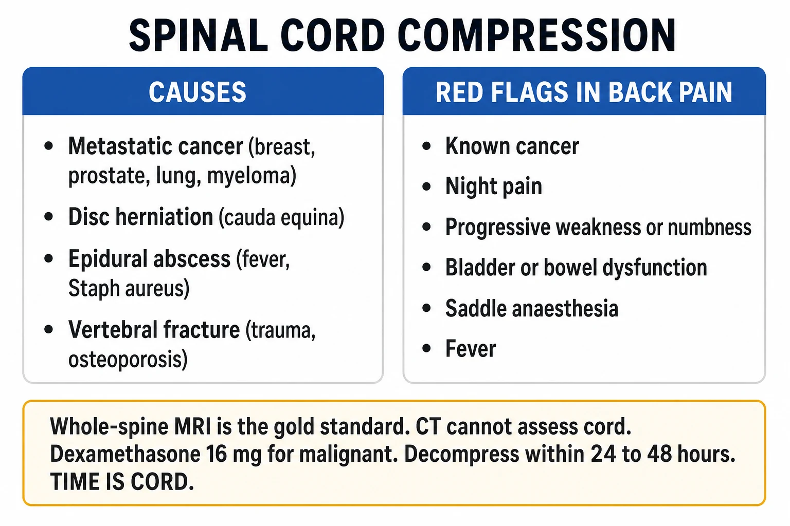
- Deficit: asymmetric LMN weakness and reflex loss, saddle anaesthesia, bladder/bowel retention, sexual dysfunction — a SURGICAL EMERGENCY
- Purely a nerve-root (LMN) injury, so recovery is possible IF decompressed early — the red flag is new urinary retention (check the post-void residual)
Spinal shock vs neurogenic shock — do not confuse them
These are two distinct phenomena with overlapping names that examiners use to probe whether you understand the pathophysiology. Spinal shock is a transient NEURAL phenomenon (loss of reflexes); neurogenic shock is a sustained CARDIOVASCULAR phenomenon (loss of vasomotor tone). They can coexist in a cervical/upper-thoracic injury but they resolve on different timescales and are managed differently.[1]
- The spinal shock — the temporary the areflexia, the flaccidity, the loss of the reflexes below the level (the from the loss of the supraspinal the input). The resolves over the days to the weeks. The during this the period the ASIA the assessment the may the underestimate the eventual the deficit.[1]
- The neurogenic shock — the hypotension + the bradycardia from the loss of the sympathetic the tone (the T6 and above). The vasodilation → the warm, the dry skin; the bradycardia (the unopposed the parasympathetic). The treat with the noradrenaline or the phenylephrine (the vasopressor for the vasodilation) + the atropine or the isoprenaline for the bradycardia. The do NOT the confuse with the hypovolaemic the shock (the neurogenic the warm, the hypovolaemic the cold).[1]
Spinal shock
Transient neural — loss of reflexes below the lesion
- Mechanism: sudden loss of supraspinal excitatory input → areflexia and flaccidity below the lesion
- Onset: immediate; duration: hours to days to (rarely) weeks; resolves as spinal neurons regain intrinsic excitability
- End-point: return of the BULBOCAVERNOSUS reflex (S3-S4) — squeeze the glans or pull the Foley → anal sphincter contracts; usually back by 24-48h
- Clinical impact: the ASIA exam during spinal shock may UNDERESTIMATE the eventual deficit — do not declare an ASIA A as final until spinal shock has resolved
- Management: none specific — supportive; serial re-examination
Neurogenic shock
Sustained cardiovascular — loss of vasomotor tone
- Mechanism: loss of sympathetic outflow (T1-L2) → unopposed parasympathetic → vasodilation + bradycardia
- Onset: within minutes to hours; duration: days to 1-3 weeks; the higher the lesion the longer and the deeper
- Triad: HYPOTENSION + BRADYCARDIA + WARM DRY SKIN (vasodilated); only occurs with lesions at or above T6
- Management: vasopressor (noradrenaline preferred, or phenylephrine) for the MAP 85-90; atropine/isoprenaline for symptomatic bradycardia; DO NOT treat with crystalloid alone
- Confusion trap: the multiply-injured patient may have BOTH neurogenic AND haemorrhagic shock — exclude a bleed first with FAST/CT
Cervical spine clearance — NEXUS vs the Canadian C-spine rule
Cervical spine injury occurs in 2-5 per cent of blunt trauma patients and in up to 10 per cent of those with a closed head injury or a high-energy mechanism. The objective of clearance is to identify the small minority who need imaging without irradiating or immobilising the large majority who do not. Two validated clinical decision rules exist; both allow a clinically well patient to be cleared without imaging when their criteria are met, and both have a sensitivity high enough that a missed injury is, in practice, a failure to apply the rule.[2][3][4]
NEXUS (Hoffman, NEJM 2000)
5 low-risk criteria — ALL must be met to withhold imaging
- Withhold imaging ONLY if ALL FIVE are present:
- (1) No midline cervical tenderness
- (2) No focal neurological deficit
- (3) Normal level of alertness (no intoxication, no altered mental status, GCS normal)
- (4) No intoxication (alcohol, drugs)
- (5) No painful distracting injury (long-bone fracture, burn, etc.)
- Sensitivity ~90.7% (misses ~1 in 10); specificity ~36% — simple and applied to all comers; miss ONE criterion → image
Canadian C-spine rule (Stiell, JAMA 2001)
A stepwise branching algorithm — more specific
- Apply ONLY to alert (GCS 15), stable trauma patients where C-spine injury is a concern
- STEP 1 — any HIGH-RISK factor mandates imaging: age >=65, dangerous mechanism (fall >1 m/5 stairs, axial head load, high-speed MVC/rollover/ejection, motorised recreational vehicle, bicycle collision), or paraesthesia in the extremities
- STEP 2 — if no high-risk factor, any LOW-RISK factor that allows SAFE assessment of range of motion: simple rear-end MVC (excludes being pushed into oncoming traffic, hit by a bus/large truck, rollover, high speed), sitting position in the ED, ambulatory at any time, delayed onset of neck pain, absence of midline cervical spine tenderness
- STEP 3 — if a low-risk factor is present, can the patient rotate the neck 45° left AND right? If YES → no imaging needed; if NO → image
- Sensitivity ~99.4%; specificity ~40-45% — MORE sensitive and more specific than NEXUS, so it images fewer patients
NEXUS (Hoffman, NEJM 2000)
Prospective, multi-centre, observational validation study in 34,069 blunt-trauma patients
Population: All blunt-trauma patients undergoing cervical-spine imaging at 21 US centres
Key finding
The criteria identified 99.0% of all injuries (sensitivity 99.0%, NPV 99.8%, specificity 12.9% for the original 34,069) — later pooled sensitivity ~90.7%. Only 2 of 818 significant injuries were missed. Specificity is low, so many patients are still imaged.
Practice change
If ALL FIVE NEXUS criteria are met, cervical-spine imaging can safely be withheld. The rule is simple but low-specificity; miss one criterion and you must image. The Canadian C-spine rule performs better but applies only to alert, stable patients.
Canadian C-spine rule (Stiell, JAMA 2001)
Prospective cohort derivation and validation in 8,924 alert, stable blunt-trauma patients
Population: Adults with GCS 15 and stable vital signs, in whom C-spine injury was a concern
Key finding
Sensitivity 99.4% (1 of 169 important injuries missed); specificity 45.1%. The rule was more sensitive AND more specific than NEXUS applied to the same population, and would have reduced imaging by 15-25%.
Practice change
In the ALERT, STABLE blunt-trauma patient, the Canadian C-spine rule outperforms NEXUS: fewer patients are imaged for the same (negligible) miss rate. It requires a cooperative, sober, awake patient — it does NOT apply to the obtunded.
Cervical spine clearance pathway — the three routes
Route 1 — CLINICAL clearance (the awake, low-risk patient)
For the alert (GCS 15), sober, stable patient with a low-energy mechanism: apply the Canadian C-spine rule (preferred) or NEXUS. If the patient satisfies the low-risk criteria and (Canadian rule) can rotate 45° left and right without pain, CLEAR the cervical spine clinically — remove the collar and document. No imaging needed.
Route 2 — CT FIRST-LINE clearance (the high-risk or obtunded patient)
Any high-risk feature (dangerous mechanism, age >=65, focal neurological deficit, intoxication, painful distracting injury, GCS <15) → obtain a helical CT cervical spine from occiput to T1 with sagittal and coronal reconstructions. CT is now FIRST-LINE in adults — three-view plain films miss up to 15% and are obsolete for adult clearance. A normal, high-quality CT clears the cervical spine from a BONY stability standpoint in the overwhelming majority.
Route 3 — MRI for the NEUROLOGICAL DEFICIT or the CT-negative concern
Indications for MRI: (a) a new or evolving neurological deficit (cord compression, haematoma, contusion); (b) a CT that is suspicious but non-diagnostic (possible ligamentous injury); (c) the obtunded/uncooperative patient in whom the clinician cannot reliably clear on CT alone and who is to be mobilised (controversial — see below). MRI shows cord oedema, haematoma, compression, disc disruption and PLC injury that CT cannot. Do NOT let MRI delay decompression in a deteriorating patient.
The obtunded-patient controversy — CT-only is the modern default
For the intubated/unconscious patient the clinical rules cannot be applied. The current EAST and AANS/CNS consensus is that a normal high-quality CT cervical spine clears the bony column in the vast majority; routine MRI purely to 'clear the ligaments' is NOT mandatory and adds false positives (oedema from resuscitation and immobility). Reserve MRI for a deficit, an equivocal CT, or persistent clinical concern before mobilisation. A purely ligamentous unstable injury with a completely normal CT is rare in adults.
The imaging
- The CT first (the bony — the fracture, the dislocation, the canal the compromise). The rapid; the standard for the bony.[1]
- The MRI for the cord (the oedema, the haematoma, the compression, the ligamentous the injury). The for the neurological the deficit the with the normal the CT; the for the surgical the planning.[1]
The CT answers the bony question — is there a fracture, a dislocation, canal compromise, instability? The MRI answers the soft-tissue and cord question — is there cord oedema, contusion, haematoma, an extruded disc, a torn posterior ligamentous complex, an epidural haematoma? In practice: CT first, always and immediately, for the unstable patient and the high-risk patient alike; MRI second, urgently if there is a neurological deficit or a CT that does not explain the deficit, and semi-electively for surgical planning of an unstable injury. Do NOT send an unstable patient to the MRI scanner — it is a remote, slow, ferromagnetic-no-go zone.[1][10]
CT cervical/thoracolumbar spine
First-line, fast, for the bone
- Indication: ANY high-risk blunt-trauma patient; the obtunded; first-line for clearance
- Strengths: rapid, available, high sensitivity for bony injury and canal compromise; the reconstructions are read on a workstation
- Looks for: fracture (burst, wedge, hangman, teardrop, Jefferson), facet dislocation/perching, canal compromise (retropulsion), vertebral translation, three-column involvement
- Weakness: misses pure ligamentous injury and ALL cord pathology
MRI spine
For the cord and the ligaments
- Indication: a neurological deficit (esp. with normal CT); a suspicious CT; surgical planning; the obtunded patient with persistent concern before mobilisation
- Strengths: the ONLY modality that sees the cord — oedema, contusion, haematoma, compression, infarction; also the PLC, disc, ligaments
- Looks for: cord oedema (T2 hyperintensity), cord haematoma (T1/gradient-echo — a poor prognostic sign), extruded disc, epidural haematoma, PLC disruption (STIR/fat-sat)
- Weakness: slow, remote, ferromagnetic; cannot be used for an unstable patient; over-reads trivial oedema that may be clinically meaningless
Plain films (3-view C-spine)
Obsolete for adult clearance
- Historical standard; now replaced by CT in adults
- Miss ~15% of injuries, especially at the cervicothoracic junction; require repeat "swimmer" views
- Retained role: paediatric clearance (radiation-sparing), intra-operative localisation, resource-limited settings, and follow-up of known fractures
The management
1. The ABC + the immobilisation
- The ABC (the airway — the C-spine the). The cervical the collar (the until the C-spine the cleared).[1]
Airway management in the suspected cervical-spine injury uses manual in-line stabilisation (MILS) during intubation (a second rescuer holds the head neutral without traction) and rapid-sequence induction. Do NOT let the collar obstruct intubation: the front is opened and MILS applied. The classic error is to delay intubation for fear of the spine — the dying airway kills before the cord does. Maintain SpO₂ >= 94 per cent from the outset.[1][11]
2. The MAP 85 to 90 (the cord perfusion)
- The maintain the MAP over 85 to 90 mmHg for the 7 days (the improve the cord the perfusion, the reduce the secondary the injury).[1]
- The noradrenaline (the preferred — the alpha and the beta). The phenylephrine the alternative.[1]
- The avoid the hypotension (the worsens the secondary the injury).[1]
3. The early decompression
- The surgery within 24 hours (the decompression + the stabilisation). The improves the neurological the outcome (the especially the incomplete).[1]
For the patient with an incomplete SCI and ongoing cord compression, decompression and stabilisation within 24 hours (the STASCIS trial) approximately doubled the odds of a two-grade ASIA improvement compared with late surgery. This is a level 2 recommendation and one of the few interventions proven to change the neurological outcome. The corollary: do NOT delay surgery for "optimisation" — the 24-hour window matters.[6]
STASCIS (Fehlings, PLoS ONE 2012) — Surgical Timing in Acute Spinal Cord Injury Study
Multicentre, prospective, observational cohort; 313 adults with acute cervical SCI enrolled 2002-2009
Population: Adults with traumatic cervical SCI and cord compression, treated at 6 North American spine centres
Key finding
Early surgery nearly trebled the odds of a >=2 grade AIS improvement at 6 months: 19.8% (early) vs 8.8% (late); adjusted OR 2.83 (95% CI 1.10-7.28). Early surgery was safe, with no excess complication rate.
Practice change
For the patient with an incomplete cervical SCI and ongoing cord compression, decompression and stabilisation WITHIN 24 hours approximately trebles the odds of a clinically meaningful neurological recovery. Do not delay surgery for 'optimisation'.
4. The steroids — the controversial
- The methylprednisolone (the NASCIS trials — the 30 mg per kg the bolus then 5.4 mg per kg per h for 23 to 48 h). The controversial — the modest the benefit the but the increased the infection, the GI bleed, the hyperglycaemia. The NOT the standard the of the care in the most the units (the individual the decision).[1]
NASCIS II (Bracken, NEJM 1990)
The original 24-hour protocol
- Protocol: methylprednisolone 30 mg/kg IV bolus over 1 h, then 5.4 mg/kg/h for 23 hours
- Therapeutic window: benefit only if started WITHIN 8 HOURS of injury
- Reported: a small motor-score improvement in the within-8h subgroup (a post-hoc subgroup analysis)
- Criticism: the benefit emerged only in post-hoc subgroups; the primary analysis was negative
NASCIS III (Bracken, JAMA 1997)
Duration depends on timing
- Started within 3 h of injury: 24-hour infusion (5.4 mg/kg/h)
- Started 3-8 h after injury: 48-hour infusion (5.4 mg/kg/h) — longer infusion
- Reported: a marginal motor benefit of the 48-h regimen in the 3-8h group
- Harms: a clear increase in severe sepsis, pneumonia, wound infection, GI bleed, hyperglycaemia, and longer ICU stay; mortality not reduced
Current guideline position (AANS/CNS 2013)
Steroids are an OPTION, not a recommendation
- The 2013 AANS/CNS guideline: high-dose methylprednisolone is an OPTION (level C), with confounded and contradictory evidence
- Most ANZ, UK, European and many North American units do NOT give steroids
- If given, it is the NASCIS II protocol started within 8 hours; NEVER after 8 hours (no benefit, all harm)
- Contraindications: pregnancy, active sepsis, uncontrolled diabetes — and most units add "multisystem trauma" because of the sepsis/wound risk
NASCIS II (Bracken, NEJM 1990)
Multicentre, randomised, double-blind, placebo-controlled trial; 487 acute SCI patients
Population: Adults with acute SCI within 12 hours, all grades
Key finding
No significant difference in the primary analysis. A POST-HOC subgroup analysis suggested a small motor improvement in patients treated within 8 hours of injury. The trial is criticised for relying on post-hoc subgroups.
Practice change
If steroids are used, use the NASCIS II protocol (30 mg/kg bolus, then 5.4 mg/kg/h for 23 h) and ONLY within 8 hours of injury. The benefit is marginal, methodologically fragile, and offset by infection, GI bleeding, hyperglycaemia and longer ICU stay. Steroids are NOT standard of care.
NASCIS III (Bracken, JAMA 1997)
Multicentre, randomised, double-blind trial; 499 acute SCI patients
Population: Adults with acute SCI within 8 hours
Key finding
In patients starting treatment 3-8 h after injury, the 48-h infusion showed a small motor improvement; within 3 h, the 24-h infusion sufficed. The 48-h infusion caused significantly MORE severe sepsis, pneumonia and GI complications.
Practice change
The longer the infusion, the greater the harm. The 48-h regimen (3-8 h window) added sepsis and pneumonia without changing mortality. Reinforced that steroids are marginal and risky; modern guidelines downgrade them to an option.
5. The supportive
- The DVT prophylaxis (the high the risk — the LMWH after 48 to 72 h). The bladder the care (the urinary the retention — the catheter). The bowel the care. The skin (the pressure the areas). The pneumonia (the respiratory the muscle the weakness).[1]
SCI carries one of the highest VTE risks in all of medicine (50-100 per cent without prophylaxis). Mechanical prophylaxis (sequential compression) from admission; pharmacological prophylaxis (LMWH) at 24-72 hours once there is no active haemorrhage on serial imaging. An IVC filter is reserved for the patient in whom anticoagulation is absolutely contraindicated — it does NOT replace pharmacological prophylaxis. Early enteral nutrition, meticulous skin care (turn q2h with log-roll), aggressive pulmonary toilet, and a planned early tracheostomy for high cervical injuries complete the supportive bundle.[1][15]
[1]SAQ — C5 complete spinal cord injury with neurogenic shock
10 minutes · 10 marks
A 28-year-old man is brought to ED after a diving accident; he hit his head on the bottom of a shallow pool. He is GCS 15 but cannot move his arms or legs, has no sensation below the clavicles, and has a flaccid areflexic rectal examination with absent bulbocavernosus reflex. HR 48, BP 78/50, warm dry peripheries, SpO₂ 96% on room air. CT shows a C5 burst fracture with retropulsion into the spinal canal.
SAQ — Incomplete spinal cord injury syndromes
10 minutes · 10 marks
A 50-year-old woman involved in a motor vehicle crash presents with a flexion-injury pattern. Neurological examination shows: (i) profoundly weak upper limbs, especially the hands (more than proximal arms); (ii) preserved motor power in the lower limbs; (iii) variable sensory disturbance but preserved proprioception and vibration in the lower limbs. MRI shows a hyperintense cord lesion at C4–C5 with central cord haemorrhage.
Red flags
References
- [1]Karnad DR, Nor MBM, Richards GA, et al. Intensive care in severe malaria: Report from the task force on tropical diseases by the World Federation of Societies of Intensive and Critical Care Medicine. Journal of critical care, 2018.PMID 29132978
- [2]Como JJ, Diaz JJ, Dunham CM, et al. Practice management guidelines for identification of cervical spine injuries following trauma: update from the eastern association for the surgery of trauma practice management guidelines committee. The Journal of trauma, 2009.PMID 19741415
- [3]Hoffman JR, Mower WR, Wolfson AB, et al. Validity of a set of clinical criteria to rule out injury to the cervical spine in patients with blunt trauma. National Emergency X-Radiography Utilization Study Group. The New England journal of medicine, 2000.PMID 10891516
- [4]Stiell IG, Wells GA, Vandemheen KL, et al. The Canadian C-spine rule for radiography in alert and stable trauma patients. JAMA, 2001.PMID 11597285
- [5]Kirshblum SC, Burns SP, Biering-Sorensen F, et al. International standards for neurological classification of spinal cord injury (ASIA standards, revised 2011). Journal of Spinal Cord Medicine, 2011.PMID 22330108
- [6]Fehlings MG, Vaccaro A, Wilson JR, et al. Early versus delayed decompression for traumatic cervical spinal cord injury: results of the Surgical Timing in Acute Spinal Cord Injury Study (STASCIS). PloS one, 2012.PMID 22384132
- [7]A randomized, controlled trial of methylprednisolone or naloxone in the treatment of acute spinal-cord injury. The New England journal of medicine, 1990.PMID 2278580
- [8]Bracken MB, Shepard MJ, Holford TR, et al. Administration of methylprednisolone for 24 or 48 hours or tirilazad mesylate for 48 hours in the treatment of acute spinal cord injury. Results of the Third National Acute Spinal Cord Injury Randomized Controlled Trial. National Acute Spinal Cord Injury Study. JAMA, 1997.PMID 9168289
- [9]Hadley MN, Walters BC, Grabb PA, et al. Pharmacological therapy after acute cervical spinal cord injury. Neurosurgery, 2002.PMID 12431289
- [10]Malhotra A, Durand D, Wu X, et al. Utility of MRI for cervical spine clearance in blunt trauma patients after a negative CT. European radiology, 2018.PMID 29450715
- [11]Galvagno SM Jr, Nahmias JT, Young DA Advanced Trauma Life Support(®) Update 2019: Management and Applications for Adults and Special Populations. Anesthesiology clinics, 2019.PMID 30711226
- [13]Kwon BK, Tetreault LA, Martin AR, et al. A Clinical Practice Guideline for the Management of Patients With Acute Spinal Cord Injury: Recommendations on Hemodynamic Management. Global spine journal, 2024.PMID 38526923
- [15]Hachem LD, Ahuja CS, Fehlings MG Assessment and management of acute spinal cord injury: From point of injury to rehabilitation. The journal of spinal cord medicine, 2017.PMID 28571527
- [16]Consortium for Spinal Cord Medicine Acute management of autonomic dysreflexia: individuals with spinal cord injury presenting to health-care facilities. The journal of spinal cord medicine, 2002.PMID 12051242