MedVellum
MCQsExamsAtlas
DashboardPricing
MBBS / Core medicine✳Dermatology✳ICU Fellowship (CICM)✳Anaesthesia✳Emergency Medicine✳Psychiatry Fellowship✳Paediatrics Fellowship✳Physician Medicine✳MCQs✳SAQs✳Vivas✳OSCE✳Evidence-first✳MBBS / Core medicine✳Dermatology✳ICU Fellowship (CICM)✳Anaesthesia✳Emergency Medicine✳Psychiatry Fellowship✳Paediatrics Fellowship✳Physician Medicine✳MCQs✳SAQs✳Vivas✳OSCE✳Evidence-first✳

MedVellum.

The folio

Exam-exhaustive medical education across every specialty — evidence-graded topics, engraved plates, and practice in every written and oral format. Educational content only — not medical advice.

llms.txt · psychiatry LLM catalog · sitemap

Atlas

  • Specialty atlas
  • MBBS / Core medicine
  • Dermatology
  • ICU Fellowship (CICM)
  • Anaesthesia
  • Emergency Medicine
  • Psychiatry Fellowship
  • Paediatrics Fellowship
  • Physician Medicine

Study & account

  • MCQ practice
  • Practice alias
  • Exam tools
  • Dashboard
  • Pricing
  • Sign in

© 2026 MedVellum. For education only — not a substitute for clinical judgement.

Folio edition · Set in Instrument Serif & Archivo

ICU TopicsTrauma

ICU · Trauma

Chest trauma and blunt cardiac injury in ICU

Also known as Chest trauma · Blunt cardiac injury · Cardiac contusion · Flail chest · Pulmonary contusion · Traumatic aortic injury · Thoracic trauma

Chest trauma: blunt (motor vehicle crash, fall, crush — 80%) or penetrating (stab, gunshot — 20%). ICU-relevant: (1) FLAIL CHEST — ≥3 consecutive ribs fractured in ≥2 places → paradoxical chest wall movement → respiratory failure. (2) PULMONARY CONTUSION — lung parenchymal injury → alveolar haemorrhage/oedema → hypoxia, may worsen over 24-48h. (3) BLUNT CARDIAC INJURY (BCI) — myocardial contusion → arrhythmia, troponin elevation, rarely cardiac rupture/failure. (4) TRAUMATIC AORTIC INJURY — deceleration → aortic transection (90% die at scene, survivors need urgent repair). (5) TENSION PNEUMOTHORAX/HAEMOTHORAX — life-threatening, needle decompression/chest drain. Management: ABCDE, analgesia (epidural/paravertebral for rib fractures), lung-protective ventilation if needed (flail chest/pulmonary contusion), chest drains for pneumo/haemothorax, surgical fixation of flail chest (controversial).

high24 referencesUpdated 3 July 2026
On this page & tools

Your progress

Saved locally on this device.

Target exams

CICMFFICMEDIC

Red flags

Pulmonary contusion WORSENS over 24-48h — monitor closely, may need intubationTension pneumothorax — immediate needle decompression (don't wait for CXR)Traumatic aortic injury — widened mediastinum on CXR → urgent CT angiography → surgical/TEVAR repairBlunt cardiac injury — monitor ECG 24-48h, troponin, echocardiographyCommotio cordis — blunt precordial impact triggering instant VF in a structurally normal heart → immediate CPR + defibrillationRetained haemothorax — undrained blood after 2-3 days → empyema/fibrothorax → early VATSTracheobronchial injury — persistent massive air leak / lung fails to re-expand → bronchoscopy, surgical repairBlunt oesophageal injury — delayed diagnosis → mediastinitis/sepsis → contrast oesophagography + early surgical repair

Your progress

Saved locally on this device.

Target exams

CICMFFICMEDIC

Red flags

Pulmonary contusion WORSENS over 24-48h — monitor closely, may need intubationTension pneumothorax — immediate needle decompression (don't wait for CXR)Traumatic aortic injury — widened mediastinum on CXR → urgent CT angiography → surgical/TEVAR repairBlunt cardiac injury — monitor ECG 24-48h, troponin, echocardiographyCommotio cordis — blunt precordial impact triggering instant VF in a structurally normal heart → immediate CPR + defibrillationRetained haemothorax — undrained blood after 2-3 days → empyema/fibrothorax → early VATSTracheobronchial injury — persistent massive air leak / lung fails to re-expand → bronchoscopy, surgical repairBlunt oesophageal injury — delayed diagnosis → mediastinitis/sepsis → contrast oesophagography + early surgical repair

In one line

Chest trauma ICU: FLAIL CHEST (≥3 ribs × ≥2 breaks → paradoxical movement → ventilation). PULMONARY CONTUSION (worsens 24-48h → hypoxia). BLUNT CARDIAC INJURY (ECG monitoring 24-48h, troponin, echo). TRAUMATIC AORTIC INJURY (widened mediastinum → CT angiography → TEVAR). Management: ABCDE, analgesia (epidural), lung-protective ventilation, chest drains, surgical rib fixation (selected).

[10]
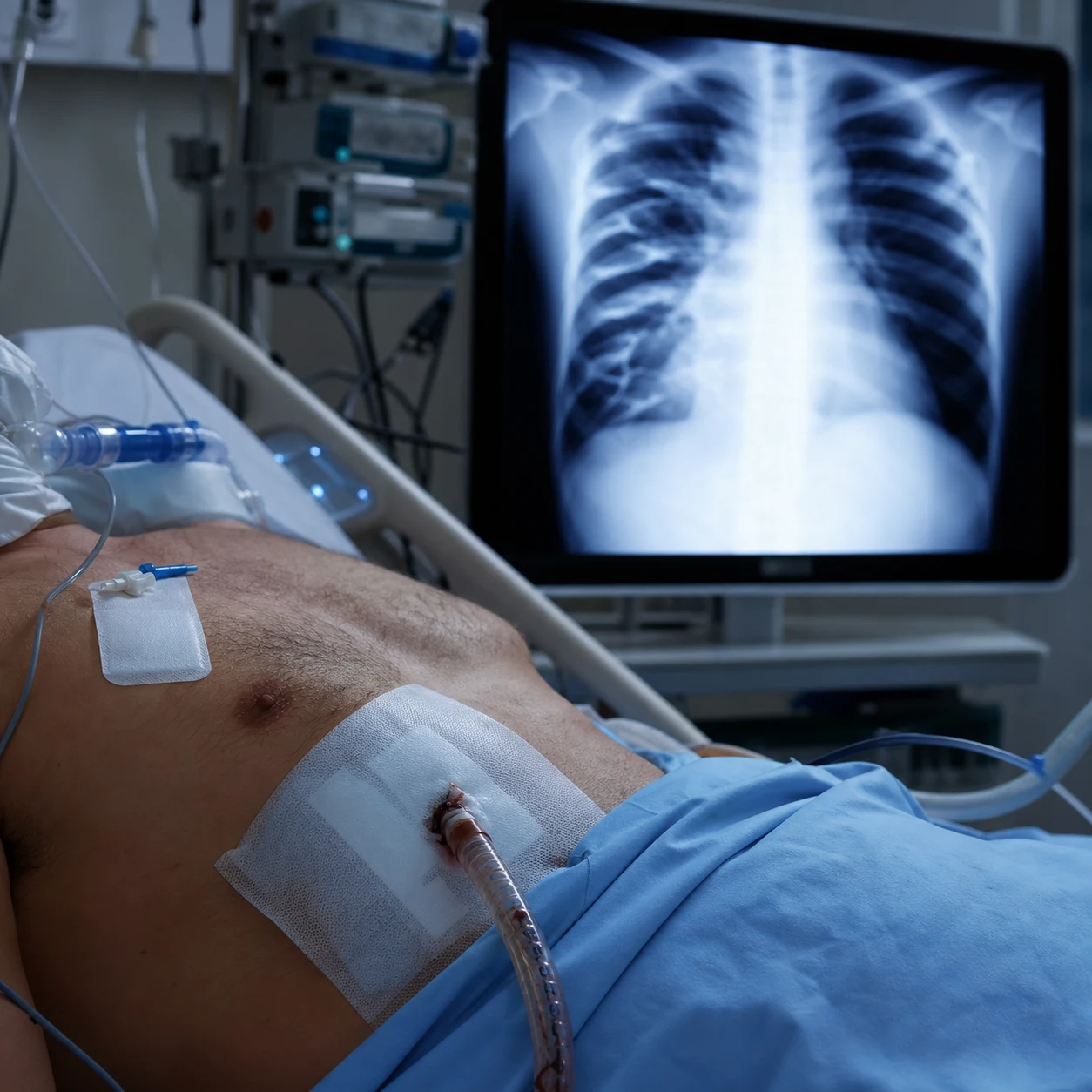
Cinematic ICU scene of a blunt chest trauma patient with a flail segment showing paradoxical movement, a pulmonary contusion on the CT, an elevated troponin with new AF on the monitor, chest tube and analgesia running, clinical-blue lighting, no faces, no text
FigureChest trauma — flail chest (three or more ribs fractured in two or more places, paradoxical movement, ventilation for respiratory failure), pulmonary contusion (hypoxia worsening at 24-48 h), and blunt cardiac injury (troponin rise, arrhythmia, rarely pump failure). Adequate analgesia and selective ventilation drive outcome.
[5] [10]

SAQ — Severe blunt chest trauma with pulmonary contusion and BCI

10 minutes · 10 marks

A 62-year-old man (60 kg) is admitted after a high-speed motor vehicle crash in which he struck the steering wheel. Initial GCS 15, RR 28, SpO₂ 90% on 15 L O₂, BP 105/68, HR 110 (sinus), ST elevation of 1 mm in V2–V4 on ECG, troponin 0.4 μg/L, CXR with patchy opacification in the right mid-zone. He has right-sided rib fractures 4–7. CT chest confirms right pulmonary contusion and possible myocardial contusion.

[2]

SAQ — Traumatic aortic injury — beta-blocker first

10 minutes · 10 marks

A 35-year-old man is admitted after a high-speed motorcycle crash with a steering-wheel-like handlebar impact to the chest. GCS 15, BP 165/95, HR 122, RR 26, SpO₂ 96%. Contrast-enhanced CT chest shows a peri-aortic haematoma at the isthmus with intimal flap and pseudoaneurysm consistent with a grade III blunt thoracic aortic injury (BTAI).

[5]

Clinical pearls

High-yield chest trauma points for CICM/FFICM exam

  1. Pulmonary contusion WORSENS over 24-48 hours. Initial CXR may be NORMAL (or minimal changes). Over 24-48h: progressive infiltrates (alveolar haemorrhage + oedema) → hypoxia → may need intubation. MONITOR: serial CXR, SpO2, ABG. DON'T be reassured by normal early CXR. CT (more sensitive) detects contusion earlier.[2]
  2. Rib fractures in elderly = HIGH mortality. ≥3 rib fractures in age >65: mortality 10-20% (from pneumonia, respiratory failure). KEY: AGGRESSIVE analgesia (epidural/paravertebral), early mobilisation, pulmonary physiotherapy, monitor in ICU/HDU (even if initially stable). ADEQUATE ANALGESIA is the MOST IMPORTANT intervention for rib fractures.[6]
  3. Blunt cardiac injury (BCI) — ECG is the key screening test. OTA guideline: (a) Admission ECG: if NORMAL → BCI unlikely (no further cardiac monitoring needed — low risk). (b) If ABNORMAL (arrhythmia, ST changes, conduction abnormality): admit for cardiac monitoring 24-48h. (c) Troponin: if elevated → echo (assess wall motion). (d) MOST BCI: arrhythmia (AF, PVCs, heart block) — self-limited. Rare: cardiac rupture (tamponade — fatal), severe pump failure (cardiogenic shock).[3]
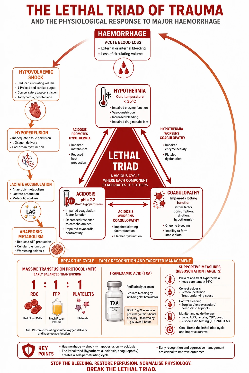
  4. Conservative fluid management in pulmonary contusion. Aggressive fluid resuscitation → increased pulmonary capillary pressure → WORSEN contusion (more oedema, more hypoxia). Use: MINIMUM fluid to maintain perfusion (MAP ≥65). Monitor: urine output, lactate. If hypovolaemic from blood loss: TRANSFUSE (blood, not crystalloid — avoids volume overload). 'Dry' strategy preferred in isolated pulmonary contusion.[2]
  5. Surgical rib fixation — selected patients. Flail chest or severely displaced fractures → surgical fixation (plates/screws to stabilise rib cage). Benefits: reduced pain, faster recovery, earlier extubation, fewer complications (pneumonia). EVIDENCE: meta-analyses show benefit (reduced ICU stay, ventilation days, pneumonia). INDICATIONS: (a) Flail chest with respiratory failure. (b) Severely displaced fractures (chest wall deformity). (c) Refractory pain (non-operative management failed). (d) Thoracotomy for other reason (fix ribs while in chest). NOT for: all rib fractures (most heal without surgery).[4]
  6. Traumatic aortic injury — 90% die at scene. Survivors (10%): partial transection (contained by adventitia/mediastinal tissue). PRESENTATION: (a) Widened mediastinum (>8 cm or >25% of thoracic width) on CXR. (b) Apical cap (blood pleural, left apex). (c) Depressed left main bronchus. (d) NG tube deviation. (e) Loss of aortic knob. (f) BP differential (upper extremity SBP > lower — partial obstruction). CT ANGIOGRAPHY: definitive. MANAGEMENT: (a) BP control (SBP 100-120 — reduce shear stress on tear). (b) TEVAR (thoracic endovascular aortic repair) — preferred (stent graft — less invasive than open). (c) Open repair (if TEVAR not suitable).[5]
  7. Tension pneumothorax — IMMEDIATE decompression (don't wait for CXR). CLINICAL: hypotension, hypoxia, tachycardia, tracheal deviation (late), absent breath sounds (affected side), distended neck veins (from increased intrathoracic pressure). MANAGEMENT: (a) NEEDLE DECOMPRESSION immediately: 14G needle, 2nd intercostal space, midclavicular line (or 5th ICS, mid-axillary line — newer guidelines). (b) Release of air = rush of air = diagnosis confirmed. (c) Then: formal CHEST DRAIN (large bore — 28-36 Fr — tube thoracostomy). DON'T WAIT for CXR (patient may die).[1]
  8. Haemothorax — chest drain, monitor output. CHEST DRAIN: large bore (28-36 Fr — blood is viscous — small drains block). THORACOTOMY indications (emergency): (a) Initial drainage >1500 mL (massive — ongoing bleeding). (b) Ongoing drainage >200 mL/h for 2-4 hours (active bleeding — surgery needed). (c) Persistent blood transfusion needed. (d) Clotted haemothorax (drain can't evacuate — needs surgery to evacuate clot).[1]
  9. Epidural analgesia — BEST for rib fracture pain. Epidural (thoracic — T5-T8 level): local anaesthetic (bupivacaine/ropivacaine) + opioid (fentanyl). Benefits: (1) Superior analgesia (allows deep breathing, coughing, clearance of secretions). (2) Reduced pneumonia (better chest expansion). (3) Reduced need for systemic opioids (less sedation, delirium). (4) May reduce need for intubation (if pain-controlled, can breathe adequately). CAUTIONS: coagulopathy (trauma — check platelets/INR before inserting), hypotension (sympathetic block — vasopressors may be needed), neurological assessment (epidural may mask spinal injury). PAR avertebral block: alternative (less hypotension, unilateral).[6]
  10. Stemal fracture — assess for cardiac injury. Sternal fracture (from direct impact — steering wheel, seatbelt): high association with BCI (blunt cardiac injury — 20-40%). ASSESS: ECG (arrhythmia), troponin (elevation), echo (wall motion). MOST sternal fractures: stable (no displacement) — managed conservatively (analgesia). Unstable (displaced): surgical fixation. MECHANISM: direct compression of heart between sternum and spine → myocardial contusion.[3]
  11. Diaphragmatic injury — often missed. Blunt (rare) or penetrating. Left diaphragm more common (liver protects right). PRESENTATION: delayed (herniation of abdominal contents into chest — bowel sounds in chest, respiratory compromise). DIAGNOSIS: CT (often missed on CXR — nasogastric tube curled in chest). MANAGEMENT: surgical repair (laparotomy/thoracotomy). IMPORTANT: consider in ALL penetrating thoracoabdominal trauma (lower chest/upper abdomen — diaphragm traversed).[1]
  12. Tracheobronchial injury — rare but life-threatening. MECHANISM: direct crush or deceleration (bronchus tears at carina — most common site). PRESENTATION: massive air leak (continuous bubbling in chest drain — doesn't resolve), pneumothorax that doesn't re-expand with drain, subcutaneous emphysema (extensive). DIAGNOSIS: bronchoscopy (visualise tear). MANAGEMENT: surgical repair (thoracic surgery). May need: double-lumen tube (isolate injured lung).[1]
  13. Multiple rib fractures = significant injury. Even without flail chest: ≥3 rib fractures → significant pain, respiratory compromise, pneumonia risk. ICU/HDU admission for: (a) Age >65. (b) ≥3 rib fractures. (c) Pre-existing lung disease (COPD). (d) Hypoxia (SpO2 <92% despite oxygen). (e) Inability to cough/breathe deeply (poor analgesia). AGGRESSIVE management: analgesia (epidural), pulmonary physiotherapy, HFNC/NIV if hypoxic, monitor closely.[6]
  14. Chest trauma + concurrent injuries. 30-50% of chest trauma patients have MULTI-SYSTEM injuries: head injury (50%), abdominal injury (30%), extremity fracture (40%). FULL TRAUMA SURVEY (primary + secondary): don't focus on chest alone — miss abdominal bleeding, spinal injury, TBI. PRIORITISE: life-threatening injuries first (ABC — airway, breathing, circulation). CHEST TRAUMA is often the cause of early death (tension pneumo, aortic injury, massive haemothorax).[6]

Red flags

Critical chest trauma red flags

  • Pulmonary contusion worsens 24-48h → monitor closely, may need intubation.[2]
  • Tension pneumothorax → immediate needle decompression (don't wait for CXR).[1]
  • Traumatic aortic injury (widened mediastinum) → CT angiography → TEVAR.[5]
  • Blunt cardiac injury → ECG + troponin, monitor 24-48h (arrhythmia).[3]
  • ≥3 rib fractures in elderly → HIGH mortality (10-20%) → aggressive analgesia (epidural).[6]
  • Conservative fluids in pulmonary contusion (avoid overload → worsens hypoxia).[2]

Prognosis

Chest trauma outcomes (Battle 2019)

[2]

Commotio cordis

[9] [9]

High-yield commotio cordis points for CICM/FFICM exam

  1. Commotio cordis = sudden VF from a blow to a NORMAL heart. The defining feature is a structurally and histologically normal heart — there is no contusion, no ischaemia, no structural disease. The blow (e.g. baseball, hockey puck, knee, airbag) must land DIRECTLY OVER THE PRECORDIUM during a 10-30 ms vulnerable window on the T-wave upstroke. The mechanism is mechanically activated K(ATP) channels and stretch-induced depolarisation causing R-on-T. Mean age ~15 years, male predominance, but reported from infancy to the seventh decade.[7]
  2. Outcome is time-to-shock dependent — bystander CPR + AED saves lives. In the US Commotio Cordis Registry, survival improved from ~10% in the 1990s to >50% in recent eras, driven entirely by prompt CPR and early defibrillation at sporting venues. Survival if the first shock is delivered within 1 minute approaches 90%; beyond 3 minutes it is <5%. Practical point: an AED and someone willing to use it is the single most important determinant of survival.[9]
  3. Commotio cordis is NOT only a sport phenomenon. A scoping review confirms cases from assault, motor-vehicle airbag deployment, falls, and even fist bumps. In any ICU patient with unwitnessed VF arrest and a bruise over the sternum, commotio cordis is in the differential. The diagnosis is one of exclusion after echo + cardiac MRI show a structurally normal heart.[8]

Blunt cardiac injury — diagnostic and arrhythmia management
Pathophysiology of blunt chest trauma: pulmonary contusion evolving over 24-48 hours, myocardial contusion with arrhythmia risk, aortic isthmus injury from deceleration
FigureContusion worsens over a day; BCI is mostly electrical; BTAI is graded for TEVAR versus repair.
[3]

High-yield blunt cardiac injury (contusion) management pearls

  1. EAST 2012 (Clancy): ECG is the screening test, troponin is adjunctive. A normal admission ECG combined with a normal troponin effectively EXCLUDES clinically significant BCI — the patient does NOT need prolonged cardiac monitoring. An abnormal ECG mandates 24-48 h of continuous monitoring; troponin elevation prompts echocardiography to look for wall-motion abnormality. Routine serial troponins in an asymptomatic patient with a normal ECG are NOT recommended and do not change management.[3]
  2. The serious complications of BCI are mechanical, not arrhythmic. Most arrhythmias (AF, PVCs, sinus bradycardia, heart block) are benign and self-limiting within 24-48 h. The killers are: (1) free-wall rupture → tamponade (hours to days), (2) ventricular septal rupture → acute VSD murmur + pulmonary oedema, (3) papillary muscle/chordal rupture → acute mitral regurgitation, (4) coronary artery laceration/thrombosis → infarction, (5) severe myocardial stunning → cardiogenic shock. Bedside echo is the key diagnostic tool in any haemodynamically unstable BCI patient.[3]
  3. Haemopneumothorax is the strongest independent predictor of BCI. In a nationwide analysis, haemopneumothorax, sternal fracture, and high-energy mechanism independently predicted BCI. Practically: any patient with a sternal fracture OR a haemopneumothorax should be screened with ECG + troponin and monitored, even if asymptomatic on arrival. Conversely, an isolated minor chest wall contusion with a normal ECG needs no cardiac workup.[12]
  4. Arrhythmia management in BCI follows standard ACLS. Treat symptomatic bradycardia with atropine → transcutaneous/transvenous pacing (consider beta-blockade-driven conduction disease). AF with rapid ventricular response: rate-control with amiodarone or beta-blocker if haemodynamically stable; synchronised cardioversion if unstable. Sustained VT/VF: defibrillate per ACLS. Beta-blockade is logical (reduces automaticity and ischaemia) once hypotension and bradycardia are excluded. Avoid class Ic agents in structural heart disease.[3]
  5. Mortality from BCI itself is driven by pump failure, not the arrhythmia. Nationwide data show that BCI patients who die usually have cardiogenic shock, valvular injury, or rupture rather than refractory arrhythmia. Independent predictors of mortality include age, ISS, need for vasopressors, and the presence of cardiac tamponade. This argues for early echocardiography and, in the unstable patient, a low threshold for inotropic/mechanical circulatory support.[6]

Sternal fracture and pulmonary contusion — deeper detail

Sternal fracture and pulmonary contusion pearls

  1. Sternal fracture: isolated, stable, and conservative — but screen for BCI. Most sternal fractures (steering-wheel, seatbelt, direct blow) are UNDISPLACED and managed with analgesia alone; they heal without surgery. Displaced/unstable fractures or those with overlapping costal injury warrant surgical fixation. The crucial point: sternal fracture is a marker for transmitted cardiac energy — ECG + troponin are mandatory, and BCI is documented in roughly one-fifth to one-third of sternal fractures depending on definition and screening intensity.[10]
  2. Pulmonary contusion — radiology LAGS the physiology. The initial CXR underestimates contusion in up to 50% of cases; CT is far more sensitive and shows non-segmental, peripheral, posterior opacities often within hours. Progression of infiltrates and worsening hypoxaemia peak at 24-48 h, then plateau. The volume of contused lung on CT (a rough rule: >20% of total lung volume) predicts the need for mechanical ventilation and progression to ARDS. Serial CXRs and ABGs (not a single early film) drive decisions.[2]
  3. 'Permissive under-resuscitation' and lung-protective ventilation in pulmonary contusion. Two parallel strategies: (a) FLUID-RESTRICTIVE — minimise crystalloid (blood/products for haemorrhage, vasopressors for perfusion), guided by lactate clearance and urine output, because positive fluid balance worsens alveolar oedema and hypoxia; (b) if intubated, LOW TIDAL VOLUMES (Vt 4-6 mL/kg PBW), plateau pressure <30 cm H2O, and titrated PEEP to avoid both shear (volutrauma) and cyclic collapse (atelectrauma). APRV (airway pressure release ventilation) is an attractive alternative mode and has been associated with a lower incidence of ventilator-associated pneumonia and fewer ventilator days in pulmonary contusion.[2]
  4. HFNC/NIV in pulmonary contusion and flail chest — select carefully. High-flow nasal cannula is useful for moderate hypoxaemia and reduces work of breathing without the barotrauma of invasive ventilation. NIV (CPAP or BiPAP) can avert intubation in flail chest when hypoxia is driven by splinting/atelectasis rather than parenchymal failure, BUT it is contraindicated in the patient who cannot protect their airway, is exhausted, has rising PaCO2, has a facial/maxillary fracture, or has an unstable cervical spine. Failure of NIV within 1-2 h (worsening PaO2/FiO2 or PaCO2) mandates prompt intubation rather than escalating NIV settings.[1]

Flail chest and surgical rib fixation

[1]
[1]

Flail chest and rib-fracture pearls

  1. Rib fractures in the elderly are a disease of failure to breathe, not of the bones. A patient >65 with ≥3 rib fractures has a 10-20% mortality, predominantly from splinting → atelectasis → pneumonia → respiratory failure, not from the fracture itself. The single highest-yield intervention is AGGRESSIVE analgesia (epidural first-line if no contraindication) plus early mobilisation, incentive spirometry, and physiotherapy. ICU/HDU admission is warranted even when the patient 'looks well' on arrival — deterioration is common over the first 24-48 h.[1]
  2. Epidural analgesia is preferred — but check the coagulation profile first. Trauma patients are often coagulopathic (dilutional, on anticoagulants, or with liver injury) — check platelets, INR, and fibrinogen before inserting a thoracic epidural. Hypotension from sympathectomy is common and may need vasopressors; epidural can mask an evolving spinal cord injury, so document a pre-block neuro exam. If epidural is contraindicated, a paravertebral, ESP, or serratus anterior catheter is a reasonable alternative, with PCA opioid as back-up.[22]
  3. Liposomal bupivacaine intercostal injection is an evidence-based alternative to epidural. A 2022 randomised trial showed that ultrasound-guided intercostal injection of liposomal bupivacaine provided comparable pain control and opioid-sparing to thoracic epidural over 72 h, without the hypotension or coagulopathy restrictions. This is particularly useful in the anticoagulated trauma patient or where epidural expertise is unavailable. Network meta-analyses consistently show regional techniques (any) outperform opioid-only analgesia for pneumonia and ventilator-free days.[21]
  4. SSRF reduces ventilation days, pneumonia, ICU stay, and pain — but only in selected patients. Multiple randomised trials and the 2024 meta-analysis with trial sequential analysis confirm that operative fixation of flail chest/severe rib fractures reduces duration of mechanical ventilation, ICU length of stay, pneumonia, tracheostomy rate, and chronic chest-wall pain. Benefit is greatest when fixation is performed EARLY (within 72 h) in patients with a true flail segment or severe displacement. SSRF is NOT indicated for most isolated rib fractures, which heal with analgesia alone.[19]

Haemothorax, pneumothorax and retained haemothorax

[5] [13]

Haemothorax, pneumothorax and retained haemothorax pearls

  1. Tube thoracostomy technique — over the UPPER border of the rib. The intercostal neurovascular bundle runs in the costal groove along the LOWER border of each rib. To avoid injuring it, make the incision and blunt-dissect OVER THE UPPER BORDER OF THE RIB BELOW the selected intercostal space. Landmark: 5th ICS, anterior axillary line (within the 'safe triangle' bounded by the anterior border of latissimus dorsi, the posterior border of pectoralis major, and a line superior to the 5th rib). Always perform a finger sweep before inserting the tube to confirm pleural entry, break adhesions, and exclude diaphragmatic (intra-abdominal) placement.[14]
  2. Massive haemothorax — the numbers that trigger thoracotomy. Initial drainage >1500 mL, ongoing drainage >200 mL/h for 2-4 h, persistent haemodynamic instability despite drainage + resuscitation, or persistent transfusion requirement → emergency thoracotomy. Clotted haemothorax (the drain cannot evacuate solid clot) also requires surgery because it will become retained haemothorax/empyema. Use a LARGE-BORE (28-36 Fr) straight tube placed posteroinferiorly for blood.[14]
  3. Retained haemothorax → early VATS prevents empyema and fibrothorax. A retained haemothorax detected on day 2-7 CT should trigger early VATS (ideally within 7 days), which evacuates clot and early peel with >80% success and dramatically reduces empyema rates. Delaying beyond 10 days allows organisation of the cortex, making VATS difficult and forcing open decortication. The single best preventive step is a correctly positioned large-bore tube at initial management, with prompt re-imaging.[13]
  4. Occult pneumothorax can usually be observed — until positive-pressure ventilation. A small pneumothorax seen only on CT (occult) in a spontaneously breathing, stable patient can be observed with serial examination and delayed tube only if it enlarges or becomes symptomatic. The moment the patient requires positive-pressure ventilation (intubation, NIV, GA for surgery), the risk of tension conversion rises sharply — prophylactic tube thoracostomy is then indicated.[1]

Tracheobronchial injury and oesophageal injury

[16] [16]

Tracheobronchial, oesophageal and diaphragmatic injury pearls

  1. The cardinal sign of tracheobronchial injury is a persistent massive air leak. A chest drain that bubbles continuously and never settles, or a pneumothorax that will not re-expand despite a correctly placed tube, is a tracheobronchial tear until bronchoscopy proves otherwise. Most blunt tears occur within 2.5 cm of the carina, where the trachea is fixed and the mobile bronchi shear during deceleration. Flexible bronchoscopy is diagnostic and also allows bronchial-blocker placement and airway toilet.[15]
  2. Management of tracheobronchial injury ranges from observation to one-lung ventilation and surgical repair. Small partial-thickness tears in a ventilated patient can sometimes be managed conservatively with low airway pressures, chest drainage, and antibiotics. Full-thickness injuries need surgical repair, often via a double-lumen tube to isolate the injured lung. Outcome is good when recognised early (primary repair within hours); delayed repair of strictures/complete disruptions requires resection/anastomosis or sleeve resection.[16]
  3. Blunt oesophageal rupture kills by delay — image early. Because the oesophagus is well protected, blunt rupture is rare and easily missed; the patient presents with disproportionate chest/epigastric pain, pneumomediastinum, and evolving sepsis. Water-soluble contrast oesophagography (with barium back-up) plus CT confirm the leak. Urgent repair (<24 h) and broad-spectrum antibiotics dramatically improve survival compared with delayed management, where diversion and drainage become necessary and mortality climbs above 30%.[17]
  4. Diaphragmatic rupture — left more common, often delayed. The liver protects the right hemidiaphragm, so 75-90% of blunt ruptures are LEFT-sided. Many are missed acutely (CXR is often non-diagnostic) and present days-to-years later with herniation of abdominal viscera into the chest (bowel/omentum strangulation, respiratory compromise). Clues: NG tube tip curling in the chest, an apparent 'elevated hemidiaphragm' with bowel gas above it, or a new pleural effusion after trauma. CT (coronal/sagittal reformats) and (in chronic cases) MRI are diagnostic. Surgical repair is always required (laparotomy for acute, thoracic or abdominal approach for chronic).[1]

Blunt thoracic aortic injury (BTAI) and TEVAR
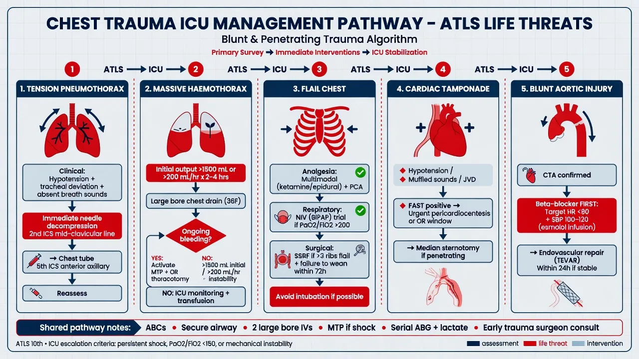
Management pathway for major chest trauma: tube thoracostomy, aggressive analgesia for ribs and flail, beta-blocker-first control for BTAI, TEVAR for higher-grade aortic injury
FigureAnalgesia prevents pneumonia; BTAI needs heart-rate control before pure vasodilators.
[5]
[5]

Blunt thoracic aortic injury and TEVAR pearls

  1. 90% of BTAI die at the scene — survivors have a contained (adventitial) injury. The classic survivor is a partial transection at the isthmus held by adventitia and mediastinal tissue. Without treatment, most survivors rupture within hours-to-days; the widening mediastinum on the trauma CXR is therefore an emergency that mandates CT angiography. Any high-energy deceleration mechanism with an abnormal mediastinum should be assumed to be BTAI until proven otherwise.[5]
  2. Beta-blockade first, then vasodilator — before and during repair. The single most important medical intervention is reducing the rate of pressure rise (dp/dt) in the aorta: start with a short-acting beta-blocker (esmolol, target SBP 100-120 and HR <100) and only THEN add a vasodilator (nicardipine or GTN) if needed. Giving a vasodilator first causes reflex tachycardia that increases shear and may extend the tear. Analgesia and avoidance of hypertension/pain are part of this strategy.[5]
  3. TEVAR is the standard of care for grade II-IV BTAI. Endovascular repair is associated with significantly lower mortality (~5-9%) and paraplegia than open repair (~20-30% mortality), and is feasible in patients with severe concomitant injuries who would not tolerate open thoracotomy. The SVS 2021 guideline recommends TEVAR for most injuries requiring repair. Lifelong CT surveillance is mandatory for endoleak, migration, and device-related complications. Open repair is reserved for unsuitable anatomy or when thoracotomy is needed for other injuries.[5]
  4. Grade I (intimal tear) BTAI is increasingly managed without an operation. Contemporary series show that small intimal tears heal on serial CT under pharmacological BP control, with a low rate of progression. Surgery (TEVAR) is reserved for grade I injuries that enlarge on surveillance imaging or that present with haemodynamic instability. This non-operative strategy avoids the morbidity and lifelong surveillance burden of a stent graft in young patients.[5]

Resuscitative thoracotomy — survival evidence

Resuscitative (emergency department) thoracotomy pearls

  1. Survival after EDT depends almost entirely on mechanism and signs of life. Rhee's 25-year review of published data showed overall survival of ~7.4%, but with a clear hierarchy: penetrating cardiac stab wound ~30-35%, penetrating cardiac gunshot ~15-20%, penetrating non-cardiac torso ~10-15%, and blunt trauma ~1-2%. The decisive factor is the presence of signs of life (SOL) at scene or on arrival: SOL present throughout → 15-35% survival; SOL lost before arrival → <5%; no SOL at scene → <1% (futile). Modern Trauma Quality Improvement Program data confirm overall survival around 8-9%, with the best neurological outcomes in penetrating injury and short arrest-to-thoracotomy intervals.[23]
  2. Patient selection: who should NOT get an EDT. EDT is indicated for: penetrating chest/torso trauma with arrest <15 min, or penetrating injury with peri-arrest (SBP <60, tamponade, massive haemothorax); blunt trauma with WITNESSED arrest and <10 min of arrest, particularly with a treatable intrathoracic cause. It is generally futile (and should not be performed) for: blunt trauma with unwitnessed arrest or arrest >10 min, penetrating trauma with no signs of life and arrest >15 min, or any patient without signs of life at the scene. A practical evidence-based selection algorithm minimises futile EDTs while capturing survivors.[24]
  3. Goals of the clamshell: relieve tamponade, control bleeding, cross-clamp, internal massage. Through a left anterolateral (or bilateral clamshell) incision at the 4th-5th ICS: (1) release pericardial tamponade (longitudinal pericardiotomy anterior to the phrenic nerve); (2) control intrathoracic haemorrhage (compress/repair cardiac or great-vessel injury — finger control first, sutures/staples/ Foley balloon as temporising); (3) cross-clamp the descending aorta to redistribute the limited blood volume to the heart and brain; (4) perform open cardiac massage (bimanual compression); (5) clamp the pulmonary hilum if a pulmonary hilar injury. Survival depends on rapidity — every minute counts.[23]

Red flags (expanded)

Critical chest trauma red flags (expanded)

  • Commotio cordis → instant VF after precordial impact in a young athlete → CPR + AED within minutes (survival halves every minute without shock).[7]
  • Tension pneumothorax → immediate needle/finger decompression (do not wait for CXR).[1]
  • Massive haemothorax → >1500 mL initial or >200 mL/h → emergency thoracotomy.[14]
  • Retained haemothorax (residual blood on day 2-7 CT) → early VATS to prevent empyema/fibrothorax.[13]
  • Blunt aortic injury (widened mediastinum) → CTA → beta-blockade → TEVAR.[5]
  • BTAI grade IV (rupture/extravasation) → emergency TEVAR/open repair; highest mortality.[5]
  • Tracheobronchial injury → persistent massive air leak / lung fails to re-expand → bronchoscopy → surgical repair.[15]
  • Blunt oesophageal injury → delayed diagnosis → mediastinitis/sepsis → contrast oesophagogram + early repair.[17]
  • Diaphragmatic rupture (left > right) → may present late with visceral herniation/strangulation → surgical repair.[1]
  • Blunt cardiac injury → ECG + troponin; abnormal ECG or haemodynamic instability → echo + 24-48 h monitoring.[3]
  • Sternal fracture + abnormal ECG → strong marker for BCI; screen and monitor.[11]
  • ≥3 rib fractures in age >65 → 10-20% mortality → aggressive regional analgesia (epidural).[1]
  • Pulmonary contusion worsens 24-48 h → fluid-restrictive strategy, lung-protective ventilation, consider APRV.[2]

Prognosis (expanded)

Commotio cordis — outcomes (US Minneapolis Commotio Cordis Registry)

[24]

Blunt thoracic aortic injury — TEVAR vs open repair (SVS 2021 guideline)

[5]

Surgical rib fixation (SSRF) — meta-analysis evidence (Hisamune 2024; Sharma 2024)

[20]

Retained haemothorax — VATS evidence (Chou 2015)

[24]

Blunt cardiac injury — outcomes (El-Qawaqzeh 2023; Clancy/EAST 2012)

[10]

References

  1. [1]Griffard J, et al. Management of blunt chest trauma. Surgical clinics of North America, 2024.PMID 38453306
  2. [2]Walkey AJ, et al. Use of airway pressure release ventilation is associated with a reduced incidence of ventilator-associated pneumonia in patients with pulmonary contusion. Journal of trauma, 2011.PMID 20526208
  3. [3]Clancy K, et al. Screening for blunt cardiac injury: an Eastern Association for the Surgery of Trauma practice management guideline. Journal of trauma and acute care surgery, 2012.PMID 23114485
  4. [4]Sharma VJ, et al. Surgical stabilisation of rib fractures: a meta-analysis of randomised controlled trials. Injury, 2024.PMID 38945079
  5. [5]Lee WA, Matsumura JS, Mitchell RS, et al. Endovascular repair of traumatic thoracic aortic injury: clinical practice guidelines of the Society for Vascular Surgery. Journal of vascular surgery, 2011.PMID 20974523
  6. [6]El-Qawaqzeh K, et al. Predictors of mortality in blunt cardiac injury: a nationwide analysis. Journal of surgical research, 2023.PMID 36108535
  7. [7]Maron BJ, et al. Clinical profile and spectrum of commotio cordis. JAMA, 2002.PMID 11879111
  8. [8]Sohail S, et al. Commotio cordis in non-sports-related injury: a scoping review. Current problems in cardiology, 2024.PMID 37890546
  9. [9]Salzillo C, et al. Commotio cordis in sudden cardiac death in the young: a state-of-the-art review. Reviews in cardiovascular medicine, 2025.PMID 41089808
  10. [10]Klei DS, et al. Epidemiology and outcomes of traumatic sternal fractures and associated blunt cardiac injury. European journal of trauma and emergency surgery, 2026.PMID 42223595
  11. [11]Fokin AA, et al. Blunt cardiac injury in patients with sternal fractures. Cureus, 2022.PMID 35382179
  12. [12]Grigorian A, et al. National risk factors for blunt cardiac injury: hemopneumothorax is the strongest predictor. American journal of surgery, 2019.PMID 30060913
  13. [13]Chou YP, et al. Video-assisted thoracoscopic surgery for retained hemothorax in blunt chest trauma. Current opinion in pulmonary medicine, 2015.PMID 25978625
  14. [14]Broderick SR, et al. Hemothorax: etiology, diagnosis, and management. Thoracic surgery clinics, 2013.PMID 23206720
  15. [15]Shemmeri E, et al. Blunt tracheobronchial trauma. Thoracic surgery clinics, 2018.PMID 30054080
  16. [16]Grewal HS, et al. Treatment of tracheobronchial injuries: a contemporary review. Chest, 2019.PMID 30059680
  17. [17]Fabien JJ, et al. Blunt traumatic esophageal injury: decreased mortality with urgent repair. American surgeon, 2022.PMID 35466715
  18. [18]Bryant AS, et al. Esophageal trauma. Thoracic surgery clinics, 2007.PMID 17650698
  19. [19]Hisamune R, Kobayashi M, Nakasato K, et al. A meta-analysis and trial sequential analysis of randomised controlled trials comparing nonoperative and operative management of chest trauma with multiple rib fractures. World journal of emergency surgery : WJES, 2024.PMID 38504282
  20. [20]Beks RB, et al. Fixation of flail chest or multiple rib fractures: current evidence and how to proceed — a systematic review. European journal of trauma and emergency surgery, 2019.PMID 30276722
  21. [21]Wallen TE, et al. Intercostal liposomal bupivacaine injection for rib fractures: a prospective randomised controlled trial. Journal of trauma and acute care surgery, 2022.PMID 34789700
  22. [22]Gamberini L, et al. Regional anaesthesia modalities in blunt thoracic trauma: a systematic review and Bayesian network meta-analysis. American journal of emergency medicine, 2025.PMID 39740311
  23. [23]Rhee PM, et al. Survival after emergency department thoracotomy: review of published data from the past 25 years. Journal of the American College of Surgeons, 2000.PMID 10703853
  24. [24]Joseph B, et al. Improving survival after an emergency resuscitative thoracotomy: a 5-year review of the Trauma Quality Improvement Program. Trauma surgery and acute care open, 2018.PMID 30402559