ICU · Trauma
Fat embolism syndrome: diagnosis, ventilation, and management in trauma
Also known as Fat embolism syndrome · FES · Fat embolism · Orthopaedic embolism · Gurd criteria
Fat embolism syndrome (FES) is a clinical syndrome (triad: respiratory distress, neurological dysfunction, petechial rash) occurring 12-72 hours after TRAUMA (especially long bone/pelvic fractures) or occasionally orthopaedic procedures (joint replacement, intramedullary nailing). PATHOPHYSIOLOGY: (1) MECHANICAL — fat globules from marrow enter venous circulation - embolise to lungs (and through PFO to brain). (2) BIOCHEMICAL — free fatty acids released - toxic to capillary endothelium - inflammatory cascade - ARDS-like lung injury, cerebral oedema. CLASSIC TRIAD (within 72h): (1) RESPIRATORY DISTRESS (dyspnoea, hypoxaemia, ARDS — 85% of cases). (2) NEUROLOGICAL (confusion, altered consciousness, seizures — 80%). (3) PETECHIAL RASH (upper chest, axillae, conjunctivae — 50-60% — pathognomonic but often late/absent). DIAGNOSIS: clinical (Gurd criteria — major + minor features). MANAGEMENT: SUPPORTIVE (oxygen, ventilation — lung-protective), early fracture fixation (reduces FES risk), corticosteroids controversial. MORTALITY: 5-10% (with supportive care). PREVENTION: early fracture fixation, operative stabilisation.
On this page & tools
Your progress
Saved locally on this device.
Target exams
Red flags
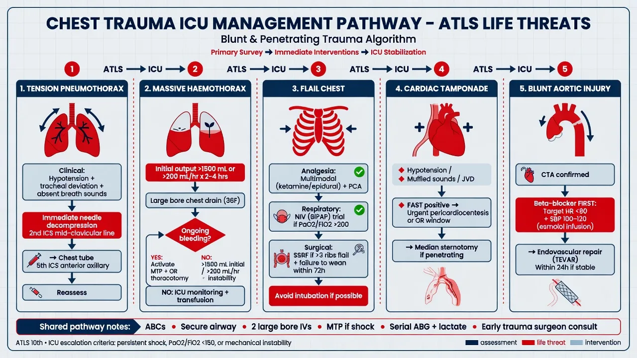
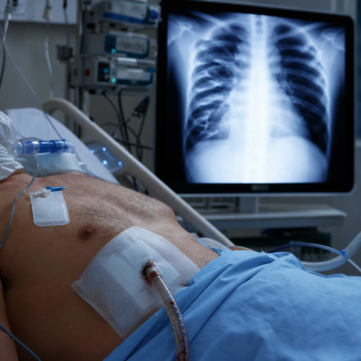
FES differential — acute deterioration after trauma
| Condition | Timing | Key features | Diagnosis |
|---|---|---|---|
| Fat embolism (FES) | 12-72h post-fracture | Triad (resp + neuro + rash), hypoxaemia | Clinical (Gurd); exclude others |
| Pulmonary embolism | Any time (esp. immobilised) | Unilateral leg swelling, sudden dyspnoea, pleuritic pain | CTPA, D-dimer, echocardiography |
| Tension pneumothorax | Immediate (or delayed) | Unilateral hyperresonance, tracheal deviation | Clinical; CXR/POCUS |
| Aspiration pneumonitis | During/after intubation | Post-intubation hypoxaemia, infiltrate | Clinical; CXR |
| **ARDS (non-fat) } | Days | Lung injury from sepsis/trauma/pneumonia | 柏林定义 |
| Sepsis | Any time | Fever, source, rising lactate | Cultures, source identification |
| Intracranial injury | Immediate/delayed | Focal deficit, deteriorating GCS | CT brain |
Management of suspected fat embolism syndrome
- RECOGNISE THE CLINICAL PICTURE — 12-72 HOURS after long bone/pelvic fracture (or orthopaedic surgery): sudden RESPIRATORY DISTRESS (dyspnoea, hypoxaemia — SpO2 drop), NEUROLOGICAL (confusion, agitation, decreased consciousness, seizures), PETECHIAL RASH (upper chest, axillae, conjunctivae — pathognomonic but may be absent). ALSO: fever, tachycardia, jaundice. Apply Gurd criteria (1 major + 4 minor, or 2 major alone). EXCLUDE other causes (PE, pneumothorax, sepsis, intracranial injury)
- SUPPORTIVE RESPIRATORY CARE (MAINSTAY) — (a) OXYGEN (high-flow — target SpO2 >92%). (b) HFNC or NIV if moderate hypoxaemia. (c) INTUBATE + MECHANICAL VENTILATION if severe (PaO2/FiO2 <200, refractory hypoxaemia, exhaustion) — LUNG-PROTECTIVE (Vt 6 mL/kg, plateau <30, PEEP titrated). (d) FES causes ARDS-like lung injury (capillary leak from free fatty acids) -> manage as ARDS (proning if severe, permissive hypercapnia). (e) AVOID fluid overload (worsens pulmonary oedema)
- NEUROLOGICAL SUPPORT — (a) Cerebral FES: confusion, seizures, coma — from embolic fat (through PFO or via pulmonary venous shunts) + cerebral oedema. (b) CT brain (exclude traumatic brain injury, haemorrhage). (c) Seizures: benzodiazepines. (d) Raised ICP (if severe): head elevation, osmotic agents (mannitol, hypertonic saline) — if evidence of oedema. (e) Most neurological changes are TRANSIENT (resolve over days). (f) Sedation: minimise (to assess neurology)
- FRACTURE MANAGEMENT (PREVENT ONGOING EMBOLISATION) — (a) EARLY OPERATIVE FIXATION (intramedullary nailing, plating) — stabilises fracture -> reduces ongoing fat/marrow embolisation. (b) AVOID delayed fixation or prolonged traction (higher FES risk). (c) TIMING: within 24-48h of injury (if haemodynamically stable). (d) CAVEAT: intramedullary nailing itself can cause FES (reaming + pressurisation -> embolisation) — use unreamed nails or plating for high-risk. (e) Support fracture (splint) before definitive fixation
- ADJUNCTS (CONTROVERSIAL) — (a) CORTICOSTEROIDS: (i) PROPHYLACTIC (high-dose methylprednisolone at admission) — some studies reduce FES incidence in high-risk (multiple long bone fractures). (ii) THERAPEUTIC (once FES established) — no proven benefit. (iii) NOT routine — controversial. (b) ASPIRIN, HEPARIN, DEXTRAN: no proven benefit (avoid — bleeding risk in trauma). (c) ALBUMIN: theoretical (binds free fatty acids) — no proven benefit. (d) NO SPECIFIC ANTIDOTE — supportive care is mainstay
- MONITORING + SUPPORTIVE CARE + RECOVERY — (a) Continuous SpO2, cardiac monitor, urine output. (b) Serial ABG (hypoxaemia, acidosis). (c) Serial GCS (neurological trend). (d) FBC (anaemia, thrombocytopenia), coagulation (DIC), LFTs (jaundice), renal function. (e) SUPPORTIVE: nutrition, DVT prophylaxis, stress ulcer prophylaxis, infection surveillance. (f) EXPECTED COURSE: respiratory distress peaks 24-72h, then improves over days; neurology resolves over days-weeks; most recover fully. (g) MORTALITY: 5-10% (with supportive care). (h) REHABILITATION: orthopaedic (fracture healing), neuro/cognitive (if deficits persist)
SAQ — Fat embolism syndrome after femoral nailing
10 minutes · 10 marks
A 24-year-old man was admitted 36 h ago after a motorcycle crash with an isolated closed left femoral shaft fracture; he underwent intramedullary nailing within 6 h. He now has acute onset of dyspnoea (RR 36, SpO₂ 86% on 15 L O₂), confusion and agitation, and a petechial rash over the upper chest and conjunctivae. HR 128, BP 100/70, temp 38.6°C. ABG shows pH 7.30, PaO₂ 52, PaCO₂ 32. Platelets 95 ×10⁹/L.
SAQ — Cerebral fat embolism in the multiply-injured patient
10 minutes · 10 marks
A 30-year-old man with bilateral femoral shaft fractures from a motor vehicle crash (no head injury on initial CT) develops acute confusion, agitation and decreased level of consciousness 30 h after admission, with respiratory failure requiring intubation. MRI brain shows a 'starfield pattern' of diffuse punctate T2/FLAIR hyperintense lesions in the deep white matter. Petechiae over the axillae are noted.
Clinical pearls
Red flags
Prognosis
Fat embolism syndrome evidence and outcomes
Incidence: 0.5-3% of long bone fractures; higher (10-20%) in multiple fractures/pelvic. Mortality: 5-10% (with supportive care). Early fixation (vs delayed/traction): reduces FES risk (multiple observational studies — Lever 2018 review). Corticosteroids (Bemelman 2019 meta-analysis): possible prophylactic benefit in high-risk; not routine. Ventilation: lung-protective (as ARDS) — reduces mortality (ARDSNet principle applies). Neurological: usually transient (days-weeks); some persistent cognitive deficits. Respiratory: most recover fully (no long-term lung damage). Diagnosis: clinical (Gurd criteria); MRI brain (starfield pattern), BAL (fat-laden macrophages) — supportive.
Pathophysiology — mechanical and biochemical mechanisms in detail
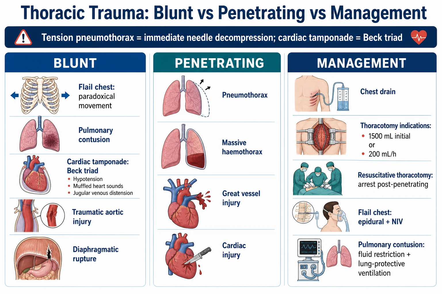
Mechanical vs biochemical mechanisms of fat embolism syndrome
| Feature | Mechanical theory | Biochemical (physicochemical) theory |
|---|---|---|
| Core event | Embolised marrow fat globules (40-200 μm) physically occlude pulmonary and systemic capillaries | Lipase hydrolyses triglycerides → free fatty acids (oleic, linoleic) released locally |
| Source of fat | Medullary cavity of long bones (femur, tibia); also subcutaneous/omental fat in severe trauma | Same embolised fat globules, broken down in lung and tissues |
| Onset | Immediate at moment of fracture / instrumentation | Delayed 12-72h — needs time for hydrolysis + inflammatory amplification |
| Pulmonary effect | RV afterload rise, V/Q mismatch, shunt; gross fat in pulmonary vessels at autopsy | FFA-toxic endothelial injury → capillary leak → ARDS-like diffuse alveolar damage with neutrophil infiltrate |
| Systemic effect | Passage via PFO (25% of adults) or pulmonary shunts → brain, retina, skin, kidney, coronary microcirculation | Circulating FFAs activate complement + neutrophils, aggravate coagulopathy (DIC-like), spread injury to distant beds |
| Therapeutic implication | Reduce ongoing embolisation: early stabilisation, unreamed nails, venting of the nail canal | Support the capillary leak: lung-protective ventilation, conservative fluids; no proven FFA scavenger |
| Histology | Fat microglobulin in pulmonary, cerebral and renal capillaries (Oil-Red-O stain) | Endothelial disruption, perivascular haemorrhage, neutrophilic infiltrate, microthrombi |
Pathophysiological cascade from fracture to ARDS-pattern lung injury
- FRACTURE / INSTRUMENTATION RELEASES MARROW FAT — Medullary pressure rises sharply with reaming or fracture displacement; liquid marrow fat is forced into the thin-walled venous sinusoids of the long bone and enters the systemic venous circulation as 40-200 μm droplets
- PULMONARY VASCULAR OBSTRUCTION (mechanical phase) — Fat droplets are too large to traverse the pulmonary capillary bed (8-9 μm diameter) and embolise distally; acute rise in pulmonary vascular resistance, RV afterload, and right-heart strain; some fat lodges in cerebral, retinal, renal and dermal capillaries via a PFO (paradoxical embolism) or pre-capillary shunts
- LIPASE-MEDIATED HYDROLYSIS (biochemical phase) — Pulmonary and tissue lipase cleaves triglycerides into FFAs (oleic acid is the most endotheliotoxic) — this step takes 12-72h and explains the latency of the clinical syndrome
- ENDOTHELIAL INJURY + NEUTROPHIL ACTIVATION — FFAs directly injure capillary endothelium; complement (C5a) and cytokine (TNF-α, IL-1, IL-6) cascades recruit and activate neutrophils; free radicals and proteases damage the basement membrane
- CAPILLARY LEAK → ARDS-LIKE PATTERN — Alveolar-capillary barrier disruption → protein-rich pulmonary oedema, hyaline membranes, refractory hypoxaemia with bilateral infiltrates — histologically indistinguishable from ARDS of any other cause
- SYSTEMIC ORGAN INJURY — The same capillary-leak process in the brain (cerebral oedema, petechiae), skin (petechial rash), kidneys (oliguria, lipiduria), and heart (microinfarcts) produces the multisystem FES phenotype
Gurd's and Lindeque criteria — diagnostic frameworks in detail
Gurd (1970/1974) vs Lindeque (1987) diagnostic criteria for FES
| Feature | Gurd & Wilson criteria | Lindeque criteria |
|---|---|---|
| Year / design | 1970/1974 — clinical scoring system | 1987 — simplification emphasising respiratory criteria |
| Major criteria | (1) Respiratory insufficiency (dyspnoea, tachypnoea, cyanosis, ARDS); (2) Cerebral involvement (confusion, drowsiness, seizures, coma unrelated to head injury or hypoxaemia); (3) Petechial rash (chest, axillae, conjunctivae, neck) | Not divided into major/minor |
| Minor criteria | (1) Pyrexia >38°C; (2) Tachycardia >110/min; (3) Retinal changes (fat, petechiae); (4) Jaundice; (5) Renal signs (oliguria, anuria, fat, tubular casts); (6) Anaemia; (7) Thrombocytopenia; (8) High ESR; (9) Fat macroglobinaemia | — |
| Diagnostic rule | 1 major + 4 minor, OR 1 major + fat macroglobinaemia, OR 2 major (in setting of long-bone/pelvic trauma 12-72h prior) | A patient with a long-bone fracture who develops RR >35, PaO2 <60 mmHg on room air, declining PetCO2, or unexplained tachycardia/pyrexia/thrombocytopenia has FES by definition |
| Strength | More comprehensive; widely used in research and exam answers | Captures respiratory-predominant FES earlier; no exclusion workup required |
| Limitation | Petechial rash may be absent/late; anaemia and thrombocytopenia are non-specific in trauma | Over-diagnoses FES — many multiply-injured patients meet Lindeque criteria without true FES |
| In practice | Use Gurd as the framework; Lindeque as a 'low threshold to act' principle | — |
Cerebral fat embolism — the "starfield" MRI pattern
Approach to neurological dysfunction in suspected FES
- DETERMINE TIMING AND EXCLUDE TBI — Onset 12-72h after the insult, in the absence of direct head injury or worsening on repeat CT brain. Obtain a non-contrast CT brain early — this is to EXCLUDE traumatic brain injury (extradural, subdural, contusional haemorrhage) and anoxic injury, NOT to diagnose FES (CT is often normal or shows only non-specific oedema in early cerebral FES)
- PERFORM MRI BRAIN WITH DIFFUSION (DWI) IF AVAILABLE — The characteristic 'STARFIELD PATTERN' on DWI/FLAIR is a diffuse scattering of punctate, 2-10 mm, non-confluent, restricted-diffusion lesions throughout the cerebral white matter, basal ganglia, brainstem and cerebellum — microinfarcts from fat emboli. Susceptibility-weighted imaging (SWI) shows numerous microhaemorrhages. The pattern is highly suggestive of cerebral FES
- EXCLUDE PARADOXICAL EMBOLI — BUBBLE CONTRAST ECHO / TCD — If neurological involvement dominates with mild pulmonary features, search for a PFO with bubble-contrast echocardiography or transcranial Doppler with agitated saline. Right-to-left shunting supports paradoxical fat embolism
- MANAGE SUPPORTIVELY — Most cerebral FES is self-limiting: (a) treat seizures with benzodiazepines then levetiracetam; (b) maintain head of bed 30°; (c) if signs of raised ICP (coma, pupillary changes, imaging oedema), use osmotic therapy (mannitol 0.5 g/kg or 3% saline boluses to Na 145-150); (d) maintain CPP >60 mmHg; (e) avoid hyperglycaemia, hyperthermia and agitation
- ANTICIPATE RECOVERY OVER DAYS TO WEEKS — Confusion and agitation typically resolve within 5-10 days; deeper deficits may persist for weeks. MRI abnormalities resolve slowly (weeks-months); a small minority are left with persistent cognitive impairment. Avoid early prognostication — 'wait and see' is appropriate
Cerebral FES vs traumatic brain injury (TBI) — distinguishing features
| Feature | Cerebral FES | Traumatic brain injury (TBI) |
|---|---|---|
| Mechanism | Fat microemboli via PFO/shunts → diffuse microinfarcts + oedema | Direct impact/deceleration → contusion, haemorrhage, axonal injury |
| Timing | 12-72h after long-bone fracture | At the moment of injury |
| Consciousness | Fluctuating confusion, agitation → depressed consciousness | Immediate depressed GCS (or lucid interval then deterioration in extradural) |
| Focal signs | Rare — usually diffuse | Common — focal deficit relates to lesion site |
| CT brain (early) | Often normal or diffuse oedema | Abnormal — haemorrhage, midline shift, fracture |
| MRI brain | 'Starfield' pattern (punctate DWI/SWI lesions) | Focal contusions, DAI (corpus callosum, brainstem), haematoma |
| Concurrent | Respiratory distress, petechial rash, fever | May have scalp laceration, skull fracture, other injuries |
| Prognosis | Usually transient (days-weeks) | Variable — depends on injury severity |
Early vs delayed fracture fixation — the central preventive intervention
Early total care (ETC) vs damage control orthopaedics (DCO) in polytrauma
| Strategy | Early total care (ETC) | Damage control orthopaedics (DCO) |
|---|---|---|
| Definition | Definitive fixation (usually reamed intramedullary nail) of major long-bone fractures within 24h of injury | Temporary external fixation now, conversion to definitive fixation in 5-14 days once physiology stabilises |
| Patients | Stable, no severe head/chest injury, normal lactate, no coagulopathy | 'Borderline' or unstable: shock, severe TBI, severe chest trauma (AIS ≥3), coagulopathy, hypothermia, severe acid-base derangement |
| Effect on FES risk | Stabilises the fracture and HALTS ongoing marrow embolisation — reduces FES incidence | Also stabilises and halts embolisation; smaller secondary inflammatory hit than definitive nailing in the unstable patient |
| Secondary inflammatory hit | Definitive nailing in a shocked patient adds a surgical 'second hit' that may worsen lung injury | External fixator is minimal 'second hit' — lung-protective in the unstable polytrauma patient |
| Risk of reaming itself causing FES | Reaming pressurises the canal and releases marrow fat — measurable embolic shower on TEE; clinical FES uncommon but possible | External fixation avoids canal pressurisation — minimal embolic load |
| Practical principle | STABLE patient → early definitive fixation (within 24-48h) — reduces FES, pneumonia, DVT, mortality | UNSTABLE / borderline patient → external fixator, return for definitive fixation once stabilised |
Choosing fixation strategy in a polytrauma patient with long-bone fracture
- ASSESS STABILITY AT PRESENTATION AND AFTER RESUSCITATION — Determine: shock (lactate >2.5, base deficit >-5, transfusion need), TBI (GCS ≤8 or significant intracranial lesion on CT), chest trauma (AIS ≥3, pulmonary contusion, PaO2/FiO2 <300), coagulopathy (INR >1.5, platelets <95), hypothermia (<35°C)
- STABLE PATIENT → EARLY TOTAL CARE — Definitive fixation (reamed or unreamed nail, plating) within 24-48h. Reduces FES, ARDS, pneumonia, DVT, mortality. Reamed nailing is acceptable and is the standard for femoral shaft fractures
- BORDERLINE / UNSTABLE PATIENT → DAMAGE CONTROL ORTHOPAEDICS — Apply a spanning external fixator rapidly (≤2h), with minimal blood loss and physiological insult. The fracture is stabilised (stopping ongoing embolisation) without the second-hit inflammatory load of reaming. Convert to definitive fixation in 5-14 days once lactate clears, coagulopathy resolves, and chest/TBI stabilise
- PATIENT WITH ISOLATED SEVERE CHEST TRAUMA + FEMUR FRACTURE — Special case: even a 'stable' patient with a severe pulmonary contusion can deteriorate with early reaming. Many centres prefer DCO (external fixator) in this combination, then convert after 5-7 days when lung function improves
- PATIENT WITH SEVERE TBI + FEMUR FRACTURE — Avoid hypotension (SBP <110) and hypoxia at all costs (each worsens secondary brain injury). DCO with rapid external fixation and meticulous haemodynamic management is preferred; early reamed nailing is associated with worse neurological outcomes in some series
- MINIMISE THE EMBOLIC LOAD DURING NAILING (whichever strategy) — (a) Vent the nail canal where possible (when the proximal femur or tibia allows); (b) Use unreamed nails or slow, low-pressure reaming in high-risk cases; (c) Avoid sustained high intramedullary pressure (tight fit, back-slapping); (d) Monitor with transoesophageal echocardiography or pulmonary artery pressure in the highest-risk cases
Corticosteroid prophylaxis — the evidence
Corticosteroid prophylaxis for FES — key trials and meta-analysis
Schonfeld 1983 (Ann Intern Med, n=64, high-risk patients): methylprednisolone 7.5 mg/kg q6h × 12 doses starting within 12h — FES reduced from 22% to 0% in the treated group; no increase in infection. Establishes the conceptual role of prophylactic steroids.[9] Kallenbach 1987 (J Trauma, n=80): low-dose methylprednisolone (1.5 mg/kg) × 4 doses — FES reduced from 22% to 8%; no infection signal. Argued for a lower, safer dose.[10] Lindeque 1987 (J Bone Joint Surg Br, n=54): high-dose methylprednisolone (1 g × 3 doses) — no difference in FES or ARDS rates. A negative trial.[11] Bederman 2009 meta-analysis (Can J Surg, 7 RCTs, 389 patients): pooled RR for FES 0.40 (95% CI 0.22-0.72) favouring prophylactic corticosteroids; significant heterogeneity, variable definitions of FES, no mortality benefit, possible trend to more infection. Concluded: prophylactic steroids REDUCE FES incidence in high-risk patients but no mortality benefit; not adopted as routine due to concern for infection in trauma.[4] Clinical bottom line: steroids have a plausible mechanism (suppress FFA-induced neutrophil inflammation), a consistent prophylactic signal in meta-analysis, and no mortality benefit. Most trauma guidelines do NOT recommend routine prophylactic steroids; some centres use them for the highest-risk patients (multiple long-bone fractures, severe pelvic fracture) in whom FES incidence is 10-20%. THERAPEUTIC steroids (after FES has developed): NO trial evidence of benefit; not recommended.
Supportive investigations — bronchoalveolar lavage and others
Supportive investigations in suspected FES
| Test | Finding in FES | Sensitivity | Specificity | Practical role |
|---|---|---|---|---|
| BAL fat-laden macrophages (Oil-Red-O / Sudan stain) | Lipid-laden macrophages in alveolar lavage | ~80% | Low — also positive in aspiration, chronic steroid use, ARDS | Supportive only; FES is a clinical diagnosis |
| Frozen-section plasma / fat macroglobinaemia (Oil-Red-O on centrifuged plasma) | Fat microglobules in plasma | ~50-70% | Moderate | Rarely performed; sometimes in research/medicolegal |
| MRI brain DWI / SWI | 'Starfield pattern' — punctate restricted-diffusion lesions and microhaemorrhages | High (when present) | High (in context) | Best for cerebral FES; confirms neuro involvement |
| CT brain (non-contrast) | Often normal early; may show diffuse oedema or petechial haemorrhage later | Low early | Low | Used to EXCLUDE TBI; not to diagnose FES |
| Fundoscopy | Retinal fat globules, petechial haemorrhages, cotton-wool spots | Moderate | High (in context) | Bedside, supportive — a Gurd MINOR criterion |
| Skin biopsy (petechial lesion) | Intravascular fat on Oil-Red-O | ~80% | High | Rarely needed; used when diagnosis genuinely uncertain |
| Transoesophageal echo during nailing | Echogenic 'snowstorm' material in right heart | High (procedural) | High for embolic shower | Predicts the inflammatory load; not routinely used |
| Platelet count trend | Falling platelets over 24-48h | Non-specific | Non-specific | Cheap, available, supportive when falling |
Iatrogenic / perioperative fat embolism
Recognition and prevention of perioperative FES (orthopaedic and liposuction contexts)
- RECOGNISE THE NON-TRAUMA SETTINGS — FES is well-described after: (a) intramedullary nailing (femur, tibia); (b) total hip and total knee arthroplasty (pressurisation of cement or marrow); (c) liposuction, especially large-volume (>5 L) or combined with fat-grafting; (d) medullary reaming of any long bone; (e) percutaneous vertebroplasty/kyphoplasty (rare). Maintain a low index of suspicion in any patient 12-72h after these procedures who develops hypoxaemia, confusion, or a petechial rash
- PHYSIOLOGICAL MONITORING DURING HIGH-RISK PROCEDURES — In high-risk orthopaedic procedures (reamed femoral nailing in polytrauma, long bilateral nailing) consider transoesophageal echocardiography or pulmonary artery pressure monitoring to detect the embolic shower in real time. A precipitate rise in pulmonary artery pressure or RV strain pattern on TEE heralds a major embolic load and should slow the procedure, vent the canal, or pause
- CANAL VENTING AND UNREAMED TECHNIQUES — Venting the femoral canal (e.g. via the greater trochanter) reduces intramedullary pressure during reaming by 30-50% and lowers the embolic shower. Unreamed nails and low-pressure reaming also reduce the load. Plating avoids canal pressurisation altogether and is preferred for high-risk polytrauma cases (see DCO above)
- CEMENT IMPLANTATION SYNDROME — the arthroplasty analogue — During cemented hip/knee arthroplasty, pressurised cement can force marrow contents into the venous circulation → acute hypoxaemia, hypotension, bronchospasm, occasionally cardiac arrest. The mechanism is identical to FES (mechanical embolisation + vasoactive mediator release) but compressed in time to seconds-minutes. Manage with high-flow oxygen, vasopressors, and (if severe) resuscitation and ventilation. Use uncemented components in patients at high cardiac risk
- LIPOSUCTION-ASSOCIATED FES — Large-volume liposuction (>5 L total aspirate) can produce an FES-like illness (hypoxaemia, confusion, coagulopathy) 12-72h postoperatively. Prevention: staged procedures, avoid mega-volume aspiration, maintain hydration, monitor overnight. Treatment is supportive and indistinguishable from trauma-related FES
- POST-MORTEM IDENTIFICATION — FES may be a contributor to death after trauma or surgery; pathologists stain pulmonary, cerebral and renal sections with Oil-Red-O to demonstrate intravascular fat. A diagnosis that may be missed on routine H&E staining is therefore confirmed histologically
Red flags (expanded)
Prognosis (expanded)
FES outcomes — incidence, mortality, and recovery
Incidence: 0.5-3% of isolated long-bone fractures; 10-20% in multiple long-bone or severe pelvic fractures; up to 30% in polytrauma with combined femoral + chest injury. A systematic review (Lempert 2021) found femur-fracture FES incidence around 1-2% in modern practice, lower than historical estimates, attributed to early fixation and modern nailing techniques.[16] Mortality: 5-10% with modern supportive care (lung-protective ventilation, conservative fluids); historical mortality was 10-20% before ARDS-era ventilation. Most deaths are from ARDS or multi-organ failure rather than FES itself.[15] Predictors of poor outcome: severe hypoxaemia (PaO2/FiO2 <100), RV failure on echo, severe cerebral involvement (coma >48h, seizures), multi-organ failure, age >60, pre-existing cardiopulmonary disease.[19] Respiratory recovery: peak distress at 24-72h, then improvement over 5-7 days; most survivors have no residual pulmonary impairment. A minority progress to fibrotic lung disease. Neurological recovery: most cognitive deficits resolve over 1-4 weeks; a small subgroup (~5-10% of cerebral FES) has persistent cognitive impairment, fine-motor disturbance or mood disorder detectable at 6-12 months. Skin: petechiae resolve in 5-7 days without scarring. Preventable fraction: early operative fixation vs prolonged traction reduces FES incidence by 3-5× — the single highest-yield preventive intervention.[5]
FES prevention — evidence for early fixation and steroids
Bone 1989 (JBJS Am, RCT): early (<24h) vs delayed (>48h) stabilisation of femoral shaft fractures — significant reduction in pulmonary complications (ARDS, pneumonia, FES) and ICU stay with early fixation. Foundational trial for early total care.[5] Pape 2007 (Ann Surg, 'borderline' polytrauma patients): compared ETC (reamed nailing) vs DCO (external fixation then conversion) in borderline polytrauma patients with femoral shaft fractures. DCO showed a trend to fewer pulmonary complications in the borderline group, supporting damage control strategy for the unstable/borderline patient.[12] Bederman 2009 meta-analysis (Can J Surg, 7 RCTs, 389 patients): prophylactic corticosteroids reduced FES incidence (pooled RR 0.40, 95% CI 0.22-0.72) but with significant heterogeneity, no mortality benefit, and a non-significant trend to more infection. NOT adopted as routine.[4] Practical synthesis: in the STABLE polytrauma patient, fix early and definitively (ETC); in the BORDERLINE/UNSTABLE patient (shock, severe TBI, severe chest trauma, coagulopathy, hypothermia), apply a damage control external fixator and return for definitive fixation once stabilised. Steroids are reserved for discussion in the highest-risk subgroup.[15]
Diagnostic test performance — MRI, BAL, fundoscopy
MRI brain DWI 'starfield' pattern: most specific supportive test for cerebral FES; sensitivity limited by timing (best at 24-72h) and access in the ICU. SWI adds microhaemorrhage detection.[2] BAL fat-laden macrophages: sensitivity ~80% but specificity low (also positive in aspiration, chronic steroid use, ARDS); supportive only.[2] Fundoscopy (retinal fat, petechiae, cotton-wool spots): bedside, free, a Gurd MINOR criterion; underused.[1] Skin biopsy of petechial lesion (Oil-Red-O): ~80% positive in FES; rarely needed clinically.[14] Plasma fat macroglobinaemia: sensitivity ~50-70%; specificity moderate; rarely performed.[13]
Anaesthetic and perioperative considerations
Anaesthetic technique in known/at-risk cases: avoid hypoxaemia and hypotension (worsen both pulmonary and cerebral FES); maintain adequate perfusion pressure; lung-protective ventilation if intubated; careful fluid balance (conservative).[7] Intraoperative detection: TEE shows the embolic shower in real time during reaming — useful in high-risk cases for procedural modification.[8] PFO considerations: a known PFO is a relative risk-amplifier for systemic (cerebral, retinal, dermal) FES; bubble-contrast echo if paradoxical embolism suspected. PFO closure not routinely indicated after a single FES episode.[13] Cement implantation syndrome: the time-compressed arthroplasty analogue of FES — pressurised cement forces marrow into venous circulation in seconds. Prevent with uncemented components in high-risk patients; treat with oxygen, vasopressors and ventilation if severe.[7]
References
- [1]Gurd AR, Wilson RI The fat embolism syndrome. Journal of bone and joint surgery (British volume), 1974.PMID 4547466
- [2]Newbigin K, Souza CA, Torres C, et al. Fat embolism syndrome: State-of-the-art review focused on pulmonary imaging findings. Respiratory medicine, 2016.PMID 26895808
- [3]Timon C, Keady C, Murphy CG Fat Embolism Syndrome — A Qualitative Review of its Incidence, Presentation, Pathogenesis and Management. Malaysian orthopaedic journal, 2021.PMID 33880141
- [4]Bederman SS, Bhandari M, McKee MD, et al. Do corticosteroids reduce the risk of fat embolism syndrome in patients with long-bone fractures? A meta-analysis. Canadian journal of surgery, 2009.PMID 19865573
- [5]Bone LB, Johnson KD, Weigelt J, et al. Early versus delayed stabilization of femoral fractures. A prospective randomized study. Journal of bone and joint surgery (American volume), 1989.PMID 2925704
- [6]Rothberg DL, Makarewich CA Fat Embolism and Fat Embolism Syndrome. Journal of the American Academy of Orthopaedic Surgeons, 2019.PMID 30958807
- [7]Luff D, Hewson DW Fat embolism syndrome. BJA education, 2021.PMID 34457354
- [8]Mellor A, Soni N Fat embolism. Anaesthesia, 2001.PMID 11167474
- [9]Schonfeld SA, Ploysongsang Y, DiLisio R, et al. Fat embolism prophylaxis with corticosteroids. A prospective study in high-risk patients. Annals of internal medicine, 1983.PMID 6354030
- [10]Kallenbach J, Lewis M, Zaltzman M, et al. 'Low-dose' corticosteroid prophylaxis against fat embolism. Journal of trauma, 1987.PMID 3312625
- [11]Lindeque BG, Schoeman HS, Dommisse GF, et al. Fat embolism and the fat embolism syndrome. A double-blind therapeutic study. Journal of bone and joint surgery (British volume), 1987.PMID 3818718
- [12]Pape HC, Rixen D, Morley J, et al. Impact of the method of initial stabilization for femoral shaft fractures in patients with multiple injuries at risk for complications (borderline patients). Annals of surgery, 2007.PMID 17717453
- [13]Parisi DM, Koval K, Egol K Fat embolism syndrome. American journal of orthopedics (Belle Mead, N.J.), 2002.PMID 12650535
- [14]Cantu CA, Pavlisko EN Liposuction-Induced Fat Embolism Syndrome: A Brief Review and Postmortem Diagnostic Approach. Archives of pathology and laboratory medicine, 2018.PMID 29939780
- [15]Kwon J, Coimbra R Fat embolism syndrome after trauma: What you need to know. Journal of trauma and acute care surgery, 2024.PMID 39213184
- [16]Lempert M, Halvachizadeh S, Ellanti P, et al. Incidence of Fat Embolism Syndrome in Femur Fractures and Its Associated Risk Factors over Time — A Systematic Review. Journal of clinical medicine, 2021.PMID 34205701
- [17]Taviloglu K, Yanar H Fat embolism syndrome. Surgery today, 2007.PMID 17186337
- [18]Richards RR Fat embolism syndrome. Canadian journal of surgery, 1997.PMID 9336522
- [19]Miyake T, Okada H, Kanda N Advances and uncertainties in fat embolism syndrome: a review. Trauma surgery and acute care open, 2026.PMID 42375760