ICU · Trauma
Massive haemothorax and emergency thoracotomy (resuscitative)
Also known as Massive haemothorax · Emergency thoracotomy · Resuscitative thoracotomy · Emergency department thoracotomy · EDT · Clamshell thoracotomy · Trauma thoracotomy
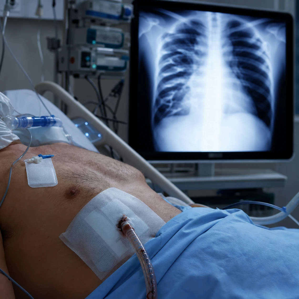
Massive haemothorax: 1500 mL blood in pleural space OR 200 mL/hr ongoing drainage. Life-threatening: impairs ventilation + circulation (hypovolaemia + tension physiology). Management: IMMEDIATE large-bore chest tube (28-36 Fr) at 5th ICS mid-axillary line (BATLS/ATLS). Resuscitative thoracotomy (EDT / clamshell): emergency department procedure for arrested/peri-arrest trauma — releases tamponade, controls bleeding, open cardiac massage, cross-clamps aorta. INDICATIONS: penetrating chest trauma with <15 min arrest; penetrating torso trauma with signs of life; blunt trauma with witnessed arrest (<10 min) — selective. Contraindicated: blunt trauma prolonged arrest (15 min), no signs of life. Survival: penetrating cardiac stab ~30%, gunshots ~15%, blunt <2%.
On this page & tools
Your progress
Saved locally on this device.
Target exams
Red flags
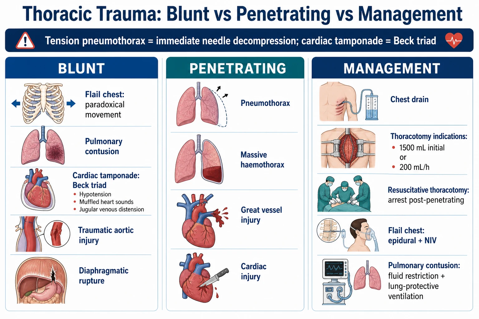
Resuscitative thoracotomy indications and survival
| Scenario | Indicated? | Survival |
|---|---|---|
| Penetrating cardiac injury — stab, with signs of life (SOL) | YES — immediate | ~30-35% |
| Penetrating cardiac injury — gunshot, with SOL | YES — immediate | ~15-20% |
| Penetrating torso (non-cardiac), arrest <15 min | YES | ~10-15% |
| Penetrating trauma, no SOL at scene | NO — futile | <1% |
| Blunt trauma, witnessed arrest <10 min, SOL | SELECTIVE (consider) | ~1-5% |
| Blunt trauma, arrest >15 min, no SOL | NO — futile | <1% |
| Penetrating extremity trauma, arrest | NO (not thoracic — different approach) | Very low |
Resuscitative thoracotomy (clamshell) — procedure steps
- DECISION — confirm indication: penetrating chest/torso trauma + arrest OR peri-arrest (SBP<60, massive haemothorax, tamponade). Time since arrest <15 min (penetrating) or <10 min (blunt). If no signs of life and prolonged arrest → DO NOT proceed (futile). Call for surgical backup
- AIRWAY + ACCESS — (if not already): intubate (RSI — assume full stomach, C-spine). Two large-bore IV (14-16G) or IO. Activate massive transfusion protocol. Oxygen
- INCISION — Left anterolateral thoracotomy: 4th or 5th intercostal space (ICS) — in MALE, just below nipple; in FEMALE, inframammary fold. Curved incision from sternum to mid-axillary line. Extend through skin, subcutaneous, intercostal muscles, into pleura. May extend as CLAMSHELL (right thoracotomy — across sternum — for bilateral access if needed)
- EVACUATE — scoop out blood/clots. Identify injuries: lung laceration, cardiac wound, great vessel, pulmonary hilum
- TAMPONADE — if pericardium tense (cardiac injury): make pericardiotomy (longitudinal incision, AVOID phrenic nerve laterally). Release clot. Find cardiac wound — control with finger pressure or Foley catheter balloon or suture (3-0 Prolene)
- THORACOTOMY AORTA CROSS-CLAMP — if exsanguination/need proximal control: open mediastinal pleura, identify descending thoracic aorta, cross-clamp (targets: subdiaphragmatic bleeding; also increases coronary/cerebral perfusion). LIMIT clamp time (<30 min — spinal cord ischaemia)
- OPEN CARDIAC MASSAGE — if arrest: bimanual compression (two hands around heart, from apex toward base) — NOT single-handed (ineffective). If ventricular fibrillation — internal paddles at 10-20J. Epinephrine IV/IO
- CONTROL BLEEDING — lung laceration: stapler (GIA) or tractotomy; hilar clamp if pulmonary artery/vein injury; great vessel: proximal/distal control, repair. Temporise → transfer to OR for definitive repair
- TEMPORISE + TRANSFER — once perfusion restored (ROSC, SBP>70): rapid transfer to OR for definitive surgery. Pack the chest, temporary skin closure, continue resuscitation en route
SAQ — Massive haemothorax from penetrating chest trauma
10 minutes · 10 marks
A 32-year-old man is brought to ED by ambulance 20 minutes after a single stab wound to the right posterior chest. He is pale, diaphoretic, RR 28, HR 130, BP 78/50, GCS 14. Breath sounds are absent at the right base with dullness to percussion. A 32 Fr chest drain placed in the ED drains 1400 mL of blood immediately and continues to drain at 250 mL/hr.
SAQ — Resuscitative (ED) thoracotomy — indications and outcomes
10 minutes · 10 marks
A 25-year-old man is brought to ED in cardiac arrest after a stab wound to the precordium. Paramedics report he had a pulse and was talking at scene 8 minutes ago; on arrival there is pulseless electrical activity at 50/min, no palpable pulse, GCS 3, with a 2 cm stab wound left of the sternum at the 4th intercostal space.
Clinical pearls
Red flags
Prognosis
Resuscitative thoracotomy and haemothorax evidence
Survival by mechanism (Rhee 2000, 25-year review, 2598 patients): penetrating stab 33%, GSW 18%, blunt 2%. Signs of life at scene: 15-35% survival; no SOL: <1%. EAST practice management guidelines (Seamon 2017 review): strongly recommend EDT for penetrating torso trauma with SOL; blunt trauma with witnessed arrest — selective. Passos (2019, JTACS) — population-based: overall survival 9.6% (penetrating 15.9%, blunt 1.7%). Survivors had better neurological outcomes (88% good CPC). Massive transfusion (PROPPR 2013, JAMA): 1:1:1 trended toward fewer exsanguination deaths vs 1:1:2. Tranexamic acid (CRASH-2 2010): within 3h reduces mortality; >3h increases mortality. Retained haemothorax: VATS within 7 days reduces empyema (Meyer 2005).
Massive haemothorax — detailed management
Definition and pathophysiology
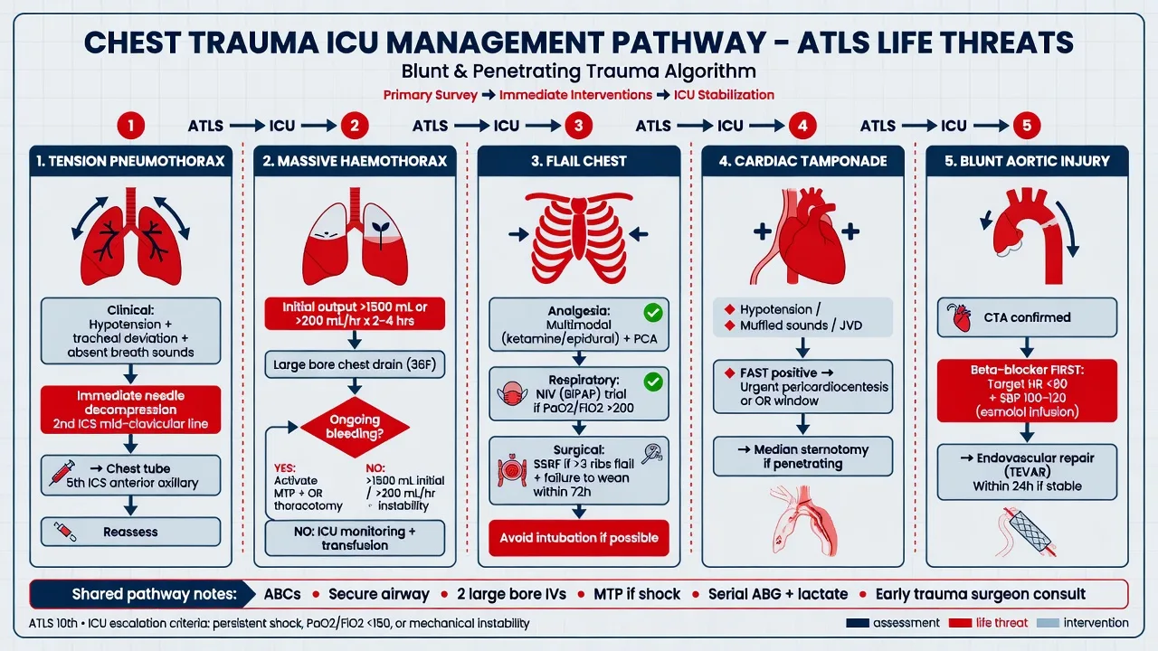
Massive haemothorax is defined (ATLS 10th edition, BATLS) by ANY ONE of the following criteria applied to the initial chest tube drainage: [1]
- >1500 mL of blood evacuated immediately on insertion of the first chest tube (in an adult ~70 kg, this is >25% of circulating volume), OR
- >200 mL/hr of ongoing drainage for 2–4 consecutive hours (some sources use >250 mL/hr in the first 4 hours), OR
- Persistent haemodynamic instability (lactate not clearing, ongoing vasopressor requirement, SBP <90) despite adequate drainage and resuscitation, OR
- Requirement of ongoing transfusion to maintain haemoglobin after evacuation. [1]
Pathophysiology — three concurrent insults: [1]
- Hypovolaemic shock — loss of intravascular volume reduces preload, cardiac output and organ perfusion.
- Ventilation failure — the affected hemithorax fills with blood, collapsing the lung, paradoxically shifting the mediastinum and impairing gas exchange (V/Q mismatch, shunt).
- Tension physiology (if large enough) — increasing intrathoracic pressure on the ipsilateral side compresses the contralateral lung and great vessels, reducing venous return — a stiff, blood-filled hemithorax behaves mechanically like a tension pneumothorax in the late stage. [1]
Sources of bleeding — determine whether bleeding will stop spontaneously: [1]
- Lung parenchyma (most common, low-pressure pulmonary circuit) — tends to tamponade itself as the haemothorax fills the pleural space; usually settles with chest tube drainage alone.
- Intercostal/internal mammary arteries (systemic pressure) — can bleed briskly and persistently; usually require surgical ligation.
- Great vessels (aorta, subclavian, SVC/IVC, pulmonary hilum) — exsanguinating; require immediate thoracotomy.
- Heart/pericardium — if pericardium open, drains into pleural space; if pericardium intact → tamponade. [1]
Initial management (the first 5 minutes)
- ABCDE primary survey — assume C-spine injury until excluded; intubate early if GCS <8 or respiratory distress.
- Two large-bore IV (14–16 G) or intraosseous (IO) access; draw blood for crossmatch, VBG/lactate, coagulation.
- Activate massive transfusion protocol — target RBC:plasma:platelets 1:1:1; administer tranexamic acid 1 g IV bolus then 1 g over 8 h WITHIN 3 h of injury (CRASH-2).
- Insert LARGE-BORE chest tube (28–36 Fr) at 5th ICS anterior axillary line — drain and quantify. DO NOT needle-decompress (blood will not flow through a cannula).
- Chest X-ray / eFAST — confirm tube position, exclude contralateral injury or tamponade, identify pericardial fluid.
- Decide: does the patient meet criteria for thoracotomy? If yes → OR (or resuscitative thoracotomy if arrested). [1]
Large-bore chest tube insertion (28–36 Fr) for massive haemothorax — step-by-step
- PREPARE — full aseptic technique (chlorhexidine, drapes, gown, gloves, mask). Gather: scalpel (No. 10/11 blade), Kelly clamps, large-bore tube (28–36 Fr for haemothorax — SMALLER tubes clot), underwater seal drain, sutures (0 silk/nylon for secure, 2-0 for closure), occlusive dressing. Patient supine, arm abducted above head on affected side (opens intercostal spaces)
- LANDMARK — 5th ICS, anterior axillary line (safe zone: avoids internal mammary artery medially, long thoracic nerve, diaphragm/liver/spleen). In male: just below the nipple. In female: inframammary fold
- ANAESTHESIA — if conscious: 1% lidocaine up to 3 mg/kg (with adrenaline 1:200,000), infiltrate skin, periosteum of rib above and below, pleura. Allow time to work (2-3 min)
- SKIN INCISION — 3–5 cm incision parallel to and ALONG the rib, over the UPPER border of the LOWER rib (i.e. walk over the top of the rib below) — the neurovascular bundle runs in the costal groove on the LOWER border of the rib ABOVE; this avoids damaging it
- BLUNT DISSECTION — Kelly clamp through subcutaneous tissue and intercostal muscles, passing OVER the upper border of the lower rib. Spread the clamp repeatedly to widen the tract. Continue until the parietal pleura is breached — you feel/see a 'give' and often a rush of air/blood
- FINGER SWEEP — insert index finger into pleural space. CONFIRM intrathoracic position (feel lung, ribs, no abdominal contents). Sweep to break down adhesions and exclude diaphragmatic injury (liver/spleen if finger enters abdomen)
- INSERT TUBE — guide the 28–36 Fr tube (with the clamp through the tract or by mounting it on a blunt trocar — never a sharp trocar) DIRECTED POSTEROAPICALLY (towards the apex and posteriorly — that is where blood pools). Advance 8–12 cm so the last side-hole is well inside the pleural space
- CONNECT — attach to underwater seal drainage. Apply suction (−20 cmH₂O). Observe: blood volume, colour, ongoing rate (record 5-minutely). Swing/tidaling in the drain confirms intrapleural position
- SECURE — heavy (0) non-absorbable suture: purse-string or 'U'-stitch around tube entry; tie firmly. Tape the tube to chest wall with wide tape (prevents dislodgement). Dress with occlusive dressing
- CONFIRM — chest X-ray (tube position, residual haemothorax, lung re-expansion, mediastinal position). Assess drained volume against thoracotomy criteria
- REASSESS — if >1500 mL immediately OR >200 mL/hr ongoing OR persistent instability → call surgery for thoracotomy (OR, or resuscitative if arresting). DO NOT insert a second tube hoping to drain more — the criteria have been met
Thoracotomy indications for massive haemothorax
The or-thoracotomy (formal operative thoracotomy) is indicated if the patient meets ANY of: [1]
- >1500 mL initial drainage, OR
- >200 mL/hr ongoing for 2–4 h (or >250 mL/hr in first 4 h — institutional variation), OR
- Persistent haemodynamic instability despite adequate drainage and resuscitation, OR
- Imaging/clinical evidence of great vessel, cardiac, hilar, or tracheobronchial injury requiring surgical repair. [1]
The resuscitative (ED) thoracotomy is a DIFFERENT procedure — reserved for the arrested or peri-arrest patient (see below). The two should not be conflated: a stable patient with a massive haemothorax goes to the operating theatre, not for a clamshell on the ED trolley. [1]
Autotransfusion (cell salvage) in traumatic haemothorax
Principle — blood drained from the pleural cavity can be reinfused into the patient after washing and concentration, reducing allogeneic transfusion. [1]
Autotransfusion systems for traumatic haemothorax
| System | Mechanism | Advantages | Limitations |
|---|---|---|---|
| Simple collection + reinfusion (e.g. Atrium Ocean, Pleur-evac ATS) | Chest drain connects to a collection chamber; blood anticoagulated (CPD or citrate) and reinfused via filter (170 µm) without washing | Cheap, fast, can be set up at bedside, no special equipment, ABO-identical | Does NOT wash — reinfusion of activated clotting factors, cytokines, microthrombi; risk of coagulopathy and DIC if >2 L reinfused |
| Cell saver (e.g. Haemonetics Cell Saver, Sorin Xtra) | Blood collected, centrifuged, washed with saline, packed to Hct ~50-60%, reinfused via filter | Removes contaminants (tissue factor, cytokines, fat, free Hb); safer for large volumes (>2 L) | Expensive, requires technician and machine, ~10–15 min setup, cannot be used if contamination (bile, faeces, urine, malignancy present) |
| Direct auto-transfusion via fenestrated chest tube (Belt, Sorensen) | Blood collected in sterile reservoir with anticoagulant, reinfused immediately through inline filter | Rapid, simple, ideal for austere/military settings | Same risks as simple system; limited by anticoagulant dosing accuracy |
Indications — massive haemothorax with anticipated large-volume loss (especially penetrating trauma in fit young patients with no comorbidities), field/military setting where blood bank unavailable. [1]
Contraindications — contamination of chest cavity with bowel contents (ruptured diaphragm + visceral injury), malignant pleural effusion, active infection. Relative contraindication — coagulopathy already present (autotransfusion may worsen if unwashed). [1]
Risks — coagulopathy/DIC (especially >2 L unwashed reinfusion), sepsis (if contaminated), air embolism (always reinfuse through filter, never directly via central line), haemolysis, hypocalcaemia (citrate). [1]
Practical point — most modern trauma centres use cell-saver technology for ongoing surgical blood loss, but the first 1–2 L from a chest tube can be reinfused directly via a 170 µm filter with citrate anticoagulation (1:7 citrate:blood) when blood bank products are delayed. [1]
Resuscitative thoracotomy — comprehensive review
Indications (WTA 2024 / EAST consensus)
The Western Trauma Association (WTA) 2024 algorithm and EAST practice management guidelines converge on these indications: [1]
Resuscitative thoracotomy — WTA 2024 indications by scenario
| Scenario | Indicated? | Action | Survival |
|---|---|---|---|
| Penetrating thoracic trauma, <15 min prehospital arrest, SOL at scene | YES — STRONG | Immediate EDT | 25–35% |
| Penetrating thoracic trauma, profound shock (SBP <60), still conscious | YES — STRONG | Immediate EDT | 20–30% |
| Penetrating thoracic trauma, SBP <70, deteriorating despite resuscitation | YES | EDT after airway/control | 15–25% |
| Penetrating non-thoracic (neck/abdomen/extremity) trauma, <15 min arrest, SOL | YES — for aortic occlusion/REBOA alternative | EDT or REBOA | 5–15% |
| Blunt trauma, witnessed arrest <10 min, SOL on arrival | SELECTIVE | EDT if SOL — discuss with team | 1–5% |
| Blunt trauma, no SOL at scene, prolonged arrest | NO — futile | Do NOT perform | <1% |
| Penetrating trauma, no SOL at scene, prolonged arrest (>15 min) | NO — futile | Do NOT perform | <1% |
| Catastrophic brain injury (gaping skull, semi-decapitation) | NO — futile | Do NOT perform | 0% |
Signs of life (SOL) — for the purposes of this decision, SOL = ANY of: palpable pulse, measurable blood pressure, spontaneous respiratory effort, pupillary response, purposeful movement, organised cardiac electrical activity on ECG. Absent SOL + arrest >15 min = futility. [1]
Time thresholds — penetrating cardiac arrest <15 min from loss of pulse to scalpel on skin. Blunt cardiac arrest <10 min. These are NOT transport times — they are from the witnessed loss of pulse. Document carefully. [1]
Contraindications and futility
- Blunt trauma with no SOL at scene and prolonged downtime — survival essentially 0%.
- Penetrating trauma with no SOL and arrest >15 min.
- Catastrophic non-survivable brain injury (massive cranial destruction).
- Downtime clearly >15 min (penetrating) or >10 min (blunt) without CPR.
- Absence of immediately available surgical capability to complete definitive repair after EDT — opening the chest is the easy part; closing it is the hard part. [1]
Technique — left anterolateral thoracotomy
Left anterolateral thoracotomy (initial) — anatomical technique
- POSITION — patient supine, arm abducted on affected side (usually left; if right-sided injury suspected, plan clamshell from the start). Operator on patient's left side. Skin prep with chlorhexidine (rapid, painted broadly). Drape minimally (no time for elaborate draping)
- LANDMARK — 4th or 5th intercostal space (ICS) on LEFT: in male, just below the nipple (5th ICS); in female, inframammary fold. The 4th/5th ICS corresponds to the level of the cardiac apex and gives best access to pericardium, left lung and descending aorta
- INCISION — curved incision starting just lateral to the sternum, extending in a gentle curve to the mid-axillary line. Skin → subcutaneous fat → pectoralis major/serratus anterior → intercostal muscles. Use scalpel for skin, heavy scissors or Mayo scissors for muscle and intercostal layers
- ENTER PLEURA — incise the intercostal muscles along the UPPER border of the 5th rib (avoids the neurovascular bundle on the lower border of the 4th rib). Open the parietal pleura. Often you will encounter a gush of blood (confirming haemothorax)
- INSERT FINOCHIETTO RETRACTOR (or hand retraction) — spread ribs apart. If no retractor, manually retract with retractors or assistant's hands
- EVACUATE BLOOD — scoop/ladle out blood and clots into a bowl (autotransfusion if cell saver available). Inspect: left lung, pericardium, descending aorta, left subclavian, oesophagus (NG tube palpable)
- ASSESS PERICARDIUM — if tense/bluish, perform pericardiotomy (see below). If normal, leave alone
- CONTROL LUNG BLEEDING — peripheral laceration: stapled wedge (GIA). Deep/through-and-through: tractotomy. Hilar: Satinsky clamp
- AORTIC CROSS-CLAMP — if subdiaphragmatic bleeding or to augment perfusion: open mediastinal pleura, bluntly dissect descending thoracic aorta, apply vascular clamp (DeBakey)
- CONVERT TO CLAMSHELL IF NEEDED — if right-sided injury or right heart/great vessel involvement: extend incision across sternum to right 4th ICS (bilateral). Cut sternum transversely with Lebsche knife or heavy scissors. Ligate internal mammary arteries (they bleed profusely, frequently missed) with clips or suture ligation
Pericardiotomy and cardiac repair
Pericardiotomy and cardiac wound control
- LIFT PERICARDIUM — identify the tense pericardial sac. Pick up with DeBakey forceps in the midline/anterior aspect. Identify the PHRENIC NERVE (runs longitudinally along the LATERAL pericardium — preserve it)
- LONGITUDINAL INCISION — make a vertical (craniocaudal) incision with scissors in the anterior pericardium, away from phrenic nerve. Often 4–6 cm long. Evacuate clot (often with finger breaking down adhesions)
- DELIVER HEART — gently deliver the heart into the pericardial well to inspect all four chambers and identify the wound(s). Stab wounds may be multiple (entry + exit)
- CONTROL BLEEDING — options (in order of speed):
- FINGER PRESSURE — initial tamponade with index finger over the wound
- FOLEY CATHETER — pass 16-18 Fr Foley through the wound INTO the ventricle, inflate 30 mL balloon, gentle traction — balloon tamponades from inside (works well for thin-walled ventricular wounds). Caution: do not over-pull — may enlarge wound
- SKIN STAPLER — fire staples along wound edges (fast, temporising — needs definitive repair later)
- SUTURE — 3-0 Prolene on a non-cutting (taper) needle, continuous or interrupted. Use Teflon pledgets if tissue friable. Horizontal mattress sutures UNDER coronary arteries if wound adjacent to LAD/PDA/circumflex (do not strangulate the artery)
- AVOID CORONARY ARTERIES — LAD runs in the anterior interventricular groove. Suturing across it infarcts the territory. If wound traverses coronary artery: may need CPB for coronary bypass (rare in this setting — temporise, transfer)
- DEFIBRILLATE IF VF/VT — internal paddles (10–20 J biphasic) placed on either side of heart. Epinephrine 1 mg IV/IO (or intracardiac if no access). Amiodarone 300 mg IV for refractory VF
- VOLUME LOADING + INOTROPES — once bleeding controlled: fill the heart (warm fluids, blood products), epinephrine/norepinephrine infusion, calcium (ionised), bicarbonate if severe acidosis (pH <7.1)
Aortic cross-clamping during resuscitative thoracotomy
Indications in the ED thoracotomy context: [1]
- Subdiaphragmatic haemorrhage with shock — to control retroperitoneal/abdominal bleeding while laparotomy proceeds (aortic occlusion at the diaphragmatic hiatus).
- Augment coronary and cerebral perfusion during cardiac arrest (clamps above the renals concentrate the limited cardiac output to brain and heart).
- Pelvic/lower extremity exsanguination (alternative or adjunct to REBOA — Resuscitative Endovascular Balloon Occlusion of the Aorta). [1]
Technique: [1]
- Open mediastinal pleura overlying the descending thoracic aorta (between the vertebral bodies and the oesophagus, left of midline).
- Identify the aorta: thick-walled, muscular, pulsatile (or flaccid if arrested). The oesophagus is to the right — distinguish by an in-situ NG tube (palpable).
- Bluntly dissect around the aorta with a finger or right-angle clamp — develop an anterior-posterior plane.
- Apply a DeBakey or Satinsky vascular clamp across the aorta. Confirm occlusion (no distal pulse). [1]
Time limit — <30 min: [1]
- Beyond 30 min: spinal cord ischaemia (anterior spinal artery compromised) → paraplegia; liver/bowel/renal ischaemia → reperfusion injury, acidosis, hyperkalaemia.
- Release slowly: clamp reperfusion releases acidotic, hyperkalaemic blood into the circulation → hypotension, arrhythmia. Pre-empt with calcium, bicarbonate, vasopressors. [1]
REBOA — the less-invasive alternative
Resuscitative thoracotomy (RT) vs REBOA for traumatic arrest
| Feature | Resuscitative thoracotomy (RT) | REBOA |
|---|---|---|
| Access | Open chest — left anterolateral/clamshell | Femoral artery (CFA) percutaneous or cut-down |
| What you can do | Release tamponade, control thoracic bleeding, internal massage, thoracic aortic clamp | Aortic occlusion ONLY (zones I/III) — no cardiac access, no thoracic control |
| Time to effective occlusion | 3–5 min (incision to clamp) | 5–10 min (femoral access + balloon inflation) |
| Best for | Penetrating thoracic trauma with tamponade/cardiac arrest | Subdiaphragmatic bleeding with shock (pelvic, abdominal, retroperitoneal); not for thoracic source |
| Survival | Penetrating 15–35%, blunt 1–5% | Mixed evidence — equivalent or slightly worse than RT in some series; benefit unclear in blunt arrest |
| Complications | Pain, infection, bleeding from chest wall, internal mammary arteries, spinal cord ischaemia (clamp) | Limb ischaemia (occluded leg), spinal cord ischaemia, reperfusion injury, vascular injury (CFA dissection) |
| Operator skill | Surgeon / ED consultant with trauma experience | ED/intensivist trained in REBOA (shorter learning curve) |
| Contraindication | No surgical backup; futility | Thoracic source of bleeding; cannot gain femoral access |
Pragmatic position — REBOA does NOT replace RT for penetrating thoracic trauma (where the benefit of pericardiotomy + thoracic control dominates). REBOA is an alternative for non-thoracic haemorrhage in shock/arrest, where thoracotomy provides no benefit. Choice depends on suspected bleeding source: think chest → RT; think abdomen/pelvis → REBOA. [1]
Internal cardiac massage
The advantage over closed-chest compressions: with the chest open, bimanual cardiac compression generates 40–60% of normal cardiac output (vs 20–30% with closed compressions). Critical for neurological survival. [1]
Open (internal) cardiac massage
- POSITION — open pericardium first (pericardiotomy as above). Deliver heart gently into pericardial well
- BIMANUAL TECHNIQUE — cup heart between two palms. Right hand BEHIND the heart (posterolateral, against the diaphragmatic surface) — lifts/anteriorly displaces. Left hand ANTERIOR (over the RV). Compress from APEX toward BASE (mimicking systole). Avoid compressing only the thin-walled RV (risk of rupture)
- RATE 80–100/min — coordinated, smooth. Avoid 'milking' the heart with one hand (ineffective, may injure)
- DEFIBRILLATION — if VF/VT: internal paddles at 10 J initially (escalate 20-30 J). Place paddles on either side of heart (anteroposterior or two sides). Shock, resume massage
- EPINEPHRINE 1 mg IV/IO every 3–5 min during arrest. VASOPRESSIN 40 U IV as alternative. AMIODARONE 300 mg IV for refractory VF
- VOLUME LOADING — if heart empty (hypovolaemic arrest): STOP compressions briefly, fill with blood products/fluid via rapid infuser, then resume. Compressing an empty heart is futile
- MONITOR FOR EFFICACY — palpable carotid/femoral pulse, rising ETCO₂, narrowing pupils, return of organised rhythm on monitor
Survival data — what to tell the team and the family
Survival after resuscitative thoracotomy — major series
| Study / series | N | Overall survival | Penetrating survival | Blunt survival | Good neuro (CPC 1–2) |
|---|---|---|---|---|---|
| Rhee 2000 — 25-year review (Denver + 4 US centres) | 2598 | 7.4% | Stab 33%, GSW 18% | 2% | ~92% of survivors |
| Seamon 2017 — EAST review | — | — | Penetrating torso 15–30% | <5% | Most survivors good CPC |
| Passos 2019 — population-based (Canada) | 246 | 9.6% | 15.9% | 1.7% | 88% good CPC |
| Slessor 2018 — UK military + civilian (BATLS) | — | 12% overall | Stab 28%, GSW 14% | 3% | ~80% good CPC |
| Bartowski 2017 (US Level 1 trauma centres) | — | 8% | 19% (with SOL) | 2% | 90% good CPC |
Key takeaways: [1]
- Survival is mechanism-dependent: penetrating >> blunt. Stab > GSW > blunt.
- Signs of life at scene is the strongest predictor — SOL present throughout: 15–35% survival. SOL lost before ED: <5%. No SOL: <1%.
- Among survivors, neurological outcomes are surprisingly good — 80–92% have good CPC (1–2). The brain is generally preserved because in traumatic arrest the insult is recent (unlike medical arrest where 'down time' is longer).
- Time matters — every minute from arrest to scalpel reduces survival ~5%. Decisive action is paramount. [1]
Complications of resuscitative thoracotomy
ICU management after resuscitative thoracotomy
Post-resuscitative thoracotomy ICU management (first 24 h)
- ADMIT to ICU intubated, ventilated, sedated (propofol/fentanyl — short-acting to allow neuro assessment). arterial line + central venous access + urinary catheter + nasogastric tube + temperature probe
- HAEMODYNAMIC OPTIMISATION:
- MAP >65 mmHg (lower if bleeding uncontrolled — permissive hypotension SBP 80–90)
- Continuous vasopressor infusion (norepinephrine first-line; vasopressin adjunct)
- Calcium — ionised Ca >1.0 mmol/L (calcium gluconate 10 mL 10% IV PRN; citrate in blood products chelates Ca)
- Tranexamic acid — if within 3 h of injury
- CORRECT LETHAL TRIAD:
- Rewarm: forced-air warmer (Bair Hugger), warmed fluids, ambient temperature 28°C, heated humidified ventilator circuit. Target 36–37°C
- Correct acidosis: improve perfusion; consider sodium bicarbonate if pH <7.1 (and ventilation adequate)
- Correct coagulopathy: 1:1:1 transfusion; cryoprecipitate if fibrinogen <1.5 g/L; consider prothrombin complex concentrate (PCC) if on anticoagulants
- VENTILATION — lung-protective: tidal volume 6 mL/kg ideal body weight, plateau pressure <30 cmH₂O, PEEP 5–10 cmH₂O. Single-lung ventilation may be needed if one chest packed or post-pneumonectomy
- CHEST DRAINS — monitor output (record hourly). Sudden increase >200 mL/hr → surgical review (active bleeding). Decrease in output + rising air leak → possible tube blockage/clot
- NEUROLOGICAL ASSESSMENT — once stable: serial GCS, pupils. Consider CT head/CT trauma series (cervical, chest, abdomen, pelvis) to identify missed injuries. Continuous EEG if concerned about non-convulsive status (post-arrest)
- RENAL — monitor UO (target >0.5 mL/kg/hr); rising creatinine + persistent oliguria despite resuscitation → AKI; consider CRRT early if hyperkalaemic/acidotic/fluid overloaded
- GLYCAEMIC CONTROL — target 6–10 mmol/L (avoid hypoglycaemia and severe hyperglycaemia)
- STRESS ULCER PROPHYLAXIS — proton pump inhibitor if ventilated >48 h or coagulopathic
- VTE PROPHYLAXIS — mechanical (compression stockings, sequential compression devices) once bleeding controlled; chemical (LMWH) once haemostasis confirmed (~24–48 h post-op)
- NUTRITION — early enteral feeding within 24–48 h if no contraindication (post-laparotomy ileus may delay)
- PLAN DEFINITIVE SURGERY — return to OR at 24–48 h for chest closure, definitive repair of injuries, abdominal washout if damage-control laparotomy also performed
Additional high-yield clinical pearls
Examination favourites — what examiners ask
Common CICM/FFICM/EDIC viva questions — model answers
| Examiner question | High-yield model answer |
|---|---|
| "Define massive haemothorax" | >1500 mL initial drainage OR >200 mL/hr ongoing for 2–4 h OR persistent haemodynamic instability despite drainage + resuscitation. |
| "Where do you put the chest tube?" | 5th ICS, anterior axillary line. Along upper border of lower rib (avoids neurovascular bundle). Direct tube posterapically. |
| "What size chest tube for haemothorax?" | Large bore — 28–36 Fr. Smaller tubes (e.g. pigtail 8–14 Fr) clot and block; reserved for small effusions/pneumothoraces. |
| "When would you do a thoracotomy?" | Massive haemothorax criteria met (volume) → OR. Penetrating chest trauma with arrest <15 min + SOL → resuscitative (ED) thoracotomy. |
| "Describe the incision for resuscitative thoracotomy" | Left anterolateral, 4th/5th ICS, curved from sternum to mid-axillary line. In male just below nipple, in female inframammary fold. Convert to clamshell if right-sided access needed. |
| "Survival rates?" | Penetrating cardiac stab 30–35%, GSW 15–20%, penetrating non-cardiac 10–15%, blunt 1–5%. SOL at scene 15–35%, no SOL <1%. |
| "Contraindications to RT" | Blunt trauma no SOL prolonged arrest; penetrating no SOL >15 min; catastrophic brain injury; no surgical backup. |
| "Complications of aortic cross-clamp" | Spinal cord ischaemia → paraplegia; renal/hepatic/bowel ischaemia; reperfusion (acidosis, hyperkalaemia); limit <30 min. |
| "What is REBOA?" | Resuscitative Endovascular Balloon Occlusion of the Aorta — percutaneous femoral arterial balloon inflated in zone 1 or 3 to occlude aorta for subdiaphragmatic bleeding. Alternative to aortic cross-clamp; cannot replace RT for thoracic source. |
| "How is cardiac massage different in open chest?" | Bimanual, apex-to-base, 80–100/min, generates 40–60% of normal CO vs 20–30% closed. Internal defibrillation 10–20 J. |
| "How do you fix a ventricular stab wound?" | Finger pressure → Foley balloon tamponade → suture 3-0 Prolene (Teflon pledgets if friable). Horizontal mattress UNDER coronary arteries if near LAD. Avoid strangulating coronaries. |
| "What is the lethal triad and how do you correct it?" | Hypothermia, acidosis, coagulopathy. ICU: rewarm to 36–37°C, correct pH >7.2, transfuse 1:1:1 + cryoprecipitate + calcium + TXA (within 3 h). |
Key trials and guidelines — massive haemothorax and resuscitative thoracotomy
- Rhee PM et al. (2000) — Journal of Trauma 25-year multicentre review of 2598 resuscitative thoracotomies. Overall survival 7.4%; penetrating stab 33%, GSW 18%, blunt 2%. Established the modern survival-by-mechanism framework. [6]
- Seamon MJ et al. (2017/2018) — EAST Practice Management Guidelines. Strongly recommend EDT for penetrating thoracic trauma with SOL; selective for blunt trauma with witnessed arrest. Defines futility thresholds.[2]
- Passos EM et al. (2019) — Journal of Trauma and Acute Care Surgery. Population-based Canadian study (n=246): overall survival 9.6% (penetrating 15.9%, blunt 1.7%); 88% of survivors good CPC.[4]
- PROPPR Trial (Holcomb et al., 2013, JAMA). Pragmatic Randomized Optimal Platelet and Plasma Ratios. 1:1:1 vs 1:1:2 transfusion in severe trauma. No difference in 24-h or 30-d mortality (primary), but trend toward fewer exsanguination deaths at 24 h with 1:1:1. Establishes 1:1:1 as standard of care.
- CRASH-2 (2010, Lancet). Tranexamic acid in trauma bleeding: 1 g bolus + 1 g over 8 h. Reduced all-cause mortality if given within 3 h. Subsequent analysis showed INCREASED mortality if given after 3 h — do not give late.
- MATTERs (Morrison et al., 2012, Arch Surg). Military Application of Tranexamic Acid in Trauma. TXA reduced mortality in combat casualties (mostly penetrating) requiring massive transfusion. Confirmed CRASH-2 in military setting.
- Meyer DM et al. (2005, Ann Thorac Surg). Retained haemothorax: early VATS (within 7 days) reduces empyema and length of stay vs conservative management.
- WTA 2024 Algorithm for Resuscitative Thoracotomy. Western Trauma Association consensus: indications, technique, futility thresholds — current authoritative reference for trauma teams.
- ATLS 10th edition (2018/2019). American College of Surgeons — defines massive haemothorax criteria (>1500 mL or >200 mL/hr) and initial management algorithm.[3]
- BATLS (UK Joint Services Publication). Battlefield algorithm adaptations including CABC, whole blood, permissive hypotension, RT indications in military setting.[5]
Quick reference — key thresholds and numbers
Numbers to remember — high-yield for exams
| Parameter | Threshold / value |
|---|---|
| Massive haemothorax — initial drainage | >1500 mL on first chest tube |
| Massive haemothorax — ongoing drainage | >200 mL/hr for 2–4 h (or >250 mL/hr in first 4 h) |
| Chest tube size for haemothorax | 28–36 Fr |
| Chest tube insertion site | 5th ICS, anterior axillary line |
| Underwater seal suction | −20 cmH₂O |
| RT time threshold (penetrating) | <15 min arrest to scalpel |
| RT time threshold (blunt) | <10 min arrest to scalpel |
| Aortic cross-clamp time limit | <30 min (spinal cord ischaemia) |
| Internal defibrillation energy | 10–20 J biphasic |
| TXA — give within | 3 h of injury (1 g bolus + 1 g over 8 h) |
| TXA — harm after | 3 h (do not give late) |
| Transfusion ratio (PROPPR) | 1:1:1 (RBC:FFP:platelets) |
| Fibrinogen target | >1.5 g/L (Clauss) |
| Ionised calcium target | >1.0 mmol/L |
| Permissive hypotension | SBP 80–90 mmHg until bleeding controlled |
| Perimortem Caesarean (pregnant arrest) | At 4 min (≥20 weeks) |
| Survival — penetrating cardiac stab | 30–35% |
| Survival — penetrating cardiac GSW | 15–20% |
| Survival — penetrating non-cardiac torso | 10–15% |
| Survival — blunt trauma | 1–5% |
| Neurological outcome — survivors | 80–92% good CPC (1–2) |
| Retained haemothorax — VATS within | 3–7 days (≤7 days) |
| Lung-protective ventilation | Vt 6 mL/kg IBW, Pplat <30 cmH₂O |
| REBOA zone 1 | Left subclavian → celiac (intra-abdominal bleeding) |
| REBOA zone 3 | Below renals → bifurcation (pelvic/extremity bleeding) |
| REBOA zone 2 | NO-OCCLUSION zone (celiac → renals) |
References
- [1]Wall MJ, et al. Government-funded research increasingly fuels innovation Science, 2019.PMID 31221848
- [2]Seamon MJ, et al. Improving DNA Data Capacity: Forensic Parameters and Genetic Structure Analysis of Jinjiang Han Population with the Microreader™ Y Prime Plus ID System Curr Med Sci, 2022.PMID 35403953
- [3]Advanced Trauma Life Support (ATLS), et al. Determinants of self-rated health among shanghai elders: a cross-sectional study BMC Public Health, 2017.PMID 29029627
- [4]Passos EM, et al. Can sand nourishment material affect dune vegetation through nutrient addition? Sci Total Environ, 2020.PMID 32278174
- [5]Slessor D, et al. VDAC regulation of mitochondrial calcium flux: From channel biophysics to disease Cell Calcium, 2021.PMID 33529977
- [6]Rhee PM, et al. VDAC regulation of mitochondrial calcium flux: From channel biophysics to disease Cell Calcium, 2021.PMID 33529977