Paeds · cardiology
Patent ductus arteriosus
Also known as Patent ductus arteriosus · PDA · Persistent ductus arteriosus · Ductus Botalli
Paediatric cardiology fellowship guide to patent ductus arteriosus in term infants, children and adolescents: the persistent fetal communication between pulmonary artery and descending aorta, the continuous machinery murmur, the haemodynamic burden grading, the echocardiographic and angiographic assessment, the observe–transcatheter–surgical management ladder with the Amplatzer Duct Occluder, the infective-endocarditis-prophylaxis reframe, and the Eisenmenger boundary beyond which closure becomes harmful.
On this page & tools
Your progress
Saved locally on this device.
Practise this topic
Target exams
Red flags
Life stages
Care settings
Clinical exam formats
Board mappings
Overview & Definition
A four-year-old is referred by a general practitioner who heard a "continuous machinery murmur" at a routine check. The child is thriving, the pulses are a little full, and the echocardiogram shows a 2 mm duct with continuous left-to-right flow and a mildly dilated left atrium. This everyday referral is the face of the congenital patent ductus arteriosus in modern paediatric cardiology, and the decisions — whether to close, when, and how — are made on haemodynamic grounds, not on the presence of a murmur alone.
[1] [5]The ductus arteriosus is the fetal vessel connecting the main pulmonary artery to the descending aorta, allowing the right ventricular output to bypass the high-resistance lungs and reach the placenta via the umbilical circulation. It is meant to close after birth — functionally within the first day as oxygen rises and prostaglandins fall, and anatomically over two to three weeks as intimal proliferation converts it to the ligamentum arteriosum. A patent ductus arteriosus is persistence of this channel beyond the expected closure window in a term infant or older child. It is one of the commonest congenital heart defects and exists on a spectrum from an incidental silent echo finding to a large shunt causing heart failure and, eventually, pulmonary vascular disease.
[1] [2]The cardiology PDA must be distinguished from the preterm PDA, which is a failure of the structurally immature duct to close and is managed with cyclo-oxygenase inhibitors and, rarely, ligation. The congenital PDA discussed here is the term-infant and childhood entity managed by paediatric cardiology with surveillance, transcatheter device occlusion or, less often now, surgery. Holding the two apart matters because the management ladders, the evidence base and the complications are different, and a candidate who blurs them answers badly.
[1]The modern story of the congenital PDA is one of devices replacing surgery and surveillance replacing reflex closure. The Amplatzer Duct Occluder, introduced in the late 1990s, made outpatient same-day closure the default for the moderate duct, while the realisation that infective endocarditis prophylaxis did not work for the duct pushed practice away from closing every silent duct. Today the burden of proof sits with the decision to intervene, and the skill is matching the haemodynamic burden to the right rung of the ladder.
[4] [8] [11]Epidemiology & Risk Factors
Patent ductus arteriosus is among the commonest congenital heart lesions. The Atlanta birth-defects surveillance programme found a birth prevalence of roughly 0.7 per 1000 live births for PDA in term infants, and it accounts for 5–10% of all congenital heart disease. Worldwide meta-analysis places the overall birth prevalence of congenital heart disease near 9 per 1000, with left-to-right shunts — atrial septal defect, ventricular septal defect and PDA — dominating the totals.
[2] [3]The risk profile for a congenital (term) PDA differs from that of the preterm duct. Gestational immaturity is the dominant driver of the preterm entity, whereas the term PDA clusters around genetic and environmental causes. Recognised associations include maternal rubella infection (a classic and still-tested cause), high-altitude residence (chronic hypoxia relaxes the ductal smooth muscle), female sex (roughly a two-to-one female preponderance), and a family history of congenital heart disease. Several syndromes carry a higher rate of PDA, including Char syndrome (TFAP2B mutation, with facial dysmorphism and a fifth-finger anomaly), Noonan syndrome and Down syndrome.
[1] [3]The epidemiology is also shaped by case finding. The widespread use of echocardiography for any murmur, and increasingly for incidental indications, has inflated the apparent incidence of small and silent PDAs — ducts that would once have been called "innocent" or never detected at all. This detection bias is central to the modern debate over whether to close a silent duct, because the denominator of detected ducts has grown faster than the numerator of ducts that genuinely warrant closure.
[5] [11]Pathophysiology
Two forces hold the fetal duct open: low oxygen tension and high circulating prostaglandin E2, chiefly from the placenta. Both relax the ductal smooth muscle. At birth the cord is clamped, the placenta is lost, oxygen tension rises and prostaglandin levels fall. The ductal smooth muscle is now free to contract — functional closure follows within 12 to 24 hours, driven by oxygen-sensitive potassium-channel closure, calcium influx and contraction, and anatomical closure completes over two to three weeks through intimal proliferation and medial necrosis, leaving the ligamentum arteriosum.
[1]The congenital PDA represents a failure of this closure in a structurally mature duct. The mechanisms are partly structural (a defect in the smooth-muscle layer or the intimal cushions that should obliterate the lumen) and partly biochemical (a persistence of ductal sensitivity to the relaxing prostaglandins). Genetic factors modify both — the TFAP2B and CITED2 pathways that govern ductal development are implicated in familial and syndromic cases. Whatever the mechanism, the result is a persistent channel between the pulmonary artery and the descending aorta.
[1]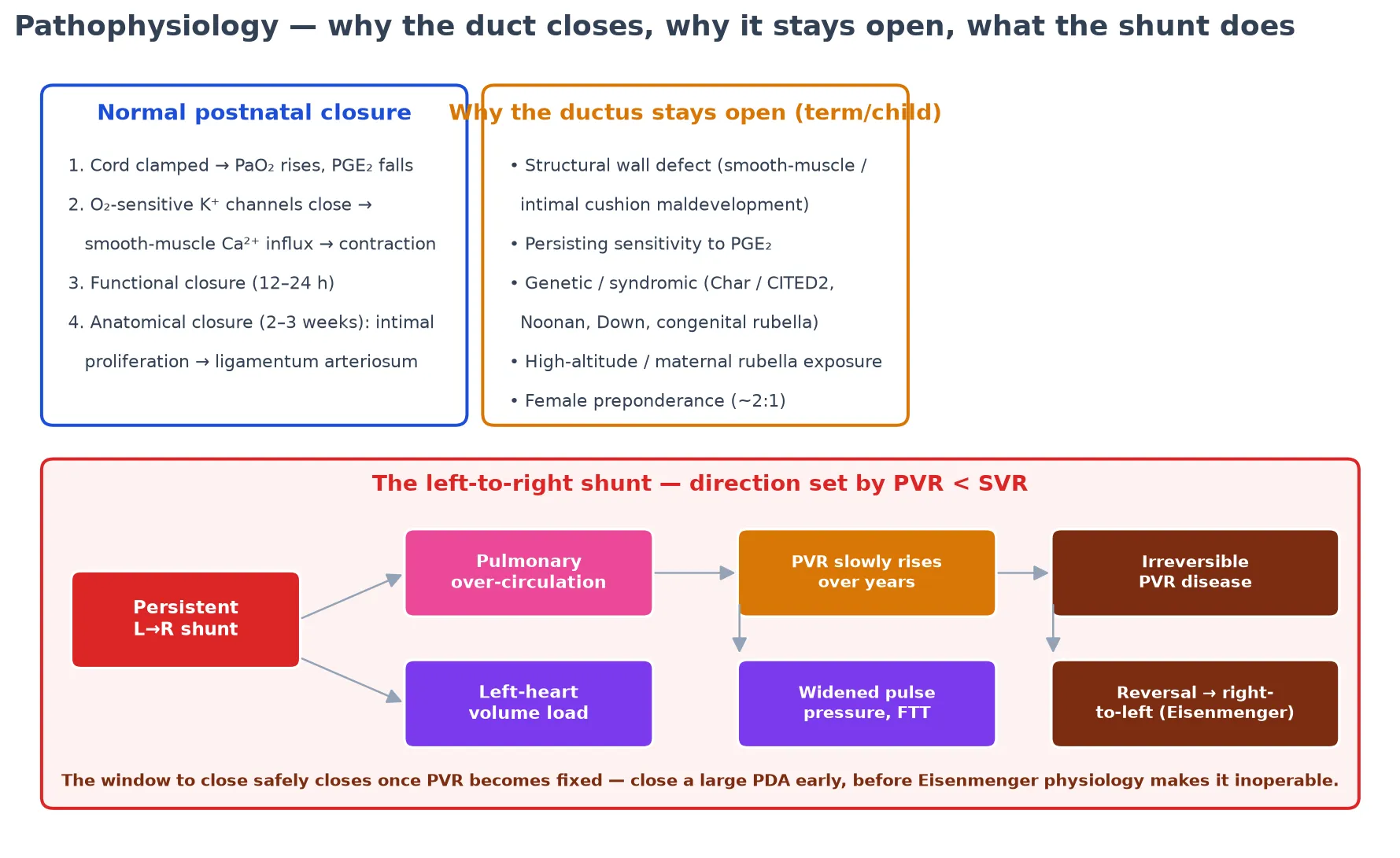
Once the duct persists, the direction and size of the shunt are governed by the balance of systemic and pulmonary vascular resistance. After birth the pulmonary vascular resistance falls, so flow is from the aorta (high pressure) to the pulmonary artery (low pressure), in both systole and diastole — the basis of the continuous murmur. The magnitude depends on the duct's diameter and length: a small restrictive duct produces a loud murmur but a trivial shunt, while a large non-restrictive duct produces a large shunt whose murmur may paradoxically soften as pulmonary pressure rises.
[1]The haemodynamic consequences are the heart of the clinical picture. Pulmonary over-circulation floods the lungs, causing tachypnoea, recurrent lower-respiratory infections, pulmonary oedema and failure to thrive. Left-heart volume load dilates the left atrium and ventricle, producing a hyperdynamic precordium, bounding pulses and a widened pulse pressure as the stroke volume runs off into the low-resistance pulmonary bed during diastole. Over years, a large unrepaired shunt raises the pulmonary vascular resistance until it equals and then exceeds systemic pressure — the shunt reverses, differential cyanosis appears, and the lesion crosses into Eisenmenger physiology, beyond which closure is harmful.
[1] [9]Classification
The classification that drives every management decision separates a PDA by haemodynamic burden — the size of the shunt and its effect on the heart and lungs — rather than by any single measurement. A silent PDA is an incidental echo finding with no murmur and no load; a small PDA gives a continuous murmur with normal pulses and a normal heart; a moderate PDA adds bounding pulses, a widened pulse pressure and mild left-heart dilation; a large PDA causes pulmonary hypertension, failure to thrive and recurrent respiratory illness. This gradient maps directly onto whether and when to close.
[1] [5]Significance is graded by echocardiography, integrating several measures rather than relying on one. The duct size and flow pattern on colour and continuous-wave Doppler, the left atrial to aortic root ratio (a marker of left-heart volume load, with values above 1.5 suggesting overload), the peak systolic flow velocity across the duct (a high velocity means a restrictive duct and normal pulmonary pressures, a low velocity warns of elevated pulmonary pressure), and the left ventricular size together define the burden. A normal-heart, high-velocity, small duct is the benign end; a dilated-heart, low-velocity, large duct is the dangerous end.
[1]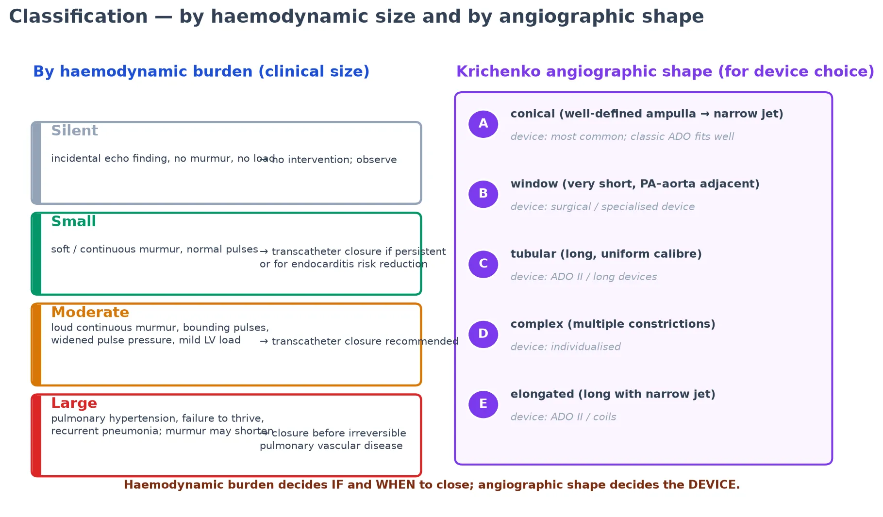
A separate and complementary classification is angiographic shape, after Krichenko. Most ducts are type A — conical, with a well-defined aortic ampulla narrowing to a pulmonary jet — and this is the shape the standard Amplatzer Duct Occluder was designed to occlude. Type C tubular ducts and the elongated shapes suit the ADO II, while the short type B window duct and the complex type D may need surgery or a bespoke device. This morphological classification matters at the catheter table, because the device is chosen to the shape, and the shape can make a transcatheter closure impossible.
[1] [11]The PDA spectrum — from incidental to inoperable
- No or soft continuous murmur
- Normal pulses, normal heart size
- High-velocity restrictive flow on echo
- Observe; closure is discretionary
- Low absolute endocarditis risk
- Loud continuous machinery murmur
- Bounding pulses, widened pulse pressure
- Left atrial and ventricular dilation
- Transcatheter ADO closure recommended
- Close before school age in most units
- Heart failure, failure to thrive, recurrent LRTI
- Pulmonary hypertension, loud P2
- Low-velocity flow, risk of Eisenmenger
- Close urgently if PVR still reversible
- Inoperable once Eisenmenger established
The clinical and echocardiographic pictures should agree, and discordance is informative. A loud murmur with a small, high-velocity, restrictive duct is a benign lesion; a quiet chest with a large, low-velocity duct and a dilated left heart is the dangerous one. The lesson examiners draw from this agreement is that the stethoscope over-calls the small restrictive duct and under-calls the large pressure-equalising duct — the treating team must integrate the murmur, the pulses and the echo before deciding.
[1]Clinical Presentation
The classic presentation is the thriving preschool child with a continuous machinery murmur found at a routine check. The murmur is unmistakable once heard — a rough, continuous crescendo–decrescendo sound peaking around the second heart sound, loudest at the upper left sternal edge, often with a thrill and radiating to the left infraclavicular area and the back. It does not respect the cardiac cycle, running through systole and diastole, because the aorta-to-pulmonary-artery pressure gradient persists throughout.
[1]The cardiovascular examination reveals the run-off. Feel the peripheral pulses — they are bounding, collapsing or "water-hammer" in quality, palpable at the wrists and ankles, because the large stroke volume escapes into the low-resistance pulmonary bed during diastole. Measure the blood pressure — a widened pulse pressure with a characteristically low diastolic is the signature of a significant shunt, and a wide pulse pressure with bounding pulses in a thriving child is a PDA until proven otherwise. Watch the precordium — it is hyperdynamic when the shunt is large.
[1]The end-organ presentation of a large duct is the face of heart failure in infancy. Tachypnoea, especially with feeds, diaphoresis, poor weight gain and recurrent lower-respiratory infections reflect the pulmonary over-circulation. The infant may be admitted repeatedly for "bronchiolitis" or "pneumonia" before the duct is recognised, and the heart failure of a large left-to-right shunt is one of the causes of failure to thrive that the examiner expects you to hold.
[1] [9]Not every presentation is loud. A silent PDA has no murmur and is found on echocardiography done for another reason. A large PDA with established pulmonary hypertension may have a shortened or absent murmur as the pressure gradient across the duct falls, a loud pulmonary component of the second sound, and — once the shunt reverses — differential cyanosis (dusky lower limbs with pink upper body, best seen in the toes versus the fingers). A continuous murmur that disappears in a child who is not improving is a danger sign, not reassurance.
[1] [10]Differential Diagnosis
A continuous murmur at the upper left sternal edge is a PDA in most cases, but the differential is the discipline that separates the confident candidate from the guesser. The other continuous murmurs of childhood include a coronary arteriovenous fistula (continuous, but often lower sternal border, with a broader radiation), a systemic arteriovenous malformation (e.g. a cerebral or hepatic AV fistula, producing a run-off lesion with bounding pulses and heart failure but no localised murmur), a venous hum (an innocent continuous murmur in the neck that abolishes with head turning or pressure on the neck veins), and an aortopulmonary window (a rare but large left-to-right shunt behaving like a massive PDA).
[1]The bounding-pulse, widened-pulse-pressure presentation widens the differential further. Aortic regurgitation produces a water-hammer pulse and a diastolic murmur (not a continuous one). A large arteriovenous malformation anywhere can cause high-output bounding pulses. Aortic coarctation produces bounding upper-limb pulses with weak femorals and a blood-pressure differential — a critical mimic to exclude, because a PDA proximal to a coarctation may mask the femoral pulse deficit, and the two lesions coexist more often than chance.
[1]The resolving question in the infant with heart failure is not "is there a shunt?" but "is the PDA the cause, or is something else?". A ventricular septal defect, an atrioventricular septal defect and a large atrial septal defect all produce pulmonary over-circulation and failure to thrive, and they coexist with a PDA in many children. The echocardiogram resolves the differential definitively, and it is the investigation that converts a clinical suspicion into an anatomical and haemodynamic diagnosis before any closure decision.
[1] [5]The innocent murmurs deserve a specific mention because they are the commonest reason a child is referred with a murmur that turns out to be nothing. A venous hum is the close mimic of the PDA — continuous, but infraclavicular, abolished by turning the head or compressing the neck veins, and not associated with bounding pulses or a widened pulse pressure. A still's murmur or a pulmonary flow murmur is systolic, not continuous. The discipline of examining the pulses and measuring the blood pressure distinguishes the run-off lesion from the innocent murmur at the bedside.
[1]Clinical & Bedside Assessment
Begin at the bedside, because the echocardiogram is interpreted in the light of the clinical state, not the other way round. The focused cardiovascular examination has four targets. Listen for the continuous murmur at the upper left sternal edge, characterising its timing, radiation and the presence of a thrill. Feel the peripheral pulses for the bounding, collapsing quality of a run-off lesion, comparing radial and femoral pulses to exclude coarctation. Watch the precordium for the hyperdynamic impulse of a volume-loaded left ventricle. Measure the blood pressure for the widened pulse pressure and the low diastolic.
[1]Then assess the territories the shunt burdens. For pulmonary over-circulation, document the respiratory rate and work of breathing, the feeding pattern and weight trend, and the history of recurrent respiratory illness. For left-heart volume load, the examination is largely echocardiographic, but the degree of precordial hyperactivity and the presence of a gallop in an infant with a large shunt signal heart failure. Plot the growth chart — failure to thrive is the single most useful longitudinal marker of a haemodynamically important duct.
[1] [9]Examine for the syndromic associations, because the duct does not exist in isolation. Look for the dysmorphic features of Noonan, Down or Char syndrome, the cataracts and hearing deficit of congenital rubella, and the fifth-finger and facial features of Char syndrome. These findings reframe the workup from a single lesion to a multisystem assessment, and they are the kind of detail an examiner rewards in the long case.
[1]The candidate's job at the bedside is to assemble the case for or against significance, so that when the echocardiogram is done its findings can be weighed against a clear clinical question. A thriving child with a soft continuous murmur and full-but-not-bounding pulses has a small duct until the echo proves otherwise; an infant with heart failure, bounding pulses and a hyperdynamic precordium has a large duct until the echo proves otherwise. The bedside picture sets the urgency, and the echo confirms the anatomy.
[1] [5]Investigations
Echocardiography is the gold standard, and it does three jobs. First, it confirms normal cardiac structure and excludes the duct-dependent lesions and coarctation that must never be closed. Second, it grades the duct's haemodynamic burden through the constellation of measures — duct size, flow pattern and peak velocity, left atrial and ventricular size, and pulmonary artery pressure estimation. Third, it defines the duct's morphology for device selection, should closure be planned. A single, complete echocardiogram answers the three questions that frame every PDA decision.
[1] [5]The estimation of pulmonary artery pressure is the echocardiographic finding that changes everything. The peak systolic velocity across the duct is inversely related to the pressure gradient between aorta and pulmonary artery — a high velocity (above roughly 4 m/s) means a large gradient and normal pulmonary pressures, while a low velocity warns that pulmonary pressure is climbing. Tricuspid regurgitation velocity estimates right ventricular systolic pressure, and septal geometry confirms the pressure load. A duct with a low flow velocity, raised right-heart pressure and a flattened septum has crossed towards Eisenmenger territory, and closure may be contraindicated.
[1] [9]In Australia and Aotearoa New Zealand, paediatric cardiology services are centralised, with echocardiography performed and reported by cardiologists and transcatheter closure offered as day-stay procedures for suitable ducts. In the United Kingdom, the same model applies under paediatric cardiology networks. In North America and Europe, Amplatzer Duct Occluder closure is the default for the moderate duct, with surgical ligation reserved for the small or device-unfriendly duct. In low-resource settings without transcatheter facilities, surgical ligation by thoracotomy remains the standard, and the priority is to close the large, pressure-loading duct before pulmonary vascular disease becomes fixed.
[10][11]The chest radiograph and electrocardiogram support but do not define the diagnosis. The radiograph may show cardiomegaly, increased pulmonary vascular markings (pulmonary plethora) and, in the older child with established pulmonary hypertension, pruning of the peripheral pulmonary vessels. The electrocardiogram may show left atrial and left ventricular hypertrophy in a moderate duct, and biventricular or right-heart dominance once pulmonary hypertension supervenes. Both are non-specific and lag the echocardiogram, but they frame the chronicity and the burden.
[1]In selected cases — typically the older child or adult with a large duct where pulmonary vascular disease is suspected — cardiac catheterisation measures the pulmonary vascular resistance directly and tests reversibility with vasodilators or oxygen. A calculated pulmonary vascular resistance above a threshold (commonly 8 Wood units indexed, or non-reactive to vasodilator challenge) places the patient beyond the operable boundary, and closure is withheld. This is the investigation that converts the closure decision from an echo judgement into a physiological verdict.
[1] [9]Management — Resuscitation
The infant presenting in heart failure from a large duct needs stabilisation while the closure is planned. The priorities are to support the circulation and the breathing, not to reach for a drug that closes the duct — the cyclo-oxygenase inhibitors used for the preterm duct have no established role in the term-infant congenital PDA. Give diuretics (frusemide) to reduce the pulmonary over-circulation and volume load, optimise the feeding and caloric intake to address the failure to thrive, and add an afterload-reducing agent where appropriate to lower the shunt.
[1] [9]Respiratory support is reserved for the decompensated infant. Continuous positive airway pressure or nasal high-flow reduces the work of breathing and the pulmonary oedema, and mechanical ventilation is reserved for the infant in respiratory failure. The aim of this phase is to stabilise the child sufficiently for definitive closure, which is the only therapy that reverses the shunt — medical therapy buys time, it does not treat the lesion.
[1]The resuscitative phase is also the safety phase. Before any closure, the echocardiogram must confirm a structurally normal heart and exclude a duct-dependent circulation, and the pulmonary pressure must be documented. The single worst error in PDA management is closing a duct that the child depends on for systemic or pulmonary flow, and the resuscitative assessment is where that error is prevented. A structurally normal heart and a measured, reversible pulmonary pressure are the prerequisites for every closure.
[1] [9]Management — Definitive & Stepwise
The stepwise ladder runs from observation through transcatheter device closure to surgical ligation, with the rung chosen to match the haemodynamic burden. The evidence and the technology now weight the transcatheter rung far more heavily than the surgical one, and the observation rung far more heavily than either — most detected ducts are not closed, and most that are closed are closed with a device.
[1] [5]Step one is observation. The silent or small PDA with no haemodynamic load — normal pulses, normal heart size, high-velocity restrictive flow — is observed, with interval echocardiography to confirm stability or spontaneous closure. Many small ducts close spontaneously in infancy, and the absolute risk of endocarditis on a small duct is now considered too low to justify closure for prophylaxis alone. The burden of proof sits with the decision to intervene, and observation is an active, evidence-based strategy, not neglect.
[1] [8]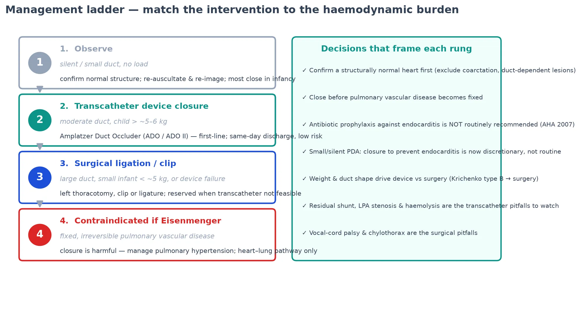
Step two is transcatheter device closure, the default for the moderate duct in a child large enough for the delivery sheath. The Amplatzer Duct Occluder, introduced by Masura and colleagues and refined through the ADO II, is a self-expanding nitinol mushroom-shaped device deployed from a femoral venous approach across the duct, occluding flow by a combination of mechanical obstruction and thrombus formation. Closure rates exceed 95% in suitable ducts, the procedure is day-stay, and same-day discharge is standard. The Krichenko type A conical duct is the shape the device was designed for.
[4] [5] [12]The Amplatzer Duct Occluder era (Masura 1998 → ADO II)
Catheter closure of moderate-to-large PDAs with a new self-expanding nitinol device, extended in the ADO II to smaller, tubular and elongated ducts in smaller children.
Key finding
Device closure achieves > 95% complete occlusion in suitable ducts with low complication rates, displacing surgery as the default for the moderate duct.
The complications of transcatheter closure are uncommon but specific, and naming them signals a candidate who understands the procedure. Residual shunt and haemolysis within the first weeks reflect incomplete occlusion and usually settle as the device endothelialises. Left pulmonary artery stenosis and aortic coarctation from device protrusion are the pitfalls in small infants, where the device may obstruct the adjacent LPA or aortic isthmus — the Tomasulo data quantify their incidence and fate. Device embolisation is rare and usually retrievable. These are the trade-offs that justify choosing the right device for the right duct.
[6] [11]Step three is surgical ligation or clip occlusion, reserved for the duct that is too large, too small (a tiny premature infant in whom the device sheath cannot be accommodated), or the wrong shape for a device — the type B window duct and the complex type D. Surgery is via a left thoracotomy, with a clip or double ligature, and carries its own complications: left recurrent laryngeal nerve injury (vocal-cord paralysis, which may persist), chylothorax, phrenic nerve injury, bleeding and the risks of thoracotomy. The Orb data document the long-term outcome of post-ligation vocal-fold paralysis.
[7] [1]The fourth rung is the contraindication. Once the pulmonary vascular resistance is fixed and non-reactive — established Eisenmenger physiology — closing the duct removes the low-pressure pop-off into the pulmonary bed and can precipitate right-heart failure. Closure is withheld, and management shifts to pulmonary-arterial-hypertension therapy and, in selected end-stage cases, heart–lung transplantation. Recognising the Eisenmenger boundary is the single most important judgement in the long case, because it is the line between a curative and a harmful procedure.
[9] [10]The synthesis the examiner wants is the explicit matching of the rung to the burden. The silent or small duct is observed; the moderate duct in a suitable child is closed with an Amplatzer device; the device-unfriendly duct is ligated; the Eisenmenger duct is not closed at all. Each rung carries evidence, device and risk, and the skill is fitting the intervention to the haemodynamic state of the child rather than to the presence of a murmur.
[1] [11]Specific Subtypes & Scenarios
The silent PDA is the scenario that generates the most debate. Detected on an echocardiogram done for another reason, with no murmur and no load, it presents the question of whether to close purely to abolish the small lifetime risk of endocarditis. The modern consensus, anchored in the AHA 2007 endocarditis-prophylaxis revision, is that the absolute risk is too low to justify routine closure, and the decision is individualised — shared with the family, weighing the small procedural risk against the small infection risk, with observation as the default.
[8] [12]The large PDA presenting in infancy with heart failure is the urgent scenario. The child is failing to thrive with recurrent respiratory illness and a hyperdynamic precordium, the echo shows a large duct with a volume-loaded left heart, and the pulmonary pressure is rising but still reversible. This is the duct to close — surgically if the infant is too small for a device, transcatheter once the size allows — before the pulmonary vascular resistance becomes fixed. The window of operability is the central concept.
[1] [9]The late-presenting large PDA in the older child or adolescent is the long-case scenario. The child has been "well" with a murmur for years, the echo now shows pulmonary hypertension, and the question is whether the resistance is still reversible. Cardiac catheterisation with vasodilator testing decides operability, and the candidate who can articulate the threshold (a pulmonary vascular resistance index above the operable boundary, or non-reactivity to oxygen or nitric oxide) and the consequence (closure withheld, pulmonary-hypertension therapy commenced) answers this scenario correctly.
[9] [10]The PDA with a syndromic association reframes the management. Char syndrome (TFAP2B) carries a duct that may coexist with other cardiac and extra-cardiac features; Down syndrome and Noonan syndrome carry their own cardiac lesion profiles alongside the duct. The duct is managed on its haemodynamic merits, but the workup is broadened to the syndrome, and the genetic counselling is part of the encounter. These are the long cases where the duct is the entry point to a multisystem assessment.
[1]The PDA in the transitioning adolescent and adult is the scenario that bridges paediatric and adult congenital cardiology. The 2018 AHA/ACC and 2020 ESC adult-congenital guidelines address the late PDA directly — closure is recommended for the significant left-to-right shunt with preserved pulmonary vascular resistance, deferred for the small silent duct, and contraindicated for the Eisenmenger duct. The transition is planned, the contraception and pregnancy counselling is given, and the long-term follow-up is handed to the adult congenital service.
[9] [10]Complications & Pitfalls
The complications arise from two sources — the untreated significant shunt and the closure itself. From the shunt: failure to thrive, recurrent respiratory illness, pulmonary oedema, and, over years, progressive pulmonary vascular disease culminating in Eisenmenger physiology with differential cyanosis, right-heart failure and the shortened survival of pulmonary hypertension. From the closure: the procedural risks of transcatheter and surgical closure, and the harm of treating a duct that would have been better left alone.
[1] [9]The classic pitfall is closing a duct that did not need closing. A silent or small duct with no haemodynamic load exposes the child to procedural risk for a shunt that caused no harm and may have closed spontaneously, and the endocarditis-prophylaxis rationale no longer justifies it. The modern evidence base exists precisely to prevent this error, and the candidate who reaches for the catheter laboratory on the strength of a murmur alone answers badly.
[8] [12]The mirror-image pitfall is missing the Eisenmenger boundary. The continuous murmur softens and shortens as the pulmonary pressure rises, the second sound develops a loud pulmonary component, and the toes become cyanotic before the fingers. Reading the disappearing murmur as "the duct is closing" rather than "the pressures are equalling" is the error that leads to an inoperable, harmful closure. The disciplined answer is to measure the pulmonary vascular resistance before any closure of a large or late-presenting duct.
[1] [10]The procedural pitfalls are specific to each rung. From transcatheter closure: residual shunt with haemolysis, left pulmonary artery stenosis and aortic coarctation from device protrusion in small infants, and device embolisation. From surgical ligation: left recurrent laryngeal nerve injury with vocal-cord paralysis (which the Orb data show can persist long-term), chylothorax, phrenic nerve injury and the general risks of thoracotomy. Naming these complications, and matching them to the procedure, is the detail that distinguishes the strong short-case answer.
[6] [7]Finally, the anatomical pitfall: closing a duct-dependent circulation. Hypoplastic left heart, critical coarctation, interrupted aortic arch and pulmonary atresia all rely on the duct, and closing it without confirming normal structure is catastrophic. The structurally normal heart must be confirmed on echocardiography before any closure, and this single check is the safety net that prevents the worst error in the topic.
[1] [9]Prognosis & Disposition
The single most important prognostic fact is that a closed duct is a cured lesion. A small or moderate PDA closed by a device in childhood carries an essentially normal long-term prognosis, with no restriction on activity, no need for endocarditis prophylaxis, and only routine cardiology follow-up. The closure is curative because it abolishes the shunt and its haemodynamic consequences, and the child returns to the normal trajectory.
[1] [11]The prognosis of the observed silent or small duct is also excellent. Most small ducts close spontaneously in infancy, and those that persist without load carry a very low absolute risk of endocarditis and no haemodynamic consequence. The follow-up is interval echocardiography until closure or stability is confirmed, after which the child is discharged from cardiology care. The natural history is benign for the small, restrictive duct.
[1] [8]The prognosis darkens sharply once the duct crosses into Eisenmenger physiology. An unrepaired large duct that has produced fixed pulmonary vascular disease carries the survival and morbidity of pulmonary arterial hypertension — exercise intolerance, right-heart failure, complications of pregnancy, and a substantially reduced life expectancy. This is the prognosis the closure-window concept exists to prevent, and it is the consequence of missing the operable boundary.
[9] [10]Disposition is to a paediatric cardiology service with echocardiography and, for closure, transcatheter facilities and surgical backup. Infants in heart failure are admitted for stabilisation and early closure; thriving children with a moderate duct are scheduled for elective day-stay device closure; silent ducts are managed in the ambulatory clinic with interval imaging. The expectation the examiner wants is that most children are managed electively as day-stay procedures and discharged to normal activity, with the few large or late-presenting ducts escalated to the catheter or theatre.
[5] [11]Special Populations
The infant too small for a device is the population that re-introduces surgery. In the very small infant (commonly below roughly 5 kg), the device delivery sheath may not be safely accommodated, and the device may obstruct the adjacent left pulmonary artery or aortic isthmus. The Tomasulo data quantify these risks, and for this population surgical ligation by thoracotomy remains the standard — a reminder that the transcatheter default has a size floor below which surgery is safer.
[6] [1]The adolescent and adult with congenital heart disease is the population that bridges to adult services. The 2018 AHA/ACC and 2020 ESC guidelines address the late PDA in this group directly, and the management turns on whether the pulmonary vascular resistance is still reversible. The transition is planned with explicit contraception and pregnancy counselling (pregnancy in Eisenmenger physiology carries a high maternal mortality), and the long-term follow-up is handed to the adult congenital cardiology service.
[9] [10]The child with a syndromic PDA is the population in whom the duct is one feature of a broader condition. Char syndrome, Noonan syndrome and Down syndrome each carry their own cardiac and developmental profile, and the management of the duct is integrated into the whole-child plan. The genetic counselling, the developmental surveillance and the family support are part of the encounter, and the long case rewards the candidate who frames the duct within the syndrome.
[1]In the low-resource setting, where transcatheter facilities and surgical expertise may be limited, the priority is to close the large, pressure-loading duct before pulmonary vascular disease becomes fixed, using surgical ligation by thoracotomy. The small and silent ducts are observed, and the emphasis is on identifying the haemodynamically significant duct and referring it for the intervention that prevents the irreversible complication. The expectant-first principle is if anything more important where the capacity to rescue from procedural complications is limited.
[1] [10]Evidence, Guidelines & Regional Differences
The defining shift in the modern evidence base is the displacement of surgery by the transcatheter device for the moderate duct. The Masura landmark study of 1998 established the Amplatzer Duct Occluder for the moderate-to-large duct, and the subsequent device generations — the ADO II and the additional-sizes variants — extended closure to smaller, tubular and elongated ducts in smaller children. The Gałeczka twenty-five-year single-centre experience and the Bhat silent-PDA series confirm closure rates above 95% with low complication rates, making device closure the default for the suitable duct.
[4] [11] [12]The second defining shift is the endocarditis-prophylaxis reframe. The AHA 2007 guideline removed routine antibiotic prophylaxis for the unrepaired PDA and for most unrepaired congenital lesions, on the evidence that prophylaxis does not reliably prevent endocarditis in these settings and that the absolute risk is low. This removed one historical rationale for closing every silent duct, and it anchored the modern preference for observation of the small, asymptomatic lesion.
[8]The adult-congenital guidelines address the late and complex PDA directly. The 2018 AHA/ACC guideline (Stout) and the 2020 ESC guideline (Baumgartner) both recommend closure for the significant left-to-right shunt with preserved pulmonary vascular resistance, defer the small silent duct, and contraindicate closure for the Eisenmenger duct. They provide the operability thresholds (the pulmonary vascular resistance index and the reversibility testing) that frame the closure decision in the older child and adult.
[9] [10]Regional practice has converged on the transcatheter-default, observation-first model, with local texture. In Australia and Aotearoa New Zealand, paediatric cardiology is centralised, with device closure as day-stay for suitable ducts and surgical ligation reserved for the small or device-unfriendly duct. In the United Kingdom, the same model applies under paediatric cardiology networks. In North America and Europe, the Amplatzer devices dominate, with surgery reserved as above. The convergence reflects the shared evidence base and the maturation of the transcatheter technology.
[10] [11]The live controversy the examiner will probe is the silent duct: whether to close a small, asymptomatic, incidentally found duct purely to abolish the lifetime endocarditis risk. The honest answer is that the absolute risk is low, the procedural risk is small but real, and the decision is shared with the family, with observation as the default and closure as a discretionary, individualised choice. The candidate who can hold this uncertainty, and name the evidence behind it, answers the controversy well.
[8] [12]Exam Pearls
State the one-liner mechanism: a PDA is persistent postnatal patency of the fetal duct between the pulmonary artery and the descending aorta, producing a left-to-right shunt whose direction is set by the PVR:SVR balance, with a continuous machinery murmur as the signature. Most are closed by a transcatheter device; the surgical and observational rungs have their own place; the Eisenmenger duct is never closed.
[1] [4]State the examination findings: a continuous machinery murmur at the upper left sternal edge, with bounding pulses and a widened pulse pressure when the shunt is significant. The murmur does not respect the cardiac cycle. A disappearing murmur with differential cyanosis and a loud P2 is Eisenmenger, not closure.
[1]State the management ladder: observe the silent or small duct; close the moderate duct with an Amplatzer Duct Occluder; reserve surgical ligation for the small or device-unfriendly duct; never close a duct past the Eisenmenger boundary. Endocarditis prophylaxis is no longer routine. Confirm a structurally normal heart before any closure.
[5] [8] [9]State the complications: from the shunt — failure to thrive, recurrent LRTI, pulmonary oedema, Eisenmenger; from the device — residual shunt, haemolysis, LPA stenosis, coarctation, embolisation; from surgery — vocal-cord paralysis, chylothorax, phrenic injury. The two trapdoors are treating a duct that needed no treatment and missing the operable window of a large duct.
[6] [7] [10]References
- [1]Schneider DJ, Moore JW Patent ductus arteriosus Circulation, 2006.PMID 17060397
- [2]Reller MD, Strickland MJ, Riehle-Colarusso T, et al Prevalence of congenital heart defects in metropolitan Atlanta, 1998-2005 J Pediatr, 2008.PMID 18657826
- [3]van der Linde D, Konings EEM, Slager MA, et al Birth prevalence of congenital heart disease worldwide: a systematic review and meta-analysis J Am Coll Cardiol, 2011.PMID 22078432
- [4]Masura J, Walsh KP, Thanopoulous B, et al Catheter closure of moderate- to large-sized patent ductus arteriosus using the new Amplatzer duct occluder: immediate and short-term results J Am Coll Cardiol, 1998.PMID 9525563
- [5]Pass RH, Hijazi ZM, Meyer DB, et al Amplatzer Duct Occluder device: a new technology for the closure of the moderate-to-large-sized patent ductus arteriosus Expert Rev Med Devices, 2006.PMID 16681450
- [6]Tomasulo CE, Gillespie MJ, Munson D, et al Incidence and fate of device-related left pulmonary artery stenosis and aortic coarctation in small infants undergoing transcatheter patent ductus arteriosus closure Catheter Cardiovasc Interv, 2020.PMID 32339400
- [7]Orb Q, Dunya G, Padia R, et al Long-term Outcomes of Vocal Fold Paralysis Following Patent Ductus Arteriosus Ligation in Neonates Laryngoscope, 2023.PMID 36054344
- [8]Wilson W, Taubert KA, Gewitz M, et al Prevention of infective endocarditis: guidelines from the American Heart Association: a guideline from the American Heart Association Rheumatic Fever, Endocarditis, and Kawasaki Disease Committee, Council on Cardiovascular Disease in the Young, and the Council on Clinical Cardiology, Council on Cardiovascular Surgery and Anesthesia, and the Quality of Care and Outcomes Research Interdisciplinary Working Group Circulation, 2007.PMID 17446442
- [9]Stout KK, Daniels CJ, Aboulhosn JA, et al 2018 AHA/ACC Guideline for the Management of Adults With Congenital Heart Disease: A Report of the American College of Cardiology/American Heart Association Task Force on Clinical Practice Guidelines J Am Coll Cardiol, 2019.PMID 30121239
- [10]Baumgartner H, De Backer J, Babu-Narayan SV, et al 2020 ESC Guidelines for the management of adult congenital heart disease Eur Heart J, 2021.PMID 32860028
- [11]Galeczka M, Szkutnik M, Bialkowski J, et al Transcatheter patent ductus arteriosus closure: what have we learned after over 25 years? A single--center experience with 1036 patients Kardiol Pol, 2021.PMID 33599452
- [12]Bhat YA, Almesned A, Alqwaee A, et al Catheter Closure of Clinically Silent Patent Ductus Arteriosus Using the Amplatzer Duct Occluder II-Additional Size: A Single-Center Experience Cureus, 2021.PMID 34589368