Paeds · clinical-assessment-and-reasoning
Age-specific normal vital signs and physiological ranges
Also known as Paediatric vital signs · Age-banded HR RR BP · Normal ranges children
Fellowship guide to measuring and interpreting age-banded paediatric vital signs, hospital versus community ranges, and escalation traps.
On this page & tools
Your progress
Saved locally on this device.
Practise this topic
Target exams
Red flags
Life stages
Care settings
Clinical exam formats
Board mappings
Quote the source before you argue a cut-off. [1] [2] [7]
M.E.A.S.U.R.E.
Overview & Definition
Vital signs are bedside numbers that estimate cardiorespiratory and thermal status. In paediatrics the same label — “tachycardia” — means different absolute rates at two months and twelve years. Fellowship practice means you can measure, age-adjust, interpret, and act without inventing a private table. [1]
Classification
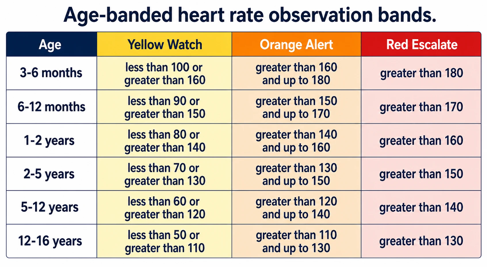
Core set: heart rate, respiratory rate, blood pressure, temperature, SpO2, plus perfusion (capillary refill, colour) and consciousness. Distinguish population reference ranges, ED/hospital centiles, and operational acceptable bands on track-and-trigger charts. [1] [2] [7]
Epidemiology & Risk Factors
Age is the dominant determinant of expected heart and respiratory rates. Fever raises heart rate further in hospitalised children. Incomplete observation sets and night gaps increase missed deterioration risk in PEWS systems. Caregiver concern remains independently important. [1] [3] [6] [7]
Pathophysiology
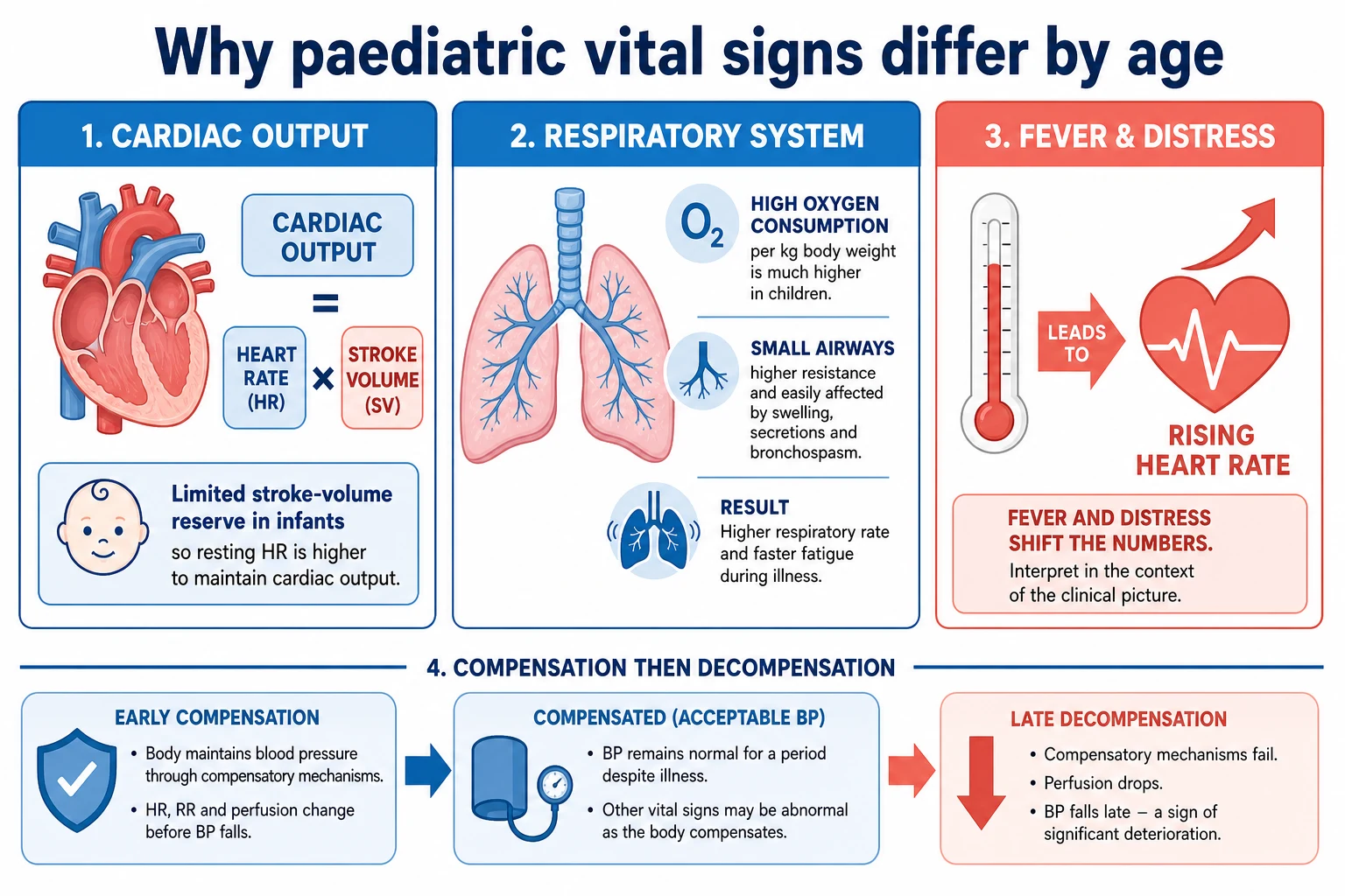
Cardiac output in infants is heart-rate dependent; stroke volume reserve is limited. Respiratory rates are higher because of higher metabolic demand and different mechanics. Children compensate with tachycardia and tachypnoea before hypotension. A falling respiratory rate in a tiring child can mean exhaustion, not recovery. [1] [2]
Clinical Presentation
A quiet infant with a high heart rate for age may look less dramatic than a screaming toddler with the same absolute rate. Always record state: sleeping, feeding, crying, febrile. Capillary refill adds perfusion context but technique matters. [3] [4] [5]
Differential Diagnosis
Tachycardia: fever, pain, fear, dehydration, shock, arrhythmia, drugs. Tachypnoea: lung disease, metabolic acidosis, fever, anxiety. Hypoxia reading: true hypoxaemia vs probe error vs skin-tone related SpO2 bias. Bradycardia: hypoxia, raised ICP, heart block, drugs — treat as threat until proven otherwise. [1] [9]
Clinical & Bedside Assessment
Count respiratory rate over adequate time; do not guess from a monitor alone when the child is moving. Use an age-appropriate BP cuff. Note temperature site. For SpO2, optimise probe position and interpret cautiously across skin tones. Capillary refill: standardise pressure and site; treat as one part of perfusion, not a solo score. [1] [4] [9]
Investigations
Vital signs drive, not replace, tests. Rising oxygen need with tachypnoea may need blood gas or imaging based on syndrome. Do not delay oxygen for a perfect chart. [2]
Management — Resuscitation
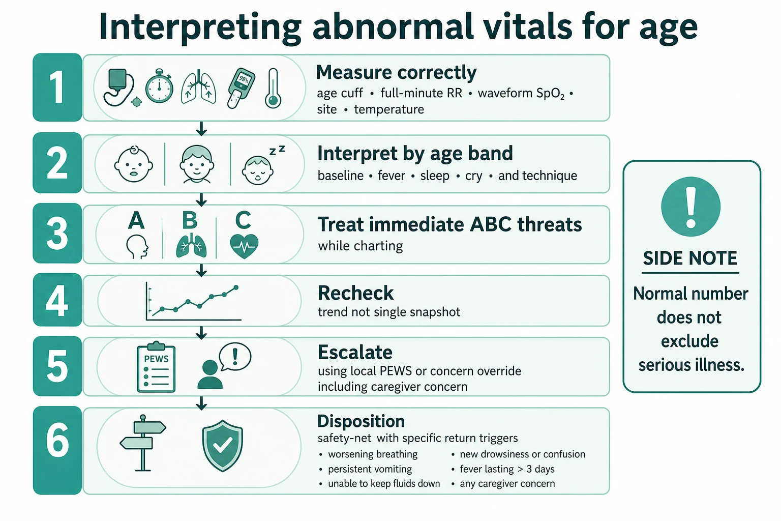
Numbers that imply airway, breathing or circulation threat trigger ABCDE and help, not longer debate about which centile table is purest. [7] [8]
Management — Definitive & Stepwise
- Measure with good technique.
- Age-band and state-adjust.
- Plot trend and PEWS if on a ward chart.
- Escalate on score, concern, or incomplete dangerous gaps.
- Hand over numbers with units, time and trajectory. [7] [10]
Specific Subtypes & Scenarios
Febrile toddler. Expect higher HR; still watch work of breathing and interaction. [3]
Hospitalised child. Prefer hospital-derived centiles and local PEWS rules over community resting ranges alone. [2]
Darker skin and SpO2. Know bias risk; correlate clinically. [9]
Caregiver concern with near-normal vitals. Do not discharge on numbers alone. [6]
Complications & Pitfalls
Adult cut-offs on children. Single normal value reassurance. Ignoring fever effect. Wrong cuff size. Treating PEWS as a diagnosis. Missing incomplete charts. [1] [7]
Prognosis & Disposition
Trajectory and response to treatment predict need for higher care more than one snapshot. EPOCH and PEWS evidence inform system design without promising immortality from a score. [8] [7]
Special Populations
Neonates, cyanotic heart disease baselines, technology-dependent children, and children with chronic lung disease need individualised expected ranges documented on the chart. [2]
Evidence, Guidelines & Regional Differences
Fleming et al. provide age-related HR/RR ranges from systematic review. [1] Bonafide et al. provide hospitalised-child HR/RR percentile curves. [2] Daymont links temperature and HR. [3] CRT evidence is summarised by Fleming. [4] [5]
Exam Pearls
- Age first, then number. [1]
- State of the child changes HR/RR. [3]
- Trend beats snapshot. [2]
- CRT is technique-dependent. [4]
- SpO2 has equity limits. [9]
- Concern can override a mild score. [6]
References
- [1]Fleming S Normal ranges of heart rate and respiratory rate in children from birth to 18 years of age: a systematic review of observational studies Lancet, 2011.PMID 21411136
- [2]Bonafide CP Development of heart and respiratory rate percentile curves for hospitalized children Pediatrics, 2013.PMID 23478871
- [3]Daymont C Heart rates in hospitalized children by age and body temperature Pediatrics, 2015.PMID 25917984
- [4]Fleming S Validity and reliability of measurement of capillary refill time in children: a systematic review Arch Dis Child, 2015.PMID 25260515
- [5]Fleming S The Diagnostic Value of Capillary Refill Time for Detecting Serious Illness in Children: A Systematic Review and Meta-Analysis PLoS One, 2015.PMID 26375953
- [6]Mills E Association between caregiver concern for clinical deterioration and critical illness in children presenting to hospital: a prospective cohort study Lancet Child Adolesc Health, 2025.PMID 40451224
- [7]Trubey R Validity and effectiveness of paediatric early warning systems and track and trigger tools for identifying and reducing clinical deterioration in hospitalised children: a systematic review BMJ Open, 2019.PMID 31061010
- [8]Parshuram CS Effect of a Pediatric Early Warning System on All-Cause Mortality in Hospitalized Pediatric Patients: The EPOCH Randomized Clinical Trial JAMA, 2018.PMID 29486493
- [9]Sharma M Racial and skin color mediated disparities in pulse oximetry in infants and young children Paediatr Respir Rev, 2024.PMID 38233229
- [10]Starmer AJ Changes in medical errors after implementation of a handoff program N Engl J Med, 2014.PMID 25372088